Obstetrics & Gynaecology · Obstetrics & Gynaecology
Menstrual Disorders
Also known as Menstrual Disorders
Menstrual disorders encompass abnormalities in menstrual cycle frequency, duration, volume, or associated symptoms. FIGO PALM-COEIN classification (2011/2018) categorises abnormal uterine bleeding: Polyps, Adenomyosis, Leiomyoma, Malignancy/hyperplasia (structural); Coagulopathy, Ovulatory dysfunction, Endometrial, Iatrogenic, Not classified (non-structural). Management follows NICE NG88: LNG-IUS first-line for HMB; tranexamic acid/mefenamic acid/COCP alternatives.
On this page & tools
Your progress
Saved locally on this device.
Exam tags
Red flags
Overview
Menstrual disorders are among the most common gynaecological presentations in primary care and gynaecology outpatient practice, accounting for roughly 20 percent of all GP consultations in reproductive-age women and over 5 percent of all referrals to gynaecology. They span four broad domains: (1) abnormalities of bleeding volume (heavy menstrual bleeding, postcoital bleeding, intermenstrual bleeding, postmenopausal bleeding); (2) abnormalities of cycle timing (amenorrhoea, oligomenorrhoea, polymenorrhoea); (3) menstrual pain (dysmenorrhoea); and (4) cyclical mood and somatic symptoms (premenstrual syndrome and premenstrual dysphoric disorder). [1]
The unifying framework for the commonest complaint — abnormal uterine bleeding (AUB) — is the FIGO PALM-COEIN classification, published in 2011 by Munro, Critchley and Broder, and adopted globally as the standard system for categorising AUB in non-gravid women of reproductive age.[1] It separates structural causes (PALM — identifiable on imaging or histology) from non-structural causes (COEIN — functional, endocrine, haematological or iatrogenic, not detectable on imaging alone).
Menstrual Physiology — The HPO Axis
A thorough understanding of the hypothalamic–pituitary–ovarian (HPO) axis is essential for classifying and managing menstrual disorders. Every amenorrhoea, oligomenorrhoea and ovulatory dysfunction question in exams ultimately tests this axis. [1]
The Hypothalamic Pulse Generator
The arcuate nucleus of the mediobasal hypothalamus houses kisspeptin neurons, which are the master regulators of the reproductive axis. Kisspeptin stimulates gonadotropin-releasing hormone (GnRH) neurons in the preoptic area to release GnRH in a pulsatile fashion — pulses every 60–90 minutes in the follicular phase (fast frequency) and every 120–240 minutes in the luteal phase (slow frequency, driven by progesterone). [1]
Pulsatility matters: a fast GnRH pulse frequency favours LH synthesis, while a slow frequency favours FSH synthesis in the same gonadotrope cells of the anterior pituitary. This is why only one dominant follicle is selected. [1]
KNDy neurons (Kisspeptin, Neurokinin B, Dynorphin) within the arcuate nucleus form the GnRH pulse generator. Neurokinin B stimulates and dynorphin (an opioid) inhibits the pulse generator, providing fine-tuning. Loss-of-function mutations in TAC3 (neurokinin B) or TACR3 cause hypogonadotropic hypogonadism — a direct exam link. [1]
The Anterior Pituitary
GnRH stimulates the gonadotropes to secrete follicle-stimulating hormone (FSH) and luteinising hormone (LH) — both glycoprotein heterodimers sharing an alpha subunit. [1]
- FSH — recruits a cohort of follicles (days 1–5), stimulates granulosa cell proliferation and aromatase activity, converting theca-derived androstenedione to oestradiol.
- LH — stimulates theca cells to produce androstenedione and testosterone (the "two-cell, two-gonadotropin" model). The mid-cycle LH surge triggers ovulation and luteinisation. [1]
The Follicular Phase (Days 1–13)
The cycle begins with the recruitment of 5–15 primordial follicles under rising FSH. Granulosa cells proliferate, and the follicle with the most FSH receptors and highest aromatase activity becomes the dominant follicle by day 6–7 — it produces exponentially more oestradiol, which exerts negative feedback on FSH, causing atresia of the subordinate follicles. This is the FSH threshold/window concept. [1]
The dominant follicle produces inhibin B, which further suppresses FSH. As oestradiol rises progressively (the growing follicle has ever-more granulosa cells), it eventually reaches a threshold of approximately 200–300 pg/mL sustained for 48 hours, at which point oestrogen switches from negative to positive feedback on the pituitary — the positive feedback mechanism triggers the massive LH surge (10-fold rise) and smaller FSH surge, leading to ovulation 24–36 hours later (typically day 14). [1]
Ovulation (Day 14)
The LH surge causes: (1) resumption of meiosis I in the oocyte (completing the first polar body); (2) prostaglandin and protease release causing follicular wall thinning (via plasminogen activator and collagenase); (3) stigma formation on the ovarian surface; (4) release of the cumulus–oocyte complex into the peritoneal cavity, captured by the fimbriae. [1]
The Luteal Phase (Days 15–28)
The ruptured follicle transforms into the corpus luteum under LH influence. Luteinised granulosa cells produce progesterone (the dominant luteal hormone) and oestradiol. Progesterone: [1]
- Converts the proliferative endometrium to a secretory endometrium (sub-nuclear vacuoles day 16–20, then supranuclear vacuoles, stromal oedema, spiral artery development).
- Raises basal body temperature by 0.3–0.5°C (thermogenic effect).
- Thickens cervical mucus, making it less penetrable to sperm.
- Exerts negative feedback on GnRH (slow pulse frequency) and LH/FSH. [1]
If pregnancy occurs, human chorionic gonadotropin (hCG) from the trophoblast rescues the corpus luteum, which continues progesterone production until the placenta takes over at 8–10 weeks (the luteoplacental shift). [1]
If pregnancy does not occur, the corpus luteum regresses (apoptosis) after 14 days, progesterone and oestradiol levels fall abruptly, and the endometrium undergoes ischaemic necrosis — vasoconstriction of spiral arteries (mediated by prostaglandin F2-alpha and endothelin-1), followed by vasodilation and bleeding. This is menstruation. The fall in oestradiol and progesterone removes negative feedback, FSH rises again, and a new cycle begins. [1]
Steroid Feedback Summary
Negative feedback
- Low–moderate oestradiol and progesterone suppress GnRH, FSH and LH
- Operates in early–mid follicular phase and throughout luteal phase
- Mediated via kisspeptin neuron suppression and direct pituitary effect
- Explains why COCP works — exogenous steroids suppress HPO axis and ovulation
Positive feedback
- Sustained high oestradiol (over 200 pg/mL for 48 h) triggers LH surge
- Operates only at mid-cycle, just before ovulation
- Requires prior 'priming' with several days of oestrogen exposure
- Basis for ovulation induction — hCG trigger after follicular maturation on letrozole/clomiphene
PALM-COEIN Classification
The FIGO classification is the single highest-yield framework for AUB in any gynaecology exam.[1] It divides causes into structural (PALM) — identifiable on imaging or histology — and non-structural (COEIN) — systemic, endocrine, haematological, or functional, not detectable on ultrasound or biopsy alone.
Structural Causes — PALM (Detailed)
Polyps (P): Endometrial polyps are benign overgrowths of endometrial glands and stroma projecting into the uterine cavity. Present in 10–24 percent of women with AUB. Risk factors: nulliparity, obesity, tamoxifen use, HRT. Most are benign (70 percent); risk of malignancy is 1–3 percent overall, rising to over 10 percent in postmenopausal women with bleeding. Presentation: intermenstrual bleeding, postcoital bleeding, HMB, infertility. Diagnosis: TVS (echogenic focal lesion); saline infusion sonohysterography (SIS) improves detection; hysteroscopy is gold standard (direct visualisation and biopsy/polypectomy). Treatment: hysteroscopic polypectomy (see-and-treat); small asymptomatic polyps in premenopausal women may be managed conservatively. [1]
Adenomyosis (A): Endometrial glands and stroma within the myometrium, causing a diffusely enlarged, boggy, tender uterus. Classic triad: HMB, dysmenorrhoea, and enlarged uterus. Typically presents in the late reproductive years and perimenopause (parous women). Diagnosis: TVS (asymmetric myometrial thickening, myometrial cysts, fan-shaped shadowing, junctional zone over 12 mm) or MRI (junctional zone over 12 mm is the hallmark; T2 hyperintense foci). Treatment: LNG-IUS first-line (reduces bleeding and pain); hysterectomy is definitive for completed family. GnRH analogues and uterine-sparing procedures (adenomyomectomy) have limited efficacy. Adenomyosis is NOT the same as endometriosis — endometriosis is outside the uterus, adenomyosis is within the myometrium. [1]
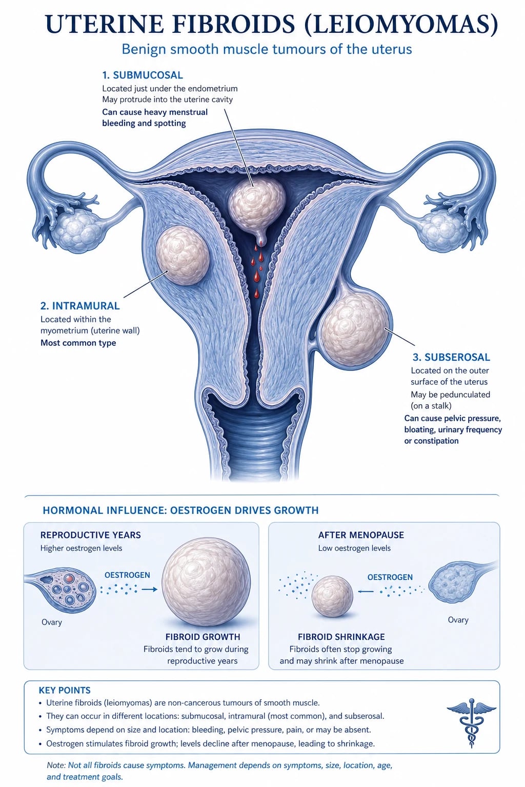
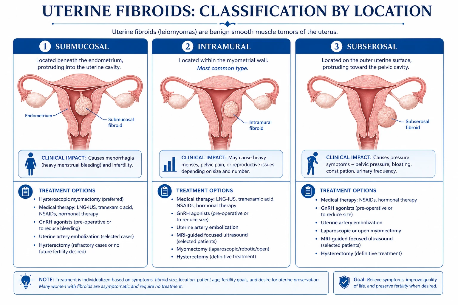
Leiomyoma (L) — Fibroids: Benign smooth-muscle tumours, the commonest pelvic tumour (present in 40 percent of women over 35, 70–80 percent by age 50). More common in women of African ancestry (3–5 times higher risk). Classification by location:
- Submucosal (project into cavity) — most likely to cause heavy bleeding and infertility; type 0 (pedunculated intracavitary), type 1 (under 50 percent intramural), type 2 (over 50 percent intramural).
- Intramural (within wall) — commonest; cause HMB, dysmenorrhoea, bulk symptoms.
- Subserosal (project outward) — may cause pressure on bladder/bowel.
- Cervical, parasitic, intraligamentous — rarer locations. [1]
Malignant transformation to leiomyosarcoma is rare (under 0.5 percent of rapidly growing fibroids; the "rapid growth" sign has a low positive predictive value). Presentation: HMB, dysmenorrhoea, pressure symptoms (frequency, constipation), abdominal mass, infertility, pregnancy complications. Diagnosis: TVS (hypoechoic well-circumscribed mass); MRI for mapping before surgery. Treatment depends on symptoms, fertility wishes and fibroid size/location (see HMB management and surgical procedures). [1]
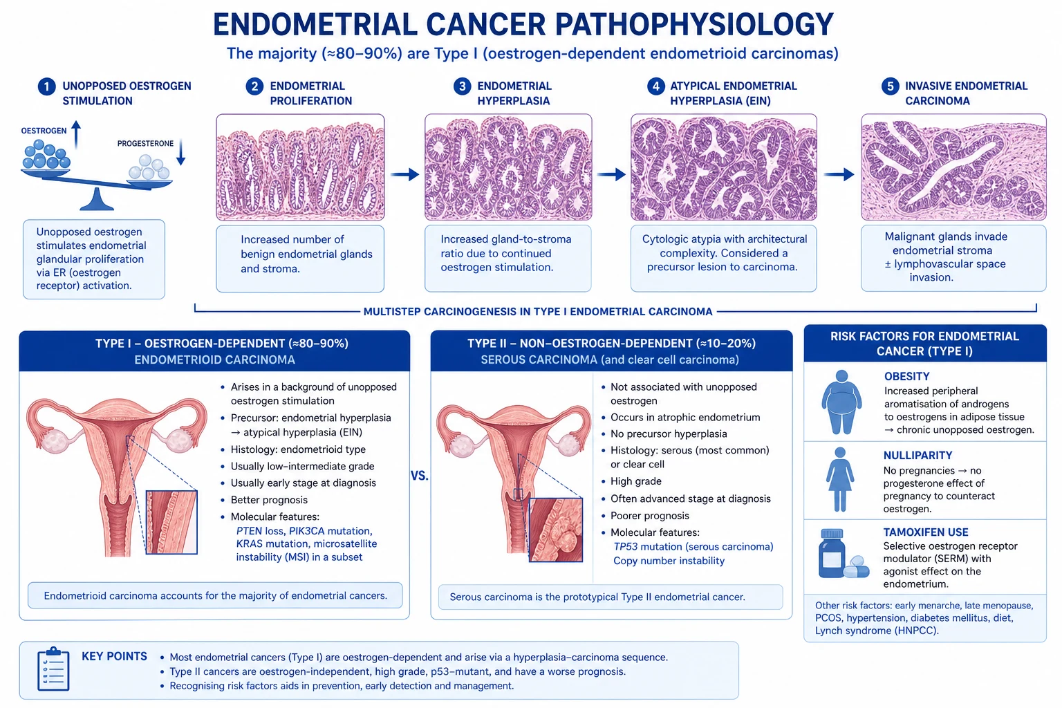
Malignancy and Hyperplasia (M): Endometrial hyperplasia and endometrial cancer are the critical diagnoses to not miss in any woman presenting with AUB, particularly postmenopausal bleeding. [1]
Endometrial hyperplasia is classified (WHO 2014) as:
- Hyperplasia without atypia — 1–3 percent progress to cancer; treat with progestogen (LNG-IUS or oral) — regression in over 80 percent.
- Atypical hyperplasia — 25–40 percent progress to cancer (or have concurrent cancer); treat with hysterectomy (or progestogen if fertility-sparing). [1]
Endometrial cancer — commonest gynaecological malignancy in high-income countries; risk factors: obesity (unopposed oestrogen), nulliparity, late menopause, PCOS, tamoxifen, diabetes, Lynch syndrome, oestrogen-secreting tumours. Histology: endometrioid (type I, 80 percent) — oestrogen-related, good prognosis; serous/clear cell (type II) — aggressive, not oestrogen-related. Staging: surgical (FIGO 2009). Treatment: total abdominal hysterectomy + bilateral salpingo-oophorectomy ± lymphadenectomy; adjuvant radiotherapy/chemotherapy depending on stage and grade. [1]
Non-Structural Causes — COEIN (Detailed)
Coagulopathy (C): Bleeding disorders underlie approximately 10–20 percent of HMB cases, with von Willebrand disease (vWD) accounting for the majority (roughly 13 percent of women with HMB since menarche). Always screen for coagulopathy when HMB has been present since menarche, there is a family history of bleeding disorder, or there are other bleeding manifestations (epistaxis, gum bleeding, easy bruising, postpartum haemorrhage, post-surgical bleeding). Workup: FBC, coagulation screen (PT, APTT), von Willebrand factor antigen (vWF:Ag), ristocetin cofactor activity (vWF:RCo), factor VIII. Other causes: immune thrombocytopenia (ITP), platelet function disorders, anticoagulant therapy (warfarin, DOACs), haemophilia carrier. [1]
Ovulatory dysfunction (O): The commonest category of non-structural AUB. Causes include PCOS (most common — see below), thyroid dysfunction (both hypo- and hyperthyroidism cause menstrual irregularity), hyperprolactinaemia (suppresses GnRH), premature ovarian insufficiency, perimenopause, stress, weight loss, eating disorders, excessive exercise, and non-classic congenital adrenal hyperplasia. The hallmark is irregular, unpredictable cycles — anovulatory or oligo-ovulatory. Management targets the underlying cause. [1]
Endometrial (E): A primary disorder of endometrial haemostasis — the endometrium fails to mount the normal local vasoconstrictor (prostaglandin F2-alpha, endothelin) and platelet plug response, leading to excessive bleeding despite a structurally normal uterus and normal systemic haemostasis. Abnormal ratios of vasodilatory (PGE2, PGI2) to vasoconstrictor prostaglandins have been demonstrated. This is essentially a diagnosis of exclusion after other causes have been ruled out. [1]
Iatrogenic (I): Medication-induced AUB — hormonal contraception (breakthrough bleeding on COCP, POP, implant, injection, IUD), anticoagulants (warfarin, DOACs), SSRIs (platelet dysfunction), tamoxifen (endometrial stimulation and polyp formation), spironolactone (menstrual irregularity), and phenytoin/anticonvulsants. Management: address the medication if possible; if treatment must continue, add a progestogen or consider switching formulations. [1]
Not yet classified (N): Rare causes not fitting the above categories — uterine arteriovenous malformations (AVMs), Caesarean section scar defects (isthmoceles) causing postmenstrual spotting, non-tuberculous pelvic tuberculosis (in endemic regions), and chronic endometritis. [1]
Definitions
| Term | Definition |
|---|---|
| Normal menstruation | Cycle 21–35 days; duration 2–7 days; blood loss 30–80 mL; regular |
| Primary amenorrhoea | No menses by age 15 with, or 13 without, secondary sex characteristics |
| Secondary amenorrhoea | No menses for 3–6 months in a previously menstruating woman |
| Oligomenorrhoea | Cycle interval over 35 days (fewer than 9 cycles/year) |
| Polymenorrhoea | Cycle interval under 21 days |
| HMB (menorrhagia) | Blood loss over 80 mL per cycle, or subjective excessive loss interfering with quality of life (PBAC over 100) |
| Dysmenorrhoea | Painful menstruation — primary (no pathology) or secondary (endometriosis, fibroids, adenomyosis, PID) |
| IMB | Intermenstrual bleeding — bleeding between periods |
| PCB | Postcoital bleeding — bleeding after intercourse (exclude cervical cancer) |
| PMB | Postmenopausal bleeding — any bleeding 12 months after the last menstrual period (exclude endometrial cancer) |
| Metrorrhagia | Irregular, frequent bleeding (often used interchangeably with IMB) |
| Menometrorrhagia | Heavy and irregular bleeding |
Clinical Assessment
History Taking
A structured menstrual history is the single most important diagnostic tool. The mnemonic "GOLIAD" captures the key parameters: Gravidity/parity, Onset, Length of cycle, Interval/duration, Amount, Dysmenorrhoea — but a more practical approach follows: [1]
Menstrual history:
- Age at menarche (normal 10–15 years; delayed suggests constitutional delay, eating disorder, or chromosomal abnormality; very early suggests precocious puberty)
- Cycle length (normal 21–35 days; under 21 = polymenorrhoea; over 35 = oligomenorrhoea)
- Cycle regularity (regular = likely ovulatory; irregular = anovulatory — PCOS, thyroid, perimenopause, POI)
- Duration of bleeding (normal 2–7 days; prolonged over 7 days)
- Volume of bleeding — quantify using PBAC (Pictorial Blood Assessment Chart) score (over 100 = HMB); ask about pad/tampon count per day, passage of clots (over 1 inch), flooding, need for double protection, and interference with daily activities
- Pain — primary dysmenorrhoea: cramping suprapubic pain, day 1–2, no pathology; secondary: deep dyspareunia, cyclical bowel/bladder symptoms (endometriosis), progressive dysmenorrhoea (adenomyosis)
- IMB, PCB, PMB — each a red flag requiring specific workup [1]
Associated symptoms (system-directed):
- Weight change — weight loss (hypothalamic amenorrhoea, anorexia); weight gain (PCOS, hypothyroidism, Cushing syndrome)
- Hirsutism, acne, alopecia — androgen excess (PCOS, non-classic CAH, androgen-secreting tumour)
- Virilisation (clitoromegaly, deep voice, temporal balding) — androgen-secreting tumour (urgent)
- Galactorrhoea — hyperprolactinaemia (prolactinoma, drug-induced)
- Thyroid symptoms — heat/cold intolerance, palpitations, weight change, tremor
- Hot flushes, night sweats, vaginal dryness — premature ovarian insufficiency (under 40), perimenopause
- Cyclical pelvic pain, dyspareunia, dyschezia — endometriosis; dysuria haematuria — bladder endometriosis
- Pressure symptoms — urinary frequency, constipation, abdominal distension (fibroids) [1]
Past medical, surgical, drug and family history:
- Thyroid disease, diabetes, coagulation disorders, autoimmune disease
- Previous gynaecological surgery (LLETZ, cone biopsy — cervical stenosis/insufficiency)
- Medications: hormonal contraception, anticoagulants, SSRIs, tamoxifen, antipsychotics (hyperprolactinaemia), valproate
- Family history: endometrial/breast/ovarian/colorectal cancer (Lynch syndrome, BRCA), bleeding disorders, PCOS [1]
Red Flags
Other red flags requiring urgent investigation:
- Persistent postcoital bleeding — exclude cervical cancer (speculum, colposcopy, biopsy)
- Intermenstrual bleeding with weight loss or systemic symptoms — exclude endometrial/cervical malignancy
- HMB with anaemia refractory to iron — exclude coagulopathy (vWD), endometrial pathology
- Sudden change in menstrual pattern in a woman over 40 — exclude endometrial pathology
- New HMB after menopause (any bleeding) — endometrial cancer
- Virilisation (clitoromegaly, deep voice, frontal balding) — androgen-secreting tumour (urgent imaging and referral)
- Cyclical haematuria or rectal bleeding — deep infiltrating endometriosis [1]
Examination
General examination:
- BMI — under 18.5 (hypothalamic amenorrhoea, eating disorder, POI with weight loss); over 30 (PCOS, endometrial cancer risk, metabolic syndrome)
- Hirsutism — Ferriman-Gallwey score (modified, over 4–6 = hirsutism); assess upper lip, chin, chest, abdomen, thighs, lower back
- Acne, acanthosis nigricans (insulin resistance in PCOS)
- Virilisation (clitoromegaly, deep voice, frontal balding, increased muscle mass — suggests androgen-secreting tumour)
- Galactorrhoea — expressible milky nipple discharge
- Thyroid — goitre, bruit, eye signs
- Stigmata of Turner syndrome — short stature, webbed neck, widely spaced nipples, low posterior hairline
- Stigmata of Cushing syndrome — moon face, buffalo hump, striae, proximal myopathy [1]
Abdominal examination:
- Distension, mass (fibroid uterus — smooth, firm, mobile, non-tender; 14–16 weeks size is palpable abdominally)
- Tenderness (endometriosis, PID)
- Hepatomegaly, ascites (in metastatic disease) [1]
Pelvic (bimanual) examination:
- Uterus — size (compare to weeks of pregnancy), mobility, contour, tenderness (enlarged/tender = adenomyosis; irregular = fibroids; fixed/retroverted = endometriosis)
- Cervix — excitation tenderness (cervical motion tenderness suggests PID/endo)
- Adnexae — masses (ovarian cyst, endometrioma), tenderness [1]
Speculum examination:
- Cervical appearance — ectropion, polyp, malignancy, discharge (infection)
- Take cervical screening if due (never assume abnormal bleeding is just a missed smear)
- Swabs if infection suspected (chlamydia, gonorrhoea NAAT) [1]
Investigation Strategy
Investigations are guided by the clinical picture, but a core panel applies to most presentations: [1]
| Test | Indication / Purpose |
|---|---|
| Pregnancy test (β-hCG) | ALL women with amenorrhoea or unexpected bleeding — rule out pregnancy and ectopic FIRST |
| FBC + ferritin | Anaemia from HMB; iron-deficiency pattern |
| TSH | Exclude thyroid dysfunction (both hypo- and hyperthyroidism cause AUB) |
| Prolactin | Galactorrhoea, amenorrhoea, oligomenorrhoea, some AUB; repeat if elevated; macroprolactin screen |
| LH, FSH, oestradiol | PCOS (LH:FSH ratio over 2, but NOT required for diagnosis); POI (FSH over 25 IU/L on two occasions 4–6 weeks apart) |
| Testosterone, SHBG, DHEAS | Hirsutism, virilisation; free androgen index (testosterone/SHBG); DHEAS for adrenal source |
| 17-Hydroxyprogesterone | Non-classic congenital adrenal hyperplasia (baseline, then ACTH stimulation if borderline) |
| Day 21 progesterone | Confirm ovulation (over 30 nmol/L = luteal; over 3 nmol/L = ovulated); day 21 of 28-day cycle, or day 21 minus 7 of longer cycle |
| AMH (anti-Mullerian hormone) | Ovarian reserve assessment; NOT for PCOS diagnosis (can be supportive but Rotterdam does not require it) |
| Coagulation screen + vWF antigen/activity | HMB since menarche, family history of bleeding, other bleeding features; vWD, platelet disorders |
| Fasting glucose / HbA1c, lipid profile | PCOS metabolic workup (insulin resistance, dyslipidaemia) |
| Transvaginal ultrasound (TVS) | Structural causes — fibroids, polyps, adenomyosis, ovarian cysts; endometrial thickness in PMB |
| Saline infusion sonohysterography (SIS) | Intracavitary pathology (polyps, submucosal fibroids) — superior to TVS for focal lesions |
| Hysteroscopy + endometrial biopsy (EB) | PMB, abnormal bleeding over 40, suspected hyperplasia/malignancy; gold standard for endometrial assessment |
| Karyotype | Primary amenorrhoea (Turner 45,X; Swyer 46,XY; androgen insensitivity 46,XY) |
| MRI pelvis | Adenomyosis (junctional zone over 12 mm), fibroid mapping before surgery, Mullerian anomalies, deep infiltrating endometriosis |
Heavy Menstrual Bleeding (HMB)
Definition and Impact
Heavy menstrual bleeding (HMB) — formerly "menorrhagia" — is excessive menstrual blood loss interfering with quality of life. Objectively, this is loss over 80 mL per cycle or PBAC score over 100, but the subjective definition (woman's perception of heaviness) is now preferred and more practical. HMB affects one in three women at some point and is the commonest reason for gynaecological referral. [1]
NICE NG88 Management Algorithm
The NICE guideline NG88 (Heavy menstrual bleeding: assessment and management) provides a stepwise pharmacological-first approach: [1]
Step 1 — Levonorgestrel intrauterine system (LNG-IUS, Mirena 52 mg):
- First-line for ALL women with HMB and no identified pathology or small fibroids (under 3 cm) not distorting the cavity
- Releases 20 micrograms/day of levonorgestrel locally to the endometrium, causing endometrial atrophy and decidualisation
- Reduces menstrual blood loss by 70–95 percent[7]
- 20 percent amenorrhoea at 1 year; 50 percent at 5 years
- Provides contraception for 5 years (8 years for contraception-only use per FSRH 2023)
- Also treats dysmenorrhoea, endometriosis pain, and provides endometrial protection in women on oestrogen HRT
- Contraindications: active pelvic infection, distorted uterine cavity (large fibroids), current breast cancer, unexplained bleeding (investigate first), gestational trophoblastic disease
- Side effects: irregular spotting/bleeding for first 3–6 months (counsel and reassure — this is the main reason for early discontinuation), hormonal symptoms (headache, breast tenderness, mood change — usually mild)
- Offer at least 3 months before judging efficacy; counsel that bleeding is irregular initially
Step 2 — If LNG-IUS declined, unsuitable, or while awaiting response (use within 3 months): Offer a non-hormonal or hormonal option based on contraceptive needs and patient preference: [1]
- Tranexamic acid — 1 g TDS (or 500 mg if under 50 kg) for up to 4 days during menses only. Antifibrinolytic; inhibits plasminogen activation. Reduces bleeding by 40–50 percent. Contraindicated in active thromboembolic disease, acquired colour vision disturbance. Start with menses, stop when bleeding stops.
- Mefenamic acid (or other NSAID) — 500 mg TDS during menses. Inhibits prostaglandin synthesis; reduces bleeding by 25–30 percent and also provides analgesia for dysmenorrhoea. Contraindicated in peptic ulcer disease, asthma (aspirin sensitivity), renal impairment. Consider gastroprotection with PPI.
- Combined oral contraceptive pill (COCP) — any standard preparation; suppresses ovulation, regularises cycle, reduces flow by approximately 30 percent. Use 30–35 microgram ethinylestradiol preparations initially. Contraindications: migraine with aura (absolute), smoker over 35, BMI over 35, personal/family history of VTE, breast cancer, uncontrolled hypertension. Use UKMEC criteria to assess.
- Oral or long-acting progestogen — norethisterone 5 mg TDS on days 5–26 of cycle, or medroxyprogesterone acetate (DMPA) depot injection 150 mg IM every 12 weeks (causes amenorrhoea in 50–70 percent by 1 year; reduces bone density — caution in adolescents). [1]
Step 3 — If pharmacological treatment fails (after 3 months of optimised therapy): Refer to specialist. Investigate for structural cause (TVS ± hysteroscopy) and consider second-line treatments: [1]
- Second-course LNG-IUS if first was not tolerated
- Add tranexamic acid + NSAID to LNG-IUS if breakthrough bleeding persists
- Consider GnRH analogues (goserelin 3.6 mg SC monthly or leuprorelin 3.75 mg monthly) — maximum 6 months without add-back HRT (tibolone 2.5 mg or estradiol/norethisterone) due to bone density loss. Use as a short-term bridge to surgery or for diagnostic confirmation of hormonal responsiveness. [1]
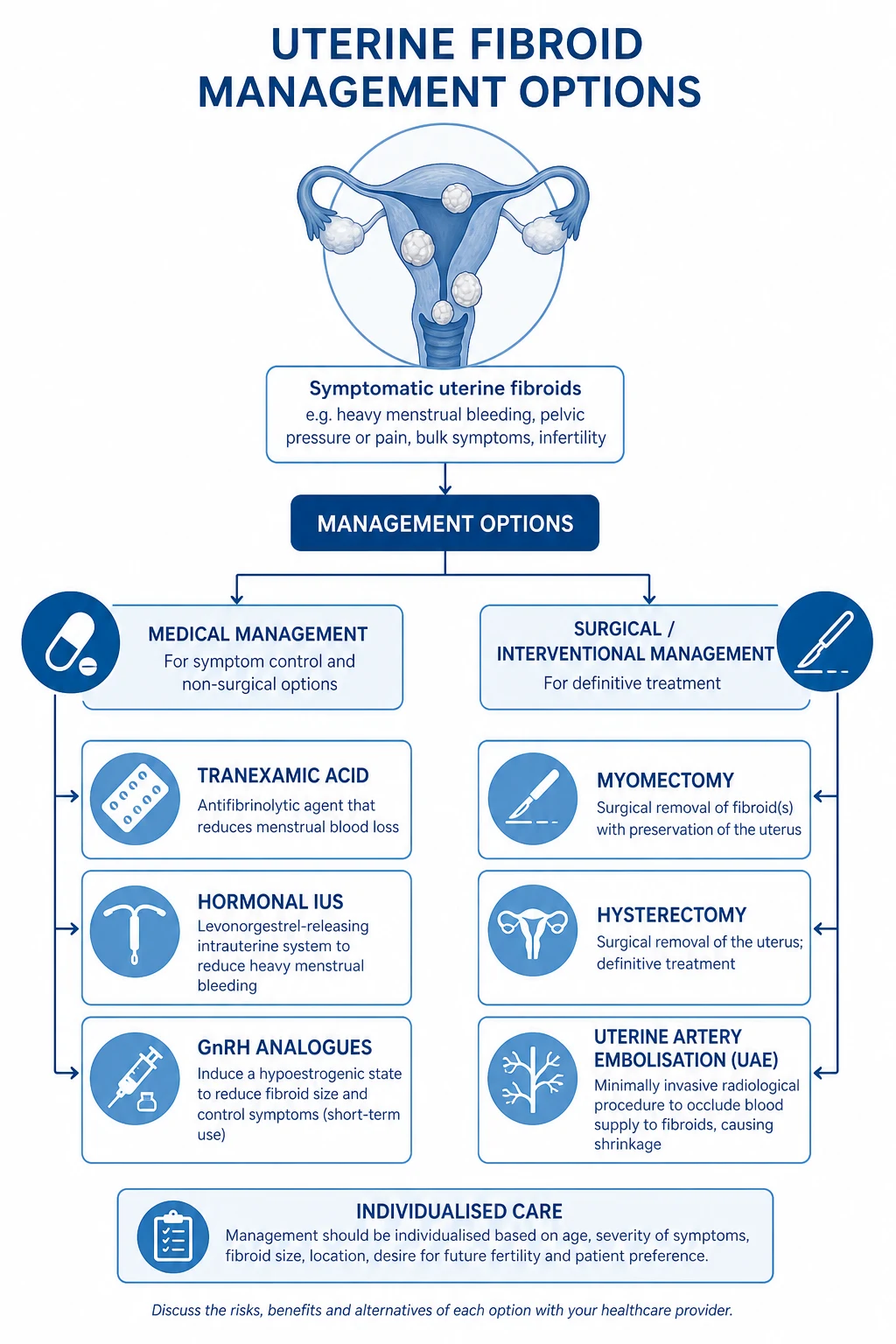
Step 4 — Surgical management: [1]
If pharmacological management fails or is not appropriate, and structural causes have been identified and addressed: [1]
- Endometrial ablation — for completed family, no desire for future pregnancy, normal cavity or small fibroids. See Surgical Procedures below.
- Hysterectomy — definitive treatment; for failed medical/ablative therapy, large fibroids, coexisting pathology (prolapse, severe dysmenorrhoea), or patient preference.
- Myomectomy — for fibroids when fertility preservation is desired.
- Uterine artery embolisation (UAE) — minimally invasive option for symptomatic fibroids. [1]
HMB with Identified Structural Pathology
Fibroids (PALM-L): Treatment depends on size, location, symptoms, fertility wishes: [1]
| Strategy | Indication | Notes |
|---|---|---|
| LNG-IUS | Small fibroids (under 3 cm) not distorting cavity | First-line; contraindicated if cavity distorted |
| Hysteroscopic myomectomy | Submucosal fibroids (types 0, 1, 2) | Fertility-sparing; day-case; resectoscope or morcellator |
| Laparoscopic/abdominal myomectomy | Intramural/subserosal fibroids over 5 cm, fertility desired | Larger blood loss; risk of rupture in future pregnancy |
| UAE | Symptomatic fibroids, completed family, declines surgery | 20–25 percent need repeat treatment within 5 years; not recommended if future fertility desired (controversial) |
| Endometrial ablation | Small submucosal fibroids with HMB, completed family | Can combine with myomectomy |
| Hysterectomy | Large fibroids, completed family, definitive treatment | Vaginal/laparoscopic/abdominal route |
| Medical (GnRH) | Pre-operative shrinkage, anaemia correction | 3–6 months only; add-back HRT |
| Ulipristal acetate (UPA) | Pre-operative fibroid shrinkage | EMA restricted use due to rare liver injury; specialist only |
Polyps (PALM-P): Hysteroscopic polypectomy (see-and-treat outpatient under local anaesthesia for small polyps; general anaesthesia for larger). Send all polyps for histology (malignancy risk higher in postmenopausal). [1]
Adenomyosis (PALM-A): LNG-IUS first-line; hysterectomy if completed family and refractory. GnRH analogues provide temporary relief. Adenomyomectomy (uterus-sparing) has high recurrence. [1]
Amenorrhoea
Definition and Classification
Amenorrhoea is the absence of menstrual periods. It is classified as: [1]
- Primary amenorrhoea — no menarche by age 15 with secondary sexual characteristics, or age 13 without secondary sexual characteristics. (Also: no menses by age 14 if no secondary sex characteristics by age 13, per some guidelines.)
- Secondary amenorrhoea — absence of menses for 3 consecutive months in a woman with previously regular cycles, or 6 months in a woman with previously irregular cycles. [1]
Approach to Amenorrhoea — The Algorithm
ALWAYS do a pregnancy test (β-hCG) FIRST. Pregnancy is the commonest cause of secondary amenorrhoea — missing it is a catastrophic clinical error. [1]
After excluding pregnancy, the workup follows a functional/anatomical localisation approach — hypothalamus, pituitary, ovary, uterus/outflow tract — using three key hormones: FSH, prolactin, TSH. [1]
Step 1: β-hCG — exclude pregnancy. [1]
Step 2: TSH and prolactin — thyroid disease and hyperprolactinaemia are common, easily treated, and should be excluded before complex workup. [1]
- If TSH high → hypothyroidism → treat with levothyroxine (typically 50–100 micrograms/day, titrate to TSH 0.4–4.0).
- If prolactin elevated (confirmed on repeat, macroprolactin excluded) → MRI pituitary. Macroprolactinoma over 10 mm: cabergoline 0.5 mg twice weekly (titrate up); microprolactinoma under 10 mm: cabergoline 0.25 mg twice weekly. Dopamine agonists are first-line for both. [1]
Step 3: FSH and oestradiol — to localise the level of the defect: [1]
- Low or normal FSH with low oestradiol (hypogonadotropic hypogonadism) → hypothalamic or pituitary cause.
- High FSH with low oestradiol (hypergonadotropic hypogonadism) → ovarian failure (primary ovarian insufficiency).
- Normal FSH with normal oestradiol (eugonadotropic) → consider PCOS (most common), or uterine/outflow tract obstruction. [1]
Step 4: Progestogen challenge test — medroxyprogesterone acetate 10 mg daily for 5–10 days, or dydrogesterone 10 mg BD for 5 days. Withdrawal bleed within 7 days indicates oestrogen is present and the outflow tract is patent (anovulation — likely PCOS). No bleed indicates either low oestrrogen (hypothalamic/pituitary/ovarian failure) or outflow obstruction. [1]
Step 5: Oestrogen-progestogen challenge — if no withdrawal bleed to progestogen alone: give oestrogen (e.g., conjugated oestrogens 1.25 mg or estradiol 2 mg daily for 21 days) followed by progestogen. Bleeding indicates the endometrium can respond — so the problem is lack of oestrogen (hypothalamic/pituitary/ovarian). No bleed indicates an outflow tract problem (Asherman syndrome, Mullerian agenesis). [1]
Causes of Amenorrhoea (Comprehensive)
Hypothalamic causes (hypogonadotropic — low FSH/LH, low oestradiol):
- Functional hypothalamic amenorrhoea — stress, weight loss (BMI under 18.5), excessive exercise (the "female athlete triad" of low energy availability, menstrual dysfunction, low bone density), eating disorders (anorexia nervosa). The hypothalamic GnRH pulse generator is suppressed. Treatment: restore energy balance — increase caloric intake, reduce exercise, psychological support; HRT for bone protection if amenorrhoea persists.
- Kallmann syndrome — isolated GnRH deficiency with anosmia (absent sense of smell); X-linked or autosomal inheritance. MRI shows absent or hypoplastic olfactory bulbs. Treatment: pulsatile GnRH or gonadotropin therapy to induce ovulation; HRT for bone/cardiovascular health.
- Constitutional delay of puberty — delayed puberty in otherwise healthy adolescents (often familial); rule out organic causes first.
- Structural lesions — craniopharyngioma, glioma, tuberculosis, sarcoidosis, head trauma, cranial irradiation.
- Drugs — antipsychotics (hyperprolactinaemia), chronic opioid use, GnRH agonists. [1]
Pituitary causes (hypogonadotropic):
- Hyperprolactinaemia — prolactinoma (micro under 10 mm or macro over 10 mm), drug-induced (antipsychotics, metoclopramide, opiates), hypothyroidism (TRH stimulates prolactin), pregnancy/lactation, renal/hepatic failure. Presents with amenorrhoea, galactorrhoea, infertility, and (macroadenoma) headaches, visual field defects (bitemporal hemianopia from optic chiasm compression). Treatment: cabergoline first-line (dopamine agonist).
- Sheehan syndrome — pituitary infarction following postpartum haemorrhage (hypovolaemic shock); panhypopituitarism. Presents with failure to lactate, persistent amenorrhoea, hypothyroidism, adrenal insufficiency. Treatment: replacement of all deficient hormones (hydrocortisone, levothyroxine, sex steroids, GH).
- Other pituitary tumours — non-functioning adenoma, Cushing disease (ACTH-secreting), acromegaly (GH-secreting).
- Empty sella syndrome — herniation of subarachnoid space into sella turcica. [1]
Ovarian causes (hypergonadotropic — high FSH over 25 IU/L, low oestradiol):
- Premature ovarian insufficiency (POI) — loss of ovarian function before age 40. Incidence: 1 percent of women under 40, 0.1 percent under 30. Causes: idiopathic (most common), autoimmune, chromosomal (Turner syndrome 45,X; fragile X premutation), iatrogenic (chemotherapy, radiotherapy, oophorectomy), galactosaemia, mumps oophoritis. Presents with amenorrhoea/oligomenorrhoea, menopausal symptoms (hot flushes, night sweats, vaginal dryness), infertility. Diagnosis: FSH over 25 IU/L on two occasions at least 4 weeks apart, with low oestradiol, under age 40. Treatment: HRT until age 51 (average age of menopause) for bone density, cardiovascular protection, symptom relief; combined HRT (oestrogen + cyclic progestogen) if uterus present; oestrogen-only if hysterectomised. COCP is an alternative. Fertility: only option is donor egg IVF; spontaneous pregnancy possible in 5–10 percent due to fluctuating ovarian function. Screen: karyotype, adrenal antibodies (Addison screen — 21-hydroxylase antibodies), TFTs, diabetes screen, bone density (DEXA).
- Turner syndrome (45,X) — streak ovaries, short stature, webbed neck, widely spaced nipples, coarctation of aorta, renal anomalies. Diagnosis: karyotype. Treatment: growth hormone in childhood, oestrogen replacement for puberty induction and bone health.
- Surgical/radiation/chemotherapy-induced ovarian failure — counsel before treatment; consider fertility preservation (egg/embryo freezing, ovarian tissue cryopreservation).
- Physiological menopause — average age 51; diagnosis clinical (over 12 months amenorrhoea with vasomotor symptoms); FSH is NOT needed routinely if over 45. [1]
Uterine and outflow tract causes (normal hormones):
- Asherman syndrome (intrauterine adhesions/synechiae) — scarring of the endometrium, typically after dilation and curettage (especially postpartum or post-miscarriage), endometritis, or uterine surgery. Presents with secondary amenorrhoea or hypomenorrhoea, cyclical pain (if outflow blocked), infertility, recurrent miscarriage. Diagnosis: hysteroscopy (gold standard — direct visualisation of adhesions); TVS, SIS, hysterosalpingography may suggest. Treatment: hysteroscopic adhesiolysis under direct vision, followed by oestrogen therapy (to regenerate endometrium) and IUD (to prevent re-adhesion). Recurrence rate high; pregnancy outcomes variable.
- Mullerian agenesis (Mayer-Rokitansky-Kuster-Hauser syndrome) — congenital absence of uterus and upper two-thirds of vagina; normal ovaries, normal secondary sex characteristics, normal karyotype (46,XX). Second commonest cause of primary amenorrhoea after gonadal dysgenesis. Associated with renal anomalies (30 percent) and skeletal anomalies. Diagnosis: karyotype (46,XX), normal hormones, absent uterus on ultrasound/MRI. Treatment: vaginal dilators or vaginoplasty (for sexual function); fertility requires surrogacy (patient's own eggs). Psychological support essential.
- Imperforate hymen — presents at puberty with cyclical pelvic pain, primary amenorrhoea, haematocolpos (bulging bluish membrane on inspection). Treatment: cruciate incision of hymen. Transverse vaginal septum and cervical atresia are rarer obstructive causes.
- Complete androgen insensitivity syndrome (CAIS) — 46,XY karyotype, non-functional androgen receptor; phenotypic female, normal breast development (aromatised testosterone), scanty/absent pubic and axillary hair, blind-ending vagina, no uterus (Mullerian structures absent due to AMH from testes), intra-abdominal testes (high malignancy risk — gonadectomy after puberty). Presents with primary amenorrhoea. Diagnosis: karyotype (46,XY), high testosterone (male range), no uterus on imaging. Treatment: gonadectomy after completion of breast development; oestrogen replacement; vaginal dilators.
- Cervical stenosis — post-surgical (cone biopsy, LLETZ), post-radiation; causes obstructive amenorrhoea with cyclical pain. [1]
Primary Amenorrhoea Workup Summary
| Finding | Likely cause | Next step |
|---|---|---|
| Uterus present, normal FSH | Constitutional delay, functional hypothalamic | Observe / lifestyle |
| Uterus present, high FSH | Gonadal dysgenesis (Turner, 46,XX gonadal dysgenesis) | Karyotype, HRT |
| Uterus present, low FSH | Kallmann syndrome, hypothalamic | Smell test, MRI pituitary |
| Uterus absent, 46,XX | Mullerian agenesis (MRKH) | Renal USS, vaginal creation |
| Uterus absent, 46,XY | CAIS | Gonadectomy post-puberty, oestrogen |
| Blind vagina / obstructed outflow | Imperforate hymen, transverse septum | Examination under anaesthesia, surgical correction |
Polycystic Ovary Syndrome (PCOS)
Definition and Epidemiology
PCOS is the commonest endocrine disorder in reproductive-age women, affecting 6–10 percent (up to 15–20 percent using broader criteria). It is the leading cause of anovulatory infertility. [1]
Rotterdam Diagnostic Criteria
The Rotterdam criteria (2003 consensus, revised) require 2 of 3 features after exclusion of other causes:[2]
- Oligo-ovulation or anovulation — irregular cycles (over 35 days or under 8 cycles/year), oligomenorrhoea, amenorrhoea.
- Hyperandrogenism — clinical (hirsutism, acne, alopecia — modified Ferriman-Gallwey score over 4–6) and/or biochemical (elevated total/free testosterone, free androgen index).
- Polycystic ovarian morphology on ultrasound — 12 or more follicles measuring 2–9 mm in diameter per ovary, and/or ovarian volume over 10 mL (using transvaginal probe; in modern high-frequency transducers the threshold is 20 follicles per ovary). [1]
Before diagnosing PCOS, exclude: congenital adrenal hyperplasia (17-OH progesterone), androgen-secreting tumour (very high testosterone/DHEAS), Cushing syndrome, thyroid dysfunction, hyperprolactinaemia. [1]
NIH criteria (1990) require hyperandrogenism plus oligo-ovulation only. AE-PCOS Society (2006) requires hyperandrogenism plus either ovulatory dysfunction or PCOM. Rotterdam is the broadest and most widely used, capturing more phenotypes. [1]
Pathophysiology
The pathophysiology is multifactorial and incompletely understood: [1]
- Insulin resistance — present in 70–80 percent of PCOS women (independent of obesity); hyperinsulinaemia stimulates ovarian androgen production and reduces SHBG (raising free testosterone). This is the rationale for metformin use.
- Hypothalamic-pituitary dysfunction — increased GnRH pulse frequency favours LH over FSH, producing elevated LH:FSH ratio (often over 2:1). High LH stimulates theca cells to produce excess androstenedione and testosterone.
- Ovarian follicular arrest — high androgen and insulin environment arrests follicular development at the 2–9 mm stage, producing the characteristic "string of pearls" appearance.
- Low SHBG — from insulin resistance and androgens — raises free (bioavailable) testosterone.
- Adrenal androgen excess — DHEAS may be elevated in 25–50 percent. [1]
Clinical Features
Reproductive
- Oligomenorrhoea/amenorrhoea
- Anovulatory infertility (leading cause)
- Endometrial hyperplasia risk (unopposed oestrogen)
- Higher miscarriage and pregnancy complication rates (GDM, pre-eclampsia)
Hyperandrogenic
- Hirsutism (Ferriman-Gallwey over 4–6)
- Acne
- Androgenic alopecia (male pattern)
- Rarely virilisation (if so, exclude tumour)
Metabolic
- Insulin resistance (70–80 percent)
- Obesity (50 percent)
- Type 2 diabetes (4x risk)
- Dyslipidaemia
- Hypertension
- Metabolic syndrome
- Non-alcoholic fatty liver disease
- Obstructive sleep apnoea
Psychological
- Anxiety, depression (3x risk)
- Reduced quality of life
- Eating disorders
- Negative body image
Management
Management is symptom-directed — there is no cure. The approach depends on the woman's primary concern. [1]
1. Lifestyle (all women):
- Weight loss — even 5–10 percent weight loss restores ovulation in 30–60 percent, improves insulin sensitivity, reduces androgens, improves fertility. This is first-line.
- Exercise — 150 minutes/week moderate intensity.
- Diet — caloric deficit for weight loss; low glycaemic index may help. [1]
2. Menstrual irregularity and endometrial protection:
- COCP — first-line pharmacological; regularises cycles, reduces ovarian androgen production (LH suppression), increases SHBG (reducing free testosterone), protects endometrium. Any standard preparation (30–35 microgram EE). Consider drospirenone-containing (anti-androgenic progestogen) for hirsutism.
- Cyclical progestogen — medroxyprogesterone 10 mg days 1–12 of each month or norethisterone 5 mg BD days 1–12 (if COCP contraindicated).
- LNG-IUS — endometrial protection without systemic hormones; useful if COCP contraindicated. [1]
3. Hirsutism and acne:
- COCP (first-line pharmacological) — wait 6 months to judge effect.
- Cosmetic — electrolysis, laser hair removal, waxing, bleaching (adjunctive).
- Anti-androgens (add to COCP — never give alone in reproductive-age women without contraception due to teratogenicity):
- Spironolactone 50–200 mg/day — competitive aldosterone antagonist with anti-androgen effect; monitor potassium.
- Cyproterone acetate 2 mg (in co-cyprindiol/ Diane-35) or 50–100 mg — direct androgen receptor blocker; higher doses for severe hirsutism.
- Finasteride 5 mg/day — 5-alpha reductase inhibitor; less commonly used.
- Eflornithine cream 13.9 percent — topical, slows facial hair growth; apply BD; works in 4–8 weeks.
- Metformin — may help hirsutism modestly via insulin reduction; less effective than COCP/anti-androgens. [1]
4. Fertility (anovulatory infertility):
- Weight loss first-line (may restore ovulation).
- Letrozole — first-line ovulation induction for PCOS infertility. Aromatase inhibitor; 2.5–5 mg days 2–6 of cycle. The PPCOS II trial (Legro et al., NEJM 2014) showed letrozole superior to clomiphene (live birth 27.5 percent vs 19.1 percent; ovulation 61.7 percent vs 48.3 percent), with fewer multiple pregnancies.[3] Letrozole is now NICE-recommended first-line for anovulatory PCOS.
- Clomiphene citrate — 50 mg days 2–6 (titrate up to 100–150 mg); previously first-line but now second-line. Risk of multiple pregnancy (8–10 percent) and OHSS. Anti-oestrogen effect on cervical mucus and endometrium.
- Metformin — 500 mg BD titrating to 1500–2000 mg/day; improves insulin sensitivity and ovulation rate; less effective than letrozole/clomiphene for live birth; useful adjunct in obese women and to reduce OHSS risk in IVF.
- Gonadotropins (FSH/LH injections) — for letrozole/clomiphene resistance; requires specialist monitoring (ultrasound folliculometry, oestradiol) due to OHSS and multiple pregnancy risk. Low-dose step-up protocol.
- Laparoscopic ovarian drilling (LOD) — surgical alternative for clomiphene-resistant PCOS; diathermy/laser to ovarian surface; reduces androgen production; lower multiple pregnancy risk than gonadotropins but adhesion risk.
- IVF — for failed ovulation induction, tubal factor, or male factor; PCOS women at higher risk of OHSS — consider agonist (not antagonist) protocol or freeze-all strategy.
5. Long-term metabolic health:
- Annual BMI, blood pressure, fasting glucose or HbA1c (diabetes risk 4x); lipids every 2–5 years.
- Screen for obstructive sleep apnoea (STOP-BANG questionnaire).
- Mental health screening for anxiety and depression. [1]
Endometriosis
Definition and Pathophysiology
Endometriosis is the presence of endometrial glands and stroma outside the uterine cavity, most commonly on the ovaries (endometriomas), peritoneum, uterosacral ligaments, pouch of Douglas, bladder, and bowel. It affects 10 percent of reproductive-age women, 30–50 percent of women with infertility, and 70 percent of women with chronic pelvic pain. [1]
Theories of pathogenesis: retrograde menstruation (Sampson, most widely accepted — menstrual tissue flows back through tubes into pelvis), coelomic metaplasia (peritoneal cells transform into endometrial cells), Mullerian remnants, lymphatic/haematogenous spread, and immune dysfunction (failure to clear ectopic endometrial cells). [1]
Clinical Features
- Dysmenorrhoea (secondary, progressive)
- Deep dyspareunia (from uterosacral/rectovaginal disease)
- Chronic pelvic pain (non-cyclical)
- Dyschezia (painful defecation, especially with deep infiltrating disease)
- Cyclical dysuria/haematuria (bladder endometriosis)
- Infertility (inflammatory environment, distorted anatomy, ovulatory dysfunction)
- Examination: tender nodularity in the pouch of Douglas, fixed retroverted uterus, tender adnexae, bluish lesions on cervix/vagina (rare) [1]
Diagnosis
- Laparoscopy with biopsy is the gold standard for definitive diagnosis (histology: endometrial glands + stroma + haemosiderin-laden macrophages).
- TVS — first-line for ovarian endometriomas (homogenous hypoechoic "ground glass" cyst with no papillations); also for deep infiltrating endometriosis (specialised technique).
- MRI — for deep infiltrating endometriosis mapping (bowel, bladder, ureter).
- CA-125 — may be elevated (non-specific; not for diagnosis or screening; useful for monitoring response).
- Empirical treatment — NICE and ESHRE recommend starting medical treatment without laparoscopic confirmation if clinical features are typical, reserving laparoscopy for diagnostic uncertainty or when medical treatment fails.[4]
Management
Pain management (NICE NG73, ESHRE guideline): [1]
- NSAIDs (ibuprofen 400 mg TDS, naproxen 500 mg BD, mefenamic acid 500 mg TDS) — first-line for pain; inhibit prostaglandin synthesis.
- Hormonal suppression — suppresses ovulation and menstruation, inducing atrophy of ectopic endometrium:
- COCP (continuous or cyclical) — first-line hormonal; suppresses ovulation. Continuous use avoids withdrawal bleeds and pain.
- Progestogen-only — LNG-IUS (reduces pain and bleeding; first-line for adenomyosis + endometriosis overlap), DMPA injection, norethisterone 5 mg TDS continuously.
- GnRH analogues (goserelin 3.6 mg SC monthly, leuprorelin 3.75 mg monthly) — induce a pseudomenopause; highly effective but maximum 6 months without add-back HRT (tibolone 2.5 mg, or estradiol/norethisterone) due to bone density loss. Third-line after COCP/progestogen failure.
- Pain modifiers — amitriptyline 10–25 mg nocte, gabapentin/pregabalin, for neuropathic component of chronic pain. [1]
Surgical management (laparoscopic):
- Laparoscopic excision (ablation) of endometriotic deposits — for pain refractory to medical therapy, or diagnostic laparoscopy with treatment.
- Ovarian endometrioma — laparoscopic cystectomy (excision preferred over drainage/ablation; higher recurrence with ablation); protect ovarian reserve (avoid excessive diathermy).
- Deep infiltrating endometriosis (DIE) — laparoscopic excision of bowel, bladder, ureteric involvement; multidisciplinary team (gynaecologist, colorectal surgeon, urologist).
- Presacral neurectomy / laparoscopic uterosacral nerve ablation (LUNA) — for refractory central pelvic pain; limited evidence. [1]
Fertility management:
- Surgical excision of mild–moderate endometriosis improves spontaneous pregnancy rates.
- Ovarian endometrioma cystectomy before IVF may improve outcomes (controversial — balance ovarian reserve loss vs improved access).
- IUI with stimulation for minimal–mild disease, unexplained infertility.
- IVF for moderate–severe disease, tubal involvement, male factor, or failed surgery/IUI. [1]
Endometriosis is a chronic disease — recurrence rates are 20–50 percent at 5 years after surgery; long-term hormonal suppression reduces recurrence. Provide psychological support and refer to endometriosis support groups. [1]
Premenstrual Syndrome (PMS) and Premenstrual Dysphoric Disorder (PMDD)
Definition
Premenstrual syndrome (PMS) is a cluster of recurrent somatic and psychological symptoms occurring during the luteal phase of the menstrual cycle, resolving within a few days of menstruation, causing significant distress or functional impairment. [1]
Premenstrual dysphoric disorder (PMDD) is the severe, predominantly psychological form of PMS, formally recognised in DSM-5 (2013). It affects 3–8 percent of menstruating women. [1]
DSM-5 Criteria for PMDD
The DSM-5 requires 5 or more of the following symptoms in the week before menses, improving within a few days after onset, with at least one being a core symptom (marked with asterisk): [1]
- Marked affective lability (mood swings, sudden sadness/tearfulness, increased sensitivity to rejection)
- Marked irritability or anger or increased interpersonal conflicts
- Marked depressed mood, feelings of hopelessness, or self-deprecating thoughts
- Marked anxiety, tension, and/or feelings of being keyed up or on edge
- Decreased interest in usual activities
- Subjective difficulty concentrating
- Lethargy, easy fatigability, marked lack of energy
- Changes in appetite (food cravings, overeating)
- Hypersomnia or insomnia
- Sense of being overwhelmed or loss of control
- Physical symptoms (breast tenderness, bloating, weight gain, joint/muscle aches, headache) [1]
Additional requirements:
- Symptoms severe enough to interfere with work, school, usual activities, or relationships
- NOT merely an exacerbation of another disorder (e.g., depression, anxiety, bipolar)
- Confirmed by prospective daily symptom ratings over at least 2 symptomatic cycles (retrospective reporting is unreliable — the symptom diary is essential) [1]
Pathophysiology
PMDD is thought to be due to altered CNS sensitivity to normal circulating levels of oestrogen and progesterone (and their neuroactive metabolites) — the hormones themselves are normal, but the brain's response is abnormal. Key mechanisms: [1]
- Allopregnanolone (a progesterone metabolite) — a positive allosteric modulator of GABA-A receptors; in PMDD, this response is paradoxical or deficient, producing irritability and anxiety.
- Serotonergic dysregulation — reduced serotonergic activity in the luteal phase.
- Calcium dysregulation — altered parathyroid hormone and calcium metabolism across the cycle (Thys-Jacobs et al.)[8]
- Genetic component: PMDD runs in families; stress and trauma are risk factors.
Management
Mild–moderate PMS
- Lifestyle: exercise 150 min/week, stress reduction, sleep hygiene
- Calcium 1200 mg/day (evidence-based; Thys-Jacobs RCT)
- Vitamin D, magnesium, B6 (pyridoxine 50–100 mg)
- Cognitive behavioural therapy (CBT) — effective for psychological symptoms
- Diet: reduce caffeine, alcohol, salt, refined sugar
- Track symptoms with diary for 2+ cycles
Moderate–severe PMS/PMDD
- SSRIs — first-line pharmacological for PMDD (sertraline, fluoxetine, citalopram, paroxetine)
- Continuous or luteal-phase dosing (start day 14, stop day 1)
- COCP with drospirenone (Yaz) — FDA-approved for PMDD
- GnRH analogues with add-back HRT — for refractory cases; 'chemical menopause' diagnostic test
Refractory PMDD
- Total abdominal hysterectomy with bilateral salpingo-oophorectomy (TAH-BSO) — definitive; 'last resort'
- Only after GnRH analogue 'chemical oophorectomy' confirms ovarian-driven symptoms
- Must be followed by oestrogen-only HRT (no cycling)
- Rarely needed but can be transformative in carefully selected cases
SSRI protocols for PMDD: The Cochrane review (Marjoribanks et al., 2013) confirmed SSRIs are effective for PMDD, with continuous and luteal-phase-only dosing both effective.[5]
- Sertraline 50–150 mg/day (continuous) OR 50–100 mg from day 14 to day 1 (luteal-phase only)
- Fluoxetine 20 mg/day (continuous) OR 20 mg luteal-phase only (weekly dosing 90 mg also effective)
- Citalopram 10–30 mg/day
- Paroxetine 20 mg/day (avoid in pregnancy)
- Start at lowest dose, titrate every 2–4 weeks
- Luteal-phase-only dosing is as effective as continuous for many women and reduces side effects (nausea, sexual dysfunction, weight gain)
- Onset faster in PMDD than depression — improvement within days, not weeks
- Side effects: nausea, headache, insomnia/somnolence, sexual dysfunction, weight change [1]
GnRH analogues for PMDD:
- "Chemical oophorectomy" — goserelin 3.6 mg monthly for 3–6 months
- Confirms ovarian-driven symptoms (if GnRH abolishes symptoms → PMDD; if symptoms persist → consider underlying psychiatric disorder)
- Not suitable long-term without add-back HRT (bone density)
- If GnRH abolishes symptoms but patient cannot tolerate it, consider TAH-BSO (definitive) [1]
Postmenopausal Bleeding (PMB)
Definition
Postmenopausal bleeding (PMB) is any vaginal bleeding occurring 12 months or more after the last menstrual period. It is always abnormal and requires urgent investigation. Approximately 10 percent of PMB is due to endometrial cancer (higher with risk factors — obesity, nulliparity, tamoxifen, late menopause, diabetes, unopposed oestrogen, Lynch syndrome). Other causes include endometrial polyps, atrophic vaginitis, endometrial hyperplasia, cervical cancer, hormone replacement therapy, and (rarely) ovarian or fallopian tube cancer. [1]
Workup Algorithm
Step 1 — Clinical assessment:
- Confirm bleeding is from the uterus (not rectal, urinary, or vaginal atrophy)
- History: timing, amount, risk factors for endometrial cancer, HRT use, bleeding pattern
- Examination: speculum (exclude cervical/vaginal source), bimanual [1]
Step 2 — Transvaginal ultrasound (TVS):
- Measure endometrial thickness (ET) — double-layer measurement
- Threshold for investigation (RCOG/NICE):
- ET over 4 mm in a woman not on HRT — proceed to hysteroscopy and biopsy
- ET over 5 mm in some guidelines (specificity higher)
- ET 4–5 mm — roughly 96 percent sensitivity for excluding endometrial cancer (Smith-Bindman et al., JAMA 1998)[6]
- On sequential HRT — threshold 4–5 mm
- On continuous combined HRT — threshold lower (3 mm or any bleeding)
- On tamoxifen — threshold 4–5 mm (higher baseline ET due to subendometrial cysts)
Step 3 — Hysteroscopy and endometrial biopsy:
- Gold standard for endometrial assessment — direct visualisation of cavity + targeted biopsy
- Outpatient hysteroscopy with Pipelle biopsy (or similar) is standard
- General anaesthesia if outpatient not tolerated or anticipated difficult procedure
- Biopsy all focal lesions (polyps) — send for histology [1]
Step 4 — Manage based on histology:
- Atrophic endometrium — reassure; consider topical oestrogen for atrophic vaginitis
- Endometrial polyp — hysteroscopic polypectomy
- Endometrial hyperplasia without atypia — LNG-IUS or oral progestogen; surveillance biopsy at 3–6 months
- Atypical hyperplasia / endometrial cancer — refer to gynaecological oncology; total hysterectomy + BSO ± staging [1]
Exam application bank (NEET-PG / INICET)
One-line answer
Menstrual disorders encompass abnormalities in menstrual cycle frequency, duration, volume, or associated symptoms. FIGO PALM-COEIN classification (2011/2018) categorises abnormal uterine bleeding: Polyps, Adenomyosis, Leiomyoma, Malignancy/hyperplasia (structural); Coagulopathy, Ovulatory dysfunction, Endometrial, Iatrogenic, Not classified (non-structural). Management follows NICE NG88: LNG-IUS first-line for HMB; tranexamic acid/mefenamic acid/COCP alternatives.
Worked stems (answer without another resource)
Stem 1 — Classic presentation. Map symptoms to mechanism; name the first investigation and first treatment step with dose/route if drug therapy is standard. [1]
Stem 2 — Unstable / complicated. List red flags that force immediate resuscitation, theatre, ICU, antidote, or reperfusion — and what you do in the first 15 minutes. [1]
Stem 3 — Atypical group. Elderly, pregnancy, child, or immunocompromised: how presentation and thresholds change. [1]
Stem 4 — Differential trap. Name the three closest mimics and one discriminator for each. [1]
Stem 5 — Disposition. Who goes home with safety-netting, who is admitted, who needs HDU/ICU/theatre, and what follow-up is mandatory. [1]
Rapid viva checklist
- Definition + classification
- Pathophysiology chain
- Bedside signs / criteria
- Score with exact components (if any)
- Emergency bundle
- Definitive therapy with doses
- Complications of disease and of treatment
- Special populations
- Guideline/trial name if classic
- Three exam traps
Coverage self-check
If you cannot answer any stem above from this page alone, re-read the matching section — the page is intended to be self-sufficient for final-prof and NEET-PG/INICET questions on Menstrual Disorders.
Dysmenorrhoea
Primary Dysmenorrhoea
Primary dysmenorrhoea is painful menstruation without pelvic pathology. It is due to increased prostaglandin (PGF2-alpha) production by the endometrium during menses, causing uterine hypercontractility, vasoconstriction, and ischaemia. Affects 50–90 percent of adolescent women. Presents as cramping suprapubic pain radiating to lower back and thighs, starting just before or at onset of menses, lasting 24–72 hours, with nausea, vomiting, diarrhoea, headache, fatigue. [1]
Management:
- NSAIDs (mefenamic acid 500 mg TDS, ibuprofen 400 mg TDS, naproxen 500 mg BD) — inhibit prostaglandin synthesis; start 1–2 days before menses if predictable. First-line.
- Combined oral contraceptive pill — suppresses ovulation, reduces prostaglandins; very effective.
- Levonorgestrel IUS — reduces menstrual flow and dysmenorrhoea.
- Non-pharmacological — heat therapy (hot water bottle), TENS, exercise, yoga, transdermal nitroglycerin, vitamin B1 (thiamine) 100 mg, magnesium, omega-3 fatty acids.
- If no response to NSAIDs + COCP → investigate for secondary causes. [1]
Secondary Dysmenorrhoea
Secondary dysmenorrhoea is painful menstruation due to pelvic pathology. Typical features: onset years after menarche (after age 20), progressive, dull aching/bearing-down pain starting days before menses, persisting throughout and after menses, not relieved by NSAIDs/COCP. [1]
| Cause | Features | Diagnosis | Treatment |
|---|---|---|---|
| Endometriosis | Cyclical pain, dyspareunia, dyschezia, infertility | Laparoscopy ± biopsy | See Endometriosis section |
| Adenomyosis | HMB, dysmenorrhoea, enlarged tender uterus | TVS/MRI (junctional zone over 12 mm) | LNG-IUS, hysterectomy |
| Fibroids | HMB, pressure, dysmenorrhoea | TVS | See HMB section |
| Pelvic inflammatory disease | Chronic pelvic pain, dyspareunia, discharge | Swabs, raised inflammatory markers | Antibiotics (doxycycline, metronidazole, ceftriaxone) |
| Copper IUD | Increased dysmenorrhoea/HMB | History | Remove/replace with LNG-IUS |
| Endometrial polyp | IMB, HMB, dysmenorrhoea | TVS, SIS, hysteroscopy | Hysteroscopic polypectomy |
| Cervical stenosis | Severe cramping with minimal flow | History (post-LLETZ/cone) | Cervical dilation |
Surgical Procedures
Hysteroscopy
Hysteroscopy is the gold standard for evaluation of the uterine cavity, allowing direct visualisation and treatment of intracavitary pathology. [1]
Technique:
- Timing — proliferative phase (day 6–10 of cycle) for best visualisation; endometrium is thin. Exception: investigation of PMB (any time).
- Pre-procedure — pregnancy test, cervical screening up to date, vaginal swabs if infection suspected. Consider misoprostol (400 micrograms PV/PR 2–4 hours pre-op) or laminaria tent for cervical priming in nulliparous/menopausal women to reduce cervical trauma.
- Anaesthesia — outpatient (no anaesthesia or paracervical block), conscious sedation, or general anaesthesia.
- Distension medium — normal saline (most modern hysteroscopes); historically glycine 1.5 percent (for monopolar diathermy — risk of hyponatraemia/TURP syndrome). Maintain pressure under 100 mmHg (or under mean arterial pressure to reduce intravasation).
- Technique — bimanual exam, sterile prep, visualise cervix, insert hysteroscope under direct vision, advance through cervical canal into cavity, systematically inspect fundus, anterior/posterior/lateral walls, tubal ostia. Identify and treat pathology: polyps (polypectomy), submucosal fibroids (resection/morcellation), septum (metroplasty), adhesions (adhesiolysis), retained products.
- Post-procedure — mild cramping and spotting for 1–2 days; resume normal activities next day. [1]
Indications: abnormal uterine bleeding (especially IMB, PMB), suspected polyps/fibroids, infertility workup, recurrent miscarriage, Asherman syndrome, suspected congenital anomaly, retained products of conception, localisation/removal of lost IUD. [1]
Complications: uterine perforation (1 percent), bleeding, infection (endometritis), cervical trauma, fluid intravasation (hyponatraemia with hypotonic media), vasovagal reaction. Perforation is the most significant — if suspected, stop, observe, and consider laparoscopy. [1]
Endometrial Ablation
Endometrial ablation destroys the endometrial lining to reduce or eliminate menstrual flow. For women with HMB who have completed their family, have no malignancy on biopsy, and have a normal-sized or slightly enlarged uterus. [1]
Pre-requisites:
- Endometrial biopsy to exclude malignancy/atypia (mandatory before ablation)
- Completed family — pregnancy after ablation is high-risk (miscarriage, ectopic, abnormal placentation); contraception is essential
- Normal or small cavity (large fibroids/cavity distortion is relative contraindication)
- No active pelvic infection
- No desire for future pregnancy [1]
First-generation (hysteroscopic):
- Transcervical resection of endometrium (TCRE) — hysteroscopic resectoscope with electrocautery loop; removes endometrium to basalis layer. Requires hysteroscopic skill; risk of perforation, fluid overload.
- Rollerball ablation — hysteroscopic electrocautery rollerball coagulates endometrium. [1]
Second-generation (non-hysteroscopic, global):
- NovaSure — bipolar radiofrequency mesh array; 90-second treatment; most widely used second-generation device.
- ThermaChoice — heated saline-filled balloon (87°C for 8 minutes).
- HydroThermAblator — heated saline free-flow (90°C for 10 minutes).
- Microwave endometrial ablation (MEA) — microwave probe (9.2 GHz). [1]
Outcomes:
- 30–50 percent amenorrhoea at 1 year
- 80–90 percent report reduced bleeding and satisfaction
- 10–20 percent require hysterectomy within 5 years
- Repeat ablation not recommended (high complication rate) [1]
Contraindications: pregnancy desire, malignancy/hyperplasia, active pelvic infection, large cavity (over 12 cm), submucosal fibroids over 3 cm (relative), previous classical Caesarean (thin isthmic scar). [1]
Hysterectomy
Hysterectomy — surgical removal of the uterus — is the definitive treatment for many benign gynaecological conditions (HMB, fibroids, adenomyosis, endometriosis, prolapse) and for gynaecological malignancy. Approximately 20,000 hysterectomies are performed annually in the UK. [1]
Types by extent:
- Total hysterectomy — uterus and cervix removed (most common)
- Subtotal/supracervical hysterectomy — uterine body removed, cervix retained (some evidence of less sexual/urinary dysfunction; cervix needs continued screening)
- Total hysterectomy with bilateral salpingo-oophorectomy (BSO) — for endometrial/cervical/ovarian cancer, prophylactic (BRCA), or menopausal women
- Radical (Wertheim) hysterectomy — uterus, cervix, upper vagina, parametrium, and pelvic lymph nodes; for early cervical cancer [1]
Routes (in order of preference per NICE): [1]
- Vaginal hysterectomy — preferred route where feasible; no abdominal incision, fastest recovery, lowest complication rate. Indications: prolapse, mobile uterus under 12 weeks size. Contraindications: very large uterus, restricted access, suspected malignancy, need for abdominal exploration.
- Laparoscopic hysterectomy — total laparoscopic (TLH), laparoscopically assisted vaginal (LAVH), or subtotal laparoscopic. Minimally invasive; faster recovery than open; longer operative time; requires advanced laparoscopic skill. Preferred over abdominal where vaginal not feasible.
- Abdominal hysterectomy — open approach (Pfannenstiel or midline incision); reserved for large masses, malignancy requiring open staging, severe adhesions, or when laparoscopic/vaginal approaches not feasible. Highest morbidity, longest recovery, most postoperative pain. [1]
Pre-operative: consent (must discuss ovaries, HRT, bladder/bowel/sexual function, prolapse risk, vaginal cuff dehiscence), anaesthetic assessment, VTE prophylaxis (LMWH, TEDS), antibiotic prophylaxis, bowel prep (selected cases), crossmatch if anticipated blood loss. [1]
Post-operative recovery: vaginal — 2–4 weeks; laparoscopic — 2–4 weeks; abdominal — 4–6 weeks. Avoid heavy lifting, intercourse, tampons for 6 weeks. [1]
Complications:
- Early — bleeding, infection (wound, urinary, pelvic), visceral injury (bladder 1–2 percent, ureter 0.5–1 percent, bowel 0.1–0.5 percent), VTE (DVT/PE)
- Late — vault prolapse, ovarian failure (if ovaries conserved, earlier menopause by 3–5 years due to compromised blood supply), sexual dysfunction, urinary incontinence, adhesions/obstruction, vault dehiscence (laparoscopic risk), pelvic pain [1]
Oophorectomy decision: If under 45, conserve ovaries (consider BSO for BRCA, Lynch, strong family history). If 45–51, discuss risks/benefits — conserving avoids surgical menopause. If over 51, BSO is generally offered (natural menopause age reached). Consensus varies by guideline. [1]
Drug Dosing Quick Reference
| Drug | Indication | Dose | Notes |
|---|---|---|---|
| Levonorgestrel IUS (Mirena) | HMB, endometrial protection, dysmenorrhoea | 52 mg device; replaced every 5 years (8 for contraception) | First-line HMB; contraindicated if cavity distorted |
| Tranexamic acid | HMB | 1 g TDS during menses (max 4 g/day); 500 mg if under 50 kg | Antifibrinolytic; avoid with active thromboembolism |
| Mefenamic acid | HMB, dysmenorrhoea | 500 mg TDS during menses | NSAID; avoid with peptic ulcer, aspirin asthma |
| Ibuprofen | Dysmenorrhoea | 400 mg TDS–QDS | NSAID; start before menses |
| Naproxen | Dysmenorrhoea | 500 mg BD | NSAID; gastroprotective |
| Combined oral contraceptive pill | Cycle regulation, HMB, dysmenorrhoea, PMS | 30–35 microgram EE preparation daily 21/7 | Check UKMEC; migraine with aura absolute CI |
| Norethisterone | HMB | 5 mg TDS days 5–26 | Cyclical progestogen |
| Medroxyprogesterone acetate (DMPA) | HMB, endometriosis, contraception | 150 mg IM every 12 weeks | Bone density caution under 18 |
| Goserelin (Zoladex) | Endometriosis, fibroids, PMDD | 3.6 mg SC monthly; max 6 months without add-back | GnRH analogue; pseudomenopause |
| Leuprorelin | Endometriosis, fibroids | 3.75 mg monthly or 11.25 mg 3-monthly | GnRH analogue |
| Letrozole | PCOS infertility | 2.5–5 mg days 2–6 of cycle | First-line ovulation induction (NEJM 2014)[3] |
| Clomiphene citrate | PCOS infertility | 50–100 mg days 2–6 | Second-line; anti-oestrogen |
| Metformin | PCOS | 500 mg BD, titrate to 1500–2000 mg/day | Insulin sensitiser; adjunct to fertility |
| Spironolactone | PCOS hirsutism | 50–200 mg/day | Anti-androgen; with COCP (teratogenic) |
| Cyproterone acetate | PCOS hirsutism | 2 mg (Diane-35) to 50 mg | Anti-androgen |
| Eflornithine cream 13.9 percent | Facial hirsutism | Apply BD | Slows hair growth; 4–8 weeks |
| Cabergoline | Hyperprolactinaemia | 0.25–0.5 mg twice weekly | Dopamine agonist; first-line |
| Sertraline | PMDD | 50–150 mg/day continuous or luteal | SSRI; first-line for PMDD |
| Fluoxetine | PMDD | 20 mg/day | SSRI; weekly 90 mg option |
| Levothyroxine | Hypothyroidism | 50–100 micrograms/day | Titrate to TSH 0.4–4.0 |
| Calcium | PMS | 1200 mg/day | Evidence-based for PMS symptoms |
| Tibolone (add-back) | GnRH add-back | 2.5 mg daily | Bone protection with GnRH |
Complications and Follow-up
Iron-deficiency anaemia — the commonest complication of chronic HMB; check ferritin, treat with oral iron (ferrous sulphate 200 mg TDS or ferrous fumarate 305 mg BD; IV iron — ferric carboxymaltose — if intolerant or severe). Recheck Hb and ferritin at 3 months. Address the underlying bleeding. [1]
Endometrial hyperplasia/cancer — women with untreated chronic anovulation (PCOS, POI, obesity, oestrogen-secreting tumour) are at risk of unopposed oestrogen-driven endometrial proliferation. All women with over 3 months amenorrhoea due to anovulation should receive cyclical progestogen or LNG-IUS for endometrial protection. Surveillance biopsy for hyperplasia: without atypia every 3–6 months until regression; atypical — refer for hysterectomy. [1]
Osteoporosis (POI, hypothalamic amenorrhoea) — hypoestrogenism accelerates bone loss. DEXA scan at diagnosis and every 2–5 years. Ensure adequate calcium (1000–1200 mg/day), vitamin D (800–1000 IU/day), weight-bearing exercise. HRT is bone-protective and should be given until age 51 in POI/hypothalamic amenorrhoea. [1]
Cardiovascular disease (POI) — premature loss of ovarian function increases cardiovascular risk. HRT until age 51 mitigates this. [1]
Infertility — PCOS (letrozole/clomiphene), POI (donor egg IVF), endometriosis (surgery/IUI/IVF), Asherman (adhesiolysis). Refer to fertility specialist after 12 months (under 35) or 6 months (over 35) of unprotected intercourse without conception. [1]
Psychological impact — menstrual disorders significantly affect quality of life, mental health, sexual function, and productivity. Screen for anxiety, depression, eating disorders, and relationship strain. Offer psychological support, CBT, and support groups. [1]
Exam Tips and High-Yield Points
Top 10 exam points: [1]
- LNG-IUS (Mirena 52 mg) is first-line for HMB (NICE NG88); reduces bleeding 70–95 percent.[7]
- PMB = endometrial cancer until proven otherwise; TVS thickness over 4–5 mm → hysteroscopy + biopsy.[6]
- β-hCG FIRST in all amenorrhoea — pregnancy must be excluded before any other investigation.
- FSH over 25 IU/L on two occasions, under age 40 = premature ovarian insufficiency; treat with HRT until age 51.
- PCOS (Rotterdam) = 2 of 3 criteria; letrozole first-line for fertility.[2][3]
- von Willebrand disease accounts for ~13 percent of HMB since menarche — screen with vWF antigen and activity.
- SSRIs first-line for PMDD (sertraline, fluoxetine); luteal-phase or continuous dosing both effective.[5]
- Endometriosis — start empirical medical treatment (NSAIDs, COCP, LNG-IUS, progestogen) without laparoscopy if clinical picture typical (NICE NG73, ESHRE).[4]
- Atypical endometrial hyperplasia = 25–40 percent cancer risk → hysterectomy (not progestogen).
- Kallmann syndrome = hypogonadotropic hypogonadism + anosmia; Sheehan syndrome = postpartum pituitary infarction → panhypopituitarism.
Summary
Menstrual disorders are common, impactful, and excellent exam topics because they test reproductive physiology, pharmacology, and clinical reasoning. The PALM-COEIN framework structures the approach to abnormal uterine bleeding; the HPO axis underpins the classification of amenorrhoea; NICE NG88 guides HMB management with LNG-IUS first-line; Rotterdam criteria diagnose PCOS with letrozole first-line for fertility; NICE NG73 guides endometriosis; DSM-5 criteria diagnose PMDD with SSRIs first-line; and PMB mandates TVS and biopsy to exclude endometrial cancer. A structured, algorithmic approach — always starting with a pregnancy test in amenorrhoea — will serve both patient and examiner well. [1]
PALM-COEIN
Rotterdam 2-of-3
Always β-hCG
FSH over 25
References
- [1]Munro MG, Critchley HO, Broder MS, et al. FIGO classification system (PALM-COEIN) for causes of abnormal uterine bleeding in nongravid women of reproductive age Int J Gynaecol Obstet, 2011.PMID 21345435
- [2]Rotterdam ESHRE/ASRM-Sponsored PCOS Consensus Workshop Group. Revised 2003 consensus on diagnostic criteria and long-term health risks related to polycystic ovary syndrome Fertil Steril, 2004.PMID 14711538
- [3]Legro RS, Brzyski RG, Diamond MP, et al. Letrozole or clomiphene for infertility in the polycystic ovary syndrome N Engl J Med, 2014.PMID 25295506
- [4]Dunselman GA, Vermeulen N, Becker C, et al. ESHRE guideline: management of women with endometriosis Hum Reprod, 2014.PMID 24435778
- [5]Marjoribanks J, Brown J, O'Brien PM, Wyatt K. Selective serotonin reuptake inhibitors for premenstrual syndrome Cochrane Database Syst Rev, 2013.PMID 23744611
- [6]Smith-Bindman R, Kerlikowske K, Feldstein VA, et al. Endovaginal ultrasound to exclude endometrial cancer and other endometrial abnormalities JAMA, 1998.PMID 9809732
- [7]Lethaby A, Hussain M, Rishworth JR, Rees MC. Progesterone or progestogen-releasing intrauterine systems for heavy menstrual bleeding Cochrane Database Syst Rev, 2015.PMID 25924648
- [8]Thys-Jacobs S, McMahon D, Bilezikian JP. Cyclical changes in calcium metabolism across the menstrual cycle in women with premenstrual dysphoric disorder J Clin Endocrinol Metab, 2007.PMID 17488795