Obstetrics & Gynaecology · Obstetrics & Gynaecology
Normal Labour and Delivery
Also known as Labour · Childbirth · Vaginal delivery · Parturition · Active management of third stage · Cardinal movements · Partograph
Normal labour = physiological expulsion of fetus, placenta, and membranes after 24 weeks gestation, via regular painful uterine contractions causing progressive cervical effacement, dilatation, descent, and expulsion. Four stages: 1st (latent 0 to 4cm + active 4 to 10cm), 2nd (full dilatation to baby), 3rd (placenta, active vs expectant management), 4th (golden hour). Eight cardinal movements: engagement, descent, flexion, internal rotation, extension, restitution, external rotation, expulsion. Three Ps: Powers, Passenger, Passage. Molecular switch: myometrial connexin-43 gap junctions plus oxytocin receptor upregulation. Active management of 3rd stage (oxytocin 10 IU IM + CCT + uterine massage) reduces PPH by 60%. APGAR at 1, 5, and 10 min.
On this page & tools
Your progress
Saved locally on this device.
Practise this topic
Exam tags
Red flags
Overview & Definition
Labour is the physiological process by which the fetus, placenta, and membranes are expelled from the uterus, classically defined as occurring after 24 completed weeks of gestation. Before 24 weeks, expulsion is termed miscarriage; between 24 and 37 weeks it is preterm labour; from 37 to 42 weeks it is term labour; and beyond 42 weeks it is post-term. The clinically useful working definition (adapted from WHO and NICE) requires four coexisting elements: regular, painful uterine contractions of progressive frequency and intensity; cervical effacement (taking up of the cervix); progressive cervical dilatation; and descent of the presenting part through the birth canal, culminating in expulsion.[12][13]
By convention, labour is divided into four sequential stages, each with a distinct physiology, set of complications, and management priority. The first stage spans the onset of regular contractions to full cervical dilatation (10 cm). The second stage runs from full dilatation to delivery of the baby. The third stage covers delivery of the baby to delivery of the placenta and membranes. The fourth stage — the golden hour — is the first hour postpartum, during which the mother is most at risk of postpartum haemorrhage (PPH) and most primed for skin-to-skin contact and initiation of breastfeeding.[13]
Labour succeeds only when the three Ps align: the Powers (effective uterine contractions of fundal origin), the Passenger (a fetus of appropriate size, presentation, position, and attitude), and the Passage (a pelvis of adequate size and shape). A failure in any one — inadequate contractions, macrosomia or malpresentation, or cephalopelvic disproportion — converts a normal labour into a prolonged, obstructed, or operative one.[12][13]
Classification
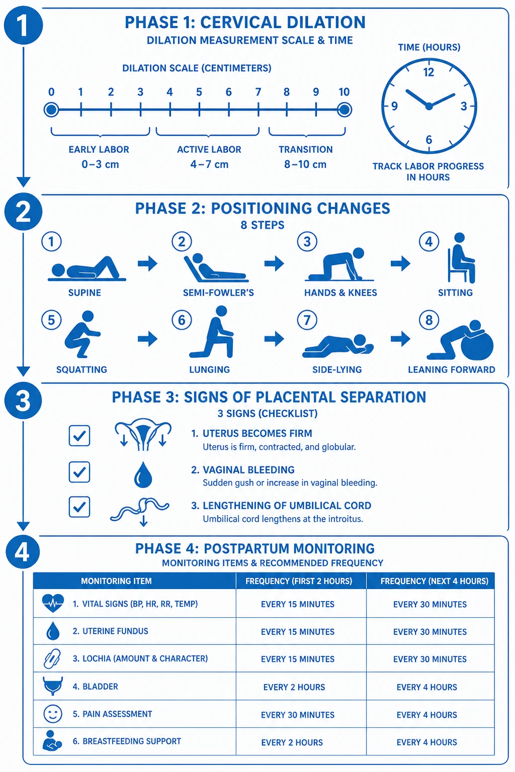
Labour can be classified along several axes that examiners reward for precision. By gestational age, it is preterm (24 to under 37 weeks), term (37 to under 42 weeks), or post-term (42 weeks and beyond). By onset, it is spontaneous (not initiated by oxytocin, prostaglandins, or amniotomy) or induced. By cervical dilation at presentation, the first stage splits into the latent phase (0 to 4 cm — slow cervical softening, effacement, and early dilatation) and the active phase (4 to 10 cm — rapid dilatation with strong regular contractions). The active phase was historically divided into the latent, active, and deceleration subphases on the Friedman labour curve, though modern data from Zhang and the WHO Labour Care Guide show that progress is more gradual than the older 1 cm/hr benchmark implies, particularly before 6 cm of dilatation.[3][4]
The third stage is also classified by management strategy: active management (prophylactic oxytocin plus controlled cord traction plus uterine massage) versus expectant (physiological) management (no prophylactic oxytocin; cord clamping delayed until pulsation ceases; placenta delivered by maternal effort). Active management is the global default for low-risk vaginal births because it reduces PPH by approximately 60%, shortens the third stage, and lowers maternal haemoglobin drop, though it slightly increases maternal blood pressure when ergometrine is added and may be associated with more retained placenta.[1][2]
Stage 1
cervical dilatation
- **Latent phase:** 0 to 4cm — effacement and softening, irregular contractions
- **Active phase:** 4 to 10cm — strong regular contractions every 2 to 3 min lasting 45 to 60 sec
- Friedman subphases: latent, active, deceleration (now reframed by Zhang data)
- Average duration: primigravida 8 to 12h, multigravida 6 to 8h
- Plot on **partogram**; alert line at 1cm/hr, action line 4h later
Stage 2
delivery of baby
- **Passive phase:** full dilatation with descent, no active pushing
- **Active phase:** mother bears down with contractions
- Cardinal movements: engagement, descent, flexion, internal rotation, extension, restitution, external rotation, expulsion
- Average: primigravida 1 to 2h, multigravida 30 to 60min
- Ritgen manoeuvre to control delivery of head; check for nuchal cord
Stage 3
placenta
- Active management (default): oxytocin 10 IU IM + CCT + uterine massage
- Expectant management: reserved for women who decline oxytocin
- Signs of separation: cord lengthens, blood gush, uterus firm and globular, uterus rises
- Usually 5 to 30min
- Inspect placenta: 2 arteries, 1 vein, all cotyledons, complete membranes
Stage 4
golden hour
- First hour postpartum — highest PPH risk
- Monitor fundus (firm, central, at umbilicus), lochia, vitals every 15min
- Skin-to-skin contact and early breastfeeding (releases oxytocin, contracts uterus)
- Perineal inspection and repair
- Confirm bladder empty
Epidemiology & Risk Factors
Globally, roughly 75 to 80% of singleton vertex deliveries at term proceed vaginally without operative intervention; the remainder require operative vaginal delivery (forceps or ventouse, approximately 10 to 15% in many high-income settings, but lower where operative skills have declined) or caesarean section. Caesarean rates vary enormously — from under 10% in low-resource settings to over 30% in parts of the United States, Brazil, China, and many private-sector Indian hospitals — driven by indication, obstetric culture, medico-legal environment, and maternal choice. The mean duration of spontaneous labour is approximately 8 to 12 hours in primigravidae and 6 to 8 hours in multigravidae, with the active phase typically lasting 4 to 6 hours in primigravidae and 2 to 4 hours in multigravidae.[3]
Risk factors for prolonged or dysfunctional labour cluster around the three Ps. Weak powers: nulliparity (longer latent and active phases), obesity, dehydration, maternal exhaustion, overuse of epidural analgesia, chorioamnionitis. Difficult passenger: fetal macrosomia (over 4,000 g or, for shoulder dystocia prediction, over 4,500 g), occiput-posterior position, deep transverse arrest, brow or face presentation, multiple gestation, polyhydramnios. Tight passage: contracted pelvis (especially android or platypelloid morphology), pelvic masses, prior pelvic trauma, short maternal stature (under 150 cm is a strong predictor in South Asian populations). Additional risk factors include induced labour (longer and more painful), advanced maternal age, and a previous prolonged labour. [1]
Risk factors for postpartum haemorrhage in the fourth stage — the most dangerous complication of normal labour — are sometimes remembered by the four Ts: Tone (uterine atony, the cause in 70% of cases; risk factors include overdistension from twins, macrosomia, polyhydramnios, prolonged labour, oxytocin use in labour), Trauma (cervical, vaginal, or perineal lacerations; uterine rupture), Tissue (retained placenta or placental fragments, abnormal placentation), and Thrombin (coagulopathy, anticoagulant therapy, pre-eclampsia with thrombocytopenia). Anaemia, common in South Asian obstetric populations, does not cause PPH but converts a 500 mL bleed into a life-threatening one.[1]
India and South Asia: Institutional delivery rates have risen sharply with the Janani Suraksha Yojana conditional cash-transfer scheme (over 90% in many states), but home deliveries by unskilled attendants persist in pockets. Anaemia in pregnancy (over 50% prevalence in many districts) lowers the threshold for symptomatic PPH. Misoprostol 600 mcg oral or sublingual is WHO-recommended for community settings where oxytocin cannot be stored cold; it is heat-stable and administable by community health workers. Skilled birth attendant coverage and partogram use remain inconsistent. The WHO Safe Childbirth Checklist has been adapted for Indian district hospitals and shown to improve adherence to essential birth practices.[11][12]
Numbers that decide a labour answer
Pathophysiology
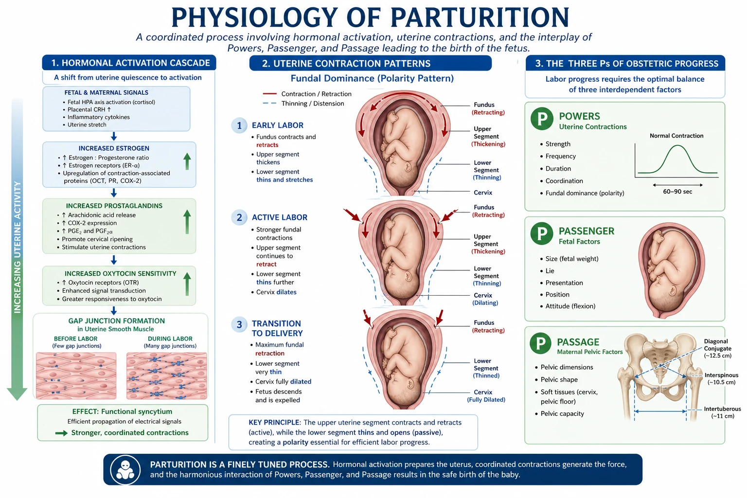
The onset of labour is the result of a coordinated endocrine and molecular switch that converts the previously quiescent uterus from a state of relaxation into a coordinated contractile organ. The first measurable event is a relative functional progesterone withdrawal: although serum progesterone does not fall in humans (unlike in sheep), the myometrium becomes less responsive to progesterone through changes in progesterone receptor isoforms. Concurrently, oestrogen rises, fetal cortisol from the maturing fetal adrenal axis increases, and prostaglandins (PGE2 and PGF2alpha) are released locally in the cervix and decidua — ripening the cervix, degrading collagen, and stimulating contractions.[7]
The molecular hallmark of the contractile switch is the appearance of myometrial gap junctions composed of connexin-43. Through most of pregnancy, the myometrium expresses a high ratio of connexin-45 to connexin-43, which produces poorly conductive gap junctions and electrical isolation of individual myocytes — keeping the uterus relaxed. Near term, this ratio inverts: connexin-43 transcription surges, gap junctions proliferate, and the myometrium becomes a functional syncytium capable of propagating coordinated action potentials. Protein kinase C and AP-1 transcription factor activation drive this switch, and pharmacological manipulation of connexin-43 is an active area of tocolysis research.[7][8]
In parallel, oxytocin receptors in the myometrium and decidua rise 100- to 200-fold toward term, sensitising the uterus to both endogenous oxytocin (released from the posterior pituitary in pulsatile fashion) and exogenous oxytocin (used for induction and augmentation). This receptor upregulation is why oxytocin is far more effective at term than in early pregnancy. As the presenting part descends and stretches the cervix and upper vagina, the Ferguson reflex is activated: afferent impulses travel via the pelvic nerves to the hypothalamus, which releases more oxytocin, which intensifies contractions, which further stretches the cervix — a positive feedback loop. This is why epidural analgesia, by blunting afferent input, can slow labour and why early amniotomy can sometimes accelerate it. [1]
Uterine contraction mechanics are defined by two key properties. Fundal dominance means each contraction originates at the fundus (the pacemaker region near the tubal ostia) and sweeps downward over 30 seconds, peaking in the mid-corpus; this arrangement pushes the fetus downward and prevents retrograde displacement of blood during a contraction. Polarity describes the differential behaviour of the two uterine segments: the upper segment contracts, retracts (muscle fibres do not relax fully back to their original length, but remain slightly shorter), and thickens — expelling the fetus and later retracting to control bleeding — while the lower segment dilates, thins, and passively accommodates the descending fetus. Retraction of the upper segment is what physically shears the placenta off the uterine wall in the third stage and what compresses the spiral arteries (the living ligatures of the uterus) after delivery to control bleeding. Failure of upper segment retraction is the basis of uterine atony and is the leading cause of PPH. [1]
The cervix itself undergoes ripening — a connective-tissue remodelling process distinct from dilatation. Hyaluronic acid content rises, collagen fibres disaggregate, and water content increases, producing a soft, compliant cervix that can be effaced (drawn up into the lower segment) and dilated. The Bishop score (0 to 13) quantifies this readiness: dilation, effacement, consistency, position, and station. A Bishop score of 8 or more predicts a favourable induction; a score of 5 or less suggests an unfavourable cervix where prostaglandin priming is needed. [1]
Clinical Presentation
The clinical onset of labour is recognised by three cardinal signs. The first is regular, painful uterine contractions of increasing frequency (every 5 to 3 minutes initially, then 2 to 3 minutes), duration (lengthening from 30 to 60 seconds), and intensity — distinguishable from the irregular, painless Braxton-Hicks contractions of late pregnancy. The second is the show — passage of a small amount of blood-stained mucus (the operculum or mucus plug that sealed the cervical canal during pregnancy) as the cervix effaces and begins to dilate. The show is also called the bloody show; fresh bleeding is not a show and warrants exclusion of placenta praevia, abruption, or vasa praevia. The third is rupture of the membranes — release of clear amniotic fluid, often as a sudden gush. ROM occurs before the onset of contractions in roughly 8 to 10% of term pregnancies (prelabour rupture of membranes, PROM at term), and during labour in the remainder. Meconium-stained liquor (green or brown) raises concern for fetal distress, particularly if thick or accompanied by CTG abnormalities; blood-stained or purulent liquor is pathological. [1]
The Friedman curve describes the sigmoidal shape of cervical dilation versus time during the first stage. The original Friedman phases were latent (slow, 0 to 3 to 4 cm), active (rapid, 3 or 4 to 8 or 9 cm), and deceleration (a brief plateau around 8 to 10 cm before full dilatation). Modern prospective data — most notably the Zhang contemporary labour curve from the Safe Labor Consortium — have refined this: dilatation is substantially slower than 1 cm/hr before 6 cm, accelerates sharply after 6 cm, and there is no consistent deceleration phase. The WHO Labour Care Guide (2020) reflects this by adopting a 4-hour threshold rather than a fixed slope, recognising that crossing a rigid action line can drive unnecessary intervention. The practical message: do not diagnose active labour, prolonged labour, or arrest of dilatation before 6 cm with confidence, and allow longer observation than older benchmarks implied.[3][4]
At the bedside, the first stage manifests as progressive cervical dilation with regular strong contractions; the second stage is heralded by full dilatation on vaginal exam, an involuntary urge to push, a bulging perineum, and finally crowning (the biparietal diameter distends the introitus and does not recede between contractions). The third stage is recognised by the four signs of separation — lengthening of the visible cord, a small gush of blood, the uterus becoming firm and globular, and the fundus rising in the abdomen. The fourth stage is asymptomatic if normal, with a firm central contracted uterus at or just below the umbilicus and moderate lochia rubra; a soft, boggy, or deviated fundus signals atony or bladder distension and is the warning sign of PPH. [1]
[1]Differential Diagnosis
The first differential is true labour versus false labour (Braxton-Hicks contractions). True labour is defined by contractions that increase in frequency, duration, and intensity, that cause progressive cervical change, and that are not relieved by sedation or hydration. False labour contractions are irregular, brief, painless or mildly uncomfortable, do not dilate the cervix, often disappear with rest or hydration, and are confined to the lower abdomen or groin rather than the fundus. The mnemonic that captures the difference is the "true labour" triad of progressive contractions, progressive dilatation, and progressive descent. [1]
A second differential is false labour versus prodromal (latent-phase) labour. Latent-phase contractions can be uncomfortable and last many hours but, unlike false labour, they produce measurable cervical change (slow dilatation and effacement). The WHO and NICE recommend admission in active labour (4 cm and above) to avoid the cascade of interventions associated with premature admission in the latent phase; women in established latent labour are best managed at home with support and reassessment. [1]
The third set of differentials concerns abnormal presentations or pathological processes mimicking labour pain. Placental abruption presents with sudden severe constant abdominal pain, often with a hard, tender, woody uterus and fetal distress — contractions are typically high-frequency but low-amplitude (couvelaire uterus). Placenta praevia presents with painless fresh bleeding, often after ROM, and is excluded by ultrasound. Uterine rupture (especially in women with a previous caesarean scar) presents with sudden cessation of contractions, loss of fetal station, severe abdominal pain, and fetal bradycardia. Chorioamnionitis presents with maternal fever, tachycardia, foul liquor, and uterine tenderness in the setting of prolonged ROM. Urinary tract infection or pyelonephritis can cause lower abdominal pain and uterine irritability but does not cause progressive dilatation. Ovarian cyst torsion or degeneration of a fibroid are less common surgical mimics. [1]
True labour
diagnosis
- Contractions: regular, increasing frequency/duration/intensity
- Cervix: progressive effacement and dilation
- Pain: across fundus, radiating to back
- Show: yes, often present
- Not relieved by sedation or rest
- Confirm with serial vaginal exams
False labour (Braxton-Hicks)
mimic
- Contractions: irregular, brief, mild
- Cervix: closed or unchanged
- Pain: lower abdomen or groin only
- Show: absent
- Relieved by rest, hydration, sedation, paracetamol
- Common in last 4 to 6 weeks
Placental abruption
emergency mimic
- Sudden constant severe abdominal pain
- Uterus: hard, tender, woody, irritable
- Bleeding: may be concealed (20%)
- Fetal distress: often present
- Risk: pre-eclampsia, trauma, cocaine
- Urgent review; emergency C-section if fetus alive
Placenta praevia
bleeding mimic
- Painless fresh bright-red bleeding after 24 weeks
- No contraction pain between bleeds
- High presenting part, abnormal lie
- Diagnosed by transabdominal ultrasound
- NO digital vaginal exam
- Deliver by planned C-section if covering os
Clinical & Bedside Assessment
The focused assessment of a labouring woman combines history, abdominal examination, vaginal examination, and continuous or intermittent fetal monitoring. The history confirms gestational age, parity, antenatal course, membrane status (intact, ruptured, time of rupture, liquor colour), onset and pattern of contractions, presence of show, and absence or presence of risk factors for labour dystocia or PPH. [1]
Abdominal examination uses the four Leopold manoeuvres. The first identifies the fetal pole in the fundus (breech feels soft and irregular; head feels hard and round and ballotts). The second locates the fetal back (smooth, curvilinear) and small parts (knees, feet) on the maternal flanks — the position of the back guides where the fetal heart is best heard. The third confirms the presenting part and whether it is engaged (the Pawlik grip): if the head is engaged, the presenting part is not movable above the pelvic brim. The fourth, with the examiner facing the mother's feet, assesses the degree of flexion of the head and the level of descent. Engagement is documented when the widest transverse diameter of the head (the biparietal diameter) has passed through the pelvic brim; in primigravidae this typically occurs 2 to 4 weeks before labour, whereas in multigravidae it may occur only in labour. [1]
Vaginal examination (sterile, with consent, and only when clinically indicated — not routine) documents five findings, remembered by some as DECS-M: Dilatation (0 to 10 cm in fingertip-widths), Effacement (from 0% — long and thick — to 100% — fully taken up), Consistency (soft, medium, or firm), Station (the level of the presenting part relative to the ischial spines, scored from -5 to +5 in centimetres, with 0 station at the spines, and +5 at the introitus), and Membrane status (intact or ruptured; liquor colour when ruptured). The examiner also identifies presentation (vertex, breech, face, brow, shoulder), position (the denominator — occiput in vertex — relative to the maternal pelvis: occipito-anterior, occipito-transverse, occipito-posterior), attitude (flexed or deflexed), and any moulding or caput succedaneum of the head. The interval between vaginal examinations should be 4 hours in active labour unless clinically indicated sooner; excessive examinations increase infection risk. [1]
Station reference card
Fetal heart rate monitoring during labour is by either intermittent auscultation (recommended for low-risk women in NICE NG190) or continuous electronic fetal monitoring with cardiotocography (CTG). Intermittent auscultation uses a Pinard stethoscope or hand-held Doppler for 60 seconds immediately after a contraction, every 15 minutes in the first stage and every 5 minutes (or after every contraction) in the second stage. Continuous CTG is indicated when there are maternal or fetal risk factors: previous caesarean, oxytocin augmentation, meconium-stained liquor, abnormal intermittent auscultation, fever, fresh bleeding, prematurity, post-term pregnancy, multiple gestation, growth-restricted fetus, or any abnormality in the fetal heart rate pattern.[13]
Investigations
The CTG is the principal intrapartum investigation. The NICE (UK) and FIGO classifications share the same conceptual framework, dividing a CTG trace into baseline rate, baseline variability, accelerations, and decelerations, and then classifying the overall trace as normal, suspicious, or pathological. The NICE NG190 framework is summarised here. [1]
Baseline fetal heart rate is the mean rate over a 10-minute window, excluding accelerations and decelerations. Normal is 110 to 160 bpm. Bradycardia (under 110 bpm, pathological if under 100) suggests fetal hypoxia, congenital heart block, or maternal beta-blocker use. Tachycardia (over 160 bpm) suggests fetal infection, maternal pyrexia, chorioamnionitis, fetal anaemia, or prematurity. [1]
Baseline variability is the band of beat-to-beat fluctuation around the baseline. Normal is 5 to 25 bpm. Reduced variability (under 5 bpm for over 30 minutes) is the single most reliable indicator of fetal hypoxia-acidosis. Sinusoidal pattern — a smooth, regular sine-wave pattern with no beat-to-beat variability — is associated with severe fetal anaemia (Rh alloimmunisation, fetomaternal haemorrhage, parvovirus infection) and is a non-reassuring sign requiring urgent action. [1]
Accelerations are abrupt rises of 15 bpm or more lasting 15 seconds or more; their presence is reassuring and reflects an intact sympathetic-autonomic nervous system. Decelerations are classified by their timing relative to contractions. Early decelerations (begin with the contraction, mirror its shape, recover by its end) are caused by head compression and are benign. Variable decelerations (variable onset, abrupt fall, often V- or U-shaped) are caused by cord compression — concerning if they are persistent, deep (under 60 bpm), prolonged (over 60 sec), show shouldering (slow return to baseline), or late return. Late decelerations (begin after the peak of the contraction, mirror its shape, recover after the contraction ends) reflect uteroplacental insufficiency and fetal hypoxia — recurrent late decelerations are the most ominous pattern. Prolonged bradycardia (under 100 bpm for over 3 minutes) is an obstetric emergency requiring immediate action. [1]
Normal CTG
reassuring
- Baseline 110 to 160 bpm
- Variability 5 to 25 bpm
- Accelerations present
- No decelerations, or only early/variable with normal characteristics
- Action: continue monitoring
Suspicious CTG
1 non-reassuring feature
- One of: baseline abnormal (100 to 109 or 161 to 180), reduced variability (under 5 for 40 to 90 min), variable decelerations with concerning features
- Action: apply conservative measures (left lateral, IV fluid, stop oxytocin, treat hypotension, paracetamol if febrile), reassess
Pathological CTG
urgent action
- Two or more non-reassuring features, or one abnormal: baseline under 100 or over 180, sinusoidal, variability under 5 for over 90 min, recurrent late decelerations, single prolonged deceleration over 3 min
- Action: escalate, conservative measures, fetal blood sampling if available, expedite delivery if persists
Conservative (intrauterine resuscitation) measures for a suspicious or pathological CTG are remembered by the mnemonic DR BRAVADO or, more practically, as a checklist: Defer oxytocin; Reposition mother to left lateral; Boost with IV fluid bolus (500 mL crystalloid); Review for cord prolapse; Atropine/anti-hypertensives reviewed; Vaginal exam to exclude cord, rapid progress, or abruption; Auscultate; Doctor review; Oxygen (now considered of limited value, but still commonly used). If the trace does not improve within 15 to 30 minutes, fetal scalp blood sampling (where available) for pH (under 7.20 = acidosis) or expedited delivery by instrumental means or caesarean is indicated.[13]
Other investigations during labour are limited. Maternal observations include pulse, blood pressure, temperature every 4 hours (or more often if febrile or hypertensive), urine output, and assessment for proteinuria (in pre-eclampsia surveillance). Bloods (group and save, full blood count) are taken on admission if not recently done; coagulation studies are added if pre-eclampsia, abruption, or chorioamnionitis is suspected. Point-of-care haemoglobin is useful in active bleeding. Ultrasound is reserved for suspected malpresentation, multiple gestation, or unexplained abnormal lie; routine ultrasound in labour is not recommended. [1]
Management — Resuscitation
Most labours are not emergencies, but several intrapartum obstetric emergencies require immediate resuscitative action while preparing for definitive intervention. These include cord prolapse, shoulder dystocia, postpartum haemorrhage, eclamptic seizure, amniotic fluid embolism, uterine rupture, and severe fetal bradycardia or prolonged deceleration. The general principles are: call for senior help, ABC for the mother (left lateral tilt to relieve aortocaval compression, high-flow oxygen, two large-bore IV cannulae, fluid resuscitation with crystalloid, bloods including group and crossmatch), position the mother appropriately, expedite delivery if the fetus is in jeopardy, and anticipate neonatal resuscitation (alert paediatric team, prepare resuscitaire). [1]
Cord prolapse — the cord descends below the presenting part after ROM, especially with a high head or transverse lie — is an obstetric emergency with fetal mortality rising steeply with delay. Management is immediate: elevate the presenting part manually off the cord, place the mother in knee-chest or steep Trendelenburg position, keep the cord moist and warm (handle it as little as possible), call for senior obstetric and anaesthetic help, and deliver by emergency caesarean section unless vaginal delivery is imminent. Cord prolapse is excluded on every vaginal examination after ROM.[13]
Shoulder dystocia — the anterior shoulder impacts behind the pubic symphysis after delivery of the head — is recognised by failure of restitution and failure of shoulders to deliver with routine traction. Time-critical (the fetus cannot breathe), it demands the sequence: call for help (senior midwife, obstetrician, paediatrician, anaesthetist); McRoberts manoeuvre (hyperflex maternal hips onto abdomen, straightening the sacrum and rotating it anteriorly); suprapubic pressure (the Rubin II manoeuvre — lateroposterior pressure behind the anterior shoulder to adduct it); episiotomy (only if internal manoeuvres are needed); internal rotational manoeuvres (Rubin I, Wood's screw, reverse Wood's screw); deliver the posterior arm first (deliver the posterior shoulder by sweeping the arm across the chest); and lastly, if all fail, the salvage manoeuvres (Gaskin manoeuvre — all-fours position; symphysiotomy; Zavanelli manoeuvre — replace the head and caesarean). Documentation of time and sequence is medico-legally critical. [1]
Postpartum haemorrhage is discussed in its own topic; the key intrapartum principle is prevention by active management of the third stage, which reduces PPH by 60%. Resuscitation during PPH follows the four Ts: address tone (uterine massage, oxytocin 10 IU IM then 40 IU in 500 mL saline over 4 hours, ergometrine 0.5 mg IM, carboprost 15-methyl-PGF2alpha 250 mcg IM every 15 min up to 8 doses, misoprostol 800 mcg sublingual), trauma (inspect cervix, vagina, perineum; repair under anaesthesia), tissue (explore uterus, remove retained placenta or fragments), and thrombin (activate massive transfusion protocol, cryoprecipitate, fresh frozen plasma, platelets).[1]
Management — Definitive & Stepwise
Stage 1 management
The goals of first-stage management are to support the labouring woman, monitor progress to detect dystocia early, monitor the fetus to detect distress early, and avoid unnecessary intervention. The cornerstones are: one-to-one midwifery support (Hodnett's Cochrane review shows continuous intrapartum support shortens labour, reduces caesarean and instrumental delivery, and improves maternal satisfaction); mobilisation and upright positions in the first stage; oral sips of water and IV fluids only if clinically indicated; bladder care (avoid distension, which can impede descent); intermittent auscultation for low-risk women and continuous CTG when risk factors are present; vaginal examination every 4 hours (or sooner if clinically indicated) with findings plotted on the partogram; maternal observations (pulse hourly, BP and temperature 4-hourly); and supportive analgesia as requested.[9][13]
The partogram — the WHO-modified graphical record of labour — plots cervical dilatation against time, with an alert line (drawn from the point of admission in active phase at the historic 1 cm/hr slope) and an action line (parallel and 2 to 4 hours to the right). Crossing the alert line prompts review of powers, passenger, and passage; crossing the action line mandates medical review, amniotomy if membranes are intact, and oxytocin augmentation if contractions are inadequate. The WHO Labour Care Guide (2020) updates this framework, replacing the rigid 1 cm/hr slope with thresholds based on individualised progress and the recognition that dilatation may be slower before 6 cm without pathology.[4]
Amniotomy (artificial rupture of membranes) is offered if labour is not progressing and membranes are intact; it shortens labour by approximately 1 hour, increases contraction intensity, and may slightly increase the risk of CTG abnormalities and chorioamnionitis if labour is prolonged. Oxytocin augmentation (per local protocol, e.g. 30 IU in 500 mL normal saline starting at 1 to 2 mU/min, titrated every 30 minutes to 4 to 5 contractions in 10 minutes, max around 20 to 32 mU/min) is offered when contractions remain inadequate after amniotomy; it requires continuous CTG and is contraindicated in previous caesarean with a trial of scar (uterine rupture risk), grand multiparity, and obstructed labour.[13]
Stage 2 management
The second stage is diagnosed at full cervical dilatation and ends with delivery of the baby. NICE NG190 distinguishes the passive second stage (full dilatation but no involuntary urge to push, particularly with epidural) from the active second stage (mother actively pushing). Allowing 1 to 2 hours of passive descent after full dilatation, particularly with epidural, reduces instrumental delivery without increasing neonatal morbidity. Active pushing is by Valsalva — sustained breath-holding for 6 to 8 seconds with each contraction, repeated two to three times per contraction. Excessive maternal exhaustion or prolonged active second stage raises the risk of maternal perineal trauma, PPH, neonatal acidosis, and shoulder dystocia. NICE recommends review for instrumental delivery or caesarean if the active second stage exceeds 1 hour in a multigravida or 2 hours in a primigravida (longer with epidural).[13]
Delivery of the head is controlled: Ritgen's manoeuvre — the operator's dominant hand covered with a sterile towel exerts forward pressure on the occiput through the perineum while the other hand controls the speed of crowning between contractions, allowing slow delivery of the head and reducing perineal trauma. The nuchal cord is checked after delivery of the head; if loose, it is looped over the head, and if tight, it is clamped and cut between two clamps. Episiotomy is not routine — NICE recommends it only when there is a clinical indication (fetal distress requiring expedited delivery, instrumental delivery, shoulder dystocia, suspected imminent severe perineal tear); when performed, a mediolateral cut at 60 degrees from the fourchette is preferred because midline episiotomies carry a higher risk of extension into the anal sphincter. [1]
Delivery of the shoulders follows restitution and external rotation: gentle downward traction delivers the anterior shoulder under the pubic symphysis, then upward traction delivers the posterior shoulder. The rest of the body follows. Cord clamping is delayed for 1 to 3 minutes in vigorous term neonates, allowing placental transfusion of up to 80 mL of blood and improving iron stores through the first 6 months; cord milking is an alternative where immediate clamping is needed ( McDonald Cochrane 2013).[5]
Stage 3 management — active vs expectant
The third stage runs from delivery of the baby to delivery of the placenta and membranes. Two strategies are recognised, both evidence-based but suited to different clinical situations. [1]
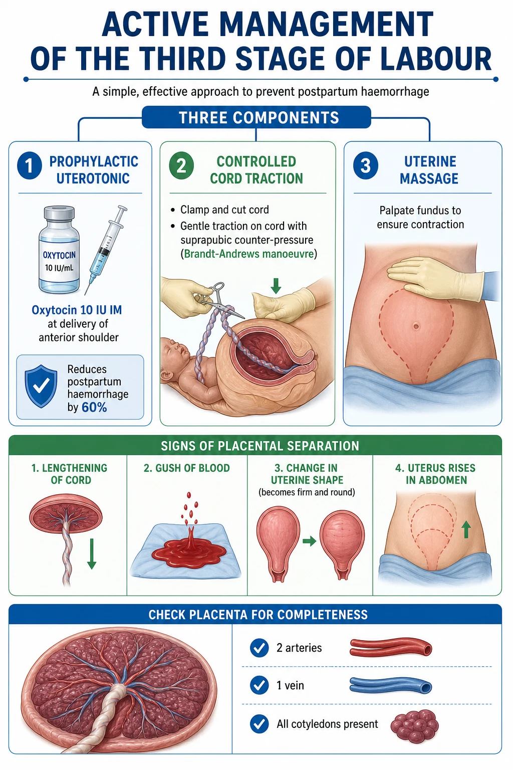
Active management (the global default for low-risk vaginal births) has three components and reduces the risk of PPH (over 500 mL) by approximately 60% and of severe PPH (over 1000 mL) by roughly 70%, shortens the third stage, and lowers postpartum maternal haemoglobin drop.[1][2]
- Prophylactic uterotonic at delivery of the anterior shoulder. The first-line agent is oxytocin 10 IU IM (onset 2 to 4 min, cheap, heat-stable, no contraindications except rare hypersensitivity). In the UK, Syntometrine (oxytocin 5 IU plus ergometrine 500 mcg IM) is commonly used; it is slightly more effective than oxytocin alone but causes vomiting and hypertension and is contraindicated in pre-eclampsia and hypertension (use oxytocin alone). Carbetocin 100 mcg IM (a long-acting oxytocin analogue) is an option where available and is heat-stable in the new formulation. Misoprostol 600 to 800 mcg oral, sublingual, or rectal is the WHO-recommended alternative in low-resource settings or when an injectable is not feasible.
- Controlled cord traction (CCT, Brandt-Andrews manoeuvre). Once signs of separation are present, clamp and cut the cord. The operator places one hand suprapubically to guard the uterus and provide counter-traction (preventing uterine inversion), and applies gentle downward and then upward traction on the cord with the other hand while the mother bears down gently. Traction is never forceful — uterine inversion is a rare but catastrophic complication of over-vigorous CCT.
- Uterine massage. Immediately after delivery, massage the fundus through the abdominal wall to ensure it is firmly contracted; continue every 15 minutes for the first hour. [1]
Expectant (physiological) management omits prophylactic oxytocin, defers cord clamping until pulsation ceases, and delivers the placenta by maternal effort with gravity and upright posture. It is associated with a higher rate of PPH and a longer third stage, but some women choose it for low-risk vaginal births and it may produce marginally higher neonatal iron stores with prolonged cord clamping. It is contraindicated in women at increased risk of PPH — previous PPH, anaemia, prolonged labour, multiple gestation, macrosomia, polyhydramnios, or any bleeding disorder. [1]
Signs of placental separation appear within 5 to 30 minutes of delivery and comprise four classic features: lengthening of the cord (the visible portion outside the vagina increases as the placenta descends); a small gush of blood from the vagina (separation bleeding); the uterus becomes firm and globular (was discoid and boggy); and the uterus rises in the abdomen (the placenta has descended into the lower segment and vagina, pushing the fundus up). A third stage longer than 30 minutes (60 minutes with expectant management) is a retained placenta and warrants manual removal in theatre with analgesia or anaesthesia and antibiotic cover. [1]
After delivery, inspect the placenta: confirm two arteries and one vein in the cord (a single umbilical artery, in 0.5 to 1% of pregnancies, is associated with renal and chromosomal anomalies), confirm all cotyledons are present, and confirm the membranes are complete. Retained cotyledons or membranes cause secondary PPH and infection. The cervix, vagina, and perineum are then inspected and perineal trauma repaired (see Complications). [1]
Stage 4 management — the golden hour
The fourth stage is the first hour postpartum, when most primary PPH occurs. Every woman is observed with: fundus assessed every 15 minutes (must be firm, central, at or just below the umbilicus; a soft boggy fundus indicates atony, a deviated fundus indicates bladder distension); lochia assessed (rubra, moderate; excessive bleeding or large clots are abnormal); vital signs (pulse, BP, respiratory rate, temperature every 15 min in the first hour, then hourly); perineum and episiotomy site inspected for haematoma; and bladder confirmed empty (catheterise if unable to void). Early skin-to-skin contact and initiation of breastfeeding within the first hour release endogenous oxytocin, which enhances uterine contraction and reduces PPH risk; this is the basis of the WHO-UNICEF Baby-Friendly Hospital Initiative. [1]
The golden-hour monitoring bundle
Every 15 min x4: pulse, BP, RR, temperature; fundus (firm, central, at umbilicus); lochia (rubra, moderate); perineum (no haematoma)
Confirm bladder empty — catheterise if unable to void (a distended bladder prevents retraction and invites PPH)
Skin-to-skin contact within 5 min; initiate breastfeeding within 1 hour — endogenous oxytocin contracts the uterus
Inspect and repair any perineal or vaginal tears before transfer
Document estimated blood loss (weigh swabs — visual estimation underestimates by 30 to 50%)
Confirm placenta complete, membranes complete, two arteries plus one vein
Before discharge from labour ward (typically 1 to 2 h postpartum): woman is haemodynamically stable, uterus firm, lochia moderate, voiding, pain controlled, bonding established
Operative delivery
When vaginal delivery cannot be safely accomplished by maternal effort alone, operative vaginal delivery (forceps or ventouse) or caesarean section is indicated. Operative vaginal delivery is appropriate only when: the head is engaged (station 0 or below); the cervix is fully dilated; the membranes are ruptured; the position is known; the pelvis is adequate; the bladder is empty; analgesia is adequate; there is a clear indication (prolonged second stage, fetal distress, maternal exhaustion, maternal medical indication to avoid Valsalva such as severe cardiac disease or uncontrolled pre-eclampsia); and consent is obtained. Trial of instrumental delivery in theatre is recommended when there is concern that delivery may fail and conversion to caesarean may be required.[13]
Ventouse (vacuum extraction) applies a soft or rigid cup to the fetal scalp, generating negative pressure that allows traction. It requires less anaesthesia (often just pudendal or perineal infiltration), is associated with less maternal perineal trauma than forceps, but carries higher risks of cephalohaematoma, scalp laceration, retinal haemorrhage, and neonatal jaundice. It is contraindicated below 34 weeks gestation (immature scalp) and relatively contraindicated at 34 to 36 weeks. The vacuum should be applied for no more than three contractions and three cup detachments before abandoning for forceps or caesarean. [1]
Forceps come in two broad families: outlet/low-cavity forceps (Wrigley's, Neville-Barnes) for delivery when the head is visible at the introitus without receding, and mid-cavity forceps (Haig-Ferguson, Kielland's for rotational delivery) for higher stations. Forceps require full analgesia (epidural, spinal, or pudendal block), are associated with more maternal perineal and anal sphincter trauma than ventouse, but produce less fetal scalp trauma. Indications, contraindications, and prerequisites are the same as for ventouse, with the additional requirement that position must be confirmed and rotational delivery performed only by an experienced operator. [1]
Lower segment caesarean section (LSCS) is performed when vaginal delivery is contraindicated or has failed, or when fetal or maternal condition demands it. Indications include failure to progress in labour, abnormal presentation (transverse lie, persistent breech if planned vaginal delivery not chosen), fetal distress not amenable to operative vaginal delivery, cord prolapse, placenta praevia, prior classical caesarean, and maternal request in selected cases. The technique involves a transverse lower abdominal skin incision (Pfannenstiel), transverse lower uterine segment incision, delivery of the head, delivery of the body, oxytocin 5 IU slow IV, delivery of the placenta by CCT, two-layer uterine closure, and peritoneal and abdominal wall closure. Emergency LSCS is classified by urgency: category 1 (immediate threat to life of woman or fetus, decision-to-delivery interval under 30 min), category 2 (maternal or fetal compromise not immediately life-threatening, under 75 min), category 3 (needs early delivery but no maternal or fetal compromise), category 4 (elective, at a scheduled time). [1]
Neonatal assessment — APGAR score
The APGAR score, introduced by Virginia Apgar in 1953, is the universal neonatal assessment at 1, 5, and (if abnormal) 10 minutes of life. It is the sum of five components, each scored 0 to 2, giving a total of 0 to 10.[6]
APGAR score components
APGAR
skin colour: blue or pale (0), acrocyanosis (1), pink all over (2)
heart rate: absent (0), under 100 bpm (1), over 100 bpm (2)
reflex irritability to suction: none (0), grimace (1), cry or cough (2)
muscle tone: limp (0), some flexion (1), active motion (2)
breathing: absent (0), slow, irregular, or weak cry (1), strong cry (2)
Interpretation: a score of 7 to 10 is reassuring, 4 to 6 is moderately depressed (the baby needs stimulation, oxygen, and possibly bag-mask ventilation), and 0 to 3 is severely depressed (immediate resuscitation per Neonatal Resuscitation Programme: warmth, position, clear airway if meconium or obstruction, dry, stimulate, positive-pressure ventilation at 40 to 60 breaths per minute with air for term infants, escalating to oxygen and chest compressions at a 3:1 ratio if heart rate remains under 60). A persistently low APGAR at 5 and 10 minutes is associated with neonatal encephalopathy, hypoxic-ischaemic injury, and cerebral palsy — though the APGAR alone does not diagnose asphyxia (other causes include prematurity, maternal sedation, congenital anomaly, sepsis).[6]
Specific Subtypes & Scenarios
Active versus expectant management of the third stage
The Bristol third stage trial (Prendiville 1988) randomised over 1,600 women to active versus expectant (physiological) management and established the 60% reduction in PPH with active management; subsequent meta-analyses have refined but not overturned this conclusion.[1] Active management is the default for women at increased risk of PPH and is offered routinely to all women; expectant management remains an option for women at low risk who request it. Practical points: omit ergometrine in hypertension and pre-eclampsia (use oxytocin alone); misoprostol is a useful alternative in community and low-resource settings; uterine massage after delivery reinforces the uterotonic effect.[2]
Prolonged and obstructed labour
Prolonged labour is defined by crossing the action line on the partogram or, in modern WHO Labour Care Guide terms, by dilatation that fails to meet the 4-hour threshold for the current cervical dilation in the active phase.[4] Causes follow the three Ps: prolonged latent phase (often a false start; managed with rest, hydration, and reassurance), prolonged active phase (review powers, passenger, passage; amniotomy then oxytocin if inadequate contractions; reassess in 4 hours), arrest of dilatation (no change for 4 hours despite adequate contractions — likely cephalopelvic disproportion; caesarean), prolonged second stage (active pushing for over 1 h multigravida or 2 h primigravida — instrumental delivery or caesarean), and deep transverse arrest (head fails to rotate at the pelvic floor — rotational forceps, manual rotation, or caesarean).
Obstructed labour — the absolute failure of the fetus to descend despite strong uterine contractions, due to mechanical disproportion or malpresentation — is a leading cause of maternal death in low-resource settings and of obstetric fistula in sub-Saharan Africa and South Asia. The clinical picture is of a bandl's ring (a pathological retraction ring between upper and lower segments, the sign of imminent uterine rupture), a moulded, caput-succedaneum-covered head at the pelvic floor, a thinned-out lower segment, maternal distress (tachycardia, dehydration, fever), and fetal distress. Management is immediate caesarean (never oxytocin, which will rupture the uterus), antibiotics, fluid resuscitation, and a high index of suspicion for bladder and ureteric injury. [1]
Trial of labour after caesarean (TOLAC / VBAC)
A woman with one previous lower-segment caesarean and a low transverse scar may be offered a planned vaginal birth after caesarean (VBAC) if she has no other contraindication. Success rates are 70 to 75% in selected women. The principal risk is uterine rupture (0.5% with one previous LSCS, higher with two or more or a previous classical scar), which presents with sudden cessation of contractions, severe abdominal pain, loss of fetal station, vaginal bleeding, and fetal bradycardia. Continuous CTG, IV access, crossmatched blood, and immediate theatre availability are mandatory. A previous classical or T-shaped uterine scar is an absolute contraindication to TOLAC.[13]
Perineal trauma
Perineal tears are graded by depth. First degree involves the skin only. Second degree involves the perineal muscles but not the anal sphincter (the typical episiotomy extension). Third degree involves the anal sphincter complex and is subdivided into 3a (under 50% of external anal sphincter thickness), 3b (over 50% of EAS), 3c (involving internal anal sphincter). Fourth degree involves the anorectal mucosa (the rectal epithelium). Repair: first-degree tears need no suture if small and not bleeding; second-degree tears are repaired in layers with absorbable suture (Vicryl Rapide); third- and fourth-degree tears require repair in theatre by a trained clinician under regional or general anaesthesia, with the EAS overlapped or approximated with monofilament absorbable suture (PDS or Monocryl), IAS repaired separately, antibiotics (co-amoxiclav or cefuroxime plus metronidazole), stool softeners (lactulose) for 7 to 10 days, and physiotherapy follow-up. Long-term sequelae include faecal incontinence, dyspareunia, and pelvic floor dysfunction. [1]
Bristol third stage trial (1988)
Key finding
Active management of the third stage (oxytocin + early cord clamping + CCT) reduced PPH over 500 mL by roughly 60% versus expectant (physiological) management. Established active management as the global default for low-risk vaginal births.
Complications & Pitfalls
The complications of normal labour are largely the complications of its abnormal forms and its iatrogenic extensions. Maternal complications include prolonged and obstructed labour (exhaustion, dehydration, chorioamnionitis, obstetric fistula, uterine rupture); perineal and anal sphincter trauma (short-term pain and infection, long-term incontinence and dyspareunia); postpartum haemorrhage (the leading direct cause of maternal death globally); perineal haematoma; deep vein thrombosis and pulmonary embolism (pregnancy is a prothrombotic state — thromboprophylaxis with LMWH for 10 days postpartum after caesarean); puerperal sepsis (endometritis, wound infection); anaemia exacerbation; postnatal depression and post-traumatic stress disorder after a difficult birth; and urinary retention. Fetal and neonatal complications include intrapartum hypoxia and acidosis (with risk of hypoxic-ischaemic encephalopathy and cerebral palsy), meconium aspiration syndrome, birth trauma (cephalohaematoma, clavicular fracture, brachial plexus injury from shoulder dystocia, skull fracture from forceps), neonatal jaundice, prematurity-related complications if iatrogenic preterm delivery, and stillbirth. [1]
Classic pitfalls in the management of normal labour include: admitting women in latent labour (drives unnecessary intervention and caesarean); diagnosing active phase arrest before 6 cm (Zhang data show slower progress before 6 cm is normal);[3] overusing continuous CTG in low-risk women (raises caesarean rate without improving neonatal outcomes); underestimating blood loss (visual estimation underestimates by 30 to 50% — weigh swabs); performing routine episiotomy (no benefit, more trauma); using ergometrine in hypertensive women (precipitates hypertensive crisis); forgetting the fourth stage (most PPH is missed in the first hour); failing to inspect the placenta and perineum (retained tissue and unrepaired tears cause secondary PPH and infection); omitting uterine massage (an essential third-stage component); delaying tocolytic failure to recognise uterine inversion (rare but catastrophic, from over-vigorous CCT); underestimating shoulder dystocia (call for help at the moment the head retracts, before any traction).
Exam application bank (NEET-PG / INICET)
One-line answer
Normal labour = physiological expulsion of fetus, placenta, and membranes after 24 weeks gestation, via regular painful uterine contractions causing progressive cervical effacement, dilatation, descent, and expulsion. Four stages: 1st (latent 0 to 4cm + active 4 to 10cm), 2nd (full dilatation to baby), 3rd (placenta, active vs expectant management), 4th (golden hour). Eight cardinal movements: engagement, descent, flexion, internal rotation, extension, restitution, external rotation, expulsion. Three Ps: Powers, Passenger, Passage. Molecular switch: myometrial connexin-43 gap junctions plus oxytocin receptor upregulation. Active management of 3rd stage (oxytocin 10 IU IM + CCT + uterine massage) reduces PPH by 60%. APGAR at 1, 5, and 10 min. [1]
Worked stems (answer without another resource)
Stem 1 — Classic presentation. Map symptoms to mechanism; name the first investigation and first treatment step with dose/route if drug therapy is standard. [1]
Stem 2 — Unstable / complicated. List red flags that force immediate resuscitation, theatre, ICU, antidote, or reperfusion — and what you do in the first 15 minutes. [1]
Stem 3 — Atypical group. Elderly, pregnancy, child, or immunocompromised: how presentation and thresholds change. [1]
Stem 4 — Differential trap. Name the three closest mimics and one discriminator for each. [1]
Stem 5 — Disposition. Who goes home with safety-netting, who is admitted, who needs HDU/ICU/theatre, and what follow-up is mandatory. [1]
Rapid viva checklist
- Definition + classification
- Pathophysiology chain
- Bedside signs / criteria
- Score with exact components (if any)
- Emergency bundle
- Definitive therapy with doses
- Complications of disease and of treatment
- Special populations
- Guideline/trial name if classic
- Three exam traps
Coverage self-check
If you cannot answer any stem above from this page alone, re-read the matching section — the page is intended to be self-sufficient for final-prof and NEET-PG/INICET questions on Normal Labour and Delivery.
Prognosis & Disposition
For an uncomplicated vaginal delivery at term, maternal mortality is extremely low in well-resourced settings (under 5 per 100,000 deliveries) and maternal morbidity is dominated by perineal trauma, urinary incontinence, and postnatal depression. Neonatal mortality is dominated by congenital anomaly, prematurity, and intrapartum hypoxia. The median length of stay is 6 to 24 hours for an uncomplicated vaginal delivery, 24 to 48 hours after instrumental delivery, and 2 to 4 days after caesarean. Discharge criteria are: maternal stability (haemodynamics, no sepsis, lochia moderate, uterus firm, voiding, mobile, pain controlled); neonatal stability (normal feeding, normal colour and tone, passed meconium and urine, no jaundice in first 24 h, APGAR recovered, normal examination); social safety (home support, transport, access to emergency care). The midwife or community health worker visits within 24 to 48 hours of discharge, and the 6-week postnatal check reviews healing, contraception, mood (Edinburgh Postnatal Depression Scale), and any complications.[12]
The disposition of a labouring woman depends on the stage and complications. Antenatal triage distinguishes latent labour (discharge home with advice) from active labour (admit to labour ward). Intrapartum escalation to obstetric-led care is triggered by any risk factor, abnormal progress, or CTG abnormality. Postpartum escalation is triggered by bleeding, fever, hypertension, or neonatal concerns. The safety-net is clear advice on warning signs (heavy bleeding, severe headache, epigastric pain, fever, calf pain, dyspnoea, neonatal poor feeding or jaundice) and 24-hour access to a midwife and emergency obstetric service.[13]
Special Populations
Women with previous caesarean are counselled antenatally between planned VBAC and planned repeat caesarean; the choice is individualised. Women with pre-eclampsia require continuous CTG, blood pressure control (labetalol, hydralazine, nifedipine), magnesium sulphate for severe features (seizure prophylaxis: 4 g IV loading then 1 g/hr infusion, monitoring for toxicity — loss of deep tendon reflexes, respiratory depression), and active management of the third stage with oxytocin alone (no ergometrine). Grand multiparae (parity 5 or more) have rapid labours but elevated risks of uterine rupture, postpartum atony, and PPH; they require active management of the third stage and a high index of suspicion for rapid progress. Women with obesity (BMI over 35) have longer labours, higher caesarean rates, and higher PPH and anaesthetic complication risks; early epidural and senior obstetric involvement are recommended. Teenage mothers have higher preterm birth rates and pre-eclampsia but generally uncomplicated vaginal deliveries if well supported. Women with diabetes in pregnancy have higher rates of fetal macrosomia (with shoulder dystocia risk) and are often induced at 38 to 39 weeks with a planned delivery plan. Women on anticoagulants (LMWH for thromboprophylaxis) should have their dose withheld at the onset of labour or 12 hours before a planned delivery to permit neuraxial anaesthesia; neuraxial block must wait 12 hours after the last prophylactic LMWH dose and 24 hours after a treatment dose. [1]
Evidence, Guidelines & Regional Differences
The foundations of modern intrapartum care are: WHO recommendations on intrapartum care for a positive childbirth experience (2018) — adopting the recognition that labour is slower before 6 cm, supporting respectful maternity care, and replacing the rigid partogram with the WHO Labour Care Guide (2020);[4][12] NICE NG190 (Intrapartum care, 2023, replacing CG190) — the UK standard, recommending midwife-led care for low-risk women, intermittent auscultation, and conservative thresholds for intervention;[13] RCOG Green-top guidelines (operative vaginal delivery, shoulder dystocia, PPH); ACOG Practice Bulletins (US); FIGO guidelines (international consensus on third-stage management, misoprostol use, operative delivery); and the WHO Safe Childbirth Checklist, a 29-item checklist that has been shown to improve adherence to essential birth practices in low-resource settings.[11]
Landmark trials and systematic reviews include the Bristol third stage trial (1988) establishing active management;[1] the Hodnett Cochrane review of continuous labour support (2012, updated) showing that one-to-one support shortens labour and reduces operative delivery;[9] the McDonald Cochrane review of delayed cord clamping (2013) showing improved neonatal iron stores without harm;[5] the Zhang contemporary labour curve (2010) redefining normal progress;[3] the Connexin-43 work by Geimonen, Sheldon and others defining the molecular basis of the myometrial contractile switch;[7][8] and the WHO Labour Care Guide development paper (2021).[4]
Regional differences are important. WHO and NICE favour 4 cm as the active-phase threshold and emphasise respectful, midwifery-led care. ACOG (US) historically used 6 cm as the active-phase threshold, aligning with Zhang data, and has more permissive thresholds for the second stage. Indian guidelines (ICMR, MoHFW) align with WHO recommendations and emphasise institutional delivery, skilled birth attendance, and active management with oxytocin or misoprostol in community settings. Operative vaginal delivery rates are highest in the UK and parts of Europe (forceps remain common), declining in the US (vacuum predominates, forceps rarely used), and low in many low-resource settings where caesarean has displaced instrumental delivery. Pain relief availability varies enormously: epidural is near-universal on request in high-income settings, scarce in low-resource ones; Entonox is common in UK midwifery-led units; pethidine, despite Cochrane evidence of limited analgesic efficacy and neonatal sedation, remains widely used in the NHS and in India.[10][13]
Exam Pearls & High-Yield Minutiae
Eight cardinal movements
EVERY DOC FRI
BPD passes through pelvic brim
head descends through pelvis — gradual, continues throughout
chin to chest — suboccipitobregmatic diameter presents
occiput rotates from transverse to anterior under symphysis
head extends around pubic symphysis, face sweeps perineum
head turns 45 degrees to undo the twist from internal rotation
shoulders rotate (anterior under symphysis), head turns further 45 degrees
rest of body delivered
WHO Safe Childbirth Checklist (29 items) covers: at admission (partograph started, antibiotics if needed, hypertension checked, labour progression reviewed); soon after birth (skin-to-skin, breastfeeding within 1 h, oxytocin given); before discharge (uterus firm, lochia moderate, bladder empty, baby feeding, immunisation started). Adapted and validated across many countries including India and Cameroon.[11]
Self-test: A primigravida at 39 weeks is in active labour at 5cm, has progressed to 7cm in 4 hours. Is this normal progress?
Yes. Zhang data show that progress of 0.5 cm/hr in the active phase before 6 cm is normal; the WHO Labour Care Guide (2020) uses a 4-hour threshold, not a rigid 1 cm/hr slope. The action line is crossed only if dilatation fails to progress by at least 1 cm over 4 hours with adequate contractions after 6 cm. Do NOT diagnose active phase arrest before 6 cm. Continue to support, monitor on the partogram, and reassess in 4 hours.[3][4]
Self-test: Name the eight cardinal movements in order.
Engagement, descent, flexion, internal rotation, extension, restitution, external rotation, expulsion. Mnemonic: EVERY DOC FRI (Engagement, Descent, Flexion, Internal rotation, Extension, Restitution, External rotation, delivery). The suboccipitobregmatic diameter (9.5 cm) presents in a well-flexed vertex; failure of flexion presents the larger occipitofrontal (11.5 cm) or mentovertical (13.5 cm) diameter and is a cause of prolonged or obstructed labour.
Self-test: A CTG shows a baseline of 145 bpm, variability 10 bpm, accelerations present, and recurrent decelerations beginning 20 seconds after the peak of each contraction and recovering after the contraction ends. What type are they and what do they signify?
Late decelerations, signifying uteroplacental insufficiency and fetal hypoxia. Recurrent late decelerations are the most ominous deceleration pattern and, with reduced variability or in a trace with two non-reassuring features, classify the trace as pathological. Apply conservative measures (left lateral, IV fluid, defer oxytocin, treat hypotension and fever) and escalate for fetal blood sampling (pH under 7.20 is acidotic) or expedited delivery if the trace does not improve within 15 to 30 minutes.[13]
References
- [1]Prendiville WJ, Harding JE, Elbourne DR, Stirrat GM. The Bristol third stage trial: active versus physiological management of third stage of labour BMJ, 1988.PMID 3144366
- [2]Prendiville WJ, Harding JE, Elbourne DR, Stirrat GM. The effects of routine oxytocic administration in the management of the third stage of labour: an overview of the evidence from controlled trials Br J Obstet Gynaecol, 1988.PMID 3277663
- [3]Zhang J, Landy HJ, Branch DW, et al. Contemporary patterns of spontaneous labor with normal neonatal outcomes Obstet Gynecol, 2010.PMID 21099592
- [4]The development of the WHO Labour Care Guide: an international survey of maternity care providers Reprod Health, 2021.PMID 33752712
- [5]McDonald SJ, Middleton P, Dowswell T, Morris PS. Effect of timing of umbilical cord clamping of term infants on maternal and neonatal outcomes Cochrane Database Syst Rev, 2013.PMID 23843134
- [6]Apgar V. A proposal for a new method of evaluation of the newborn infant Curr Res Anesth Analg, 1953.PMID 13083014
- [7]Geimonen E, Jiang W, Chou K, et al. Activation of protein kinase C in human uterine smooth muscle induces connexin-43 gene transcription through an AP-1 site in the promoter sequence J Biol Chem, 1996.PMID 8798588
- [8]Alterations in gap junction connexin43/connexin45 ratio mediate a transition from quiescence to excitation in a mathematical model of the myometrium J R Soc Interface, 2014.PMID 25401181
- [9]Hodnett ED, Gates S, Hofmeyr GJ, Sakala C, Weston J. Continuous support for women during childbirth Cochrane Database Syst Rev, 2012.PMID 23076901
- [10]Kinugasa M, Hashimoto K, Nakanishi H, et al. Safety and efficacy of a combination of pethidine and levallorphan for pain relief during labor: An observational study J Obstet Gynaecol Res, 2019.PMID 30362203
- [11]Dohbit JS, Foumane P, Tochie JN, et al. The increasing use of the WHO Safe Childbirth Checklist: lessons learned at the Yaoundé Gynaeco-Obstetric and Paediatric Hospital, Cameroon BMC Pregnancy Childbirth, 2021.PMID 34238244
- [12]Oladapo OT, Tunçalp Ö, Bonet M, Lawrie TA, Portela A, Downe S, Gülmezoglu AM. WHO model of intrapartum care for a positive childbirth experience: transforming care of women and babies for improved health and wellbeing BJOG, 2018.PMID 29637727
- [13]National Institute for Health and Care Excellence. Fetal monitoring in labour: summary and update of NICE guidance BMJ, 2022.PMID 36526275