Obstetrics & Gynaecology · Obstetrics & Gynaecology
Ovarian Cancer
Also known as Ovarian carcinoma · Epithelial ovarian cancer · High-grade serous carcinoma · Fallopian tube cancer · Primary peritoneal cancer · Krukenberg tumour · BRCA-associated ovarian cancer
Ovarian cancer is the leading cause of gynaecological cancer death in the UK and the sixth most common cancer in women (lifetime risk about 2 per cent, 1 in 50). The dominant subtype, high-grade serous carcinoma, arises from the fimbrial end of the fallopian tube (serous tubal intraepithelial carcinoma, STIC), not the ovarian surface — a paradigm shift that drives risk-reducing salpingo-oophorectomy in BRCA1/2 carriers. The classical presentation is late and non-specific — bloating, distension, early satiety — so most women are Stage III/IV at diagnosis. CA-125 plus transvaginal ultrasound triage via IOTA simple rules and the Risk of Malignancy Index (RMI), with RMI over 250 mandating specialist referral. FIGO 2014 staging is surgical. Standard care is surgical staging and maximal cytoreduction (TAH + BSO + omentectomy + peritoneal biopsies + lymphadenectomy) plus carboplatin and paclitaxel for six 3-weekly cycles. Maintenance PARP inhibitors (olaparib, niraparib) and bevacizumab have transformed first-line therapy. Stage I survival is 90 per cent; Stage IV under 15 per cent. Population screening does not work (UKCTOCS, PLCO).
On this page & tools
Your progress
Saved locally on this device.
Exam tags
Red flags
Overview & Definition
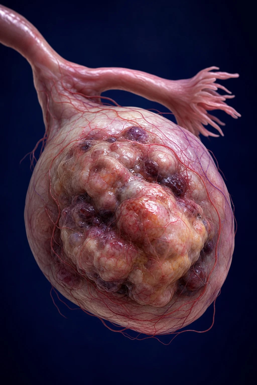
Ovarian cancer is a malignant tumour arising from the ovaries, fallopian tubes, or peritoneal surface, and is the leading cause of death from gynaecological malignancy in the United Kingdom and most high-income countries. It is the sixth most common cancer in women in the UK (after breast, lung, bowel, melanoma and uterine), with a lifetime risk of about 2 per cent (1 in 50 to 1 in 70), and an estimated 7,500 new diagnoses and 4,000 deaths per year in the UK and over 313,000 new cases and 207,000 deaths worldwide each year.[1]
The defining clinical feature — and the source of its grim reputation as the "silent killer" — is late presentation. Because the ovaries lie deep in the pelvis and because the peritoneal cavity can absorb a great deal of tumour before symptoms appear, over 70 per cent of women present at FIGO Stage III or IV, by which time tumour has disseminated across the peritoneum and prognosis is poor. This single fact — late stage at diagnosis — explains why, despite real advances in surgery and chemotherapy, the overall five-year survival has only crept from around 35 per cent to 46 per cent over the past three decades, in stark contrast to cervical and endometrial cancer.[1]
The dominant subtype is high-grade serous carcinoma, which accounts for about 70 per cent of all epithelial ovarian cancers and arises not from the ovarian surface epithelium (as taught for decades) but from the fimbrial end of the fallopian tube — a paradigm shift that began with the discovery of serous tubal intraepithelial carcinoma (STIC) in prophylactic specimens from BRCA carriers and that now drives the entire prevention strategy. The other histological subtypes — endometrioid, clear cell, mucinous, low-grade serous — have different cells of origin, different biology, different chemo-sensitivities and different routes of spread, and the modern dualistic model of ovarian carcinogenesis splits them into Type I (slow-growing, often curable, including low-grade serous, endometrioid, clear cell, mucinous) and Type II (aggressive, presenting at advanced stage, including high-grade serous and undifferentiated carcinoma).[1][2]
Because the term "ovarian cancer" lumps together several distinct diseases with distinct biology, examiners reward candidates who think in subtypes — and that thinking begins with classification. [1]
Classification
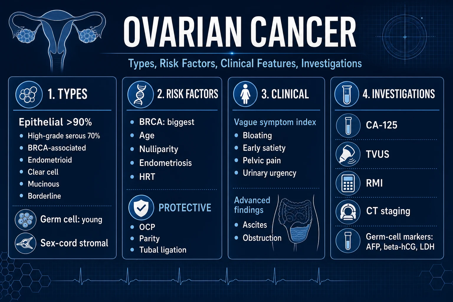
Ovarian cancer is classified along three independent axes: histological type, histological grade within type (especially for serous), and FIGO stage. Each axis drives a different clinical decision: histology for chemo-sensitivity and prognosis, grade for behaviour (a low-grade serous carcinoma behaves nothing like a high-grade one), and stage for whether the patient is a surgical candidate and what maintenance therapy will follow.[5]
Epithelial (90 per cent)
surface or tubal epithelium
- **High-grade serous carcinoma** — about 70 per cent of all ovarian cancers; aggressive, presents Stage III/IV; **TP53 mutation** in nearly 100 per cent; **BRCA1/2 mutation** in 15 to 20 per cent; arises from **fimbrial STIC**
- **Low-grade serous carcinoma** — about 5 per cent; indolent; **KRAS/BRAF mutation**; **oestrogen-receptor positive**; responds poorly to chemotherapy but well to letrozole
- **Endometrioid** — about 15 per cent; **endometriosis-associated**; often Stage I at diagnosis; **ARID1A/PTEN** mutations; frequently coexists with endometrial cancer
- **Clear cell** — 5 to 10 per cent; **endometriosis-associated**; worst prognosis stage-for-stage; **ARID1A** mutation; chemo-resistant; common in East Asian women
- **Mucinous** — about 3 per cent; large unilateral cystic mass; **KRAS** mutation; exclude a gastrointestinal primary (Krukenberg) with CEA and gastroscopy; metastatic mucinous tumours are 20 times more common than primary
- **Brenner (transitional cell)** — rare, benign in most cases; solid, fibrous cut surface
Germ cell (5 per cent)
young women under 30
- **Dysgerminoma** — most common malignant germ cell; **LDH marker**; very radiosensitive and chemosensitive; 95 per cent 5-year survival with BEP
- **Yolk sac (endodermal sinus)** — **AFP marker**; **Schiller-Duval bodies** on histology; aggressive but very chemosensitive; young women
- **Immature teratoma** — immature neural tissue; graded by amount of immature neuroepithelium;AFP may be normal or raised
- **Choriocarcinoma (non-gestational)** — **beta-hCG marker**; rare, highly aggressive; bleeds easily; chemo-sensitive
- **Mature cystic teratoma (dermoid)** — benign; contains hair, teeth, sebum; 10 to 15 per cent bilateral; 1 to 2 per cent risk of malignant transformation (usually squamous carcinoma)
Sex cord-stromal (5 per cent)
hormone-secreting
- **Granulosa cell tumour** — **oestrogen-secreting** (postmenopausal bleeding, precocious puberty in children); **inhibin B** is the tumour marker; **Call-Exner bodies** on histology; indolent but recurs decades later
- **Sertoli-Leydig** — **androgen-secreting** (virilisation, hirsutism, deepening voice); rare; usually young women
- **Fibroma-thecoma** — **oestrogen or non-functioning**; **Meigs syndrome** = ovarian fibroma plus ascites plus pleural effusion (benign — resection cures all three)
- **Sclerosing stromal tumour** — rare; young women; hormonally inactive usually
Metastatic (Krukenberg)
from a non-gynaecological primary
- **Stomach (signet ring)** — the classical Krukenberg; bilateral; **signet-ring cells** in the ovarian stroma
- **Colorectal** — also bilateral; mucin-secreting
- **Breast** — lobular breast cancer metastases commonly to the ovary; bilateral; may mimic primary ovarian cancer
- **Appendix, pancreas, biliary** — pseudomyxoma peritonei from appendix must be considered when abundant mucinous ascites is present
- **Always check CEA, do upper and lower GI endoscopy before assuming an ovarian primary in a mucinous tumour**
Ovarian cancer — the headline numbers (UK, NICE/Sung)
Epidemiology & Risk Factors
The epidemiology of ovarian cancer is dominated by a single unifying principle — the "incessant ovulation" hypothesis. Every ovulation ruptures the ovarian surface epithelium and forces it to repair; each repair cycle carries a small risk of a somatic mutation. Anything that increases the lifetime number of ovulatory cycles raises risk; anything that interrupts ovulation lowers it. This is the thread that ties nulliparity, early menarche, late menopause and infertility together as risk factors, and oral contraceptive pill use, multiparity, breastfeeding and tubal ligation as protective factors.[1]
Peak incidence is in the sixth and seventh decades (age 55 to 65 for epithelial ovarian cancer); it is rare before 40 except in BRCA carriers, in whom high-grade serous cancers appear a decade earlier. Germ cell tumours are the exception to the age rule — they are the commonest ovarian malignancy in women under 30, with peak incidence in the late teens and early twenties.[1]
Risk factors fall into four groups:[2]
- Genetic and familial (the single most powerful predictor). A family history of breast or ovarian cancer, especially at a young age, suggests an inherited cancer syndrome. BRCA1 carries an estimated 39 to 44 per cent lifetime ovarian cancer risk; BRCA2 an 11 to 17 per cent risk; both also confer a high breast cancer risk and an increased risk of prostate and pancreatic cancer. Lynch syndrome (MLH1, MSH2, MSH6, PMS2) confers a 6 to 12 per cent lifetime ovarian cancer risk and a much higher endometrial cancer risk. Together these hereditary syndromes account for 15 to 20 per cent of all epithelial ovarian cancers — a higher fraction than for almost any other solid tumour — and NICE recommends germline BRCA testing for every woman diagnosed with epithelial ovarian cancer, regardless of family history.[2]
- Reproductive. Nulliparity (never-pregnant), early menarche (before 12), late menopause (after 55), infertility and infertility treatment (especially unsuccessful IVF cycles), and never having used the oral contraceptive pill. Multiparity is protective — each full-term pregnancy reduces risk by about 15 per cent.
- Hormonal. Long-term (>5 to 10 years) menopausal hormone replacement therapy (HRT), particularly oestrogen-only preparations, increases risk by about 20 to 40 per cent; the risk falls after stopping. Endometriosis increases risk specifically of clear cell and endometrioid subtypes (relative risk 2 to 3). Polycystic ovarian syndrome modestly increases risk.
- Environmental and lifestyle. Obesity (BMI over 30) increases risk by about 20 per cent. Smoking is a weak risk factor for the mucinous subtype specifically. Talcum powder on the perineum has been associated with a marginal risk increase in some (not all) cohort studies; the evidence is contested. Asbestos exposure is firmly linked.
BRCA1 vs BRCA2 — what every candidate must memorise
Protective factors all reduce ovulation: the oral contraceptive pill reduces risk by 30 to 50 per cent after five years and the protection persists for decades after stopping — the single most important modifiable protective factor at a population level; multiparity; breastfeeding (suppresses ovulation); tubal ligation and hysterectomy (perhaps by disrupting a retrograde menstrual carcinogenic pathway); and the modern understanding that the fallopian tube is the source of high-grade serous carcinoma explains why bilateral salpingectomy at the time of routine hysterectomy or tubal sterilisation — opportunistic salpingectomy — is increasingly practised as primary prevention in the general population. [1]
India. Ovarian cancer is the third most common gynaecological cancer in Indian women (after cervical and endometrial) and ranks among the top 10 cancers in women overall. Late presentation is the rule, with over 60 to 70 per cent presenting at Stage III/IV, driven by non-specific symptoms, low awareness, lack of routine gynaecological care, and limited access to CA-125 and ultrasound in rural areas. BRCA mutations are over-represented in specific communities with founder effects (for example, BRCA1 185delAG among some Indian Jewish and Parsee populations; specific BRCA1/2 variants in some Maharashtrian and Gujarati communities). Genetic counselling, germline testing and risk-reducing surgery are underutilised because of cost, access, and cultural attitudes to prophylactic surgery in young women. Presentation with ascites is frequently mistaken initially for tuberculosis (given the high background TB prevalence), cirrhosis or ovarian hyperstimulation, leading to diagnostic delay.[1]
High-income settings (UK, USA, Australia). Population-level access to CA-125, transvaginal ultrasound and specialist gynaecological oncology has produced modest survival gains; opportunistic salpingectomy at routine gynaecological surgery, organised germline BRCA testing for every epithelial ovarian cancer patient, and cascade testing of relatives are increasingly embedded in guidelines (NICE NG181; NHS Genomic Medicine Service).
Pathophysiology

The pathogenesis of ovarian cancer is best understood through the dualistic model of Kurman and Shih, which separates the disease into two biologically distinct pathways:[2]
Type I tumours (indolent). Low-grade serous, endometrioid, clear cell, mucinous and Brenner tumours. They arise from identifiable precursor lesions — endometriosis (for endometrioid and clear cell) or ovarian cortical inclusion cysts — and they carry mutations in KRAS, BRAF, ARID1A, PTEN, CTNNB1. They grow slowly, are confined to the ovary at presentation in most cases, are often curable by surgery alone, and are relatively chemoresistant (especially clear cell). The metastatic risk is low. [1]
Type II tumours (aggressive). High-grade serous carcinoma, undifferentiated carcinoma and carcinosarcoma. These are the tumours that give ovarian cancer its grim reputation. They almost certainly arise from secretory cells of the fallopian tube fimbriae, where a precursor lesion — serous tubal intraepithelial carcinoma (STIC) — is identifiable in the prophylactic specimens of BRCA carriers years before invasive disease. They carry TP53 mutations in nearly 100 per cent of cases and are characterised by homologous recombination deficiency (HRD) — most often from a germline BRCA1 or BRCA2 mutation but also from somatic BRCA mutations, EMSY amplification or other defects in the homologous recombination DNA repair pathway. The clinical consequence of HRD is exquisite sensitivity to platinum chemotherapy and to PARP inhibitors — the foundation of modern maintenance therapy. [1]
The chain from STIC to invasive peritoneal carcinomatosis runs as follows:[2]
- BRCA mutation or other HRD is present from conception (germline) or acquired (somatic). The homologous recombination repair pathway cannot repair double-strand DNA breaks accurately.
- TP53 mutation occurs early in a fallopian tube secretory cell — the cell now lacks both its primary tumour suppressor (p53) and an accurate repair pathway.
- STIC — a flat sheet of malignant secretory cells lines the tubal lumen but does not invade. p53 staining is aberrant (diffusely positive or completely null).
- Exfoliation — malignant cells detach from the fimbrial end of the tube and are swept by peritoneal fluid onto the ovarian surface and across the peritoneum.
- Implantation and invasion of the ovarian stroma produces a complex cystic-solid adnexal mass.
- Transcoelomic spread — the hallmark route of ovarian cancer. Tumour cells shed from the ovarian surface into the peritoneal fluid, which circulates upward along the paracolic gutters to the right subphrenic space and omentum. Cells seed the omentum (the classical omental cake), the parietal peritoneum, the diaphragmatic peritoneum (which can block lymphatic drainage of peritoneal fluid), the bowel surface and the surface of the liver and spleen. This produces the characteristic triad of ascites, omental cake and diffuse peritoneal carcinomatosis — the typical Stage III presentation.
- Lymphatic spread to pelvic and para-aortic lymph nodes (along the ovarian vessels to the renal hilum) is common and is incorporated into FIGO staging.
- Haematogenous spread (liver parenchyma, lung, brain) is late and defines Stage IV. [1]
The clinical implication of this cascade is that the tumour spreads early and widely across a large serosal surface before symptoms appear — and that even when the ovary looks small on imaging, the peritoneum may already be diffusely seeded. This is why cytoreductive surgery, which removes visible peritoneal disease, is so central to outcome: chemotherapy cannot penetrate thick tumour deposits, and surgery reduces the disease to a volume at which platinum and PARP inhibitors can work. [1]
Clinical Presentation
The clinical challenge of ovarian cancer is that the symptoms are non-specific, common in healthy women, and shared with benign conditions — bloating, irritable bowel syndrome, urinary tract symptoms, weight gain, indigestion. The discriminating features are the tempo (new, persistent, and worsening over weeks) and the patient (a peri- or post-menopausal woman).[1]
[1]Atypical presentations examiners test deliberately. A young woman with a pelvic mass may have a germ cell tumour (dysgerminoma, yolk sac) rather than an epithelial cancer — and is managed very differently (fertility-sparing surgery, BEP chemotherapy). A postmenopausal woman with new-onset virilisation (deepening voice, hirsutism, clitoromegaly) has a Sertoli-Leydig tumour until excluded. A woman with precocious puberty has a granulosa cell tumour until excluded. A pregnant woman with sudden severe abdominal pain may have a torsed or ruptured ovarian mass (often a dermoid) — an acute surgical emergency. An elderly woman with bilateral solid ovarian masses and ascites may have a Krukenberg metastasis from a gastric or colonic primary, and the correct next investigation is an upper GI endoscopy and colonoscopy, not primary ovarian surgery. A woman with abdominal distension, weight loss and a palpable pelvic mass who has no ascites on imaging may have a large benign mucinous cystadenoma — but the same picture with ascites is ovarian cancer until proven otherwise. [1]
Differential Diagnosis
The differential of an adnexal mass or persistent abdominal symptoms in a woman splits into benign gynaecological causes, benign non-gynaecological causes, other gynaecological cancers, and non-ovarian malignancies metastatic to the ovary. The RMI and IOTA simple rules are designed to separate these, but the discriminating features still need to be known.[6]
| Condition | Key distinguishing feature |
|---|---|
| Functional (physiological) cyst | A simple anechoic cyst on ultrasound in a premenopausal woman, under 5 cm, unilocular, no solid component; CA-125 normal; resolves in 6 to 8 weeks on a repeat scan |
| Endometrioma | "Ground-glass" homogeneous low-level echoes on ultrasound; a history of endometriosis or dysmenorrhoea; CA-125 may be mildly raised; bilateral in 20 per cent |
| Mature cystic teratoma (dermoid) | Hyperechoic mass with echogenic focus (sebum, hair, teeth, calcification); Rokitansky nodule; shadowing from bone or teeth; bilateral in 10 to 15 per cent; CA-125 normal |
| Benign serous or mucinous cystadenoma | Large unilocular or multilocular cyst, thin septations, no solid component, no ascites; CA-125 normal |
| Pelvic inflammatory disease / tubo-ovarian abscess | Fever, pelvic pain, vaginal discharge, cervical excitation, tender adnexal mass, raised inflammatory markers; bilateral in most cases; bilateral and tubular if a hydrosalpinx is present |
| Ectopic pregnancy | Positive pregnancy test, acute lower abdominal pain, vaginal bleeding, missed period; a life-threatening emergency |
| Pedunculated fibroid | Arises from the uterus (follow the pedicle to the myometrium); firm, mobile, calcified in older women; normal CA-125 |
| Diverticular or appendiceal abscess | Right iliac fossa or left iliac fossa tender mass with sepsis; CT confirms a bowel-related collection |
| Colorectal or gastric cancer (Krukenberg) | Bilateral solid ovarian masses with a known or occult GI primary; raised CEA; ascites; omental cake; upper and lower GI endoscopy is mandatory before any surgery |
| Endometrial cancer with ovarian metastasis | Often presents with abnormal uterine bleeding; endometrial biopsy confirms; synchronous endometrioid ovarian and endometrial cancers (Lynch) also occur |
| Fallopian tube or primary peritoneal cancer | Clinically and histologically identical to high-grade serous ovarian cancer; managed identically; diagnosis rests on the location of the dominant mass |
Clinical & Bedside Assessment
The bedside assessment has two purposes: to detect the pelvic mass and its sequelae (ascites, bowel obstruction, pleural effusion), and to identify the clinical features that suggest a hereditary syndrome so that germline testing can be arranged.[6]
Abdominal examination looks for distension from ascites (shifting dullness, fluid thrill), a palpable pelvic mass (especially arising out of the pelvis), an epigastric or umbilical mass (an omental cake, or a Sister Mary Joseph nodule at the umbilicus indicating peritoneal carcinomatosis), and signs of bowel obstruction (distension, tinkling bowel sounds, colicky pain). A palpable left supraclavicular lymph node (Virchow node) is rare but classical. [1]
Bimanual pelvic (and rectovaginal) examination assesses the size, mobility and consistency of any adnexal mass, the uterus, the pouch of Douglas (a nodular, fixed mass suggests peritoneal disease), and the parametria. A fixed, irregular, bilateral adnexal mass with a separate, normal-sized uterus is highly suspicious for ovarian malignancy. [1]
General examination screens for signs of hypercoagulability (a swollen leg from DVT), pleural effusion (dullness, reduced breath sounds), cachexia and anaemia, and the cutaneous stigmata of genetic syndromes (the Cowden/PTEN trichilemmomas, Lynch-associated sebaceous tumours — though these are rare). [1]
Family history is mandatory and is a clinical investigation in its own right. A three-generation pedigree asking specifically about breast cancer (including male breast cancer), ovarian cancer, prostate cancer, pancreatic cancer, and endometrial or colorectal cancer in maternal and paternal relatives — and the age at diagnosis — identifies the woman who needs referral to clinical genetics and germline BRCA testing. The NICE threshold for genetic referral in an unaffected woman is a combined lifetime risk of breast cancer of 17 per cent or more; for a woman with ovarian cancer, germline BRCA testing is offered regardless of family history because the yield is 15 to 20 per cent. [1]
Key diagnostic thresholds at the bedside and in clinic
Investigations
The investigation strategy has three phases: a primary-care triage phase that uses symptoms to decide who to test; a diagnostic phase that confirms a mass, characterises it, and excludes other primaries; and a staging phase that defines tumour extent for surgical planning. Examiners expect you to know which test is right at which point, and why.[6]
Primary care — what NICE NG181 says. A woman over 50 (or under 50 with risk factors) with persistent (more than 2 to 3 weeks) bloating, early satiety, distension, abdominal or pelvic pain, urinary urgency or frequency, or change in bowel habit should have a serum CA-125 first, not an ultrasound. If CA-125 is over 35 U/mL, perform an ultrasound of the pelvis and abdomen and calculate the Risk of Malignancy Index (RMI). If RMI is over 250, refer under the 2-week-wait suspected-cancer pathway to a specialist gynaecological oncology multidisciplinary team. A woman with a palpable pelvic mass on examination is referred regardless of CA-125. This sequencing matters because CA-125 is cheaper, more accessible in primary care, and a better triage tool than ultrasound.[6]
Tumour markers. The markers chosen depend on the suspected subtype, the age of the woman, and the clinical picture:[6]
- CA-125 (cancer antigen 125). The primary marker for epithelial ovarian cancer. Elevated in 80 to 85 per cent of advanced epithelial cancers but only 50 per cent of Stage I cancers, and elevated in many benign conditions — endometriosis, pelvic inflammatory disease, pregnancy, fibroids, menstruation, liver disease, ascites of any cause, heart failure, pancreatitis — and in other cancers (endometrial, breast, pancreatic, gastric, colonic, lung). It is therefore non-specific as a diagnostic test but very useful for monitoring treatment response and detecting recurrence. A rising CA-125 after first-line therapy predicts recurrence a median of 3 to 6 months before imaging or symptoms, but treating an asymptomatic biochemical recurrence does not improve survival (and the patient is harmed by the psychological burden of knowing); the role of routine surveillance CA-125 is now contentious.
- CEA (carcinoembryonic antigen). Request if a mucinous ovarian tumour is suspected; a markedly raised CEA suggests a colorectal or gastric primary (Krukenberg), and upper and lower GI endoscopy is mandatory before any ovarian surgery.
- CA 19-9. Useful for mucinous tumours and to exclude a pancreatic or biliary primary.
- AFP, beta-hCG, LDH. Request in any woman under 40 with an ovarian mass, because a germ cell tumour (yolk sac — AFP; choriocarcinoma — beta-hCG; dysgerminoma — LDH) is then in the differential. These markers also drive surveillance after fertility-sparing surgery.
- Inhibin B and oestradiol. Request if a granulosa cell tumour is suspected (postmenopausal bleeding with an oestrogen-secreting solid ovarian mass). Inhibin B is the tumour marker for surveillance.
- AMH (anti-Müllerian hormone). Not a tumour marker; useful to assess ovarian reserve before fertility-sparing surgery in a young woman.
- Testosterone and DHEAS. Request if the woman is virilised (Sertoli-Leydig tumour); DHEAS excludes an adrenal source. [1]
Imaging.[6]
- Transvaginal and transabdominal pelvic ultrasound is the first-line imaging test. It characterises the mass as simple (anechoic, thin-walled, unilocular — likely benign) or complex (solid components, papillary projections, thick septations, ascites, bilocularity, irregular walls — suspect malignancy). The IOTA (International Ovarian Tumour Analysis) simple rules classify a mass as benign, malignant, or indeterminate using five benign features (B-features: unilocular, solid components under 7 mm, acoustic shadowing, smooth multilocular under 100 mm, no blood flow) and five malignant features (M-features: irregular solid, ascites, at least four papillary projections, irregular multilocular solid over 100 mm, very strong blood flow). The ADNEX model (Assessment of Different NEoplasias in the adneXa) is a more sophisticated logistic regression that estimates the probability of benign, borderline, Stage I cancer, Stage II to IV cancer, and metastatic tumour — and outperforms the RMI in meta-analysis.[6]
- CT of the abdomen and pelvis (with contrast) is performed for staging after a mass is identified — to define the extent of peritoneal disease, omental involvement, lymphadenopathy, liver or splenic surface deposits, hydronephrosis, and the presence of pleural effusion. CT thorax/abdomen/pelvis completes the radiological assessment for distant metastases. CT is also essential for assessing surgical resectability — can the surgeon achieve complete cytoreduction, or should the patient have neoadjuvant chemotherapy first?
- MRI pelvis is reserved for indeterminate ultrasound findings, for pregnant women (to avoid ionising radiation), and for characterising masses in young women where a germ cell or sex-cord tumour is suspected and organ preservation is being considered.
- PET-CT is not routine; it has a role in the recurrent setting or to evaluate equivocal lesions.
The Risk of Malignancy Index (RMI). This is a clinical decision tool that combines the three independent predictors of ovarian malignancy — ultrasound features, menopausal status, and CA-125 — into a single score. It is reproduced here in full because examiners test the formula and the thresholds:[6]
RMI = U × M × CA-125 [1]
where U (ultrasound score) is 0 (no features), 1 (one feature), or 3 (two to five features); the five ultrasound features are multilocular cyst, solid areas, metastatic disease, ascites, and bilateral lesions. M (menopausal status) is 1 for premenopausal and 3 for postmenopausal (a postmenopausal woman with an ovarian mass is much more likely to have cancer). CA-125 is the absolute serum value in U/mL. RMI over 250 is the threshold for high malignancy risk and triggers specialist gynaecological oncology referral. The RMI has a sensitivity of about 80 to 90 per cent and a specificity of about 70 to 85 per cent for ovarian cancer in the postmenopausal woman; in premenopausal women its specificity is lower because endometriosis and PID can drive both the ultrasound score and the CA-125. [1]
Diagnostic laparoscopy or laparotomy with biopsy and histology is the definitive diagnostic test for a complex adnexal mass where imaging and markers do not settle the question, and is the route by which surgical staging is performed (see Management). Cytology of ascitic fluid (or peritoneal washings if no ascites is present) is sent at the time of surgery. Germline BRCA1/2 testing is offered to every woman diagnosed with epithelial ovarian cancer regardless of age or family history (NICE, NHS Genomic Medicine Service), with cascade testing of first-degree relatives if a pathogenic variant is found.[2]
The diagnostic pathway from primary care to gynaecological oncology MDT
Symptoms (more than 2 to 3 weeks of bloating, early satiety, distension, pelvic or abdominal pain, urinary urgency or change in bowel habit) in a woman — especially over 50
Serum CA-125 FIRST (NICE NG181). If CA-125 is over 35 U/mL in a postmenopausal woman (or over 200 U/mL in a premenopausal woman) proceed to ultrasound
Transvaginal and transabdominal pelvic ultrasound — characterise the mass, apply IOTA simple rules and ADNEX model; calculate RMI = U x M x CA-125
If RMI over 250, refer under the 2-week-wait suspected-cancer pathway to a gynaecological oncology MDT
MDT pathway: CT chest/abdomen/pelvis for staging, serum tumour markers (CEA, CA 19-9, AFP, beta-hCG, LDH, inhibin if indicated), and assessment of surgical fitness
Diagnostic laparoscopy or laparotomy with surgical staging (TAH + BSO + omentectomy + peritoneal biopsies + lymphadenectomy) and ascitic cytology
Germline BRCA1/2 testing for every patient with epithelial ovarian cancer; cascade testing of first-degree relatives if a pathogenic variant is found
Management — Resuscitation
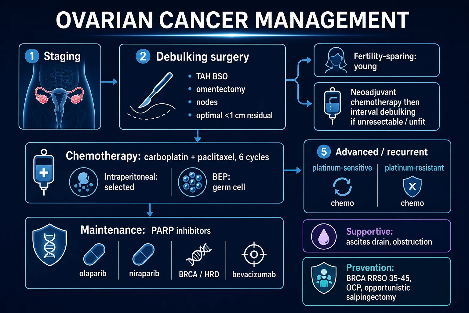
Ovarian cancer is rarely a time-critical emergency at first diagnosis, but three scenarios demand immediate action. The first is bowel obstruction from peritoneal carcinomatosis encasing the small or large bowel — managed with nasogastric decompression, intravenous fluids, parenteral nutrition if prolonged, and either surgical bypass (defunctioning stoma or resection) or, in advanced unresectable disease, a self-expanding metallic stent. The second is a large symptomatic pleural effusion or massive ascites causing respiratory compromise — managed by intercostal drain or ascitic drain (tunnelled long-term drains such as a PleurX for recurrent malignant ascites). The third is deep vein thrombosis or pulmonary embolism, common in ovarian cancer from cancer-associated hypercoagulability and from venous compression by a pelvic mass — managed with therapeutic low-molecular-weight heparin (LMWH), preferred over warfarin in active cancer. A woman who presents acutely unwell with hypercalcaemia, bowel perforation, or sepsis from a tumour-related fistula is a medical emergency needing resuscitation, antibiotics, and an early palliative-care opinion in the unresectable context.[5]
Management — Definitive & Stepwise
The definitive management of ovarian cancer is decided by three factors: stage, histology, and the patient's surgical fitness. The central principle is that the more tumour removed at surgery (the smaller the residual disease), the better the survival, and that platinum-based chemotherapy is the foundation of systemic treatment. The last decade has transformed this field with the addition of maintenance PARP inhibitors and bevacizumab to first-line therapy.[5]
Surgery — staging and cytoreduction
The standard staging laparotomy for ovarian cancer, performed through a midline incision, comprises:[5]
- Total abdominal hysterectomy (TAH) — removal of the uterus
- Bilateral salpingo-oophorectomy (BSO) — both tubes and ovaries; the tubes are removed completely including the fimbriated end, reflecting the tubal origin of high-grade serous carcinoma
- Infracolic omentectomy — removal of the omentum, the commonest site of transcoelomic spread (the omental cake)
- Peritoneal biopsies — from multiple sites (pelvic peritoneum, both paracolic gutters, the diaphragmatic peritoneum, the anterior abdominal wall)
- Pelvic and para-aortic lymphadenectomy (or sampling) — to detect nodal disease and assign stage; the 2014 FIGO revision incorporates nodal status explicitly
- Ascitic fluid for cytology, or peritoneal washings if no ascites is present
- Appendicectomy if a mucinous tumour is suspected (the appendix may be the occult primary)
- Diaphragmatic stripping, splenectomy, bowel resection as needed to achieve complete cytoreduction in advanced disease — modern ovarian cancer surgery is increasingly radical [1]
The single most important surgical outcome is the extent of cytoreduction. The goal is complete cytoreduction (CC-0) — no visible residual disease. Where complete cytoreduction is not achievable, the surgical standard for many years was optimal debulking — residual disease less than 1 cm in maximum diameter. The evidence is now unequivocal that complete (no residual) is better than optimal (less than 1 cm), which is better than suboptimal (over 1 cm), and that the survival difference between complete and any visible residual is large. The decision to attempt primary debulking versus neoadjuvant chemotherapy depends on the disease distribution on imaging, the patient's performance status and nutritional state, the surgeon's ability to achieve complete cytoreduction, and the perioperative risk.[7]
Neoadjuvant chemotherapy and interval debulking surgery. The EORTC 55971 / NCIC CTG OV.13 trial by Vergote and colleagues randomised 632 women with Stage IIIC or IV ovarian cancer to primary debulking followed by chemotherapy, or to three cycles of neoadjuvant carboplatin and paclitaxel followed by interval debulking surgery and three further cycles. Overall survival was equivalent (a median of 29 months in the primary debulking group versus 30 months in the neoadjuvant group), and the neoadjuvant group had fewer serious surgical complications and a higher rate of complete cytoreduction. Neoadjuvant chemotherapy is therefore the standard of care for women with bulky, unresectable Stage IIIC or IV disease, those who are medically unfit for radical primary surgery, or those with a low likelihood of complete cytoreduction at primary surgery. The ESMO-ESGO consensus defines the criteria for selecting primary versus neoadjuvant surgery.[7]
First-line chemotherapy
The standard first-line chemotherapy for epithelial ovarian cancer is intravenous carboplatin plus paclitaxel for six 3-weekly cycles. The regimen is:[8]
First-line chemotherapy for epithelial ovarian cancer
Standard of care for Stage II to IV after surgery, or as neoadjuvant before interval debulking
Dose
Carboplatin AUC 5 to 6 intravenously over 1 hour on day 1, plus paclitaxel 175 mg/m² intravenously over 3 hours on day 1, every 21 days for 6 cycles. Dose-dense weekly paclitaxel (80 mg/m² on days 1, 8, 15) with 3-weekly carboplatin AUC 6 is an alternative supported by JGOG 3016, especially for Japanese and East Asian populations.
Targeted and maintenance therapy — the modern revolution
After six cycles of carboplatin and paclitaxel, the question of maintenance therapy has transformed ovarian cancer care since 2018. Three landmark trials established the modern first-line maintenance options:[10][11][12]
SOLO-1 (Moore, NEJM 2018)
New England Journal of Medicine, 2018; PMID 30345884
Phase 3 randomised double-blind trial of olaparib 300 mg twice daily versus placebo as maintenance therapy in 391 patients with newly diagnosed advanced (FIGO Stage III or IV) high-grade serous or endometrioid ovarian cancer and a germline or somatic BRCA1/2 mutation, who had a complete or partial response to platinum-based chemotherapy.
Key finding
After a median follow-up of 41 months, the risk of disease progression or death was 70 per cent lower with olaparib. Three-year progression-free survival was 60 per cent with olaparib versus 27 per cent with placebo (hazard ratio 0.30, 95 per cent CI 0.23 to 0.41, p under 0.001). At 7-year follow-up the PFS benefit is sustained.
Practice change
Olaparib maintenance is the standard of care for newly diagnosed advanced ovarian cancer with a germline or somatic BRCA1/2 mutation after response to first-line platinum chemotherapy.
PRIMA (González-Martín, NEJM 2019)
New England Journal of Medicine, 2019; PMID 31562799
Phase 3 randomised double-blind trial of niraparib versus placebo as maintenance therapy in 733 patients with newly diagnosed advanced ovarian cancer (FIGO Stage III or IV) who had a response to first-line platinum chemotherapy — irrespective of BRCA status. The primary endpoint was progression-free survival, analysed by HRD status.
Key finding
Niraparib prolonged progression-free survival in the overall population (median 13.8 versus 8.2 months, HR 0.62) and most markedly in the HRD-positive subgroup (median 21.9 versus 10.4 months, HR 0.43).
Practice change
Niraparib maintenance is approved for all women with newly diagnosed advanced ovarian cancer after response to first-line platinum chemotherapy, regardless of BRCA or HRD status, with the greatest benefit in HRD-positive tumours.
PAOLA-1 (Ray-Coquard, NEJM 2019)
New England Journal of Medicine, 2019; PMID 31851799
Phase 3 randomised double-blind trial of olaparib plus bevacizumab versus placebo plus bevacizumab as first-line maintenance in 806 patients with newly diagnosed advanced ovarian cancer who had a response to platinum chemotherapy plus bevacizumab.
Key finding
Olaparib plus bevacizumab prolonged progression-free survival versus bevacizumab alone (median 22.1 versus 16.6 months, HR 0.59). The benefit was confined almost entirely to HRD-positive tumours (BRCA-mutated or genomic-instability positive; median PFS 37.2 versus 17.7 months, HR 0.33).
Practice change
The combination of olaparib plus bevacizumab is approved as first-line maintenance for HRD-positive advanced ovarian cancer.
Bevacizumab (a monoclonal antibody against vascular endothelial growth factor, VEGF) added to first-line carboplatin and paclitaxel and continued as maintenance was shown by the GOG-218 (Burger) and ICON7 (Perren) trials to improve progression-free survival (but not overall survival) in advanced disease, with the greatest benefit in patients with bulky Stage III or IV disease. The addition is now incorporated into standard first-line regimens for advanced disease, particularly in those with high-volume disease or ascites.[8][9]
The maintenance algorithm (a viva standard) is now: [1]
BRCA-mutated (germline or somatic)
any stage after response to platinum
- Olaparib maintenance for 2 years (SOLO-1) — standard of care
- OR olaparib plus bevacizumab if bevacizumab was part of first-line (PAOLA-1)
- Niraparib also approved (PRIMA)
- 5-year PFS benefit is large and clinically meaningful
HRD-positive, BRCA-wild-type
any stage after response to platinum
- Olaparib plus bevacizumab is the preferred combination (PAOLA-1)
- Niraparib alone also approved (PRIMA — strongest benefit in HRD-positive)
- HRD testing (myChoice CDx or equivalent) is now standard before deciding maintenance
HRD-negative / unknown
advanced stage after response to platinum
- Niraparib maintenance approved (PRIMA showed benefit in the overall population, including HRD-negative)
- Bevacizumab maintenance an option
- PARP inhibitor benefit is smaller than in HRD-positive tumours
- Discuss toxicity vs benefit
Low-grade serous
any stage
- Letrozole (or anastrozole) maintenance — these tumours are ER-positive and respond to endocrine therapy
- MEK inhibitor (binimetinib, trametinib) for BRAF/KRAS/NRAS-driven disease
- Poorly chemosensitive — surgery is the mainstay
Germ cell tumours
Germ cell tumours are biologically and clinically distinct from epithelial ovarian cancer — they occur in young women (under 30), grow rapidly, present at an early stage, are exquisitely chemosensitive, and have an excellent prognosis. Fertility-sparing surgery — unilateral salpingo-oophorectomy with surgical staging biopsies, preserving the uterus and contralateral ovary — is the standard surgical approach, regardless of the patient's desire for future fertility at the time, because cure is the rule and the contralateral ovary should be spared.[5]
BEP chemotherapy for ovarian germ cell tumours (Stages IC to IV or after incomplete resection)
Adjuvant chemotherapy after fertility-sparing surgery for non-localised dysgerminoma, yolk sac, immature teratoma grade 2 or 3, embryonal carcinoma, choriocarcinoma
Dose
Bleomycin 30 units IV weekly (or 15 units on days 1, 8, 15), plus etoposide 100 mg/m² IV daily on days 1 to 5, plus cisplatin 20 mg/m² IV daily on days 1 to 5, repeated every 21 days for 3 to 4 cycles (Stage I disease can be observed after complete surgical resection in selected cases — surveillance for Stage IA dysgerminoma and Stage IA grade 1 immature teratoma).
Sex cord-stromal tumours
Granulosa cell tumours are indolent and often present at Stage I (confined to the ovary). Standard surgery is TAH + BSO + staging in a postmenopausal woman; unilateral salpingo-oophorectomy in a young woman wishing to preserve fertility. Most Stage I cases require no adjuvant therapy; advanced or recurrent disease responds to platinum-based chemotherapy (BEP or carboplatin-paclitaxel), and to hormonal therapy (aromatase inhibitors, GnRH analogues) in recurrent disease. Inhibin B is the surveillance marker — it detects recurrence years before imaging because the tumour can relapse 10 to 30 years after diagnosis. Sertoli-Leydig tumours are managed similarly, with surgery as the mainstay. [1]
Palliative care
For women with recurrent platinum-resistant disease (relapse within 6 months of finishing platinum), the goal shifts to symptom control and quality of life. Single-agent chemotherapy (paclitaxel, pegylated liposomal doxorubicin, topotecan) gives modest response rates. Bevacizumab is licensed in the recurrent setting. Palliative surgery (bowel resection, defunctioning stoma) for obstruction, tunnelled ascitic drains for refractory ascites, pleurodesis or indwelling pleural catheters for effusions, opioid analgesia and an early integrated palliative-care input are all essential components of management. Advance care planning, psychological support, and discussion of the dying phase are owed to every woman with advanced ovarian cancer. [1]
FIGO 2014 Staging — Treatment by Stage
The 2014 FIGO revision (the Prat revision, derived from the older 1987 staging) unified ovarian, fallopian tube and primary peritoneal cancer into a single staging framework, recognising that they share biology and management. The system is surgical and pathological (unlike the clinical-radiological staging of cervical cancer).[5]
FIGO 2014 stage and approximate 5-year survival
Surface tumour, capsule rupture, or positive cytology
IC1 surgical spill; IC2 capsule rupture before surgery or surface tumour; IC3 malignant ascites or positive peritoneal washings. Treatment: surgical staging plus adjuvant carboplatin and paclitaxel for most cases.
Stage-specific management
Specific Subtypes & Scenarios
Borderline ovarian tumours (atypical proliferative tumours). These are epithelial tumours with cytological atypia and increased proliferation but no stromal invasion — they do not metastasise via the bloodstream and have an excellent prognosis (over 95 per cent 5-year survival for Stage I). They occur in younger women (median age 45) than invasive epithelial cancer, are often serous or mucinous, and the surgical standard is cystectomy or USO with staging biopsies in a woman wishing to preserve fertility, or TAH + BSO in a woman whose family is complete. Invasive implants (in the omentum or peritoneum) warrant more aggressive surgery and surveillance, but chemotherapy is not indicated in borderline disease because it does not respond. [1]
Low-grade serous carcinoma. Indolent, often presents in younger women (40s), is KRAS- or BRAF-mutant, is oestrogen-receptor positive, and is relatively chemoresistant. Surgery is the mainstay (maximal cytoreduction). Maintenance with letrozole or anastrozole (aromatase inhibitors) is standard. MEK inhibitors (binimetinib, trametinib, selumetinib) and BRAF/MEK combinations show promise in recurrent disease. [1]
Mucinous ovarian cancer. Rare (3 per cent of epithelial cancers), presents as a large unilateral cystic mass in a woman in her 40s or 50s, often Stage I. Always exclude a gastrointestinal primary — metastatic mucinous tumours to the ovary are 20 times more common than primary ovarian mucinous carcinomas, and management is entirely different. Request CEA, CA 19-9, and upper and lower GI endoscopy. Stage I disease is treated surgically and does not require adjuvant chemotherapy; advanced mucinous disease is poorly chemosensitive and has a poor prognosis. The appendix should be removed at surgery to exclude an occult appendiceal primary (pseudomyxoma peritonei). [1]
Ovarian cancer in pregnancy. Most adnexal masses found in pregnancy are benign — corpus luteum cyst, mature teratoma, cystadenoma. The imaging approach is ultrasound and MRI (avoid CT because of radiation to the fetus). Tumour markers are confounded by pregnancy (CA-125 rises physiologically; AFP and beta-hCG are produced by the placenta). A persistent complex mass in pregnancy is managed by a multidisciplinary team including a maternal-fetal medicine specialist and a gynaecological oncologist. If malignancy is confirmed, surgery in the second trimester (after organogenesis and before the third-trimester risk of preterm labour) is the standard for early-stage epithelial cancer and most germ cell tumours. BEP chemotherapy can be given in the second and third trimesters if needed. Krukenberg and metastatic disease in pregnancy are managed on a case-by-case basis. [1]
Recurrent ovarian cancer. The single most important factor in recurrent disease is the platinum-free interval (PFI) — the time between the last dose of platinum chemotherapy and the date of recurrence. A platinum-sensitive recurrence (PFI over 6 months) is re-treated with platinum doublet chemotherapy (carboplatin plus paclitaxel, or carboplatin plus gemcitabine, or carboplatin plus PLD) plus a PARP inhibitor or bevacizumab as maintenance. A platinum-resistant recurrence (PFI under 6 months) is treated with single-agent non-platinum chemotherapy (paclitaxel weekly, PLD, topotecan, etoposide) plus bevacizumab, with the understanding that response rates are low (10 to 30 per cent) and the goal is disease control and symptom relief. Secondary cytoreduction is an option for a platinum-sensitive recurrence if the disease is localised and completely resectable (the DESKTOP and SOCcR criteria guide selection). [1]
Complications & Pitfalls
Complications fall into disease-related, surgical, chemotherapy-related, and psychosocial, and examiners test the distinction.[5]
Disease-related complications are the consequences of peritoneal carcinomatosis: malignant ascites (large-volume, often blood-stained, requiring drainage); bowel obstruction (from serosal infiltration or kinking by tumour, presenting with colicky pain, distension, vomiting and constipation, and managed with nasogastric decompression, stenting, defunctioning stoma or palliative bypass); ureteric obstruction (with hydronephrosis and acute kidney injury — managed with nephrostomy or ureteric stent); pleural effusion (a defining feature of Stage IVA disease); deep vein thrombosis and pulmonary embolism (cancer-associated hypercoagulability, especially with mucinous tumours and in advanced disease); cachexia and asthenia (the most common terminal pathway); and spontaneous tumour rupture presenting as an acute surgical abdomen. [1]
Surgical complications of cytoreductive ovarian cancer surgery are substantial because the operation is major. They include significant blood loss requiring transfusion (especially with splenectomy, diaphragmatic stripping, bowel resection), bowel injury and anastomotic leak (with low rectal anastomosis or bowel resection), ureteric injury, bladder injury, major vascular injury (especially during para-aortic node dissection near the vena cava and aorta), wound infection and dehiscence, incisional hernia, pelvic abscess, lymphocyst and lymphoedema after lymphadenectomy, postoperative ileus, deep vein thrombosis and pulmonary embolism (mandating prophylactic LMWH and mechanical compression), and intensive care admission in a high proportion of patients. The morbidity of primary debulking must be weighed against the benefit of complete cytoreduction when deciding between primary surgery and neoadjuvant chemotherapy. [1]
Chemotherapy complications of carboplatin and paclitaxel include neutropenic sepsis (the most common serious chemotherapy emergency — needs urgent broad-spectrum antibiotics), peripheral neuropathy (paclitaxel — may be permanent in a subset), alopecia (universal with paclitaxel, often the most distressing toxicity for the patient), hypersensitivity reactions (carboplatin re-challenge after 6 or more cycles carries a 25 per cent or higher reaction rate — desensitisation protocols may be needed), thrombocytopenia (carboplatin — cumulative), anaemia, fatigue, nausea and vomiting, mucositis, and infertility with premature ovarian failure in premenopausal women. [1]
PARP inhibitor complications. Myelosuppression (especially niraparib — thrombocytopenia and anaemia; dose modification common; start at lower dose in patients under 77 kg or with platelets under 150), nausea and fatigue (olaparib and niraparib), hypertension (less common with PARP inhibitors than with bevacizumab), and myelodysplastic syndrome and acute myeloid leukaemia (rare but real — 1 to 2 per cent in long-term follow-up; baseline and annual bone marrow monitoring). [1]
Bevacizumab complications. Hypertension (often requiring treatment), proteinuria, arterial thromboembolism (stroke, myocardial infarction), impaired wound healing (hold for 28 days before and after surgery), gastrointestinal perforation (a particular concern in ovarian cancer where the bowel may be involved by tumour), and fistula formation. [1]
The classic pitfall is dismissing persistent bloating in a postmenopausal woman as irritable bowel syndrome, age-related constipation, or weight gain without doing a CA-125 and a pelvic ultrasound. The second is ordering an ultrasound first rather than a CA-125 in primary care (against NICE NG181). The third is failing to test for germline BRCA in every woman diagnosed with epithelial ovarian cancer — NICE recommends universal testing, and missing a pathogenic variant denies the patient access to PARP inhibitors and denies her relatives access to cascade testing and risk-reducing surgery. The fourth is mislabelling a Krukenberg tumour as primary ovarian cancer and operating without first excluding a gastric or colonic primary. The fifth is offering primary debulking surgery to a woman with unresectable Stage IV disease who would do better with neoadjuvant chemotherapy. [1]
Prognosis & Disposition
Prognosis is driven above all by stage at diagnosis (because most women present late), then by extent of cytoreduction (complete better than optimal better than suboptimal), histology (high-grade serous worse than endometrioid or mucinous Stage I; clear cell worse stage-for-stage), BRCA status (BRCA-mutated tumours are more chemo-sensitive and respond better to PARP inhibitors, but high-grade serous regardless of BRCA carries a worse prognosis than Type I tumours), performance status and age, and platinum sensitivity (the response to first-line platinum and the duration of the platinum-free interval are the strongest predictors of outcome in advanced disease).[1]
Five-year survival by FIGO 2014 stage (approximate)
Disposition and follow-up. A woman treated with curative intent is reviewed every 3 to 6 months for 2 years, then 6 to 12 monthly to 5 years, with clinical review, CA-125 if raised at baseline, and imaging for symptoms or rising markers. The value of routine surveillance CA-125 in asymptomatic women is contested — the MRC OV05/EORTC 55955 trial showed that treating an asymptomatic biochemical recurrence did not improve survival, and that the patient was harmed psychologically by knowing of a rising marker with no therapeutic option. Many oncologists now discuss the option of surveillance with each patient and individualise the decision. [1]
The safety-net. A woman with treated ovarian cancer who develops new symptoms (bloating, distension, abdominal pain, change in bowel habit, weight loss) at any time after treatment is presumed to have recurrence until proven otherwise — and is investigated with CA-125, CT, and clinical review. [1]
Special Populations
BRCA1/2 carriers — risk-reducing surgery. A woman with a confirmed germline BRCA1 or BRCA2 mutation is offered risk-reducing bilateral salpingo-oophorectomy (RRSO), ideally between 35 and 40 years for BRCA1 (because the risk rises sharply from the late 30s; mean age of onset 51) and 40 to 45 years for BRCA2 (mean age of onset 61; the later timing reflects the later onset and balances the menopausal risks of premature oestrogen deprivation). RRSO reduces ovarian and fallopian tube cancer risk by 80 to 95 per cent and breast cancer risk by about 50 per cent if performed premenopausally. It is performed by laparoscopy, includes the fimbriated end of the tube (the source of high-grade serous carcinoma), and includes a thorough peritoneal washings and pathological examination of the entire tubes and ovaries (serial sectioning) to detect occult early cancers. Premature menopausal symptoms (hot flushes, osteoporosis, cardiovascular risk) are managed with hormone replacement therapy (HRT) until the natural age of menopause — there is no excess breast cancer risk from HRT after RRSO in BRCA carriers without a personal history of breast cancer. BRCA carriers are also offered annual breast MRI surveillance from age 25 to 30 (alternating with mammography from age 40), and risk-reducing mastectomy is discussed.[2]
Lynch syndrome. Carriers of mismatch repair gene mutations (MLH1, MSH2, MSH6, PMS2) have a 6 to 12 per cent lifetime ovarian cancer risk and a 30 to 60 per cent endometrial cancer risk. The risk-reducing strategy is risk-reducing hysterectomy plus bilateral salpingo-oophorectomy after completion of family, typically around age 40 to 45. Colonoscopic surveillance is essential from a young age. Lynch-associated ovarian cancers tend to present at an earlier stage and have a better stage-for-stage prognosis than sporadic cases. [1]
Young women desiring fertility. For Stage IA low-grade serous, mucinous, sex-cord or germ cell tumours, fertility-sparing surgery (unilateral salpingo-oophorectomy with staging biopsies, preserving the uterus and contralateral ovary) is standard and is curative in most cases. For Stage IA high-grade serous carcinoma, the standard remains TAH + BSO because of recurrence risk in the conserved ovary, but fertility-sparing surgery is occasionally offered in selected cases after extensive counselling. Completion surgery (TAH + BSO) after childbearing is recommended for most cases. Germ cell tumours are managed with fertility-sparing surgery regardless of stage because they are chemo-sensitive and the prognosis is excellent. [1]
Pregnant women. See Specific Subtypes — most adnexal masses in pregnancy are benign; persistent complex masses are managed by a multidisciplinary team; surgery in the second trimester; chemotherapy in the second and third trimesters if required. [1]
Elderly and frail women. Frail patients with advanced ovarian cancer may not tolerate radical surgery and six cycles of platinum doublet chemotherapy; the standard alternative is neoadjuvant chemotherapy followed by interval debulking (which has lower perioperative morbidity than primary debulking) or, in the unfit, single-agent carboplatin. The decision is made by a gynaecological oncology MDT with a comprehensive geriatric assessment and an honest discussion of the goals of care. [1]
Evidence, Guidelines & Regional Differences
The evidence base for ovarian cancer has been transformed in the last decade by the introduction of PARP inhibitors as first-line maintenance therapy, but the foundation — surgery plus platinum chemotherapy — rests on trials from the 1990s.[5][10][11][12]
Vergote EORTC 55971 / NCIC OV.13 (NEJM 2010)
New England Journal of Medicine, 2010; PMID 20818904
Phase 3 randomised trial of primary debulking surgery followed by platinum chemotherapy versus three cycles of neoadjuvant carboplatin and paclitaxel followed by interval debulking surgery and three further cycles, in 632 women with Stage IIIC or IV epithelial ovarian cancer.
Key finding
Median overall survival was equivalent (29 months primary debulking versus 30 months neoadjuvant). The neoadjuvant arm had higher complete cytoreduction rates (80 per cent versus 42 per cent), less blood loss, and fewer serious adverse events (8 per cent versus 26 per cent with over 2 L blood loss; 1 per cent versus 8 per cent postoperative mortality).
Practice change
Neoadjuvant chemotherapy followed by interval debulking is the standard of care for women with bulky unresectable Stage IIIC/IV ovarian cancer, or those unfit for primary radical surgery.
GOG-218 (Burger, NEJM 2011) and ICON7 (Perren, NEJM 2011)
New England Journal of Medicine, 2011; PMIDs 22204724 (GOG-218) and 22204725 (ICON7)
Two concurrent phase 3 randomised trials of carboplatin and paclitaxel with or without bevacizumab in newly diagnosed advanced ovarian cancer. GOG-218 randomised 1,873 women to chemotherapy alone, chemotherapy plus bevacizumab during induction, or chemotherapy plus bevacizumab during induction and maintenance for 15 months. ICON7 randomised 1,528 women to chemotherapy with or without bevacizumab during induction and maintenance for 12 months.
Key finding
Both trials showed an improvement in progression-free survival (GOG-218 median PFS 14.1 versus 10.3 months; ICON7 median PFS 19.0 versus 17.3 months, with a larger benefit in the high-risk subgroup — Stage IV or incompletely resected Stage III). Neither trial demonstrated a statistically significant improvement in overall survival in the primary analysis.
Practice change
Bevacizumab is incorporated into first-line therapy for selected women with advanced ovarian cancer, especially bulky Stage III or IV disease, with continuation as maintenance.
UKCTOCS (Jacobs, Lancet 2016) and PLCO (Buys, JAMA 2011) — the negative screening trials
Lancet 2016 (UKCTOCS, PMID 26707054); JAMA 2011 (PLCO, PMID 21642681)
UKCTOCS randomised 202,638 postmenopausal women to no screening, annual multimodal screening (serial CA-125 interpreted by the Risk of Ovarian Cancer Algorithm, ROCA, with transvaginal ultrasound as a second-line test), or annual transvaginal ultrasound alone. PLCO randomised 78,216 women to annual CA-125 plus transvaginal ultrasound versus usual care.
Key finding
UKCTOCS showed a small but statistically non-significant reduction in ovarian cancer mortality (relative risk 0.89, 95 per cent CI 0.74 to 1.05) at the long-term analysis, with no mortality reduction in the ultrasound-only arm. PLCO showed no mortality reduction (relative risk 1.18) and an excess of diagnoses and complications. Both trials confirmed that the screening modalities are not sensitive enough to detect ovarian cancer at a curable stage, and that complications from false-positive results (including surgery for benign findings) cause harm.
Practice change
Population screening for ovarian cancer is not recommended in any country. Research into more sensitive early-detection strategies (multi-omic panels, cell-free DNA) continues.
Regional guideline differences reflect drug funding, access to genetic testing, and surgical centralisation. [1]
[1] [1]WHO / FIGO. The FIGO 2014 staging system is used worldwide. The NCCN Harmonised Guidelines adapt the US framework for low- and middle-income settings. In India and other resource-limited settings, late presentation, limited access to CA-125 and ultrasound, and prohibitive cost of PARP inhibitors and bevacizumab constrain implementation. Generic carboplatin and paclitaxel are widely available, and surgery remains the global mainstay of curative therapy. Opportunistic salpingectomy at the time of caesarean section or other abdominal surgery is a low-cost primary-prevention strategy being promoted in many countries.
Prevention
Prevention is the most powerful intervention available for ovarian cancer, because screening does not work and treatment of advanced disease is palliative in many patients. Prevention is delivered through three strategies. [1]
1. Risk-reducing surgery in BRCA and Lynch carriers. RRSO between 35 and 40 (BRCA1) and 40 and 45 (BRCA2) reduces ovarian and fallopian tube cancer risk by 80 to 95 per cent, and premenopausal RRSO reduces breast cancer risk by about 50 per cent. Risk-reducing hysterectomy plus BSO after family completion is offered to Lynch carriers. The identification of carriers rests on family history taking, germline testing of every woman with epithelial ovarian cancer, and cascade testing of relatives. This is the single highest-yield prevention strategy and should be embedded in every gynaecological and oncology service.[2]
2. Oral contraceptive pill chemoprevention. Five or more years of combined oral contraceptive use reduces ovarian cancer risk by 30 to 50 per cent, and the protection persists for decades after stopping. This is the single most powerful chemoprevention strategy at a population level and applies to women both with and without BRCA mutations. The benefit must be weighed against the small absolute risks of breast cancer and venous thromboembolism with the pill, but for ovarian cancer prevention the benefit is large and durable. [1]
3. Opportunistic salpingectomy. Recognition that high-grade serous carcinoma arises from the fimbrial fallopian tube has led to the recommendation that, at the time of any benign gynaecological surgery (hysterectomy for benign disease, tubal ligation, surgery for benign ovarian cysts), the fallopian tubes be removed in full (rather than the historical practice of conserving them or performing a partial salpingectomy). Population-level data from British Columbia (the OCS initiative) and the US suggest this is reducing the incidence of high-grade serous ovarian cancer, and the practice is endorsed by the Royal College of Obstetricians and Gynaecologists, ACOG, and FIGO.[2]
The prevention cascade — from carrier identification to risk-reducing surgery
Three-generation family history in every woman; identify suspected BRCA or Lynch syndrome from a pedigree of breast, ovarian, prostate, pancreatic, endometrial or colorectal cancer (especially at a young age)
Refer to clinical genetics for germline testing (multigene panel including BRCA1, BRCA2, MLH1, MSH2, MSH6, PMS2, PALB2, RAD51C, RAD51D, BRIP1)
For confirmed carriers, arrange counselling, cascade testing of first-degree relatives, and a structured surveillance plan
BRCA1 carrier: annual breast MRI from age 25 to 30, mammography from age 40, risk-reducing mastectomy discussed; risk-reducing salpingo-oophorectomy at 35 to 40
BRCA2 carrier: same breast surveillance; risk-reducing salpingo-oophorectomy at 40 to 45
Lynch carrier: colonoscopy every 1 to 2 years from age 25; risk-reducing hysterectomy plus BSO after completion of family
Population level: opportunistic salpingectomy at benign gynaecological surgery; oral contraceptive pill use as chemoprevention
Screening
Population screening for ovarian cancer does not work and is not recommended in any country. The two largest randomised trials — UKCTOCS (202,638 postmenopausal women; multimodal screening with serial CA-125 interpreted by the ROCA algorithm, or annual transvaginal ultrasound, versus no screening) and PLCO (78,216 women; annual CA-125 plus transvaginal ultrasound versus usual care) — both failed to show a clinically meaningful reduction in ovarian cancer mortality.[3][4]
The reasons screening fails are biological: most high-grade serous carcinomas (the dominant subtype) are detectable by CA-125 or imaging only when they are already advanced, because the precursor lesion (STIC) is microscopic and confined to the tube, and the transition from STIC to advanced peritoneal carcinomatosis is rapid. A multimodal strategy combining CA-125 trajectory with ultrasound finds more cancers at an earlier stage, but the gain is insufficient to reduce mortality in a screened population, and the false-positive rate causes real harm (surgery for benign findings in women who would never have developed ovarian cancer). [1]
Women at high genetic risk — confirmed BRCA1/2 or Lynch carriers — are not screened for ovarian cancer; they are offered risk-reducing surgery instead, because screening does not detect the disease at a curable stage even in this group. The only role for surveillance in a BRCA carrier who declines or defers RRSO is annual CA-125 and transvaginal ultrasound, with the explicit understanding that this is a compromise of unproven benefit. [1]
Research continues into multi-omic biomarker panels (combining CA-125 with HE4, mesothelin, osteopontin and other markers), circulating tumour DNA, and liquid biopsy approaches, in the hope that a more sensitive early-detection test will eventually make screening feasible. None is currently recommended. [1]
Exam Pearls
- Leading cause of gynaecological cancer death — the "silent killer"; 70 per cent present at Stage III/IV.[1]
- Sixth most common cancer in UK women; lifetime risk about 2 per cent (1 in 50).[1]
- High-grade serous carcinoma is the commonest subtype (70 per cent); arises from fimbrial STIC of the fallopian tube; TP53 mutated in nearly 100 per cent.[2]
- BRCA1 = 39 to 44 per cent lifetime risk; BRCA2 = 11 to 17 per cent. RRSO at 35 to 40 (BRCA1) and 40 to 45 (BRCA2).[2]
- Lynch syndrome confers a 6 to 12 per cent ovarian cancer risk and a much higher endometrial cancer risk.[2]
- CA-125 is the primary tumour marker — non-specific; raised in 80 to 85 per cent of advanced epithelial cancers. AFP for yolk sac, hCG for choriocarcinoma, LDH for dysgerminoma, inhibin for granulosa cell.[6]
- IOTA simple rules and ADNEX model characterise an adnexal mass on ultrasound.[6]
- RMI = U × M × CA-125; U (ultrasound score) is 0, 1, or 3; M (menopausal) is 1 or 3; RMI over 250 = high risk, refer to gynaecological oncology.[6]
- Standard surgical staging: TAH + BSO + omentectomy + peritoneal biopsies + pelvic and para-aortic lymphadenectomy + ascitic cytology.[5]
- Goal of surgery: complete cytoreduction (CC-0, no visible residual); "optimal debulking" is residual disease under 1 cm. The single most important surgical factor for survival.[5]
- First-line chemotherapy: carboplatin AUC 5 to 6 plus paclitaxel 175 mg/m², every 21 days for 6 cycles.[8]
- Neoadjuvant chemotherapy + interval debulking for bulky unresectable Stage IIIC/IV disease (Vergote EORTC 55971).[7]
- PARP inhibitors (olaparib, niraparib) and bevacizumab have revolutionised first-line maintenance — SOLO-1, PRIMA, PAOLA-1.[10][11][12]
- Germ cell tumours in young women — fertility-sparing surgery + BEP chemotherapy; 95 per cent 5-year survival for dysgerminoma.[5]
- Krukenberg tumour = metastatic to the ovary from a gastric (signet ring) primary; bilateral solid masses; check CEA, endoscope before operating.[6]
- Meigs syndrome = ovarian fibroma + ascites + pleural effusion; benign; resection cures all three.[6]
- Granulosa cell tumour = oestrogen-secreting (postmenopausal bleeding, precocious puberty); inhibin B is the marker; Call-Exner bodies on histology.[6]
- Population screening does not work (UKCTOCS, PLCO).[3][4]
- Opportunistic salpingectomy at benign gynaecological surgery is the modern population-level prevention strategy.[2]
- Stage I survival 90 per cent; Stage IV under 15 per cent; overall 5-year survival about 46 per cent.[1]
Ovarian cancer presentation — the 4 B symptoms
4Bs
abdominal distension — the most common presenting symptom; from ascites and a pelvic mass
urinary frequency and urgency from mass pressure on the bladder
early satiety, change in bowel habit, occasionally obstruction
abnormal vaginal bleeding — usually a sign of a hormone-secreting tumour (granulosa cell) or endometrioid subtype
Exam application bank (NEET-PG / INICET)
One-line answer
Ovarian cancer is the leading cause of gynaecological cancer death in the UK and the sixth most common cancer in women (lifetime risk about 2 per cent, 1 in 50). The dominant subtype, high-grade serous carcinoma, arises from the fimbrial end of the fallopian tube (serous tubal intraepithelial carcinoma, STIC), not the ovarian surface — a paradigm shift that drives risk-reducing salpingo-oophorectomy in BRCA1/2 carriers. The classical presentation is late and non-specific — bloating, distension, early satiety — so most women are Stage III/IV at diagnosis. CA-125 plus transvaginal ultrasound triage via IOTA simple rules and the Risk of Malignancy Index (RMI), with RMI over 250 mandating specialist referral. FIGO 2014 staging is surgical. Standard care is surgical staging and maximal cytoreduction (TAH + BSO + omentectomy + peritoneal biopsies + lymphadenectomy) plus carboplatin and paclita
Worked stems (answer without another resource)
Stem 1 — Classic presentation. Map symptoms to mechanism; name the first investigation and first treatment step with dose/route if drug therapy is standard. [1]
Stem 2 — Unstable / complicated. List red flags that force immediate resuscitation, theatre, ICU, antidote, or reperfusion — and what you do in the first 15 minutes. [1]
Stem 3 — Atypical group. Elderly, pregnancy, child, or immunocompromised: how presentation and thresholds change. [1]
Stem 4 — Differential trap. Name the three closest mimics and one discriminator for each. [1]
Stem 5 — Disposition. Who goes home with safety-netting, who is admitted, who needs HDU/ICU/theatre, and what follow-up is mandatory. [1]
Rapid viva checklist
- Definition + classification
- Pathophysiology chain
- Bedside signs / criteria
- Score with exact components (if any)
- Emergency bundle
- Definitive therapy with doses
- Complications of disease and of treatment
- Special populations
- Guideline/trial name if classic
- Three exam traps
Coverage self-check
If you cannot answer any stem above from this page alone, re-read the matching section — the page is intended to be self-sufficient for final-prof and NEET-PG/INICET questions on Ovarian Cancer.
References
- [1]Sung H, Ferlay J, Siegel RL, Laversanne M, Soerjomataram I, Jemal A, Bray F. Global Cancer Statistics 2020: GLOBOCAN Estimates of Incidence and Mortality Worldwide for 36 Cancers in 185 Countries CA Cancer J Clin, 2021.PMID 33538338
- [2]Antoniou A, Pharoah PD, Narod S, Risch HA, Eyfjord JE, Hopper JL, Loman N, Olsson H, Johannsson O, Borg A, Pasini B, Radice P, Manoukian S, Eccles DM, Tang N, Olah E, Anton-Culver H, Warner E, Lubinski J, Gronwald J, Gorski B, Tulinius H, Thorlacius S, Eerola H, Nevanlinna H, Syrjäkoski K, Kallioniemi OP, Thompson D, Evans C, Peto J, Lalloo F, Evans DG, Easton DF. Average risks of breast and ovarian cancer associated with BRCA1 or BRCA2 mutations detected in case Series unselected for family history: a combined analysis of 22 studies Am J Hum Genet, 2003.PMID 12677558
- [3]Jacobs IJ, Menon U, Ryan A, Gentry-Maharaj A, Burnell M, Kalsi JK, Amso NN, Apostolidou S, Benjamin E, Cruickshank D, Crump DN, Davies SK, Dawnay A, Dobbs S, Fletcher G, Ford J, Godfrey K, Gunu R, Habib M, Hallett R, Herod J, Jenkins H, Karpinskyj C, Leeson S, Lewis SJ, Liston WR, Lopes A, Mould T, Murdoch J, Oram D, Rabideau DJ, Reynolds K, Scott I, Seif MW, Sharma A, Singh N, Taylor J, Warburton F, Widschwendter M, Williamson K, Woolas R, Fallowfield L, McGuire AJ, Campbell S, Parmar M, Skates SJ. Ovarian cancer screening and mortality in the UK Collaborative Trial of Ovarian Cancer Screening (UKCTOCS): a randomised controlled trial Lancet, 2016.PMID 26707054
- [4]Buys SS, Partridge E, Black A, Johnson CC, Lamerato L, Isaacs C, Reding DJ, Greenlee RT, Yokochi LA, Kessel B, Crawford ED, Church TR, Andriole GL, Weissfeld JL, Fouad MN, Chia D, O'Brien B, Ragard LR, Clapp JD, Rathmell JM, Riley TL, Hartge P, Pinsky PF, Zhu CS, Izmirlian G, Kramer BS, Miller AB, Xu JL, Prorok PC, Gohagan JK, Berg CD; PLCO Project Team. Effect of screening on ovarian cancer mortality: the Prostate, Lung, Colorectal and Ovarian (PLCO) Cancer Screening Randomized Controlled Trial JAMA, 2011.PMID 21642681
- [5]Prat J; FIGO Committee on Gynecologic Oncology. Staging classification for cancer of the ovary, fallopian tube, and peritoneum Int J Gynaecol Obstet, 2014.PMID 24219974
- [6]Kaijser J, Sayasneh A, Van Hoorde K, Ghaem-Maghami S, Bourne T, Timmerman D, Van Calster B. Presurgical diagnosis of adnexal tumours using mathematical models and scoring systems: a systematic review and meta-analysis Hum Reprod Update, 2014.PMID 24327552
- [7]Vergote I, Tropé CG, Amant F, Kristensen GB, Ehlen T, Johnson N, Verheijen RH, van der Burg ME, Lacave AJ, Panici PB, Kenter GG, Casado A, Mendiola C, Coens C, Verleye L, Stuart GC, Pecorelli S, Reed NS; European Organization for Research and Treatment of Cancer-Gynaecological Cancer Group; NCIC Clinical Trials Group. Neoadjuvant chemotherapy or primary surgery in stage IIIC or IV ovarian cancer N Engl J Med, 2010.PMID 20818904
- [8]Burger RA, Brady MF, Bookman MA, Fleming GF, Monk BJ, Huang H, Mannel RS, Homesley HD, Fowler J, Greer BE, Boente M, Birrer MJ, Liang SX; Gynecologic Oncology Group. Incorporation of bevacizumab in the primary treatment of ovarian cancer N Engl J Med, 2011.PMID 22204724
- [9]Perren TJ, Swart AM, Pfisterer J, Ledermann JA, Pujade-Lauraine E, Kristensen G, Carey MS, Beale P, Cervantes A, Kurzeder C, du Bois A, Sehouli J, Kimmig R, Stähle A, Collinson F, Essapen S, Gourley C, Lortholary A, Selle F, Mirza MR, Leminen A, Plante M, Stark D, Qian W, Parmar MK, Oza AM; ICON7 Investigators. A phase 3 trial of bevacizumab in ovarian cancer N Engl J Med, 2011.PMID 22204725
- [10]Moore K, Colombo N, Scambia G, Kim BG, Oaknin A, Friedlander M, Lisyanskaya A, Floquet A, Leary A, Sonke GS, Gourley C, Banerjee S, Oza A, González-Martín A, Aghajanian C, Bradley W, Mathews C, Liu J, Lowe ES, Bloomfield R, DiSilvestro P. Maintenance Olaparib in Patients with Newly Diagnosed Advanced Ovarian Cancer N Engl J Med, 2018.PMID 30345884
- [11]González-Martín A, Pothuri B, Vergote I, DePont Christensen R, Graybill W, Mirza MR, McCormick C, Lorusso D, Hoskins P, Freyer G, Baumann K, Jardon K, Redondo A, Moore RG, Vulsteke C, O'Cearbhaill RE, Lund B, Backes F, Barretina-Ginesta P, Haggerty AF, Rubio-Pérez MJ, Shahin MS, Mangili G, Bradley WH, Bruchim I, Sun K, Malinowska IA, Li Y, Gupta D, Monk BJ; PRIMA/ENGOT-OV26/GOG-3012 Investigators. Niraparib in Patients with Newly Diagnosed Advanced Ovarian Cancer N Engl J Med, 2019.PMID 31562799
- [12]Ray-Coquard I, Pautier P, Pignata S, Pérol D, González-Martín A, Berger R, Fujiwara K, Vergote I, Colombo N, Mäenpää J, Selle F, Sehouli J, Lorusso D, Guerra Alía EM, Reinthaller A, Nagao S, Lefeuvre-Plesse C, Canzler U, Scambia G, Lortholary A, Marmé F, Combe P, de Gregorio N, Rodrigues M, Buderath P, Dubot C, Burges A, You B, Pujade-Lauraine E, Harter P, PAOLA-1 Investigators. Olaparib plus Bevacizumab as First-Line Maintenance in Ovarian Cancer N Engl J Med, 2019.PMID 31851799