Obstetrics & Gynaecology · Obstetrics & Gynaecology
Placenta Praevia
Also known as Low-lying placenta · Major praevia · Antepartum haemorrhage · Placenta accreta spectrum
Placenta praevia = placenta implanted wholly or partly in the lower uterine segment, over or near the internal cervical os. Incidence about 0.3 to 0.5 percent of pregnancies at term (higher earlier because most low placentas migrate). Classic: painless, causeless, recurrent bright-red vaginal bleeding after 20 to 24 weeks; uterus soft and non-tender (unlike abruption). Transvaginal ultrasound is the gold standard; NEVER do a digital vaginal examination until praevia is excluded. Caesarean for praevia where the placenta overlaps the os, at 36 to 37 weeks. Previous caesarean + praevia = high placenta accreta spectrum risk.
On this page & tools
Your progress
Saved locally on this device.
Exam tags
Red flags
Overview & Definition
Placenta praevia is implantation of the placenta wholly or in part within the lower uterine segment, so that it lies immediately over or alongside the internal cervical os.[1] When the placenta directly overlies the os, the expanding and thinning lower segment in the third trimester cannot stretch with it, the placental edge shears away, and the exposed maternal sinuses bleed — producing the clinical hallmark: painless, causeless, recurrent bright-red vaginal bleeding.[2]
Placenta praevia is one of the three placental causes of antepartum haemorrhage (APH), alongside placental abruption and vasa praevia. Of the three it is the commonest, and the clinical skill it tests is triage: every woman who bleeds in the second half of pregnancy must be assumed to have a praevia until transvaginal ultrasound proves otherwise, because a digital vaginal examination performed in the presence of a praevia can precipitate catastrophic, exsanguinating haemorrhage.[1][7]
The condition matters for two further reasons. First, the lower uterine segment is poorly contractile, so even after the baby is delivered the placental bed bleeds and postpartum haemorrhage (PPH) is frequent. Second, a placenta implanted over a previous caesarean scar is at high risk of the placenta accreta spectrum — morbid adherence to, or invasion through, the myometrium — which is now one of the leading reasons for peripartum hysterectomy and massive transfusion in high-income practice.[3][5]
Classification
Two classification systems coexist and the candidate must know both, because Indian, USMLE and older UK MCQs use the four-grade system while current RCOG/NICE SAQs and vivas use the two-category system. They are not interchangeable in an exam answer — state which you are using.[1][2]
Traditional 4-grade (Macafee)
I-II minor, III-IV major
- **Grade I (low-lying):** placenta in lower segment but does **not reach** the internal os
- **Grade II (marginal):** placental edge **reaches but does not cover** the internal os
- **Grade III (partial/incomplete):** placenta **partially covers** the internal os
- **Grade IV (complete/total):** placenta **completely covers** the internal os
- Grades III-IV = 'major praevia' and **always need caesarean**; I-II = 'minor praevia'
Modern RCOG/NICE (distance-based)
TVS placental edge-to-os distance
- **Placenta praevia:** placenta **overlaps the internal os** (was grades III-IV) — caesarean indicated
- **Low-lying placenta:** placental edge is **within 20 mm of the os** but does not overlap (was grades I-II)
- **Edge over 20 mm from os:** vaginal delivery appropriate (bleeding risk low)
- **Edge within 20 mm:** caesarean recommended; the closer the edge the higher the bleed risk
- Distance is measured by **transvaginal ultrasound** with a full bladder-voided technique
Placenta accreta spectrum
depth of trophoblastic invasion
- **Accreta:** chorionic villi **adhere to** myometrium (about 75% of cases)
- **Increta:** villi **invade into** the myometrium (about 15%)
- **Percreta:** villi **penetrate through** the uterine serosa, often into the bladder (about 10%)
- Risk strongly linked to **previous caesarean scar + praevia**; may need caesarean hysterectomy
The shift from a four-grade to a two-category, distance-based system reflects the dominance of transvaginal ultrasound: the placental edge to internal os distance measured in millimetres predicts bleeding risk and mode of delivery far more accurately than the older visual grades.[1] As a practical rule: an edge over 20 mm from the os permits a trial of vaginal delivery; an edge within 20 mm, and any overlap of the os, mandates caesarean. A posterior praevia is generally less hazardous than an anterior praevia over a previous scar, because the latter carries the accreta risk.[2]
Placenta praevia at a glance
Epidemiology & Risk Factors
Placenta praevia complicates about 0.3 to 0.5 percent of pregnancies at term.[2][4] The apparent incidence is far higher at the 20 to 24 week anatomy scan — up to 4 to 6 percent of placentas appear low at that stage — but more than 90 percent "migrate" upward as the lower segment forms and stretches in the third trimester ("placental migration", really differential growth of the upper segment). The term incidence has risen over the last three decades in parallel with caesarean rates, because a previous caesarean scar is the single most powerful risk factor.[4][5]
The risk factors cluster around anything that damages the endometrium or myometrium (favouring low implantation) or that enlarges the placenta (so it extends down into the lower segment).[2][4]
- Previous caesarean section — the dominant risk factor. One prior caesarean roughly triples the risk; risk rises progressively with each additional scar (odds ratio about 1.5 per scar).[4][6]
- Multiparity and increasing parity — risk rises with parity, partly confounded by prior uterine surgery.
- Advanced maternal age (over 35) — independent risk, likely from accumulated endometrial damage and higher parity.
- Multiple pregnancy and a large placenta (the larger the placental surface area, the further it extends toward the os).
- Previous uterine surgery — myomectomy (especially transcavitary), metroplasty, repeated dilatation and curettage, hysteroscopic surgery.
- Assisted reproduction / IVF — about a two- to threefold increased risk; the mechanism may relate to the embryo being transferred high or to altered endometrial receptivity.
- Smoking and cocaine use — impaired decidual vascularisation favours low implantation.
- Previous placenta praevia — a 4 to 8 percent recurrence in subsequent pregnancies.
- Male fetus and advanced gestational age — small independent effects.
India and resource-limited settings: rising institutional and private-sector caesarean rates have increased praevia and accreta incidence. High parity remains more common than in high-income settings, compounding risk. Late antenatal booking means some women present for the first time with an undiagnosed bleed. Access to ultrasound is uneven, so a 20-week low placenta may be missed, and transfusion services may be limited — every unit managing praevia must secure crossmatched blood before delivery and have a written massive-haemorrhage protocol.[7]
Pathophysiology
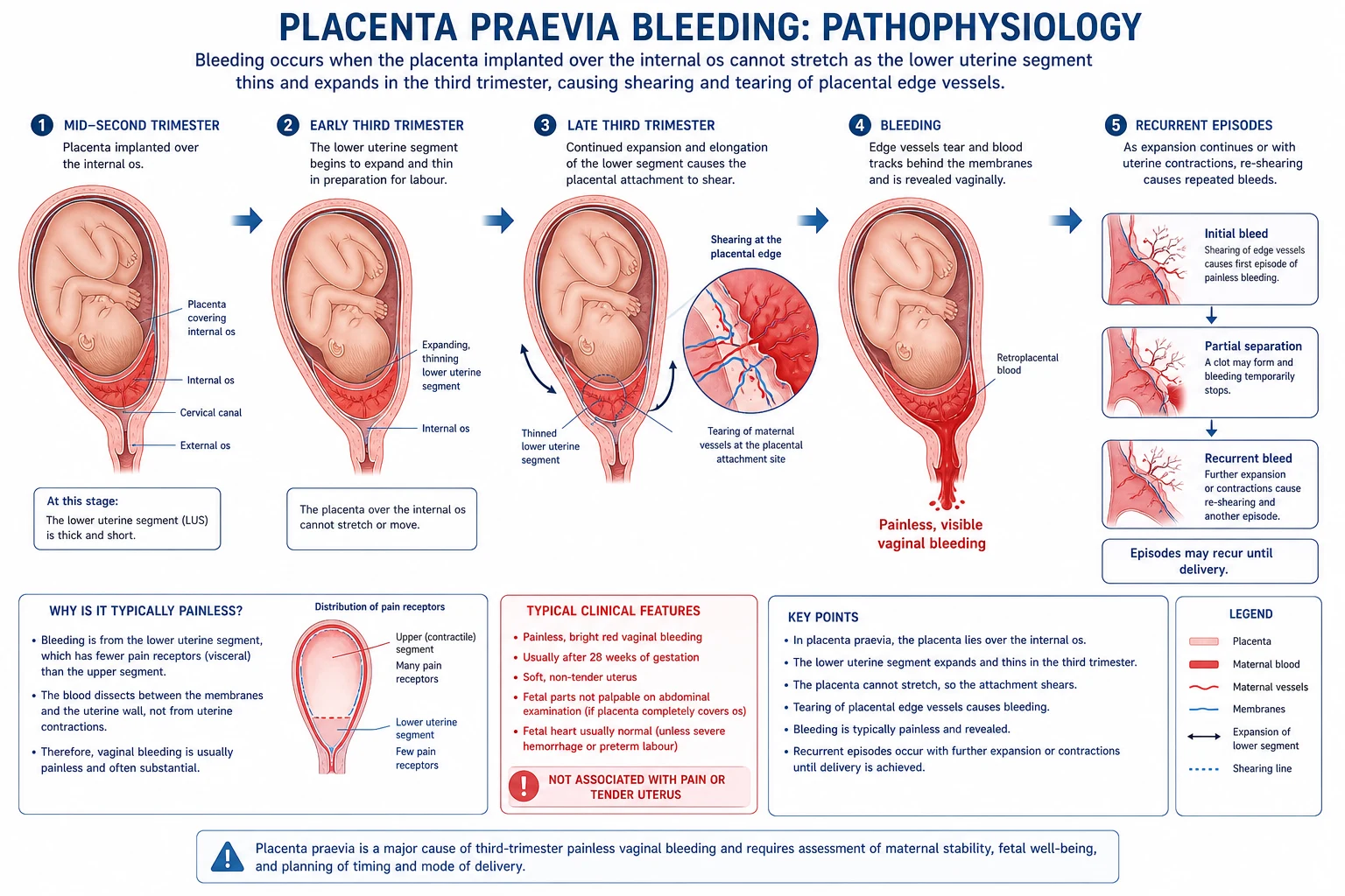
The mechanism of bleeding in praevia is mechanical shearing, which is why the clinical picture is so stereotyped.[1][2]
- Abnormal site of implantation. In a praevia the blastocyst implants low, in the lower uterine segment — the part of the uterus that forms between the isthmus and the cervix and that only develops and thins as pregnancy advances. The decidua in this region is thinner and less well vascularised than in the fundus, which also predisposes to accreta when there is a scar.
- Lower-segment formation in the third trimester. From about 28 weeks the lower segment elongates and thins (from about 1 cm to several centimetres wide) and the cervix begins to efface. The placenta, being relatively inelastic, cannot follow this expansion.
- Shearing of the placental edge. The placental edge is torn away from the uterine wall, opening the intervillous space and the large maternal marginal venous sinuses. These low-pressure venous sinuses bleed bright red, fresh blood — hence the "bright red" appearance and the typically self-limiting first episode (a "warning bleed") as the venous bleed tamponades.
- Painless. The lower uterine segment and cervix are relatively insensitive to stretch compared with the fundus and peritoneum, so the shearing is not felt — unlike the painful separation of a fundal placenta in abruption.
- Recurrent and escalating. Each subsequent episode of lower-segment stretching (or a uterine contraction, intercourse, vaginal examination, or simply standing) shears fresh edge, so bleeds recur and become heavier as pregnancy advances toward term.
- Why the fetus is usually well. Because the bleed is maternal venous blood and typically modest at first, the fetus is not acutely compromised and the fetal heart is usually normal — in contrast to abruption (acute placental separation) and vasa praevia (fetal vessel rupture, fetal blood loss).[1][2]
The placenta accreta spectrum arises by a different but related mechanism. Normal placentation requires the decidua basalis (Nitabuch's layer) to form a cleavage plane between the placenta and the myometrium. A previous caesarean scar destroys this decidua, leaving deficient endometrial re-epithelialisation; when trophoblast implants over the scar it invades directly into or through the myometrium because the normal barrier is absent.[2][3] The deeper the invasion the worse the outcome: accreta (adherent to myometrium), increta (into myometrium), percreta (through serosa, classically into the bladder posteriorly, causing haematuria). After delivery the placenta cannot separate, producing intractable PPH and, usually, the need for caesarean hysterectomy.[3]
Clinical Presentation
The classical presentation is so stereotyped that a single sentence usually secures the diagnosis in a viva, but examiners will probe the atypical and the corners.[1][7]
The classical picture
- Painless vaginal bleeding. The first bleed typically occurs after 20 weeks (often 29 to 34 weeks, sometimes after intercourse or during sleep), is bright red, fresh, and painless. There is no history of trauma, no precipitating contraction. The first episode is usually small — a "warning bleed" — and stops spontaneously within minutes to hours, which can falsely reassure patient and staff.
- Recurrent and escalating. Subsequent bleeds become heavier and more frequent as the lower segment continues to form. A woman with one praevia bleed must be assumed to be at risk of a massive bleed thereafter.
- No abdominal pain (the key differentiator from abruption). Occasionally mild, crampy, lower-abdominal aching accompanies a contraction that precipitated the shearing, but there is no severe constant pain.
- Uterus soft, relaxed, non-tender. The uterus is easily indentable on palpation; there is no hypertonus, no guarding, no woody consistency. The fundal height is appropriate for dates (fetal growth is usually normal).
- Fetal presentation often abnormal. Because the placenta occupies the lower segment, the presenting part cannot engage. Expect a transverse or oblique lie, a breech, or a high, free, mobile, non-engaged head on abdominal examination — an "unstable lie" in the third trimester is a flag for praevia.[1][2]
- No coagulopathy. Maternal clotting is normal (contrast abruption, which can trigger disseminated intravascular coagulation). There is no association with pre-eclampsia.[1][2]
Atypical presentations examiners test:[7]
- Silent/asymptomatic praevia — picked up on routine 20-week scan, never bleeding.
- Presentation as preterm labour — a contraction tears the placental edge, so the bleed is attributed to "show" and the praevia missed until a digital examination provokes torrential bleeding.
- First bleed is massive (rare) — particularly with an anterior praevia over a scar that is starting to invade (accreta), or with coagulopathy.
- Bleeding in a woman with a previous caesarean — assume accreta until disproved; the bleed may be from the accreta site itself.
- Haematuria in a woman with praevia and prior caesarean = percreta invading the bladder — an extreme obstetric emergency.
Differential Diagnosis
Any woman with vaginal bleeding after 20 to 24 weeks has an antepartum haemorrhage, and the differential is the differential of APH.[7] The candidate must generate the list and — critically — distinguish each by its discriminating features, because that is how the marks are awarded.[1][2]
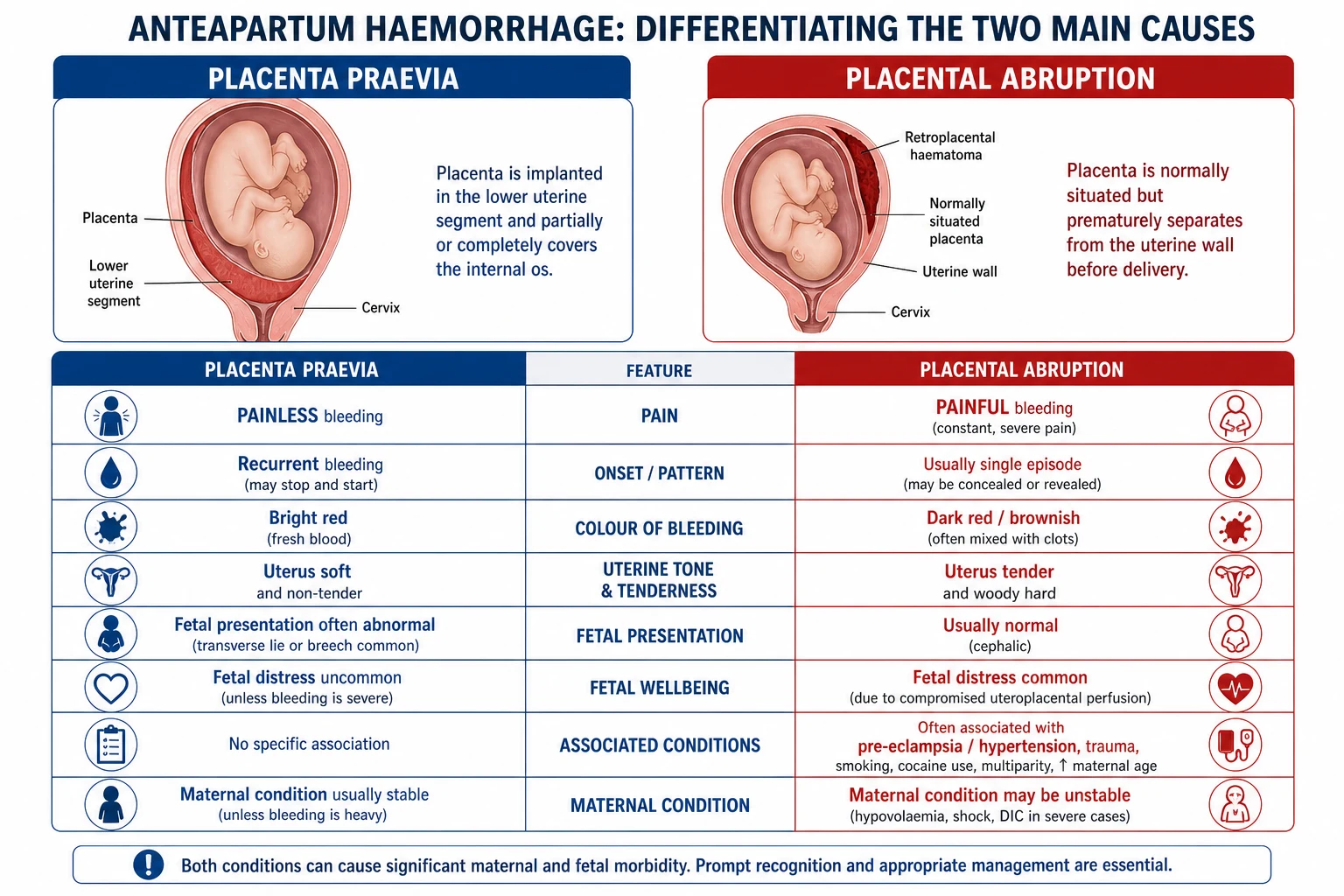
| Feature | Placenta praevia | Placental abruption | Vasa praevia |
|---|---|---|---|
| Site of placenta | Low, over/near os | Normal (fundal) | Variable (velamentous insertion) |
| Pain | Painless | Severe, constant | Painless |
| Blood | Bright red, revealed | Dark, often concealed | Bright red, fetal blood |
| Uterus | Soft, non-tender | Hard, woody, tender | Soft, non-tender |
| Fetal heart | Usually normal | Often abnormal / distress | Sudden bradycardia / sinusoidal CTG |
| Mother | Stable until large bleed | Shock disproportionate to visible loss | Mother haemodynamically normal |
| Onset | Recurrent, escalating | Sudden, single event | At rupture of membranes |
| Coagulopathy | Absent | May develop DIC | Absent |
The non-placental causes of APH that must be excluded by a speculum (never digital) examination once praevia is ruled out on ultrasound include:[1][7]
- Cervical pathology — ectropion, cervical polyp, cervicitis, and cervical cancer (any pregnant woman with postcoital or intermenstrual-type bleeding and an abnormal-looking cervix needs the praevia excluded then a cervical biopsy).[1][7]
- Preterm labour / "show" — blood-tinged mucus plug with regular painful contractions and a cervical change.
- Uterine rupture — in a woman with a previous scar presenting with severe pain, fetal distress, loss of fetal station, and maternal shock; the bleeding may be vaginal but is often intra-abdominal.
- Bloody show at term — small, mucoid, mixed with clear liquefied blood, associated with the onset of labour.
- Marginal sinus rupture of a normally-sited placenta (a minor partial separation that mimics a small praevia bleed).
The causes of antepartum haemorrhage — the 5 'P's and others
APPHIS
painful, dark, woody uterus, fetal distress
painless, bright red, soft uterus
show with contractions
cervical polyp, ectropion, cancer, varices
uterine rupture (previous scar)
vasa praevia — fetal blood, sinusoidal CTG
Clinical & Bedside Assessment
The first principle overrides everything else: in any woman bleeding after 20 weeks, assume a praevia until ultrasound proves otherwise, and do not perform a digital vaginal examination.[1]
Focused assessment in parallel with resuscitation:[1]
- Airway, Breathing, Circulation. In a heavy bleed, assess for hypovolaemic shock: pulse, blood pressure, capillary refill, peripheral perfusion. Two large-bore (14 to 16 G) intravenous cannulae immediately. Resuscitate while assessing — never serially examine a shocked woman.
- History. Gestation, parity, previous caesarean or uterine surgery, last bleed (onset, volume, colour, clots, pads soaked), associated pain or contractions, fetal movements, antenatal ultrasound results (was the placenta low at 20 weeks?), Anti-D status, blood group.
- Abdominal examination. Uterus soft, relaxed, non-tender, fundal height appropriate. Lie and presentation often abnormal — transverse, oblique, breech, or a high, free, non-engaged head that cannot be pushed into the pelvis. There is no acute abdomen, no guarding (unless rupture). Auscultate the fetal heart.
- Speculum examination (NOT digital). Once the woman is stable, inspect the cervix and vagina to identify a local cause (polyp, ectropion, cancer, trauma). This does not exclude a praevia — a speculum cannot.
- Fetal assessment. Continuous CTG to detect fetal compromise. Note that a normal CTG does not exclude abruption or praevia.
- Quantify blood loss. Weigh pads and linen; record ongoing loss. Maternal observations trended continuously.
- Triage decision. Heavy bleeding, maternal shock, or fetal compromise → resuscitate and deliver (emergency caesarean) regardless of gestation. Stable → transvaginal ultrasound to localise the placenta.[1]
Investigations
Investigation has one immediate goal — confirm or exclude a praevia — and, once confirmed, assess the placenta for accreta, gauge fetal wellbeing, and prepare for safe delivery.[1][2]
Transvaginal ultrasound (TVS) is the gold standard for placental localisation.[1] It is safe — the probe sits in the vagina and does not enter the cervical canal, so it does not provoke bleeding — and it is more accurate than transabdominal ultrasound, particularly for a posterior placenta (obscured by the fetus on transabdominal view) and for measuring the exact placental edge to internal os distance. The distance-based classification hinges on this measurement.[1]
- Transabdominal ultrasound is the first-line screening tool at the 20-week anatomy scan. A common pitfall is a false positive caused by bladder overdistension (which compresses the lower segment and makes a normal placenta look low) or a false negative for a posterior placenta hidden behind the fetus. A full bladder should be emptied before declaring a praevia.
- If low at 20 weeks, repeat TVS at 32 weeks. Over 90 percent of low placentas at 20 weeks will have migrated clear of the os by 32 to 36 weeks as the lower segment forms.[1]
- Colour Doppler and MRI for suspected accreta. In any woman with praevia plus a previous caesarean scar, screen for accreta with greyscale plus colour Doppler ultrasound looking for: placental lacunae (irregular vascular spaces), loss of the retroplacental clear (hypoechoic) zone, thinning of the myometrium to under 1 mm, bridging vessels at the uterine-bladder interface, and abnormal placental vascularity.[3][9] MRI is used as an adjunct in complex cases (e.g. posterior accreta, or to map percreta into the bladder) but is not required for routine praevia.[1]
- Pre-operative ultrasound mapping is increasingly used before caesarean in suspected accreta to plan the incision and anticipate the placental edge, reducing blood loss.[9]
- Full blood count — baseline haemoglobin, platelet count (look for the thrombocytopenia of pre-eclampsia or evolving DIC).
- Blood group, antibody screen, and crossmatch — at least 4 units crossmatched in any bleeding or planned-delivery case; have a massive-haemorrhage protocol ready.
- Coagulation screen (PT, APTT, fibrinogen) — usually normal in praevia (contrast abruption), but essential if heavy bleeding or before surgery.
- Kleihauer-Betke / flow cytometry — quantifies fetomaternal haemorrhage to calculate the Anti-D dose in Rh-negative women.
- U&E, LFT — baseline, and to screen for associated pre-eclampsia.
- Rh status — confirm, because every Rh-negative mother needs Anti-D.
Fetal: continuous CTG; consider ultrasound for fetal growth and wellbeing if stable.[1][7]
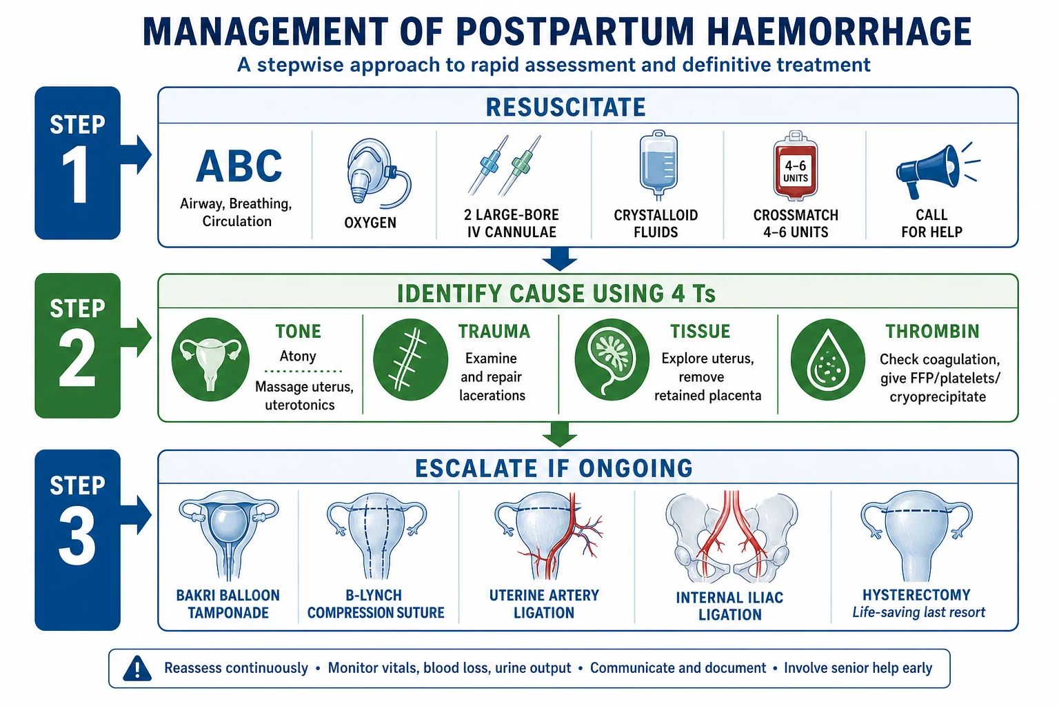
Management — Resuscitation
The APH resuscitation bundle for a heavy praevia bleed
Call for help — senior obstetrician, anaesthetist, midwife-in-charge, alert theatre and blood bank; activate the massive obstetric haemorrhage protocol
Airway + high-flow oxygen; tilt the woman 15 to 30 degrees left lateral to relieve aortocaval compression
Two large-bore (14 to 16 G) IV cannulae; take blood for FBC, coagulation, crossmatch (4 units+), group and save, Kleihauer-Betke
Fluid resuscitation — warm crystalloid (Hartmann's or normal saline 0.9 percent) boluses; target perfusion, not a number; avoid over-resuscitation that worsens bleeding
Transfuse crossmatched blood early if shocked or bleeding continues; use O-negative blood if crossmatch unavailable; give fresh frozen plasma and platelets per the massive haemorrhage protocol when over 1 blood volume is transfused
These steps are based on standard obstetric resuscitation principles and the principles of the RCOG Green-top Guideline 27a massive obstetric haemorrhage protocol.[1]
The dose of Anti-D should be adjusted upward if the Kleihauer-Betke test shows a large fetomaternal haemorrhage — give an additional 100 IU (25 microgram) per 1 mL of fetal bleed above the dose already covered.[1]
Management — Definitive & Stepwise
Definitive management divides into three streams: the stable inpatient preterm woman managed expectantly, the woman at term planned for elective caesarean, and the unstable woman needing emergency delivery. The accreta spectrum is handled as a separate high-risk pathway.[1][10]
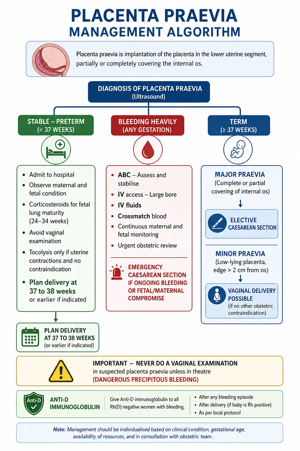
Stable, preterm (under 37 weeks) — expectant management
Once the acute bleed settles and the woman is stable, the aim is to prolong pregnancy safely to allow fetal lung maturation while being ready to deliver at any sign of deterioration.[1]
- Admit to hospital if there is any active bleeding, a major praevia, or gestation under 34 to 36 weeks; the risk of a sudden, heavy, unpredictable bleed is real. Some units allow carefully selected, asymptomatic, low-lying placentas to be managed as outpatients with strict safety-netting.
- Avoid provoking factors — no sexual intercourse, no digital vaginal examination, no strenuous activity or heavy lifting; report any bleed immediately.
- Antenatal corticosteroids for fetal lung maturation to any woman between 24 and 34 weeks 6 days at risk of preterm delivery within 7 days.[1]
- Monitor blood loss (weigh pads), maternal observations, haemoglobin, and fetal wellbeing (CTG and serial growth scans).
- Correct anaemia with oral (or IV) iron; keep haemoglobin ready to bleed.
- Plan delivery — elective caesarean at 36 to 37 weeks for praevia overlapping the os; document the plan and the crossmatch requirements.
The alternative corticosteroid is dexamethasone 6 mg IM, four doses 12 hours apart (24 mg total); efficacy is equivalent.[1]
At term — elective caesarean
- Elective caesarean section for praevia that overlaps the os (major praevia) — planned, daytime, senior obstetrician and senior anaesthetist present, crossmatched blood available, theatre and blood bank briefed.[1][10]
- Timing: 36+0 to 37+0 weeks per NICE NG201 for uncomplicated praevia (balance the small risk of an antenatal bleed against the benefit of additional gestation).[10] Some RCOG guidance allows 36+0 to 37+0; an asymptomatic low-lying placenta managed for vaginal delivery is allowed to labour, with caesarean at 38 weeks if indicated.
- Low-lying placenta (edge 1 to 20 mm from os): individualise. If the edge is over 20 mm, vaginal delivery is appropriate. If within 20 mm, caesarean is recommended because of the bleeding risk during cervical dilation.[1]
- Anaesthesia: regional (spinal or epidural) is preferred in the stable woman — she is awake, the baby is not depressed, and blood loss and thromboembolism are reduced. General anaesthesia is reserved for massive haemorrhage, maternal collapse, failed regional, or a true obstetric emergency; it carries a higher risk of aspiration and difficult airway in pregnancy.[10]
Surgical technique at caesarean for praevia
- The lower-segment transverse incision is usual, but an anterior praevia means the placenta may be incised directly — a senior surgeon is essential, the placenta may need to be cut through to reach the fetus, and bleeding from the placental bed can be torrential.[2]
- If the placenta is anterior and large, a classical (vertical upper-segment) incision may be needed to avoid cutting through it. Be ready to convert.
- After delivery of the baby, await spontaneous separation then deliver the placenta; oxytocin (5 IU IV slow at delivery, then 40 IU in 500 mL Hartmann's over 4 hours) is given to contract the upper segment — but remember the lower segment contracts poorly, so PPH is common and bimanual compression, uterotonics (oxytocin, ergometrine, carboprost, misoprostol), and uterine balloon tamponade or brace sutures may be needed.[1]
- Placenta accreta found unexpectedly at caesarean — if the placenta will not separate, do not forcibly extract it (this provokes catastrophic haemorrhage). Options are caesarean hysterectomy (definitive) or leaving the placenta in situ with planned delayed hysterectomy in selected, stable, counselled cases.[3][8]
Placenta accreta spectrum — the high-risk pathway
Suspected accreta is one of the highest-risk scenarios in obstetrics and must be managed in a multidisciplinary centre of excellence.[3][8]
Multidisciplinary plan for suspected placenta accreta spectrum
Antenatal diagnosis by expert ultrasound + MRI: lacunae, loss of clear space, thin myometrium, bridging vessels, bladder invasion
Counsel the woman explicitly about probable caesarean hysterectomy, massive transfusion, ICU admission, and possible bladder injury
Schedule elective caesarean at 35+0 to 36+6 weeks (NICE) to avoid labour and unplanned bleeding
Senior team on the day: consultant obstetrician, consultant anaesthetist, urologist on standby, interventional radiology, haematology, neonatology
Consider preoperative bilateral internal iliac artery balloon catheters or uterine artery embolisation to reduce bleeding
Two large-bore IV cannulae, arterial line, cell salvage, 6+ units crossmatched, massive haemorrhage protocol activated
Surgical approach: often classical incision avoiding the placenta; if accreta confirmed and bleeding uncontrolled — caesarean hysterectomy
Postoperative: ICU/HDU, monitor for ongoing bleeding, coagulopathy, renal failure; psychological support and debrief
Anti-D
Give Anti-D immunoglobulin to every Rh-negative, non-sensitised woman after any praevia bleed, within 72 hours, and check a Kleihauer-Betke for the size of fetomaternal haemorrhage.[1]
Specific Subtypes & Scenarios
Asymptomatic low-lying placenta at 20 weeks
the common scan finding
- Over 90 percent migrate clear of the os by 32 to 36 weeks
- Repeat **transvaginal scan at 32 weeks** before declaring praevia
- No restriction needed if asymptomatic
- Counsel: report any bleeding immediately
Minor praevia (edge within 20 mm, not overlapping)
individualised delivery
- Caesarean recommended if edge within 20 mm at term
- Trial of vaginal delivery reasonable if edge over 20 mm
- Warn of bleeding risk in labour; deliver where caesarean is immediately available
- Anti-D if Rh-negative and any bleed
Major praevia (overlaps os)
caesarean mandatory
- Elective caesarean at 36 to 37 weeks
- Senior surgeon and anaesthetist, crossmatched blood
- Anticipate cutting through an anterior placenta
- High PPH risk — uterotonics ready
Placenta accreta spectrum
morbidly adherent
- Antenatal US + MRI diagnosis; praevia + previous scar = screen
- Multidisciplinary centre of excellence
- Elective CS at 35 to 36 weeks; likely caesarean hysterectomy
- Highest maternal morbidity and transfusion need
Vasa praevia (the mimic)
fetal vessels over membranes
- Velamentous or succenturiate cord insertion
- Painless bleed **at rupture of membranes**
- **Fetal** blood loss — sudden bradycardia / sinusoidal CTG
- **Mother haemodynamically normal**; emergency caesarean to save the fetus
Praevia + previous caesarean scar
the dangerous combination
- Screen for accreta with Doppler + MRI
- Accreta risk rises with each scar (up to 60 to 70 percent with 4+ scars)
- Plan multidisciplinary, elective, daytime delivery
- Discuss hysterectomy and transfusion in advance
Complications & Pitfalls
Placenta praevia and its management carry a substantial complication burden — the candidate should be able to list maternal and fetal complications separately.[1][2][3] Maternal complications:[1][3]
-
Antepartum haemorrhage — recurrent, unpredictable, occasionally massive; the leading acute threat.
-
Postpartum haemorrhage — the lower uterine segment contracts poorly compared with the fundus, so the placental bed continues to bleed after delivery; PPH occurs in up to a third of major praevia cases.
-
Placenta accreta spectrum — morbid adherence, especially with a previous scar; may mandate caesarean hysterectomy.
-
Surgical injury at caesarean — bladder, ureter, and uterine vessel damage, particularly in the accreta/hysterectomy setting.
-
Transfusion-related complications — volume overload, transfusion reaction, infection, transfusion-related acute lung injury.
-
Thromboembolism — the combination of pregnancy, surgery, bed rest, and anaemia raises venous thromboembolism risk; prophylaxis is needed.
-
Infection — wound, endometritis, urinary.
-
Hysterectomy — for accreta or uncontrolled PPH; loss of fertility.
-
Psychological morbidity — antenatal admission, emergency surgery, neonatal unit admission, possible hysterectomy. Fetal and neonatal complications:[1][2]
-
Preterm delivery — iatrogenic (for maternal/fetal indication) or after spontaneous preterm labour; prematurity is the chief driver of perinatal morbidity.
-
Fetal growth restriction — marginally increased; the praevia placental bed is less well perfused.
-
Fetal blood loss / anaemia — rare, except with coexisting vasa praevia or large fetomaternal haemorrhage.
-
Perinatal mortality — elevated, driven almost entirely by prematurity; with term delivery, mortality approaches background.
-
Neonatal unit admission for prematurity or resuscitation.[1][2]
-
Performing a digital vaginal examination in suspected praevia → catastrophic bleeding. The cardinal error.
-
Missing a posterior praevia on transabdominal ultrasound (fetus obscures it) — always confirm with TVS.
-
Declaring a praevia on a full bladder — overdistension falsely low-reads the placenta; empty the bladder first.
-
Not repeating the 20-week low placenta scan at 32 weeks — most migrate; over-diagnosis of praevia causes unnecessary caesarean.
-
Missing accreta in a woman with praevia plus a previous scar — failure to screen with Doppler/MRI and plan a multidisciplinary delivery.
-
Operating without crossmatched blood — praevia and accreta bleed torrentially; the unit must be prepared.
-
Forgetting Anti-D in the Rh-negative mother after a bleed — sensitises future pregnancies.
-
Forcibly extracting an accreta placenta at caesarean → exsanguination.[1][3]
Prognosis & Disposition
With modern antenatal ultrasound, planned multidisciplinary delivery, and a rapid response to bleeding, maternal mortality is under 1 percent and maternal morbidity, though substantial, is manageable.[1][3] The bulk of maternal risk is concentrated in the accreta spectrum and in women who present in shock or who are managed outside a prepared unit.[3][8]
Perinatal mortality is higher than background, almost entirely attributable to prematurity (delivery for maternal or fetal indication before term) and to the rare catastrophic antepartum bleed; with delivery at 36 to 37 weeks and a stable mother, perinatal outcome is good.[1]
Outcomes and disposition
- Minor/low-lying praevia: most migrate; the woman can usually expect a vaginal delivery if the edge clears 20 mm by term.
- Major praevia: planned caesarean; good outcome if planned and prepared.
- Accreta spectrum: high morbidity — hysterectomy in the majority, large transfusion, ICU admission, longer hospital stay; outcome best in centres of excellence.[3]
- Recurrence: a 4 to 8 percent chance of praevia in a subsequent pregnancy; counsel about the cumulative risk of repeat caesarean.[2]
Disposition: any woman with a praevia bleed or a major praevia at term is managed in an obstetric unit with theatre, anaesthesia, blood bank, and neonatal facilities on site; regional transfer to a centre of excellence is recommended for suspected accreta before labour.[3] Safety-net advice on discharge: report any bleeding, contraction, or reduction in fetal movements immediately.
Special Populations
- Previous caesarean scar — the single most important modifier. Every additional scar raises both the risk of praevia in the next pregnancy and, if praevia occurs, the risk of accreta: roughly 3 percent with no scar, 11 percent after one scar, 40 percent after two, and 60 to 67 percent after four or more scars.[5][6] These women must have accreta screening and a planned, multidisciplinary delivery.[3]
- Multiple pregnancy (twins, triplets) — the larger placenta extends toward the os, increasing praevia risk; management principles are unchanged but the bleeding risk and PPH risk are higher.
- IVF / assisted reproduction — about a two- to threefold increased praevia risk; the principles of diagnosis and management are the same.
- Women with abnormal placentation discovered at term without prior scan — manage as a major praevia with accreta precautions; senior team, crossmatched blood, and a low threshold for hysterectomy.
- The Jehovah's Witness patient — blood transfusion may be refused; plan early, optimise haemoglobin antenatally with IV iron and erythropoietin, use cell salvage where accepted, and counsel explicitly about the small but real risk of maternal death from haemorrhage.[1]
- The teenager or older mother — age over 35 independently increases risk; under-18s have lower risk but higher rates of late booking and missed scans.
- Previous uterine surgery (myomectomy, metroplasty) — manage as if previous caesarean; accreta precautions apply.
Evidence, Guidelines & Regional Differences
The landmark evidence and guideline base for placenta praevia is concentrated in three documents the candidate must be able to name.[1][3][10]
RCOG Green-top Guideline 27a/27b — Jauniaux 2019
BJOG
Clinical practice guideline (Green-top 27a/27b), RCOG
Key finding
Established transvaginal ultrasound as the gold standard for placental localisation; replaced the four-grade system with a distance-based classification (praevia overlaps the os; low-lying within 20 mm); set elective caesarean at 36-37 weeks for praevia and 35-36 weeks for accreta; mandated Doppler and MRI screening for accreta in praevia plus a previous scar.
Practice change
Distance-based classification and multidisciplinary accreta pathway now standard UK and international practice.
Center of Excellence for Placenta Accreta — Silver 2015
Am J Obstet Gynecol
Multidisciplinary consensus and cohort review
Key finding
Showed that accreta spectrum managed in a prepared centre of excellence (antenatal diagnosis, senior surgical and anaesthetic team, interventional radiology, cell salvage, planned daytime delivery) had lower maternal morbidity and mortality and less massive transfusion than unprepared emergency management.
Practice change
Established accreta as a condition best managed in a multidisciplinary centre of excellence, with antenatal diagnosis and planned delivery.
Ananth 1997 meta-analysis and Wu 2005 twenty-year analysis — praevia and accreta epidemiology
Am J Obstet Gynecol
Meta-analysis (Ananth) and population-based cohort (Wu)
Key finding
Quantified that previous caesarean and previous abortion independently increase the risk of placenta praevia, and that the incidence of placenta accreta has risen over twenty years in parallel with caesarean rates — accreta risk after praevia rises from about 3 percent with no scar to 40-67 percent with four or more scars.
Practice change
Underpins modern guidance to screen every praevia plus a previous scar for accreta and to counsel on cumulative scar risk.
- UK (NICE NG201, RCOG GTG 27a, 2019): universal 20-week anomaly scan; if low, repeat TVS at 32 weeks; elective caesarean at 36+0 to 37+0 weeks for praevia; accreta screening by Doppler ± MRI; accreta delivery 35+0 to 36+6; multidisciplinary team required.
- US (ACOG): elective caesarean at 36+0 to 37+6 weeks for praevia; for suspected accreta spectrum, delivery at 34+0 to 35+6 weeks (slightly earlier than RCOG), with a multidisciplinary care team and informed consent for possible hysterectomy.
- India (ICMR/FOGSI-aligned practice): principles follow RCOG; in practice, late booking and uneven ultrasound access mean some praevias present unbooked with bleeding — every obstetric unit needs a written APH and massive-haemorrhage protocol and guaranteed crossmatched blood.[7]
- Terminology: the four-grade system (I to IV) persists in older textbooks and many Indian, USMLE, and PLAB MCQs; the modern distance-based system is used in current UK vivas and SAQs. The candidate should know both and state which is being used.
Where the evidence is weak: the optimal timing of delivery for low-lying placenta (edge within 20 mm) aiming for vaginal delivery is still debated; the best surgical strategy for accreta (immediate caesarean hysterectomy versus conservative, placenta-left-in-situ management) is individualised and evidence is largely observational.[3][8]
Exam Pearls
- PAINLESS, causeless, recurrent bright-red bleeding after 20 weeks = praevia until ultrasound proves otherwise.[1]
- Uterus SOFT and NON-TENDER (vs abruption: hard, woody, tender).[2]
- Fetal heart usually NORMAL in the early praevia bleed (maternal blood loss; contrast vasa praevia — fetal blood, sudden bradycardia).[2]
- Transvaginal ultrasound = gold standard for placental localisation; safe (probe does not enter the canal) and more accurate than transabdominal, especially for posterior placenta.[1]
- NEVER do a digital vaginal examination in suspected praevia — only in theatre with crossmatched blood and the team ready ("double setup").[1]
- Repeat the 20-week low placenta at 32 weeks — over 90 percent migrate.[1]
- Caesarean for praevia at 36 to 37 weeks (NICE); for low-lying placenta, vaginal delivery reasonable if edge is over 20 mm from os.[10]
- Previous caesarean + praevia = high accreta risk — screen with Doppler/MRI, plan multidisciplinary delivery.[3]
- Accreta: adherent. Increta: into myometrium. Percreta: through serosa (bladder).[2]
- PPH is common — the lower segment contracts poorly; have uterotonics and the PPH protocol ready.[1]
- Anti-D for every Rh-negative woman within 72 hours of any bleed; Kleihauer-Betke to dose for large fetomaternal haemorrhage.[1]
- Corticosteroids (betamethasone 12 mg IM, two doses 24 hours apart) if 24 to 34 weeks and at risk of preterm delivery.[1]
- Abruption = painful, dark, hard uterus; praevia = painless, bright red, soft uterus. The single most-tested distinction.[2]
- Vasa praevia = fetal blood, sinusoidal CTG, mother well, bleed at ROM — emergency caesarean for the fetus.[2]
Spot test: a woman at 32 weeks with a previous caesarean presents with painless bright-red bleeding, soft non-tender uterus, transverse lie, normal CTG. What is the diagnosis, the next investigation, and the one examination you must NOT do?
Diagnosis: placenta praevia (with high suspicion of accreta given the previous scar). Next investigation: transvaginal ultrasound to localise the placenta and measure the edge-to-os distance, plus Doppler/MRI to screen for accreta. Must NOT do: a digital vaginal examination — it can provoke catastrophic bleeding; only a speculum (to exclude a local cause) is permitted.[1][2]
Exam application bank (NEET-PG / INICET)
One-line answer
Placenta praevia = placenta implanted wholly or partly in the lower uterine segment, over or near the internal cervical os. Incidence about 0.3 to 0.5 percent of pregnancies at term (higher earlier because most low placentas migrate). Classic: painless, causeless, recurrent bright-red vaginal bleeding after 20 to 24 weeks; uterus soft and non-tender (unlike abruption). Transvaginal ultrasound is the gold standard; NEVER do a digital vaginal examination until praevia is excluded. Caesarean for praevia where the placenta overlaps the os, at 36 to 37 weeks. Previous caesarean + praevia = high placenta accreta spectrum risk.
Worked stems (answer without another resource)
Stem 1 — Classic presentation. Map symptoms to mechanism; name the first investigation and first treatment step with dose/route if drug therapy is standard. [1]
Stem 2 — Unstable / complicated. List red flags that force immediate resuscitation, theatre, ICU, antidote, or reperfusion — and what you do in the first 15 minutes. [1]
Stem 3 — Atypical group. Elderly, pregnancy, child, or immunocompromised: how presentation and thresholds change. [1]
Stem 4 — Differential trap. Name the three closest mimics and one discriminator for each. [1]
Stem 5 — Disposition. Who goes home with safety-netting, who is admitted, who needs HDU/ICU/theatre, and what follow-up is mandatory. [1]
Rapid viva checklist
- Definition + classification
- Pathophysiology chain
- Bedside signs / criteria
- Score with exact components (if any)
- Emergency bundle
- Definitive therapy with doses
- Complications of disease and of treatment
- Special populations
- Guideline/trial name if classic
- Three exam traps
Coverage self-check
If you cannot answer any stem above from this page alone, re-read the matching section — the page is intended to be self-sufficient for final-prof and NEET-PG/INICET questions on Placenta Praevia.
References
- [1]Jauniaux E, Alfirevic Z, Bhide AG, Belfort MA, Burton GJ, Collins SL, Dornan S, Jurkovic D, Kayem G, Kingdom J, Silver R, Sentilhes L; Royal College of Obstetricians and Gynaecologists. Placenta Praevia and Placenta Accreta: Diagnosis and Management: Green-top Guideline No. 27a BJOG, 2019.PMID 30260097
- [2]Oyelese Y, Smulian JC. Placenta previa, placenta accreta, and vasa previa Obstet Gynecol, 2006.PMID 16582134
- [3]Silver RM, Fox KA, Barton JR, et al. Center of excellence for placenta accreta Am J Obstet Gynecol, 2015.PMID 25460838
- [4]Ananth CV, Smulian JC, Vintzileos AM. The association of placenta previa with history of cesarean delivery and abortion: a metaanalysis Am J Obstet Gynecol, 1997.PMID 9396896
- [5]Wu S, Kocherginsky M, Hibbard JU. Abnormal placentation: twenty-year analysis Am J Obstet Gynecol, 2005.PMID 15902137
- [6]Silver RM. Delivery after previous cesarean: long-term maternal outcomes Semin Perinatol, 2010.PMID 20654776
- [7]Yonke N, Leeman LM, Bykerk VP, Korte JE. Late Pregnancy Bleeding Am Fam Physician, 2025.PMID 41533409
- [8]Waseem S, Fatima A, Rehman A, et al. The quintessential high-risk profile: advanced management strategies for placenta accreta spectrum in patients with advanced maternal age and IVF conception BMC Pregnancy Childbirth, 2026.PMID 42337440
- [9]Aryananda RA, Hu M, Metz TD, et al. Diagnostic ultrasound to inform the surgical approach to cesarean delivery in patients at high risk for placenta accreta spectrum disorders Am J Obstet Gynecol, 2025.PMID 40784608
- [10]Coates D, Homer C, Wilson A, Deady L, Mason E. Indications for, and timing of, planned caesarean section: A systematic analysis of clinical guidelines Women Birth, 2020.PMID 31253513