Obstetrics & Gynaecology · Obstetrics & Gynaecology
Placental Abruption
Also known as Abruptio placentae · Accidental haemorrhage · Premature separation of placenta · Couvelaire uterus · Retroplacental haemorrhage
Placental abruption (abruptio placentae) = premature separation of a normally-situated placenta from the uterine wall before delivery of the fetus. Incidence about 1 in 100 pregnancies; severe (fetal death) about 1 in 1000. Classic triad: painful vaginal bleeding + hard, woody, tender hypertonic uterus + fetal distress. Two patterns: revealed (about 80%, blood tracks via cervix, visible, less dangerous) and concealed (about 20%, blood trapped behind placenta, shock disproportionate to visible loss, more dangerous). Pathology: decidual vasculopathy (pre-eclampsia) leads to spiral artery rupture, retroplacental clot, extension of separation, and in severe cases Couvelaire uterus (blood infiltrates myometrium to serosa, uteroplacental apoplexy) and DIC (thromboplastin release, fibrinogen under 2 g/L). Top risk factor: pre-eclampsia/hypertension; recurrence 6 to 25 percent. Management: resuscitate in parallel with delivery planning; correct coagulopathy before any surgery; fetus alive + distress leads to emergency caesarean; fetus dead leads to vaginal delivery unless maternal indication. Watch for refractory atonic PPH, AKI and Sheehan syndrome.
On this page & tools
Your progress
Saved locally on this device.
Exam tags
Red flags
Overview & Definition
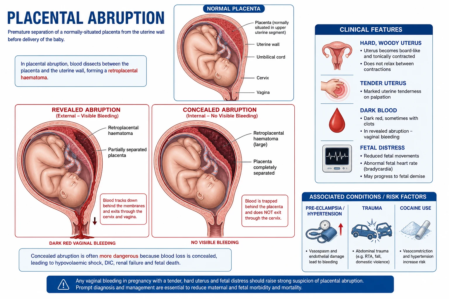
Placental abruption (Latin abruptio placentae, also called accidental haemorrhage in older British texts, and premature separation of the normally situated placenta) is the partial or complete detachment of a placenta implanted in the normal upper uterine segment from its uterine attachment before the fetus is delivered. The event tears the maternal spiral arteries of the decidua basalis, producing a retroplacental haematoma that, depending on its route of escape, may present with dramatic visible bleeding or with almost no external blood at all.[1]
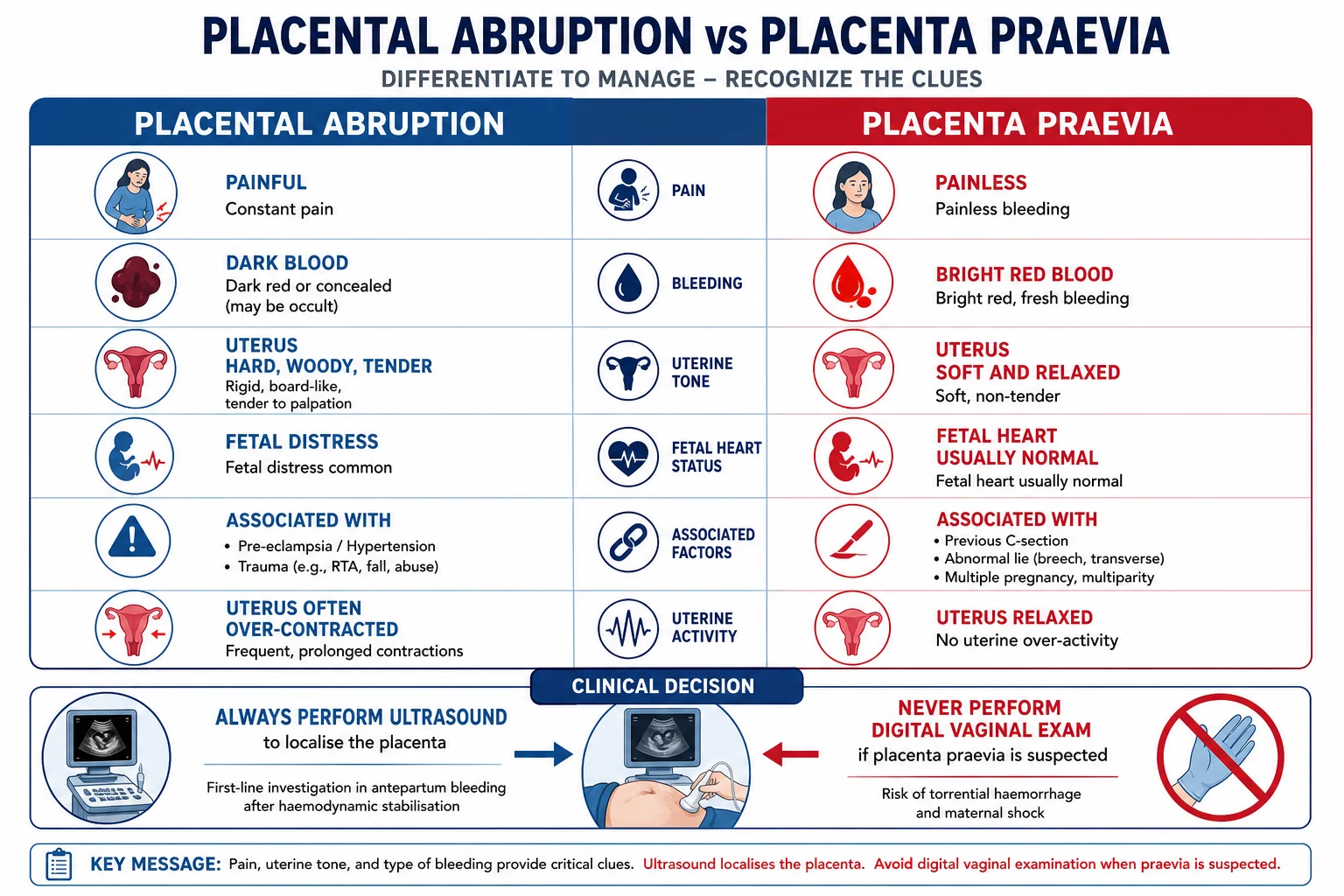
The defining word is normally situated. A placenta in the lower segment that bleeds is placenta praevia, a separate entity with a painless bright-red bleeding pattern, a soft relaxed uterus and an essentially normal fetal heart. Abruption, in contrast, classically presents with pain, a tender hypertonic uterus and fetal compromise. Distinguishing the two at the bedside is the single highest-yield discriminator examined in MBBS, USMLE and PLAB stems, and the historical "double" or "accidental" label versus praevia's "unavoidable" haemorrhage still appears in Indian textbooks.[1]
Abruption complicates roughly 0.3 to 1 percent of all pregnancies, with severe abruption (defined by fetal death) occurring in about 1 in 1000. It is one of the three leading causes of antepartum haemorrhage, alongside placenta praevia and vasa praevia, and remains a major driver of perinatal mortality, iatrogenic preterm birth and acute maternal morbidity — chiefly through coagulopathy, massive haemorrhage, the need for emergency operative delivery, and the longer-term cardiovascular consequences that are now recognised to follow affected women for life.[1][14]
Abruption — the headline numbers
Classification
Abruption is described along two orthogonal axes — the clinical pattern of bleeding (revealed vs concealed vs mixed) and the severity of separation (grade 1 mild, grade 2 moderate with fetal distress, grade 3 severe with fetal death and often coagulopathy). The clinical pattern tells you where the blood is going; the severity grade tells you what to do next.[1]
Revealed (about 80%)
external bleeding visible
- Blood dissects between membranes (chorion) and uterine wall
- Exits through cervix as **dark red, old-looking vaginal bleeding**
- Visible blood loss roughly tracks the true loss — easier triage
- Placental separation is usually marginal or partial
- Generally less dangerous; maternal shock is proportionate to visible loss
Concealed (about 20%)
blood trapped behind placenta
- Blood is locked behind an intact marginal placental edge as a **retroplacental clot**
- **No or minimal visible bleeding** despite large concealed loss
- **Shock disproportionate to visible blood** is the cardinal sign
- Uterus enlarges and becomes **woody hard, tender, hypertonic**
- Higher fetal mortality, higher rate of DIC and Couvelaire uterus
Mixed
partial escape
- Some blood trapped behind placenta, some escapes via cervix
- Common in clinical practice — pure patterns are textbook
- Severity is gauged on **clinical state**, not on visible blood alone
The severity grading most widely taught (after Sheridan and RCOG) is a three-tier clinical stratification:[1]
Grade 1 (mild)
minor separation
- Small retroplacental clot, often no fetal distress
- **No maternal shock, no coagulopathy**
- May present with light bleeding and uterine irritability
- Often only diagnosed retrospectively on placental inspection (clot on maternal surface)
Grade 2 (moderate)
fetal distress present
- Larger separation, **fetal heart abnormal** (decelerations, bradycardia, tachycardia)
- **Maternal vital signs may be abnormal** but no overt shock
- Coagulopathy is possible but uncommon
- Action: stabilise, deliver if fetus compromised
Grade 3 (severe)
fetus dead, often DIC
- Massive separation — **intrauterine fetal death** is the hallmark
- Maternal shock, often disproportionate to visible blood
- **DIC in up to 30 percent** of grade 3 abruptions
- Couvelaire uterus common — anticipate refractory atonic PPH
- Subdivided as 3a without coagulopathy, 3b with coagulopathy
The clinical pattern (revealed/concealed) and the severity grade are independent: a small revealed abruption may be grade 1, while a large concealed abruption is usually grade 3. Concealed abruption carries the worst prognosis precisely because the trapped retroplacental clot is large before any external bleeding declares itself.[1]
Epidemiology & Risk Factors
Reported incidence ranges from 0.3 percent in low-risk populations to over 1 percent in tertiary units that receive high-risk referrals, with severe abruption complicated by fetal death in roughly 0.1 percent (about 1 in 1000 births).[1][5] Population-based data show substantial international variation — rates are highest in the United States and parts of Latin America and lowest in the Nordic countries — driven by both the prevalence of risk factors (hypertensive disease, smoking, advanced maternal age) and by ascertainment and coding differences.[5] The perinatal mortality attributable to abruption is between 10 and 30 percent of cases, the variance reflecting gestational age and time to delivery; abruption accounts for around 10 percent of all perinatal deaths and a much higher fraction of late-pregnancy stillbirths.[1]
The risk factors cluster into a small number of mechanistic themes — vascular injury (the dominant pathway), mechanical shearing, uterine overdistension, acute decompression, direct placental disease, and iatrogenic triggers — and a history of abruption itself confers a very high recurrence risk. The single strongest association is with pre-eclampsia and chronic hypertension, mediated by decidual vasculopathy.[2][3]
Vascular
the dominant pathway
- **Pre-eclampsia, eclampsia, gestational hypertension** (odds ratio about 4 to 5)
- Chronic hypertension (especially poorly controlled)
- Type 1 and type 2 diabetes, chronic renal disease, autoimmune disease
- Thrombophilias — factor V Leiden, prothrombin gene mutation, antiphospholipid syndrome
- Mechanism: failed trophoblastic invasion → spiral artery vasculopathy → rupture
Prior & recurrent
highest single risk
- **Previous abruption** — recurrence 6 percent after one, rising to over 25 percent after two
- Recurrence is higher if index abruption was severe or preterm
- Counsel high-risk women and plan surveillance in next pregnancy
Mechanical / trauma
external force
- **Road traffic accidents** (deceleration, seat-belt injury, blunt abdominal trauma)
- **Domestic violence / intimate partner violence** — frequently under-asked, common
- Falls, direct abdominal blow
- External cephalic version, motor vehicle trauma in late pregnancy
- **Amniocentesis / chorionic villus sampling** (rare iatrogenic)
Uterine overdistension
stretch on placenta
- **Multiple pregnancy** (twins, higher-order)
- **Polyhydramnios** (acute or chronic)
- Macrosomia
- Mechanism: increased shearing force on the placental attachment
Sudden decompression
rapid volume loss
- **Sudden rupture of membranes** in polyhydramnios (acute uterine decompression)
- **Delivery of first twin** (rapid reduction in uterine volume)
- Therapeutic amniodrainage
- Avoid artificial rupture of membranes (ARM) where possible in at-risk women
Substance & lifestyle
modifiable
- **Cigarette smoking** — dose-dependent risk, doubles the incidence
- **Cocaine** — intense vasospasm of spiral arteries
- **Alcohol and other vasoactive drugs**
- Smoking and pre-eclampsia act synergistically
Maternal demographics
non-modifiable
- **Advanced maternal age** (over 35, and especially over 40)
- **Multiparity** — risk rises with parity, possibly confounded by age
- African, South Asian and Hispanic ethnicity (independent in some cohorts)
- Low socioeconomic status, low antenatal attendance
Placental & membrane
local factors
- **Short umbilical cord**
- **Chorioamnionitis** (intra-amniotic infection)
- **Preterm prelabour rupture of membranes (PPROM)**
- Placental anomalies, velamentous cord insertion
- **Assisted reproductive technology (ART)** conceptions — about 2-fold increased risk
The population-attributable risk is dominated by hypertensive disease and smoking, which together account for a large fraction of abruption in population studies; maternal cigarette smoking doubles the risk and acts synergistically with hypertension.[4] Trauma — particularly road traffic collisions and intimate partner violence — is an under-recognised contributor that must be asked about sensitively, and is one of the few potentially preventable precipitants.[8]
India and low- and middle-income settings: the burden of abruption is amplified by structural gaps. Domestic violence and road traffic trauma are common precipitants; pre-eclampsia is frequently diagnosed late because of poor antenatal coverage; widespread maternal anaemia converts a 500 mL retroplacental bleed into a near-fatal event by eroding physiological reserve; and delays in reaching a facility with blood, theatre and neonatal care push up both fetal and maternal mortality. Community awareness of danger signs (severe constant abdominal pain, bleeding, absent fetal movements) and a robust referral pathway are the principal public-health levers. The WHO and Government of India "Janani Shishu Suraksha Karyakram" schemes are designed to remove user-fee and transport barriers for exactly these emergencies.
Pathophysiology
The final common pathway of every abruption is the tearing of the decidual spiral arteries, producing a retroplacental haematoma that physically strips the placenta from its attachment. What initiates that tear varies — but in the great majority of cases the root pathology is decidual vasculopathy of the spiral arteries, the same lesion that underlies pre-eclampsia and fetal growth restriction.[1]
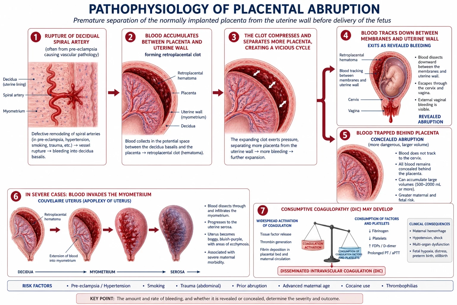
Step 1 — Decidual vasculopathy. In normal placentation, extravillous trophoblast invades and remodels the spiral arteries in two waves (early second trimester in the decidual segments, late second trimester in the myometrial segments), converting them from high-resistance narrow muscular vessels into wide low-resistance capacitance channels. When this invasion is shallow or abnormal — the central defect of pre-eclampsia — the arteries retain smooth muscle, undergo acute atherosis (fibrinoid necrosis with lipid-laden macrophages) and thrombosis, becoming structurally weak and functionally obliterative. They are primed to rupture under the shearing forces generated by uterine contractions or even minor trauma.[3]
Step 2 — Spiral artery rupture. A diseased spiral artery splits. Blood escapes from the high-pressure maternal circulation into the decidua basalis, dissecting a plane between the placenta and the myometrium. Where the dissection runs down between the membranes and the uterine wall and reaches the cervix, the result is revealed abruption; where the marginal placental edge remains sealed and the blood is locked behind the placenta, the result is concealed abruption.[1]
Step 3 — The retroplacental haematoma and the vicious cycle. The clot now sits between placenta and uterine wall. As it expands, it physically compresses the placental plate, shearing more decidua, tearing more spiral arteries, and producing more bleeding and more clot. The placenta overlying the clot infarcts, and the separating surface area grows. The uterus is progressively distended by the expanding clot, which is one reason for the increasing fundal height sometimes seen in concealed abruption.[1]
Step 4 — Uterine hypertonus. Irritation of the myometrium by the retroplacental clot produces sustained tetanic contraction — the uterus does not relax, the "woody hard" fundus is born, and palpation is exquisitely tender. The high resting tone also reduces uteroplacental perfusion (the spiral arteries are compressed by the contracted muscle), accelerating fetal hypoxia and, if unrelieved, fetal death.[2]
Step 5 — Couvelaire uterus. When the retroplacental pressure is severe, blood tracks through the myometrial wall by dissection along the myometrial fibres, reaching the serosal surface. The uterus is now stained purple-blue, suffused with blood — classically called uteroplacental apoplexy or Couvelaire uterus (after the French obstetrician who described it in 1911). The damaged muscle cannot contract effectively after delivery, which is the proximate cause of the refractory atonic postpartum haemorrhage that complicates severe abruption. Despite its dramatic appearance, Couvelaire uterus is not by itself an indication for hysterectomy — many will contract if supported with uterotonics, uterine massage and time.[1]
Step 6 — DIC. The decidua basalis and the damaged placenta are extraordinarily rich in tissue factor (thromboplastin). When a large area is breached, tissue factor pours into the maternal venous sinuses, igniting the extrinsic coagulation cascade systemically. The result is disseminated intravascular coagulation — widespread microvascular thrombosis, consumption of platelets and clotting factors (especially fibrinogen, factors V and VIII), and secondary hyperfibrinolysis. The laboratory signature is prolonged PT and APTT, falling fibrinogen (the most sensitive marker — pregnancy baseline is 4 to 6 g/L; a level under 2 g/L signals severe coagulopathy), thrombocytopenia, and markedly elevated D-dimer and fibrin degradation products.[12][13]
Pregnancy is a hypercoagulable state at baseline (fibrinogen doubled to around 5 g/L, increased factors VII, VIII, X and von Willebrand factor, depressed protein S, acquired activated protein C resistance), so a pregnant woman tolerates small amounts of tissue factor release well. This is why mild abruptions do not consume clotting factors. Once the insult exceeds hepatic synthetic capacity, fibrinogen falls sharply — the inflection point that defines severe DIC.[12]
Clinical Presentation
The textbook triad — painful vaginal bleeding, a hard woody tender hypertonic uterus, and fetal distress — is seen in fully expressed severe disease, but in clinical practice the presentation is a spectrum and missing the early signs is the commonest error.[2]
The tempo is highly variable. The classical patient presents in the third trimester (most commonly after 28 weeks, with incidence rising to term) with the sudden onset of severe abdominal pain and vaginal bleeding. A smaller group present earlier in the second trimester with what seems like preterm labour — frequent, painful contractions and a small bleed — and the diagnosis is only made when the placenta is delivered and a clot is found on the maternal surface. Atypical presentations are common and dangerous:[2]
- "Silent" concealed abruption — a woman arrives shocked and pale with little or no vaginal bleeding, in severe constant abdominal pain. The visible loss is a poor guide to severity.
- Preterm labour — abruption presenting as idiopathic preterm labour is one of the classic "missing it" scenarios. A woman who is contracting frequently and painfully, has a tender uterus, and a small dark show, may have an underlying small abruption.
- Fetal distress alone — an abnormal CTG in a woman who is not in labour, particularly with new-onset hypertension, should prompt assessment for abruption (especially retroplacental clot).
- Idiopathic fetal growth restriction or stillbirth — a small chronic abruption may present only as fetal growth restriction; a large acute abruption as sudden stillbirth with no preceding warning.
- Post-traumatic abruption — bleeding, abdominal pain or fetal heart abnormalities within 24 to 72 hours of blunt abdominal trauma should be assumed to be abruption until proven otherwise. A normal immediate assessment does not exclude delayed abruption; minimum 4 to 6 hours of CTG monitoring is mandatory after significant trauma.[8]
Symptoms & signs — frequencies
Differential Diagnosis
The differential of third-trimester painful vaginal bleeding is short and high-stakes — every cause on it is a true obstetric emergency, and the bedside distinction determines whether the woman goes to theatre, to the labour ward, or for urgent imaging.[1]
Placental abruption
painful, hard, distress
- **Painful** vaginal bleeding (constant, severe)
- **Hard, woody, tender** hypertonic uterus
- **Dark red** or concealed blood
- **Fetal distress or death** common
- Risk: **pre-eclampsia, hypertension**, trauma, smoking, cocaine
- Ultrasound: usually normal — clinical diagnosis
Placenta praevia
painless, soft, bright red
- **Painless** bright-red bleeding (recurrent, warning bleeds)
- **Soft, relaxed, non-tender** uterus
- **Normal fetal heart** usually
- **Previous caesarean**, advanced age, multiparity
- Ultrasound: **placenta in lower segment** covering/encroaching os
- **DO NOT** perform digital vaginal examination before imaging excludes praevia
Vasa praevia
fetal bleeding
- Fetal vessels run over the membranes in front of the presenting part
- **Painless bleeding at rupture of membranes**
- **Sinusoidal fetal heart** then bradycardia — fetal death rapid
- Bleeding is **fetal blood** — Apt or Kleihauer-Betke test confirms
- Sinister — small volume can kill the fetus
Uterine rupture
post-scar, cessation of contractions
- **Previous scar** (caesarean, myomectomy, perforation)
- **Severe constant pain then relief** — contractions often stop
- **Loss of fetal station** — head retracts, parts easily felt
- **Fetal distress or death**, maternal shock
- Vaginal bleeding variable — much is intra-abdominal
Preterm labour
intermittent cramping
- **Intermittent cramping** contractions, palpable and regular
- Show, then progressive cervical change
- Uterus **relaxes between** contractions
- Fetal heart usually normal
- Bleeding usually minimal — show is mucoid
Chorioamnionitis
infected, febrile
- **Pyrexia, maternal tachycardia, foul liquor**
- **Tender uterus** but usually soft, not woody
- **Fetal tachycardia** common
- Risk: prolonged rupture of membranes, GBS carrier
Cervical/vaginal causes
local lesion
- Cervical polyp, ectropion, cervical cancer
- Vaginal laceration, varices
- **Painless** bleeding, no fetal distress, uterus soft
- Diagnosed on speculum examination (after excluding praevia)
Non-obstetric abdominal pain
rare
- Appendicitis, ureteric colic, ovarian torsion, degenerating fibroid
- Usually minimal or no vaginal bleeding
- Look for the focal signs and normal uterus
The two columns that decide an MCQ or viva answer are pain (abruption yes, praevia no), uterus (abruption hard/woody/tender, praevia soft/non-tender), blood colour (abruption dark old, praevia bright fresh) and risk profile (abruption links to hypertension/trauma, praevia to previous caesarean). In the real patient, do not perform a digital vaginal examination until placenta praevia has been excluded by ultrasound — a praevia dragged onto by a finger can exsanguinate.[1]
Clinical & Bedside Assessment
The focused assessment is run in parallel with resuscitation — a senior obstetrician, anaesthetist and the haematology and neonatal teams are mobilised at the first suspicion. The objective is to make a clinical diagnosis, characterise severity, identify precipitants, and start the clock for delivery.[1]
Airway, breathing, circulation first. Two large-bore (14 to 16 gauge) cannulae, oxygen by face mask, and a rapid assessment of the shock state — pulse, blood pressure, capillary refill, peripheral temperature, conscious level. In concealed abruption the shock is disproportionate to visible blood: a woman with a small dark show may already be in Class III or IV haemorrhagic shock.[1]
The abdominal examination is diagnostic. The fundus is inspected for size (increased fundal height suggests concealed retroplacental clot), palpated for tone (hard, woody, tender, hypertonic — fails to relax between contractions), and the lie and presentation are sought (often difficult because the rigid uterine wall makes fetal parts hard to feel). The uterus is exquisitely tender on palpation. Tenderness localised to the placental site may give a clue to the side of the abruption.[1]
Vaginal loss is inspected on the pad and speculum (after excluding praevia): dark-red old blood, sometimes with clots, is typical. The amount, rate and progression of loss must be documented and weighed.[1]
Fetal assessment is non-negotiable. A cardiotocograph (CTG) is commenced immediately. The classical patterns are late decelerations, prolonged bradycardia, reduced variability, a sinusoidal pattern, tachycardia (early sign of fetal hypoxia or anaemia), or — most dramatically — an absent fetal heart consistent with intrauterine death. Abnormal CTG in a woman with painful bleeding and a tender uterus is abruption until proven otherwise.[2]
Always look for pre-eclampsia. Blood pressure, urine dipstick for protein, and (if abnormal) a full pre-eclampsia work-up (urine protein-to-creatinine ratio, full blood count, urate, transaminases, creatinine). Pre-eclampsia and abruption frequently coexist; either may precipitate the other, and the management of both must run in parallel.[3][10]
Take a focused history for risk factors — previous abruption, hypertension, diabetes, smoking, cocaine, recent trauma (including intimate partner violence, asked sensitively and privately), ART conception, multiple pregnancy, polyhydramnios. Ask about the onset, character and progression of pain and bleeding and the timing of last fetal movements.[2]
Examine for complications. Look for petechiae, oozing from cannula sites, bruising out of proportion to trauma (early DIC). Assess urine output (a urinary catheter is mandatory for monitoring — oliguria heralds acute kidney injury).[12]
Investigations
Abruption is a clinical diagnosis — investigations support resuscitation, characterise complications, exclude mimics, and prepare for delivery, but a normal ultrasound does not exclude abruption and you must not delay the operative delivery of an unstable woman for tests.[1]
Blood — essential panel
haematology and clotting
- **Full blood count** — haemoglobin (low from bleeding; baseline often normal because equilibration takes hours), platelets (low in DIC)
- **Group, save and crossmatch 4 to 6 units** (DIC risk)
- **Coagulation** — PT (prolonged), APTT (prolonged), **fibrinogen** (low — under 2 g/L signals severe coagulopathy; under 1 g/L catastrophic)
- **Fibrin degradation products and D-dimer** — markedly raised in DIC
- **Blood gas and lactate** — base deficit and lactate reflect severity of shock
Blood — additional
metabolic and renal
- **Urea and electrolytes** — creatinine rising is acute kidney injury; may be acute cortical necrosis
- **Liver function tests** — transaminitis or HELLP syndrome (coexisting pre-eclampsia)
- **Urate** — raised in pre-eclampsia
- **Blood type and RhD** — to plan anti-D prophylaxis
Kleihauer-Betke
fetomaternal haemorrhage
- Quantifies **fetomaternal haemorrhage** by acid elution (fetal cells resist acid; adult cells lyse)
- Especially important in Rh-negative women — guides the **anti-D dose**
- Flow cytometry is more accurate and is replacing the manual Kleihauer in many centres
- Large fetomaternal haemorrhage can also cause **fetal anaemia and demise** even without abruption
Ultrasound
low sensitivity
- Sensitivity for abruption is only **about 50 percent** — a normal scan does NOT exclude it
- May show a **retroplacental clot** (heterogeneous hypoechoic collection behind placenta) — becomes hyperechoic as it ages
- Useful to: **localise the placenta** (exclude praevia), assess fetal viability, presentation and growth, estimate amniotic fluid
- ALWAYS accompany with continuous CTG
Cardiotocography
fetal monitoring
- Continuous CTG is mandatory — fetal compromise may evolve in minutes
- Late decelerations, bradycardia, sinusoidal pattern, reduced variability — all worrying
- In severe disease, an absent fetal heart confirms intrauterine death
Coagulation monitoring
DIC surveillance
- Repeat fibrinogen, PT, APTT and platelets every 30 to 60 minutes in severe disease
- Fibrinogen **under 2 g/L** is the action threshold for cryoprecipitate
- Viscoelastic (ROTEM/TEG) — fast, point-of-care, increasingly used to guide blood-product ratios
The single most important laboratory number in suspected severe abruption is the fibrinogen. Pregnancy baseline is 4 to 6 g/L. A fibrinogen under 2 g/L in the setting of suspected abruption is essentially diagnostic of DIC, predicts progression to major PPH, and is the trigger for cryoprecipitate. Falling platelets and rising PT, APTT and D-dimer confirm the diagnosis.[12][13]
The Apt test (alkali denaturation) or Kleihauer-Betke is used when vasa praevia is in the differential — blood from the vagina is mixed with sodium hydroxide; fetal haemoglobin resists denaturation (stays pink) while adult haemoglobin denatures (turns brown). A pink Apt test indicates fetal bleeding and vasa praevia; a brown test confirms maternal blood and abruption.[1]
Management — Resuscitation
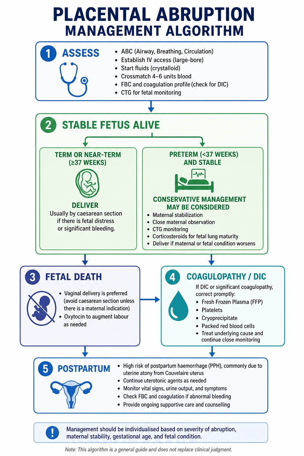
The principle is simultaneous resuscitation and delivery planning — they are run in parallel by the obstetric, anaesthetic, haematology and neonatal teams. Resuscitation is not completed before delivery is planned, because the only definitive treatment for ongoing abruption is delivery.[1]
Diagnosis is clinical. Mobilise senior obstetrician, anaesthetist, haematology lab, neonatal team, theatre. Declare a maternal collapse / obstetric haemorrhage emergency.
Oxygen 15 L/min by non-rebreather mask. Two 14 to 16 gauge IV cannulae. Take bloods (FBC, coagulation including fibrinogen, group and crossmatch 4 to 6 units, U&E, LFTs, Kleihauer-Betke).
Warmed crystalloid — Hartmann's or 0.9 percent sodium chloride, 1 to 2 L as a bolus through a fluid warmer. Avoid over-resuscitation — keep the haemoglobin over 70 to 80 g/L, do not chase a "normal" pressure at the cost of diluting clotting factors.
Crossmatch 4 to 6 units of packed red cells. Activate the massive transfusion protocol if major haemorrhage. Correct coagulopathy before surgery: FFP 12 to 15 mL per kg (about 4 units), cryoprecipitate 10 units if fibrinogen under 2 g/L (target fibrinogen over 2 g/L, ideally over 2.5 g/L), platelet pool (1 adult therapeutic dose) if platelets under 50 x10⁹ per litre. Tranexamic acid 1 g IV within 3 hours of bleeding onset (WOMAN trial) reduces death from haemorrhage.[11]
Continuous CTG. Indwelling urinary catheter — target urine output over 0.5 mL per kg per hour. Repeat fibrinogen, PT, APTT, platelets every 30 to 60 minutes. Hourly assessment of fundal height, tenderness, tone, bleeding, maternal vital signs.
Maternal left lateral tilt to relieve aortocaval compression. Adequate analgesia (intravenous opioids titrated; avoid NSAIDs). Anti-D 500 IU IM for all Rh-negative women within 72 hours — large fetomaternal haemorrhage is possible; dose is escalated per Kleihauer-Betke result.[1]
The goal of resuscitation is to make the mother safe enough to undergo an emergency operative delivery or to support her through an induced labour, not to "stabilise" her indefinitely — the source of bleeding is the separated placenta, and only delivery will control it.[1]
Management — Definitive & Stepwise
Definitive management is decided by the fetal status, the maternal condition, the gestational age, and the presence or absence of coagulopathy. Three broad pathways:[1]
Pathway 1 — Fetus alive with fetal distress
Emergency caesarean section, ideally within 30 minutes of decision (Category 1 / "code crimson" obstetric).[1] Resuscitate and correct coagulopathy first, in parallel — fibrinogen must be over 2 g/L and platelets over 50 x10⁹ per litre before incision. General anaesthesia is often preferred because regional anaesthesia is unsafe in the presence of coagulopathy and hypovolaemia (epidural haematoma risk). Neonatal team attends delivery. Prepare for atonic PPH at delivery (Couvelaire) — have uterotonics, Bakri balloon and the surgical PPH ladder ready.
Pathway 2 — Fetus alive, no fetal distress, at term or near-term (after 34 to 36 weeks)
Deliver. If the cervix is favourable and labour is established, aim for vaginal delivery with continuous CTG; augment with oxytocin if contractions are inadequate. Avoid artificial rupture of membranes if possible (it can worsen shearing forces and precipitate further separation or cord prolapse), although ARM is sometimes used in established labour to hasten delivery. If the abruption worsens or fetal distress emerges, proceed to emergency caesarean. Very preterm abruption (under 34 weeks) with a stable fetus and minimal bleeding can occasionally be managed expectantly in a tertiary unit with maternal–fetal medicine expertise — corticosteroids for fetal lung maturity, continuous fetal monitoring, serial coagulation and growth surveillance — but this is the exception, and the threshold to deliver is low.[1]
Pathway 3 — Fetus dead (intrauterine death)
Vaginal delivery is preferred. Caesarean for fetal death alone is not justified — it exposes a coagulopathic woman to surgical risk without fetal benefit, and the atonic Couvelaire uterus may not contract on the incision.[1] Induce or augment labour with oxytocin infusion (start 30 mU per minute, titrate to contractions; common range 20 to 40 mU per minute). The hypertonic, irritable uterus often means labour progresses quickly. Reserve caesarean for maternal indications — uncontrolled haemorrhage despite delivery, failed induction with ongoing maternal compromise, or other obstetric indication (e.g. transverse lie that will not turn, severe pre-eclampsia with unfavourable cervix and maternal deterioration).
The caesarean delivery itself in a coagulopathic mother should be done through a lower segment incision with careful haemostasis; the Couvelaire-affected uterus should be inspected for tone after delivery of the placenta. Uterotonics (oxytocin 5 IU slow IV then 40 IU in 500 mL Hartmann's over 4 hours; ergometrine 0.5 mg IM, avoided in hypertension/pre-eclampsia; carboprost 250 mcg IM every 15 minutes up to 8 doses, avoided in asthma; misoprostol 800 mcg rectally) are deployed early. Tranexamic acid 1 g IV within 3 hours of delivery reduces death from haemorrhage.[11] If atony persists despite uterotonics, escalate to Bakri balloon tamponade, B-Lynch or other compression sutures, internal iliac artery ligation, uterine artery embolisation, and — as the last-resort, life-saving step — subtotal hysterectomy.[1]
Operative decision tree — abruption
1
EMERGENCY caesarean (under 30 min) after correcting coagulopathy (fibrinogen over 2 g/L)
2
Vaginal delivery preferred; continuous CTG; ARM avoid if possible; caesarean if distress emerges
3
Vaginal delivery with oxytocin; caesarean ONLY for maternal indication (uncontrolled haemorrhage, failed induction, transverse lie)
4
Individualise — expectant management in tertiary unit ONLY if maternal state stable and bleeding minimal; corticosteroids; low threshold to deliver
5
Anticipate atonic PPH (Couvelaire); uterotonic ladder + TXA; escalate to Bakri → B-Lynch → artery ligation → hysterectomy
Specific Subtypes & Scenarios
A small number of scenarios dominate examiner questions because they are common, dangerous, or have specific management deltas.[1]
Traumatic abruption (after blunt abdominal trauma)
Road traffic collisions, falls and intimate partner violence are the principal causes. Abruption may be immediate (shearing force at the moment of impact) or delayed, presenting up to 24 to 72 hours after the event. All pregnant women over 20 weeks with significant trauma need a minimum of 4 to 6 hours of CTG monitoring; the threshold to extend to 24 hours is low if there is uterine tenderness, bleeding, contractions, or any abnormal CTG. A Kleihauer-Betke test is mandatory in Rh-negative women. Major trauma activates a full trauma team alongside the obstetric response.[8]
Concealed abruption
The classic and dangerous pattern — minimal or absent visible bleeding with shock disproportionate to visible loss, a woody hard tender uterus, increasing fundal height, and a high risk of DIC and Couvelaire uterus. The clinical suspicion must be high; ultrasound may be normal. Maternal mortality is higher than revealed abruption because of delayed recognition. Resuscitate aggressively, monitor fibrinogen, deliver promptly.[1]
Mild abruption at term
A small revealed abruption with a stable fetus and minimal bleeding may be managed by induction of labour and continuous CTG. The diagnosis is often only confirmed retrospectively by inspection of the delivered placenta — a dark clot adherent to the maternal surface with compression of the underlying placental tissue. Always document this finding.[1]
Recurrent abruption
Women with a previous abruption have a 6 percent recurrence risk after one episode and over 25 percent after two. They should be booked for high-risk antenatal care, low-dose aspirin (150 mg nocte from 12 weeks where indicated for pre-eclampsia prevention), serial growth scans, and an individualised delivery plan — many units deliver at 37 to 38 weeks in subsequent pregnancies to balance the risk of recurrent abruption against the risks of iatrogenic prematurity.[6][9]
Postpartum abruption (rare)
An abruption occurring after delivery (often with a retained placenta) is rare but presents as persistent atonic bleeding with a retroplacental clot found on manual removal. Treat as PPH with the uterotonic ladder and correction of coagulopathy.[1]
Abruption in a woman with a previous caesarean scar
Rare, but the combination of an abruption in a scarred uterus raises the additional question of uterine rupture — both present with abdominal pain and fetal distress. Loss of fetal station, cessation of contractions, and an easily palpable fetal part point to rupture; a hard woody tender fundus without loss of station favours abruption. Have a low threshold for laparotomy.[8]
Complications & Pitfalls
Abruption is one of the few obstetric events that can rapidly kill both mother and fetus. The complications cluster into maternal (haemorrhagic, coagulation, organ-damage, long-term) and fetal (death, prematurity, growth restriction).[1]
Haemorrhagic
bleeding consequences
- **Maternal haemorrhagic shock** — Class III/IV in concealed abruption
- **Massive transfusion** — activated protocol; coagulopathy compounds blood loss
- **Refractory atonic PPH** from Couvelaire uterus — may need hysterectomy
Coagulopathy & DIC
thromboplastin release
- **DIC in up to 10 percent of all abruptions, 30 percent of grade 3**
- Fibrinogen under 2 g/L signals severe; under 1 g/L catastrophic
- Consumption of factors V, VIII, platelets — generalised oozing, petechiae, IV-site bleeding
- Lasts hours to days after delivery — continue clotting support
Organ damage
hypoperfusion
- **Acute kidney injury** — acute tubular necrosis (reversible) or acute cortical necrosis (irreversible) from prolonged hypotension
- **Sheehan syndrome** — pituitary infarction from profound shock → postpartum amenorrhoea, failure to lactate, hypopituitarism
- **Acute respiratory distress syndrome** from massive transfusion and shock
- **Liver dysfunction** if coexisting HELLP
Fetal
perinatal
- **Intrauterine fetal death** in up to 30 percent of severe abruptions
- **Hypoxic ischaemic encephalopathy** in survivors
- **Iatrogenic preterm delivery** — large fraction of abruptions deliver prematurely
- **Fetal growth restriction** — chronic small abruption in the index or prior pregnancy
- **Fetomaternal haemorrhage** — fetal anaemia, sensitisation
Long-term maternal
future risk
- **Recurrence** in next pregnancy (6 to 25 percent)
- **Pre-eclampsia** in subsequent pregnancies
- **Cardiovascular and cerebrovascular disease** later in life — population cohort data show abruption is a sentinel marker for future vascular disease
The classic pitfalls that examiners and audit panels repeatedly flag:[1]
- Missing concealed abruption by reassurance from minimal visible blood. The shock state, fundal height, uterine tone and fetal heart — not the pad — drive severity grading.[1]
- Relying on a normal ultrasound to exclude abruption. Sensitivity is only about 50 percent.
- Not correcting coagulopathy before caesarean — catastrophic intra-operative bleeding. Aim for fibrinogen over 2 g/L.
- Performing caesarean for fetal death alone — exposes a coagulopathic woman to surgical risk without fetal benefit.
- Performing a digital vaginal examination in suspected antepartum haemorrhage before excluding praevia.
- Forgetting to give anti-D within 72 hours to Rh-negative women — large fetomaternal haemorrhage is common.
- Failing to monitor after trauma for the 24 to 72 hour window of delayed abruption.
- Under-recognising Sheehan syndrome in a woman who fails to lactate after a hypotensive delivery — it is treatable and missing it has lifelong consequences.
Prognosis & Disposition
Prognosis depends on severity at presentation, gestational age, time to delivery, and the presence of coagulopathy or Couvelaire uterus.[1]
Mild abruption (grade 1) has an excellent prognosis if managed promptly — fetal survival is near 100 percent, maternal mortality is low, and recurrence risk is moderate.[1]
Moderate abruption (grade 2) has variable fetal outcomes depending on the speed of delivery; prompt caesarean for fetal distress yields good fetal survival.[1]
Severe abruption (grade 3 with fetal death) carries a perinatal mortality of 10 to 30 percent even in modern units, with maternal mortality under 1 percent in well-resourced settings but significantly higher where access to blood, theatre and intensive care is limited.[1] Coexisting pre-eclampsia, chorioamnionitis or chronic hypertension worsen outcomes.
Long term, women who have had an abruption should be counselled that the next pregnancy is high-risk: a 6 percent recurrence after one episode, rising to over 25 percent after two; an increased risk of pre-eclampsia; and — increasingly recognised — a higher lifelong risk of cardiovascular and cerebrovascular disease, which makes cardiovascular risk-factor modification (smoking cessation, blood pressure control, lipid management) an essential part of postpartum follow-up.[14]
Disposition. After delivery, the woman is managed in a high-dependency or intensive-care setting until clotting has normalised, bleeding is controlled, urine output is established, and any coagulopathy, AKI or pre-eclampsia has stabilised. Psychological support after perinatal loss is essential. Contraception, future pregnancy planning, and the place of aspirin prophylaxis in the next pregnancy are addressed before discharge.[1]
Outcomes by severity
Special Populations
Rh-negative women. Anti-D prophylaxis is mandatory within 72 hours because the torn placental bed allows large-volume fetomaternal haemorrhage. The Kleihauer-Betke test or flow cytometry quantifies the fetal cell load and tailors additional anti-D beyond the standard 500 IU dose.[1]
Women with a previous caesarean scar. Abruption in a scarred uterus raises the differential of uterine rupture. A high index of suspicion and a low threshold for laparotomy are essential.[8]
Multiple pregnancy. Both twins and higher-order gestations are at increased risk of abruption from uterine overdistension; abruption in this group also raises the question of whether to deliver the second twin vaginally or by caesarean. Specialist input is essential.[1]
Pre-eclampsia. The two conditions frequently coexist; management must address both — control blood pressure, prevent eclampsia with magnesium sulphate, correct coagulopathy, and deliver. Magnesium sulphate is given for seizure prophylaxis in severe pre-eclampsia or eclampsia (loading 4 g IV over 5 to 10 minutes, then maintenance 1 g per hour infusion), and it slightly relaxes the uterus — but is not contraindicated.[3]
Women on anticoagulants. Therapeutic low-molecular-weight heparin or warfarin increases bleeding severity; coordinate reversal with haematology before delivery (stop LMWH 24 hours pre-planned delivery; protamine for urgent reversal; vitamin K plus prothrombin complex concentrate for warfarin).[12]
Older mothers and the chronically hypertensive. The combination of advanced maternal age, chronic hypertension and antithrombotic medication is increasingly common and represents one of the highest-risk profiles for abruption. Tight antenatal blood pressure control and aspirin prophylaxis (where indicated) are the main preventive levers.[3]
Evidence, Guidelines & Regional Differences
United Kingdom (RCOG). Green-top Guideline No. 63 (Antepartum Haemorrhage, 2011) frames abruption as a clinical diagnosis and emphasises (1) the parallel resuscitation-and-delivery pathway, (2) correction of coagulopathy before surgery, (3) caesarean for a live fetus with distress, (4) vaginal delivery for fetal death, and (5) anti-D within 72 hours in Rh-negative women.[1] NICE NG201 (Caesarean Birth, 2021) supports a 30-minute decision-to-delivery interval for Category 1 caesarean.[1]
United States (ACOG). ACOG is moving toward a quantitative, cumulative blood-loss approach and recommends aspirin 81 mg nightly from 12 weeks in women at high risk of pre-eclampsia (which is the strongest abruption risk factor).[6]
WHO and global. WHO and FIGO support low-dose aspirin (75 to 150 mg) prophylaxis for pre-eclampsia in high-risk women and emphasise access to emergency obstetric care and blood transfusion as the cornerstone of preventing maternal death from abruption in low-resource settings.[1]
India (FOGSI / Government of India). FOGSI guidance mirrors RCOG with low-dose aspirin (75 to 150 mg) for high-risk women. The Janani Shishu Suraksha Karyakram removes user-fee and transport barriers for obstetric emergencies. The high prevalence of anaemia (over 50 percent of pregnant women in many states) makes even moderate bleeding dangerous; iron-folate supplementation and detection and treatment of anaemia in pregnancy are central.[1]
Aspirin prophylaxis. Multiple systematic reviews and meta-analyses have established that low-dose aspirin (75 to 150 mg daily) started before 16 weeks of gestation reduces the incidence of pre-eclampsia, fetal growth restriction and preterm birth in high-risk women — and by reducing pre-eclampsia it indirectly reduces the incidence of abruption.[6][7] Doses below 75 mg appear less effective; doses over 150 mg do not clearly add benefit and increase bleeding risk.[6]
Tranexamic acid (WOMAN trial, 2017). The landmark international randomised trial of tranexamic acid 1 g IV within 3 hours of bleeding onset in over 20,000 women with postpartum haemorrhage (including abruption-related PPH) showed a significant reduction in death from bleeding and no increase in thromboembolic complications. It is now standard in massive obstetric haemorrhage protocols worldwide.[11]
DIC management. Modern DIC management in obstetrics prioritises treatment of the underlying trigger (delivery) plus targeted blood-product replacement guided by fibrinogen, PT, APTT, platelet count, and viscoelastic testing rather than empirical ratios — fibrinogen replacement with cryoprecipitate (target over 2 g/L, ideally over 2.5 g/L) is the cornerstone.[12][13]
Future cardiovascular risk. Population-based cohort data (over 800,000 women) show that abruption is an independent sentinel marker for future cardiovascular and cerebrovascular disease, with a 1.5- to 2-fold increased hazard — the postpartum period is increasingly recognised as a window for cardiovascular risk-factor modification.[14]
Exam Pearls & High-Yield Minutiae
Abruption vs praevia — the bedside discriminator
ABRUPT
abruption = severe constant pain. Praevia = painless
abruption = dark old blood. Praevia = bright fresh red
abruption = hard, tender, hypertonic. Praevia = soft, relaxed
in abruption US is often normal — clinical diagnosis. Praevia shows low placenta
abruption — hypertension/trauma. Praevia — previous caesarean
abruption = resuscitate + deliver. Praevia = stabilise + plan delivery
Test yourself: concealed abruption and fibrinogen
A 33-year-old at 36 weeks presents with severe constant abdominal pain, BP 90/60, pulse 130, minimal dark vaginal bleeding, a woody hard tender uterus, fetal bradycardia, fibrinogen 1.6 g/L, platelets 45 x10⁹/L. What is the diagnosis, the next step, and the fibrinogen target before incision?
Answer: Severe (grade 3) placental abruption with concealed bleeding and DIC. Resuscitate (ABC, two large-bore cannulae, warmed crystalloid, crossmatch 4 to 6 units, activate massive transfusion) in parallel with preparing for emergency caesarean (fetus alive with bradycardia). Correct coagulopathy before incision — cryoprecipitate to bring fibrinogen over 2 g/L (ideally 2.5 g/L) and platelet pool to over 50 x10⁹/L. Give tranexamic acid 1 g IV. Anti-D if Rh-negative.[1][12]
Exam application bank (NEET-PG / INICET)
One-line answer
Placental abruption (abruptio placentae) = premature separation of a normally-situated placenta from the uterine wall before delivery of the fetus. Incidence about 1 in 100 pregnancies; severe (fetal death) about 1 in 1000. Classic triad: painful vaginal bleeding + hard, woody, tender hypertonic uterus + fetal distress. Two patterns: revealed (about 80%, blood tracks via cervix, visible, less dangerous) and concealed (about 20%, blood trapped behind placenta, shock disproportionate to visible loss, more dangerous). Pathology: decidual vasculopathy (pre-eclampsia) leads to spiral artery rupture, retroplacental clot, extension of separation, and in severe cases Couvelaire uterus (blood infiltrates myometrium to serosa, uteroplacental apoplexy) and DIC (thromboplastin release, fibrinogen under 2 g/L). Top risk factor: pre-eclampsia/hypertension; recurrence 6 to 25 percent. Management: resus [1]
Worked stems (answer without another resource)
Stem 1 — Classic presentation. Map symptoms to mechanism; name the first investigation and first treatment step with dose/route if drug therapy is standard. [1]
Stem 2 — Unstable / complicated. List red flags that force immediate resuscitation, theatre, ICU, antidote, or reperfusion — and what you do in the first 15 minutes. [1]
Stem 3 — Atypical group. Elderly, pregnancy, child, or immunocompromised: how presentation and thresholds change. [1]
Stem 4 — Differential trap. Name the three closest mimics and one discriminator for each. [1]
Stem 5 — Disposition. Who goes home with safety-netting, who is admitted, who needs HDU/ICU/theatre, and what follow-up is mandatory. [1]
Rapid viva checklist
- Definition + classification
- Pathophysiology chain
- Bedside signs / criteria
- Score with exact components (if any)
- Emergency bundle
- Definitive therapy with doses
- Complications of disease and of treatment
- Special populations
- Guideline/trial name if classic
- Three exam traps
Coverage self-check
If you cannot answer any stem above from this page alone, re-read the matching section — the page is intended to be self-sufficient for final-prof and NEET-PG/INICET questions on Placental Abruption.
Severe abruption with DIC — describe the management
10 minutes · 10 marks
A 35-year-old para 2 woman at 37 weeks with known chronic hypertension presents with sudden severe abdominal pain, dark vaginal bleeding, BP 95/60, pulse 128, woody tender fundus, CTG showing late decelerations progressing to bradycardia. Fibrinogen 1.5 g/L, platelets 42 x10⁹/L, PT 18 s, APTT 48 s. (10 marks, 10 minutes)
Abruption vs praevia — distinguish at the bedside
10 minutes · 6 marks
A 28-year-old at 36 weeks presents with painless bright-red vaginal bleeding. Distinguish placenta praevia from placental abruption at the bedside. (6 marks, 10 minutes)
“A 30-year-old at 38 weeks, para 1, presents at 02:00 with severe constant abdominal pain for 1 hour. On arrival: BP 88/56, pulse 132, minimal dark vaginal bleeding, fundus woody tender, CTG shows fetal bradycardia.”
References
- [1]Oyelese Y, Ananth CV. Placental abruption Obstet Gynecol, 2006.PMID 17012465
- [2]Tikkanen M, Nuutila M, Hiilesmaa V, Paavonen J, Ylikorkala O. Clinical presentation and risk factors of placental abruption Acta Obstet Gynecol Scand, 2006.PMID 16752262
- [3]Ananth CV, Savitz DA, Williams MA. Placental abruption and its association with hypertension and prolonged rupture of membranes: a methodologic review and meta-analysis Obstet Gynecol, 1996.PMID 8692522
- [4]Ananth CV, Savitz DA, Luther ER. Incidence of placental abruption in relation to cigarette smoking and hypertensive disorders during pregnancy: a meta-analysis of observational studies Obstet Gynecol, 1999.PMID 10214847
- [5]Ananth CV, VanderWeele TJ, Hernández-Díaz S. An international contrast of rates of placental abruption: an age-period-cohort analysis PLoS One, 2015.PMID 26018653
- [6]Roberge S, Bujold E, Nicolaides KH. The role of aspirin dose on the prevention of preeclampsia and fetal growth restriction: systematic review and meta-analysis Am J Obstet Gynecol, 2017.PMID 27640943
- [7]Roberge S, Bujold E, Nicolaides KH. Meta-analysis on the effect of aspirin use for prevention of preeclampsia on placental abruption and antepartum hemorrhage Am J Obstet Gynecol, 2018.PMID 29305829
- [8]Huls CK. Trauma in pregnancy Semin Perinatol, 2018.PMID 29463389
- [9]Ananth CV, Wisser J, Dzengofera M, Tüzün B, Heimstad R, Bhide A, Thilaganathan B, Hanson H, Solt T, Kruse M. Change in paternity, risk of placental abruption and confounding by birth interval: a population-based prospective cohort study in Norway, 1967-2009 BMJ Open, 2015.PMID 25670732
- [10]Parker SE, Nembhard WN, Bilodeau E, Ghassabian A, Werler MM, Mitchell AA, Tinker SC. Placental abruption and subsequent risk of pre-eclampsia: a population-based case-control study Paediatr Perinat Epidemiol, 2015.PMID 25761509
- [11]WOMAN Trial Collaborators. Effect of early tranexamic acid administration on mortality, hysterectomy, and other morbidities in women with post-partum haemorrhage (WOMAN): an international, randomised, double-blind, placebo-controlled trial Lancet, 2017.PMID 28456509
- [12]Levi M. How I treat disseminated intravascular coagulation Blood, 2018.PMID 29255070
- [13]Levi M. Disseminated intravascular coagulation: an update on pathogenesis and diagnosis Expert Rev Hematol, 2018.PMID 29999440
- [14]Ananth CV, Patrick HS, Ananth S, Zhang Y, Kostis WJ, Schuster M. Maternal Cardiovascular and Cerebrovascular Health After Placental Abruption: A Systematic Review and Meta-Analysis (CHAP-SR) Am J Epidemiol, 2021.PMID 34263291
- [15]Tanaka H, Matsunaga S, Yamashita T, Okutomi T, Sakurai A, Sekizawa A, Hasegawa J, Terui K, Miyake Y, Murotsuki J, Ikeda T. A systematic review of massive transfusion protocol in obstetrics Taiwan J Obstet Gynecol, 2017.PMID 29241907
- [16]Kogutt BK, Vaught AJ. Postpartum hemorrhage: Blood product management and massive transfusion Semin Perinatol, 2019.PMID 30527516