Obstetrics & Gynaecology · Obstetrics & Gynaecology
Uterine Fibroids (Leiomyomas)
Also known as Uterine fibroids · Leiomyoma · Uterine myoma · Fibroid uterus · FIGO classification
Uterine fibroids (leiomyomas) are benign monoclonal smooth-muscle tumours of the myometrium — the commonest pelvic tumour in women (up to 70 to 80 percent by age 50; most asymptomatic). They are oestrogen- and progesterone-responsive (grow in reproductive years, shrink after menopause). Each fibroid is monoclonal, driven by MED12 mutations in about 70 percent. Risk factors: age, African ancestry (2 to 3x, earlier and more severe), family history, nulliparity, early menarche, obesity; protective: parity, OCP use, vitamin D sufficiency. The FIGO location classification (0 to 8) drives symptoms and treatment: submucosal (0 to 2) cause heavy menstrual bleeding and infertility; intramural (3 to 4) are commonest; subserosal (5 to 7) cause bulk/pressure. Diagnosis: transvaginal ultrasound first-line, MRI for mapping/sarcoma suspicion, hysteroscopy for submucosal. Management: expectant if asymptomatic; medical (tranexamic acid/NSAIDs, LNG-IUS, GnRH agonist/antagonist with add-back — elagolix, relugolix); surgical (hysterectomy definitive, myomectomy fertility-sparing, hysteroscopic resection for submucosal); interventional (uterine artery embolisation, MRgFUS).
On this page & tools
Your progress
Saved locally on this device.
Exam tags
Red flags
Overview & Definition
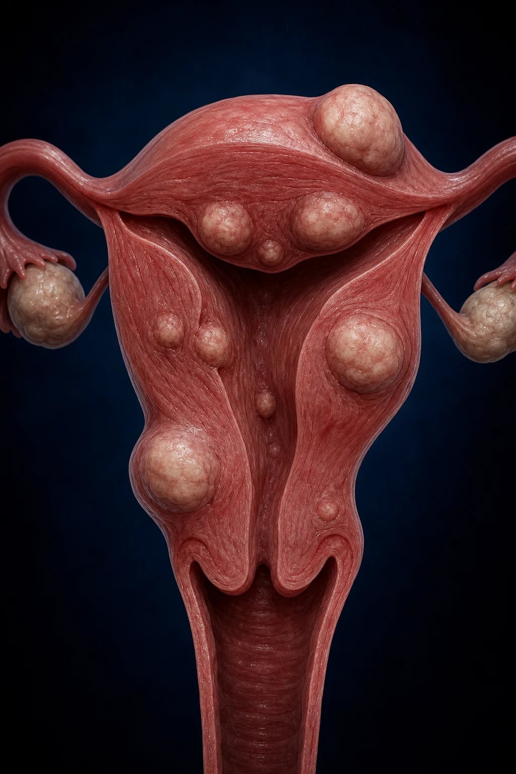
Uterine fibroids (synonyms: leiomyomas, myomas, fibromyomas, fibroids) are benign, monoclonal neoplasms arising from the smooth-muscle cell of the myometrium (the myocyte), with a variable accompanying deposition of extracellular matrix (collagen and elastin). They are the single commonest pelvic tumour in women and the leading benign indication for hysterectomy worldwide.[1][2]
Three principles make this topic high-yield and examinable from every angle: [1]
- They are oestrogen- and progesterone-responsive. Fibroids grow during the reproductive years and in pregnancy, and shrink after the menopause when ovarian steroid output falls. This single fact explains the epidemiology (peak 30 to 50 years), the protective effect of parity and smoking, the response to GnRH suppression, and the pivotal red flag: a fibroid that grows in a postmenopausal woman is not behaving like a fibroid — suspect leiomyosarcoma.[1]
- Location — not size — determines symptoms. The FIGO PALM-COEIN-type classification (0 to 8) records where each fibroid sits relative to the endometrial cavity, myometrium, and serosa. A 2 cm submucosal fibroid can cause severe menorrhagia and infertility, whereas a 10 cm subserosal fibroid may be silent apart from a mass. Matching the symptom to the FIGO type is the central clinical skill.[2]
- Every management decision is filtered through the patient's desire for future fertility and uterine preservation. A 45-year-old with completed family and severe menorrhagia is offered a hysterectomy (definitive, curative). A 28-year-old with primary infertility and a submucosal fibroid is offered a hysteroscopic myomectomy. The same disease, opposite operations.[1]
Although the historical term fibroid implies a fibrous tumour, the neoplastic cell is smooth muscle, and the preferred pathological term is leiomyoma. They are almost always benign; the relationship with leiomyosarcoma is now considered to be one of separate de novo origin rather than malignant transformation of a pre-existing fibroid (a critical point addressed under Red Flags and Complications).[1]
Classification
Uterine fibroids are classified primarily by anatomical location using the FIGO (International Federation of Gynecology and Obstetrics) classification, originally proposed by Munro and Parker and now the universal language for describing fibroids. The system uses types 0 through 8, where lower numbers denote proximity to the endometrial cavity.[2]
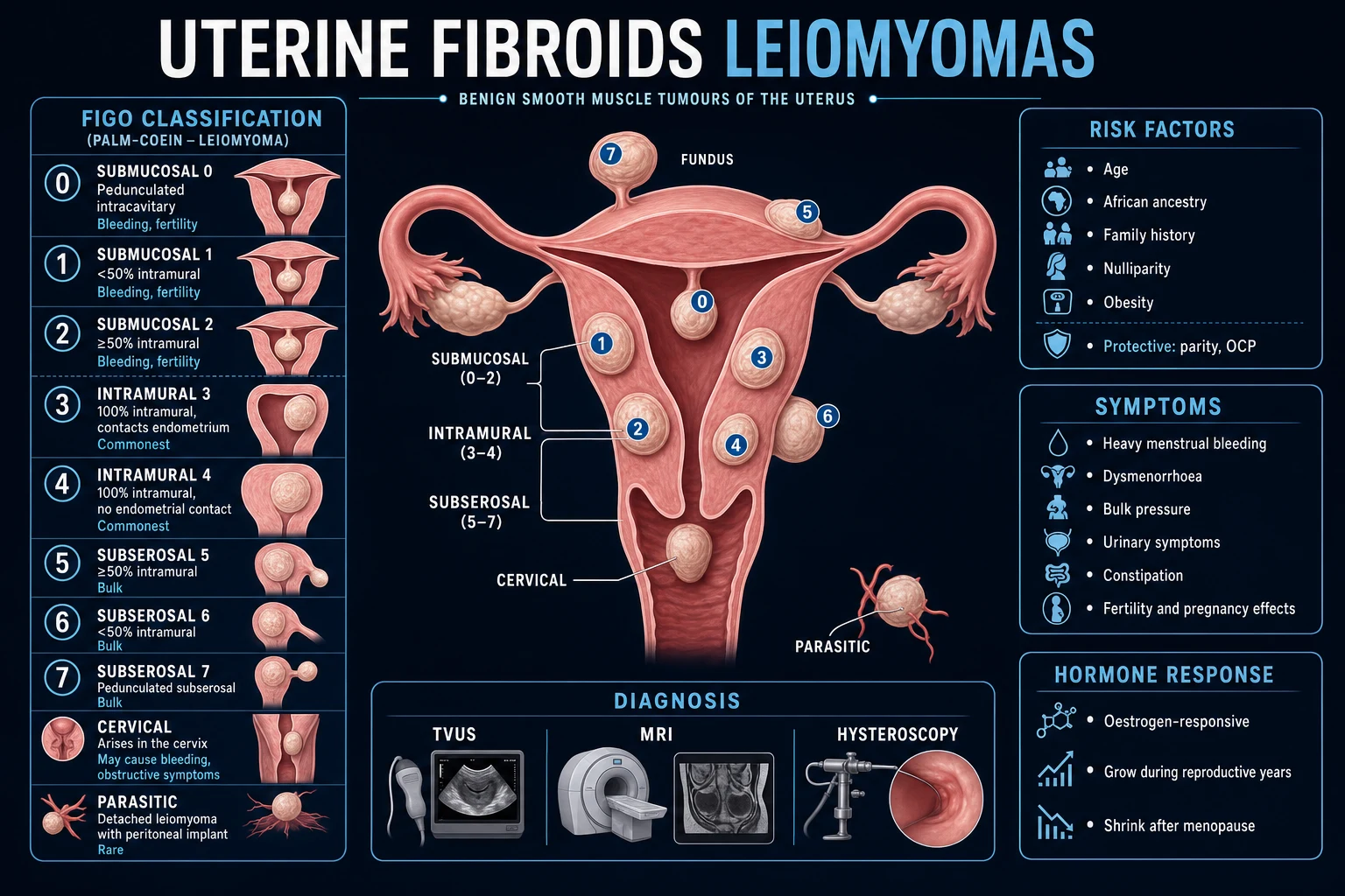
The classification groups neatly into three functional families that the examiner uses as the spine of every question: [1]
Submucosal (FIGO 0 to 2)
cavity-distorting
- Distorts the endometrial cavity
- Most symptomatic per unit size
- Heavy/prolonged menstrual bleeding, dysmenorrhoea, anaemia
- Infertility, recurrent miscarriage, implantation failure
- Treatment: hysteroscopic resection (fertility-sparing)
Intramural (FIGO 3 to 4)
commonest
- Within the myometrial wall
- Commonest location overall
- Menorrhagia + uterine enlargement
- Bulk symptoms when large
- Treatment: medical, myomectomy if fertility desired
Subserosal (FIGO 5 to 7)
bulk-pressure
- Protrude from the outer uterine surface
- May be pedunculated (torsion risk)
- Bulk and pressure: urinary, bowel, back pain
- Often silent with respect to bleeding
- Treatment: myomectomy, UAE, MRgFUS
Type 8 / special
cervical, parasitic
- Cervical, broad-ligament, parasitic
- Anatomical distortion, ureter at risk
- Parasitic derives blood supply from extra-uterine structures
- Diffuse uterine leiomyomatosis = innumerable confluent fibroids
- Needs individualised surgical planning
A few special pathological/descriptive subtypes also appear in exams: [1]
- Red (carneous) degeneration — acute haemorrhagic infarction of a fibroid, classically in pregnancy or the mid-trimester, presenting with severe localised abdominal pain, low-grade fever and sometimes mild vomiting. Managed conservatively with analgesia — surgery is avoided.
- Parasitic fibroid — a subserosal fibroid that has detached from the uterus and acquired an extra-uterine blood supply (omentum, mesentery).
- Diffuse uterine leiomyomatosis — innumerable confluent fibroids replacing the myometrium; uterine-sparing treatment is difficult.
- Lipoleiomyoma — a fibroid with a dominant fatty component; usually an incidental postmenopausal finding.
- Cellular leiomyoma, mitotically active leiomyoma, bizarre/atypical leiomyoma (symplastic) — histological variants that are still benign but can mimic sarcoma; diagnosis on histology with attention to mitotic count, necrosis, and atypia. [1]
Epidemiology & Risk Factors
Uterine fibroids — epidemiology at a glance
- Prevalence. Up to 70 to 80 percent of women will have one or more fibroids by age 50 in ultrasound/autopsy series; only about 25 to 30 percent are symptomatic. In symptomatic women aged 30 to 44, fibroids are found in over 60 percent.[2][5]
- Age and menopausal status. Peak incidence 30 to 50 years; fibroids are rare before menarche and typically regress after the menopause (with the fall in ovarian steroids) — the basis of the "postmenopausal growth is a red flag" reflex.[1]
- Ethnicity. Women of African (sub-Saharan) ancestry have a 2 to 3-fold higher prevalence, an earlier age of onset, and larger, more numerous and more severe fibroids — leading to higher hysterectomy rates at younger ages. Theorised contributors: lower median vitamin D status, genetic/epigenetic factors, hair-relaxer exposure (debated).[1][5]
- Reproductive history. Nulliparity is a strong risk factor (each pregnancy reduces risk); multiparity is protective. Early menarche and late menopause prolong unopposed steroid exposure and increase risk.
- Family history. A first-degree relative with fibroids roughly doubles a woman's risk (heritability estimated at 50 to 70 percent in twin studies).
- Metabolic/lifestyle. Obesity (peripheral aromatisation of androgens to oestrogen in adipose), hypertension, and polycystic ovary syndrome all increase risk. Smokers have a slightly reduced risk (anti-oestrogenic effect, debated).
- Diet. High intake of red meat and ham and low intake of green vegetables and fruit has been associated with higher risk; alcohol (especially beer) and high glycaemic load may increase risk. Vitamin D deficiency is a reproducible correlate.
- Protective factors. Parity, combined oral contraceptive use (long-term), and vitamin D sufficiency are protective.
India / South Asia. Fibroids are a leading cause of hysterectomy in India, with prevalence in community and hospital ultrasound studies of roughly 4 to 20 percent (varies widely by population, parity, and diagnostic threshold). Compared with Caucasian populations, South Asian women develop symptomatic fibroids at a smaller uterine size and lower BMI — a "thin woman with fibroids" phenotype is common. Vitamin D deficiency is highly prevalent in this population and is an active area of chemoprevention research. Hysterectomy rates are high and often performed early; there is growing emphasis on uterus-conserving options (myomectomy, medical therapy, UAE) given the cultural importance of fertility and the menstrual body. Access to MRI and minimally-invasive options is uneven, and laparoscopic power morcellation practices have shifted after the global sarcoma-safety concerns.
Pathophysiology
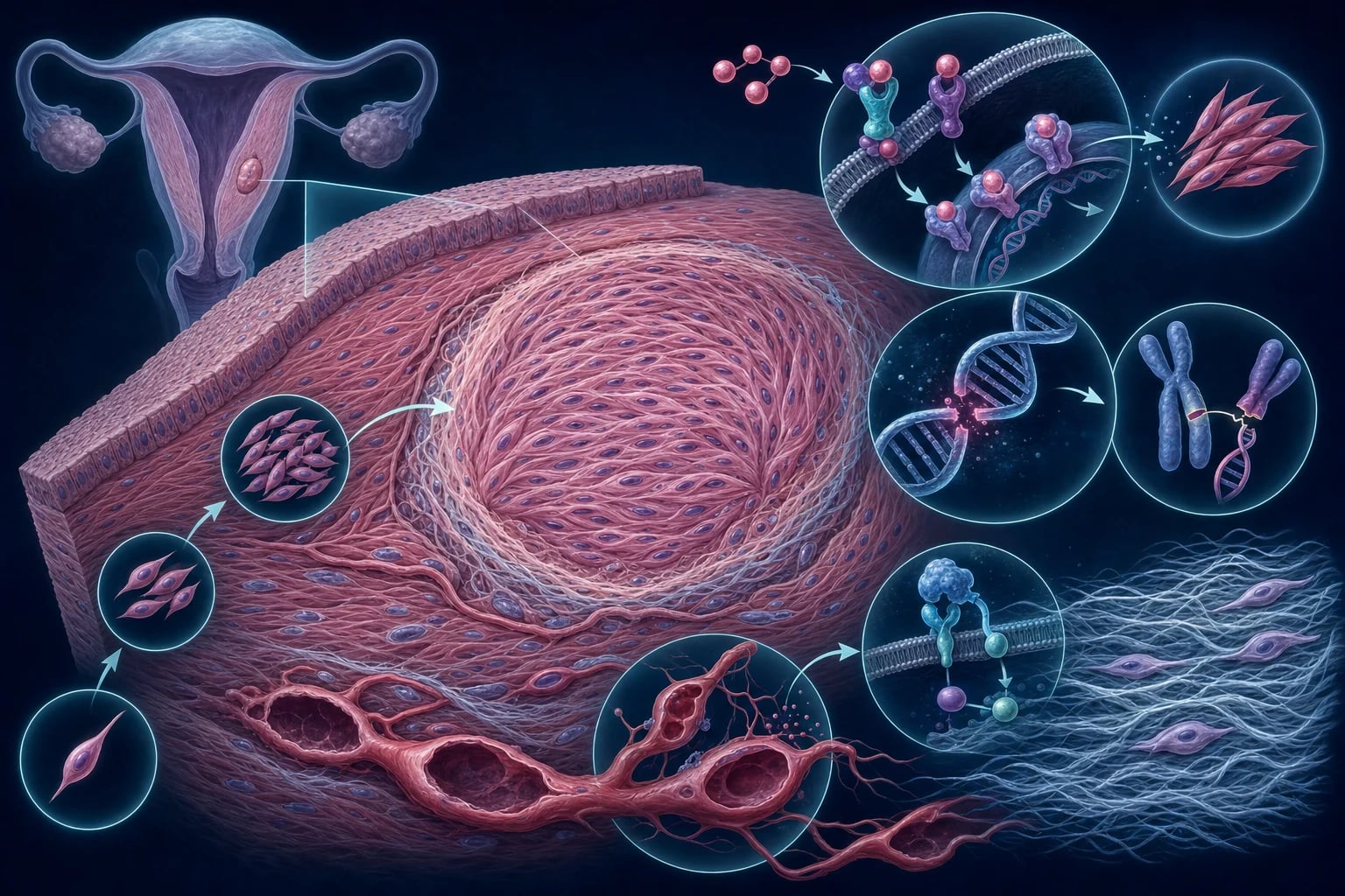
Understanding the molecular pathogenesis directly explains the symptoms, the risk factors, and every modern drug: [1]
1. Each fibroid is MONOCLONAL. Using glucose-6-phosphate dehydrogenase (G6PD) isoenzyme and X-chromosome inactivation studies, every fibroid in a given uterus is derived from a single transformed myometrial smooth-muscle cell. Multiple fibroids in the same woman are genetically distinct from one another — i.e., they do not metastasise; each is an independent neoplasm. This is why "recurrence" after myomectomy is really the emergence of new clones, not regrowth of resected ones. [1]
2. Somatic genetic drivers. Several reproducible mutations drive the clone:
- MED12 mutations (Xq15) in about 70 percent of fibroids — the mediator complex subunit 12 is part of the RNA polymerase II transcription machinery; mutations dysregulate transcription of proliferative and Wnt/beta-catenin target genes. This is the single commonest driver.
- HMGA2 over-expression/rearrangement (12q14-15) — especially in large fibroids; HMGA2 is an architectural transcription factor that drives growth.
- COL4A5/COL4A6 deletions and fumarate hydratase (FH) loss — rarer subsets; FH loss (also seen in hereditary leiomyomatosis and renal-cell cancer syndrome) produces pseudohypoxia.
- Cytogenetically normal in about 40 percent. [1]
3. Hormonal responsiveness. Fibroids over-express both oestrogen receptors (ER-alpha) and progesterone receptors (PR) relative to the surrounding myometrium. Oestrogen drives proliferation directly; progesterone is paradoxically the dominant mitogen in vivo — it upregulates growth factors (EGF, IGF-1, VEGF) and the anti-apoptotic protein BCL2. This is the rationale for selective progesterone receptor modulators (SPRMs) such as ulipristal acetate, and for the GnRH agonists/antagonists that suppress both steroids.[2][3]
4. Extracellular matrix (ECM) and growth factors. Fibroids accumulate a dense, disordered ECM rich in collagen and fibronectin with abnormal elastin — driven principally by TGF-beta signalling. The ECM is not inert: it sequesters growth factors, stiffens the tissue (mechanotransduction further stimulating proliferation), and contributes to the firm, pale, whorled cut surface seen grossly. Fibroids also produce bFGF (basic fibroblast growth factor) which promotes the dilated, disordered vascular network (thin-walled veins, deficient smooth muscle) characteristic of fibroids.[1]
5. Why the symptoms happen:
- Heavy menstrual bleeding (HMB): (i) the disordered, fragile vasculature overlying a submucosal fibroid bleeds easily; (ii) the enlarged, stiff uterus contracts poorly so spiral artery haemostasis is impaired; (iii) a large endometrial surface area overlying fibroids sheds more; (iv) local imbalance of vasoconstrictors (endothelins) and vasodilators (prostaglandins, VEGF).
- Bulk/pressure: sheer mass displaces the bladder (frequency, urgency, retention), rectum (constipation), ureters (hydronephrosis with very large fibroids), and the venous plexus (leg oedema, deep vein thrombosis risk).
- Infertility/miscarriage: cavity distortion impairs sperm transport and embryo implantation; local inflammatory and contractile changes; altered expression of implantation genes (HOXA10) in the endometrium overlying submucosal fibroids.
- Dysmenorrhoea: prostaglandin-mediated uterine contractions against a stiff, fibroid-laden wall. [1]
6. Natural history. Fibroids grow slowly over years during the reproductive years, accelerate in pregnancy, and involute after the menopause (calcification is common in postmenopausal fibroids). They outgrow their blood supply episodically, producing the various forms of degeneration: hyaline (commonest), cystic, fatty (lipoleiomyoma), calcific, and red (carneous) degeneration. [1]
Clinical Presentation
[1]Symptoms track the FIGO location, not the size. Submucosal fibroids produce disproportionate menorrhagia, intermenstrual bleeding, dysmenorrhoea, and reproductive dysfunction. Intramural fibroids cause a mixture of bleeding and bulk. Subserosal fibroids are silent with respect to bleeding but produce pressure symptoms and a palpable mass, and a pedunculated subserosal fibroid may torsion (acute severe pain, vomiting, peritonism). Cervical fibroids may cause acute urinary retention (a "globular" mass behind the symphysis) and dramatically distort the bladder and ureters.[1]
Menstrual pattern in fibroids. Classically menorrhagia with regular cycles — the cycle remains ovulatory, but the period is heavier and longer. Intermenstrual and postcoital bleeding can occur with submucosal fibroids. Postmenopausal bleeding in a woman known to have fibroids is never attributed to the fibroids until endometrial malignancy has been excluded.[1]
Atypical presentations (high-yield):
- Postmenopausal woman with an enlarging "fibroid" or new bleeding — suspect leiomyosarcoma and endometrial carcinoma; investigate urgently (endometrial sampling, MRI, LDH). Do not morcellate.[1]
- Pregnant woman in the second trimester with acute severe localised abdominal pain, low-grade fever, nausea and mild leucocytosis — red (carneous) degeneration: managed with analgesia and reassurance; surgery is avoided in pregnancy unless there is a clear surgical emergency (torsion, infarction of a pedunculated fibroid).[1]
- Young nulliparous woman with severe menorrhagia and a low ferritin — fibroids (often submucosal) but always exclude a bleeding diathesis (von Willebrand disease is common in women with HMB since menarche).
- Rare paraneoplastic: massive fibroids occasionally cause polycythaemia (ectopic erythropoietin) and very rarely systemic amyloidosis.
Differential Diagnosis
A reproductive-age woman presenting with heavy bleeding and/or an enlarged uterus has a focused differential. The clinical skill is to triage by age, parity, cycle regularity, and the imaging appearance. [1]
| Condition | Key distinguishing feature |
|---|---|
| Adenomyosis | Diffusely enlarged, boggy, tender uterus; severe dysmenorrhoea with menorrhagia; TVUS shows asymmetric myometrial thickening, myometrial cysts, fan-shaped shadowing; MRI shows junctional zone thickness over 12 mm. Often co-exists with fibroids. |
| Pregnancy (intrauterine or ectopic) | Amenorrhoea, nausea, breast tenderness; beta-hCG is mandatory in any reproductive-age woman with bleeding or pain before assuming fibroids. |
| Endometrial polyp | Focal intracavitary echogenic lesion on TVUS/SIS; intermenstrual or postmenopausal bleeding; confirmed on hysteroscopy with biopsy. |
| Endometrial hyperplasia / carcinoma | Perimenopausal or postmenopausal bleeding, anovulatory cycles, obesity, PCOS, tamoxifen, nulliparity, diabetes. Pipelle endometrial biopsy is mandatory in at-risk women. |
| Ovarian neoplasm | Mass is mobile and in the adnexa, separate from the uterus on bimanual and ultrasound examination; a solid ovarian mass raises concern for malignancy (CA-125, imaging). |
| Leiomyosarcoma | Rapid growth, postmenopausal enlargement, LDH elevated; MRI features (T1/T2 signal patterns, restricted diffusion) but no reliable pre-operative test — diagnosis is on final histology (mitoses, coagulative tumour-cell necrosis, severe atypia). |
| Pelvic congestion syndrome | Dilated pelvic veins on imaging, post-coital ache, varices; dull chronic pelvic pain, worse on standing. |
| Uterine sarcoma (endometrial stromal sarcoma, adenosarcoma) | Rare; rapid growth, postmenopausal; biopsy/histology. |
| Endometriosis (adnexal/extra-uterine) | Cyclical pain, dyspareunia, endometriomas on imaging; coexists with fibroids. |
Two non-negotiable reflexes: (i) any reproductive-age woman with abnormal bleeding or pelvic pain gets a beta-hCG; (ii) any woman over about 40 to 45 with HMB, or any woman with postmenopausal bleeding, gets an endometrial sample.[1]
Clinical & Bedside Assessment
History establishes the symptom complex (HMB, pressure, fertility), the cycle pattern, reproductive intent, and red flags (rapid growth, postmenopausal change, family history of sarcoma/cancer). Quantify bleeding with the alkaline haematin method or the simpler pictorial blood-loss assessment chart (PBAC); ask about anaemia symptoms and urinary/bowel compromise. [1]
General examination:
- Pallor (conjunctiva, palmar creases) — iron-deficiency anaemia.
- BMI, blood pressure — obesity and hypertension are risk factors and influence anaesthetic/peri-operative risk.
- Abdominal examination — a palpable firm, irregular, non-tender pelvic mass arising from the pelvis (the "uterus moves with the cervix" sign on bimanual confirms a uterine origin). The size is conventionally expressed as gestational-week equivalents ("a 16-week-size fibroid uterus").
- Speculum examination — visualise the cervix to exclude a cervical polyp, ectropion, or visible malignancy; a cervical fibroid may be seen as a fleshy mass distorting the os. [1]
Bimanual pelvic examination:
- An enlarged, irregular, mobile, non-tender, firm uterus with nodular surfaces is the classical finding of intramural/subserosal fibroids.
- A diffusely enlarged, boggy, tender uterus suggests adenomyosis.
- Adnexal fullness separate from the uterus suggests an ovarian mass.
- The cervix may be displaced by a cervical fibroid; urethral catheterisation may be needed if retention. [1]
There is no pathognomonic bedside sign for fibroids, but the combination of an irregular firm mobile uterus in a reproductive-age woman with menorrhagia is highly suggestive. [1]
Investigations
The diagnostic pathway is tiered: confirm and map the fibroids, characterise the bleeding cause, exclude pregnancy and malignancy, and stage the patient for treatment. [1]
1. Transvaginal ultrasound (TVUS) — FIRST-LINE. TVUS defines the number, size, location (FIGO type), and calcification of fibroids. Fibroids appear as well-circumscribed, hypoechoic, heterogeneous, rounded myometrial masses with acoustic shadowing; submucosal fibroids distort the endometrial echo. TVUS also assesses the endometrial thickness and the ovaries.[1][2]
2. Saline-infusion sonohysterography (SIS, sonohysterogram). The most sensitive ultrasound method for evaluating the endometrial cavity and for distinguishing a submucosal fibroid from an endometrial polyp, and for measuring the intramural extension of a submucosal fibroid (critical before hysteroscopic resection — the FIGO 0/1/2 distinction).[1]
3. Hysteroscopy. Diagnostic and therapeutic for submucosal (FIGO 0 to 2) fibroids — direct visualisation and same-setting resection (see Management). Also allows targeted biopsy of any suspicious endometrium.[1]
4. Pelvic MRI. Not first-line, but indicated for: mapping before complex surgery or UAE; distinguishing fibroids from adenomyosis; suspected leiomyosarcoma (with LDH and diffusion-weighted imaging); very large or complex uteri where TVUS is limited; suspected parasitic/broad-ligament fibroids; and in women where TVUS is technically difficult. The junctional zone thickness distinguishes adenomyosis (over 12 mm). MRI features suggesting sarcoma include T1 high signal (haemorrhage), T1 post-contrast enhancement, restricted diffusion on DWI, and rising LDH/LDH-3 — none individually definitive.[1]
5. Endometrial sampling (Pipelle biopsy) or hysteroscopic biopsy. Mandatory in:
- Postmenopausal bleeding (any cause) — to exclude endometrial carcinoma.
- Women over about 40 to 45 years presenting with new or changed HMB, or with risk factors for endometrial hyperplasia/cancer (obesity, PCOS, nulliparity, tamoxifen, diabetes, anovulatory cycles, family history Lynch).
- Persistent intermenstrual bleeding, or persistent/atypical bleeding on treatment. A submucosal fibroid does not exempt the endometrium from sampling in an at-risk woman.[1]
6. Laboratory tests:
- Full blood count and ferritin — iron-deficiency anaemia (baseline and to monitor therapy).
- Beta-hCG — exclude pregnancy before any intervention/medication in a reproductive-age woman.
- Thyroid function and prolactin — if cycle irregularity suggests a coexisting endocrine cause.
- Coagulation screen and von Willebrand panel — if HMB since menarche, bruising/bleeding history, or family history of a bleeding disorder.
- LDH and LDH isoenzyme (LDH-3) — proposed biomarker for leiomyosarcoma (sensitive but not specific); used adjunctively with MRI when sarcoma is suspected. [1]
7. Pre-operative assessment before surgery/UAE: renal function and electrolytes (ureteric compromise from large fibroids), group and save/cross-match, ECG and anaesthetic review. Before UAE, a recent negative pregnancy test and normal Pap/cervical screening are confirmed, and endometrial sampling is considered in at-risk women. [1]
Fibroid investigations — thresholds and key values
Management — Resuscitation
Most fibroids present electively, but acute heavy bleeding and acute severe pain are genuine emergencies. [1]
Acute severe heavy menstrual bleeding (haemodynamically compromising):[2]
- ABC — large-bore IV access, full blood count, group and save/cross-match, coagulation screen.
- Fluid resuscitation and blood transfusion if Hb is critically low (under 70 g/L, or under 80 g/L with symptoms) or haemodynamically unstable.
- IV tranexamic acid 1 g (over 10 minutes, repeated after 30 minutes if needed, max 4 g/24 h) — antifibrinolytic.
- IV conjugated equine oestrogens (e.g. 25 mg IV every 6 to 12 hours) or a high-dose oral or IV progestin (e.g. norethisterone 5 to 10 mg TDS, or medroxyprogesterone acetate) to arrest acute bleeding.
- A GnRH agonist may be used to shut down ovarian steroids in the short term (takes days).
- Foley catheter balloon tamponade of the uterine cavity in refractory torrential bleeding as a bridge to definitive surgery.
- Definitive: emergency hysteroscopic resection of a bleeding submucosal fibroid, uterine artery embolisation, or hysterectomy if medical measures fail and the woman has completed her family.
Acute severe pain (degeneration or torsion):[1]
- Analgesia — paracetamol, then an NSAID (mefenamic acid/ibuprofen/diclofenac; avoid NSAIDs beyond 32 weeks in pregnancy), escalating to an opioid.
- Red (carneous) degeneration in pregnancy — analgesia, rest, reassurance; the pain settles over days to weeks. Do not operate.
- Torsion of a pedunculated subserosal fibroid — surgical emergency (laparoscopic myomectomy or excision).
- Acute urinary retention from a cervical/anterior fibroid — urethral catheterisation; definitive treatment of the fibroid once drained.
Management — Definitive & Stepwise
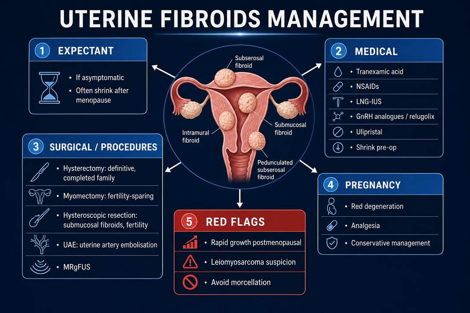
The guiding principles: (i) treat only symptomatic patients; (ii) match the treatment to the symptom, the FIGO type, and the patient's reproductive intent; (iii) choose fertility-sparing options for women desiring future pregnancy; (iv) suspect sarcoma and avoid morcellation in any rapidly growing or postmenopausal fibroid.[1][2]
Step 1 — Expectant management
Asymptomatic fibroids, or small fibroids with no anaemia or fertility concern: no treatment. Reassure the patient that fibroids are benign and typically shrink after the menopause. Offer annual review and report red flags (rapid growth, new pain, heavy bleeding, postmenopausal bleeding).[1]
Step 2 — Medical therapy (symptom control and pre-operative volume reduction)
A. Symptomatic control of heavy menstrual bleeding (does not shrink fibroids):
- Tranexamic acid — 1 g orally three times daily during heavy days of menstruation (maximum 4 days per cycle). Antifibrinolytic; reduces menstrual loss by 40 to 50 percent. Avoid in active thromboembolic disease.
- NSAIDs — mefenamic acid 500 mg TDS during menses; reduce prostaglandin-mediated flow and dysmenorrhoea.
- Combined oral contraceptive pill (OCP) or cyclical progestogens (norethisterone 5 mg TDS days 5 to 25) — regulate cycles and reduce flow.
- Levonorgestrel-releasing intrauterine system (LNG-IUS, Mirena) — first-line for HMB in NICE guidance; reduces menstrual loss by 70 to 95 percent and offers contraception. Limitation: less effective and higher expulsion rate when the cavity is distorted by a submucosal fibroid (evaluate cavity before insertion).[2]
B. Volume reduction and amenorrhoea (shrink fibroids; used pre-op, peri-menopause, or for women unsuitable for surgery):
- GnRH agonists (goserelin 3.6 mg SC monthly or leuprolide 3.75 mg IM monthly, 3 to 6 months). Induce a hypo-oestrogenic state, shrinking fibroids by 30 to 60 percent and inducing amenorrhoea — correcting anaemia and easing surgery. Limitations: rebound regrowth within months of stopping, and hypo-oestrogenic side effects (hot flushes, bone loss) — limit to 3 to 6 months unless add-back therapy (e.g. estradiol/norethisterone) is given. Used pre-operatively to reduce fibroid/uterine size and correct anaemia before myomectomy/hysterectomy.[1]
- GnRH antagonists — oral, no initial flare (unlike agonists), rapid onset:
- Relugolix combination — relugolix 40 mg + estradiol 1 mg / norethindrone acetate 0.5 mg once daily, FDA-approved for long-term use (up to 24 months) for heavy menstrual bleeding associated with fibroids. The add-back mitigates bone loss.
- Elagolix + add-back — elagolix 300 mg twice daily + estradiol 1 mg / norethindrone acetate 0.5 mg once daily. Elaris UF-1 and UF-2 trials (NEJM 2020): primary endpoint met in 68.5 percent (UF-1) and 76.5 percent (UF-2) of women on elagolix plus add-back versus 8.7 percent and 10 percent on placebo.[5][6]
- Selective progesterone receptor modulators (SPRMs):
- Ulipristal acetate 5 mg orally once daily for 3 months (repeat courses possible). PEARL I (NEJM 2012): bleeding control in 91 percent (5 mg) and 92 percent (10 mg) versus 19 percent placebo; amenorrhoea in 73 to 82 percent; fibroid volume reduction of 12 to 21 percent. PEARL II: non-inferior to leuprolide for bleeding control with significantly fewer hot flushes (10 to 11 percent versus 40 percent).[3][4]
- Safety caveat: the European Medicines Agency (EMA) suspended ulipristal (Esmya) in 2020 to 2021 and restricted/reintroduced it with strict liver-function monitoring after rare cases of severe liver injury requiring transplantation. Always check current local licensing before prescribing.
Step 3 — Surgical management
A. Hysterectomy — DEFINITIVE. Removal of the uterus (with or without ovaries); no recurrence; for women who have completed their family and want definitive cure, or with severe refractory symptoms. Routes: total abdominal (TAH), vaginal, laparoscopic, or robotic-assisted — minimally invasive routes offer faster recovery. Surgical menopause if ovaries removed; counsel on HRT.[1]
B. Myomectomy — FERTILITY-SPARING. Removal of fibroids with conservation of the uterus. For women desiring future fertility or uterine preservation. Routes:
- Hysteroscopic myomectomy — for submucosal fibroids (FIGO 0 to 2). Resectoscope (monopolar with glycine, or bipolar saline). Improves fertility and reduces miscarriage when the cavity is distorted. Limitation: for FIGO 2 (at least 50 percent intramural) a two-stage procedure or pre-operative GnRH shrinkage may be needed; risk of uterine perforation, fluid overload (glycine → hyponatraemia), and intra-uterine adhesions (Asherman).[1]
- Laparoscopic myomectomy — for intramural/subserosal fibroids; morcellation required to extract the specimen (see sarcoma warning); meticulous uterine repair (risk of scar rupture in future pregnancy — counsel about mode of delivery).[1]
- Abdominal (open) myomectomy — for large, numerous, or deep intramural fibroids; allows strong multilayer uterine repair.
- Recurrence of new fibroids after myomectomy: about 15 to 30 percent at 5 years (higher with multiple fibroids).[1]
C. Endometrial ablation. An option for small submucosal fibroids (FIGO 0 to 1) with completed family and no cavity distortion; less effective than hysteroscopic myomectomy and does not preserve fertility (contraception still needed; pregnancy after ablation is high-risk). [1]
Step 4 — Interventional / radiological (uterus-sparing, non-surgical)
- Uterine artery embolisation (UAE). Interventional radiology — catheter embolises the uterine arteries (polyvinyl alcohol particles/gelatin) to cut the fibroid's blood supply, producing 40 to 60 percent volume reduction and symptom relief in 80 to 90 percent. For symptomatic women not desiring future fertility (controversial in fertility seekers — ovarian reserve and pregnancy outcomes debated). Risks: post-embolisation syndrome (pain, fever, leucocytosis for days), infection/sloughing of a submucosal fibroid, premature ovarian failure (especially over 45 years), passage of fibroid tissue, need for re-intervention (surgery) in about 15 to 30 percent by 5 years.[1]
- Magnetic resonance-guided focused ultrasound (MRgFUS / HIFU). Non-invasive thermal ablation of a focal fibroid under real-time MRI thermometry; no incision; suitable for accessible, non-calcified fibroids. Limited by depth/size/calcification and the need for dedicated equipment.
- Laparoscopic radiofrequency ablation (Lap-RFA, Acessa system). Laparoscopic ultrasound-guided insertion of a needle electrode into each fibroid; thermal ablation preserving myometrium. Outpatient/short-stay option.
Choice driven by the patient profile
Completed family, severe HMB
definitive
- Hysterectomy — definitive, no recurrence
- Or UAE if wishes to avoid surgery
- Or LNG-IUS if prefers non-surgical first
Desires fertility + submucosal
FIGO 0 to 2
- Hysteroscopic myomectomy
- Improves pregnancy rates and reduces miscarriage
- Pre-op GnRH if FIGO 2/large
Desires fertility + intramural/subserosal
fertility-sparing
- Laparoscopic or abdominal myomectomy
- Counsel on scar rupture + delivery
- Avoid UAE/MRgFUS if possible
Perimenopausal, symptomatic
temporise
- Medical therapy (GnRH antagonist + add-back)
- Bridge to natural menopause (shrinkage)
- Surgery if refractory
Escalation triggers and red flags
- Suspected leiomyosarcoma (rapid growth, postmenopausal enlargement, LDH elevated, MRI features) → do NOT morcellate; refer to gynaecologic oncology; use contained morcellation or en-bloc extraction if surgery is needed.[1]
- Failure of medical therapy with persistent anaemia or severe symptoms → surgical or interventional treatment.
- Pressure complications (hydronephrosis, recurrent retention, bowel obstruction) → operative relief.
Specific Subtypes & Scenarios
- Submucosal fibroid (FIGO 0 to 2). Most symptomatic per unit size; causes HMB, dysmenorrhoea, infertility, miscarriage. Hysteroscopic resection is the procedure of choice and improves fertility. The degree of intramural extension (FIGO 0 vs 1 vs 2) determines resectability and the need for staging or pre-operative shrinkage.[1]
- Cervical fibroid. Distorts pelvic anatomy; the ureter is at risk at surgery; may cause acute urinary retention; usually requires laparotomy and ureteric stenting. The cervix is displaced and the uterine vessels are pulled upwards — anticipate a difficult hysterectomy.[1]
- Pedunculated subserosal fibroid (FIGO 6). Risk of torsion (acute severe pain, peritonism) — emergency laparoscopic excision. Easy to remove with minimal myometrial disruption.[1]
- Broad-ligament (intraligamentous) fibroid (FIGO 8). Lies between the leaves of the broad ligament; ureter and uterine vessels may be intimately related; careful dissection, identify the ureter first.[1]
- Parasitic fibroid. Detached from the uterus, supplied by omental/mesenteric vessels; rare; managed by excision of the parasitic blood supply and tumour.
- Red (carneous) degeneration. Acute haemorrhagic infarction, classically in pregnancy; severe localised pain, low-grade fever, mild nausea. Managed with analgesia, rest and reassurance — avoid surgery.[1]
- Diffuse uterine leiomyomatosis. Innumerable confluent fibroids replacing the uterus; uterus-sparing treatment is difficult; often ends in hysterectomy.
- Hereditary leiomyomatosis and renal cell cancer (HLRCC, Reed syndrome). Autosomal dominant fumarate hydratase (FH) mutation — cutaneous and uterine leiomyomas with aggressive papillary renal-cell carcinoma; screen the family.
Complications & Pitfalls
Disease-related:
- Iron-deficiency anaemia from chronic HMB (the most common complication; may be severe and the presenting feature).
- Infertility, recurrent miscarriage, implantation failure (cavity-distorting submucosal fibroids).
- Pregnancy complications: miscarriage, preterm labour, fetal malpresentation, intrauterine growth restriction, placental abruption, obstructed labour, increased caesarean rate, postpartum haemorrhage, and uterine rupture after prior myomectomy.[1]
- Acute events: torsion of a pedunculated fibroid; acute urinary retention; hydronephrosis/renal impairment from a massive fibroid; deep vein thrombosis/pelvic venous stasis.
- Rare: polycythaemia (ectopic erythropoietin), systemic amyloidosis (very rare).
Treatment-related:
- Hysterectomy: bleeding, infection, vault prolapse, bladder/bowel/ureteric injury, premature ovarian failure if ovaries removed.
- Myomectomy: adhesion formation, recurrence (15 to 30 percent at 5 years), uterine rupture in future pregnancy (especially after deep intramural or laparoscopic myomectomy — counsel on mode of delivery), intra-operative bleeding sometimes necessitating conversion to hysterectomy.
- Hysteroscopic resection: uterine perforation, fluid overload (glycine → dilutional hyponatraemia, cerebral oedema), intra-uterine adhesions (Asherman syndrome).[1]
- UAE: post-embolisation syndrome, infection/sloughing and passage of a submucosal fibroid, premature ovarian failure (especially over 45 years), need for re-intervention.
- Power morcellation → occult leiomyosarcoma spread (FDA boxed warning, 2014). The single most important peri-operative pitfall in modern fibroid surgery — always consider sarcoma in any rapidly growing or postmenopausal fibroid, and use contained morcellation or en-bloc extraction.[1]
- Ulipristal acetate hepatotoxicity — rare but serious (hepatic failure requiring transplant); EMA suspension/restricted reintroduction with mandatory LFT monitoring.[3]
Prognosis & Disposition
- Benign disease with an excellent long-term prognosis. Fibroids shrink after the menopause; symptomatic disease is effectively controlled with the modern armamentarium.[1]
- Recurrence after myomectomy: about 15 to 30 percent at 5 years (higher with multiple fibroids); hysterectomy is curative (no recurrence).[1]
- Quality of life improves markedly after appropriate treatment; anaemia corrects once bleeding is controlled.
- Leiomyosarcoma is rare (estimated under 1 in 500 to 1 in 1000 women undergoing surgery for presumed fibroids) and is now considered a separate de novo neoplasm. Prognosis is poor when it occurs (5-year survival roughly 30 to 50 percent if confined to the uterus at diagnosis, worse with extra-uterine spread).[1]
- Disposition: most women are managed as outpatients; emergency admission is for acute severe bleeding (transfusion and haemostatic therapy), acute severe pain (degeneration/torsion), or acute urinary retention. Referral to gynaecology (and gynaecologic oncology if sarcoma is suspected) is standard.
Special Populations
- Women seeking fertility. Cavity-distorting submucosal fibroids (FIGO 0 to 2) → hysteroscopic resection (improves pregnancy and live-birth rates — clear evidence). Intramural fibroids that do not distort the cavity have a debated effect on fertility; myomectomy is considered when other causes are excluded and the fibroids are large or numerous. Avoid UAE and MRgFUS where possible in women seeking fertility (ovarian reserve/fertility outcomes debated). IVF may be needed for non-cavity lesions or coexisting tubal/male factor.[1][2]
- Pregnancy. Most pregnancies with fibroids are uneventful. Red (carneous) degeneration is managed conservatively with analgesia (avoid NSAIDs beyond 32 weeks; opioids if needed). Increased surveillance for preterm labour, malpresentation, and growth restriction. Route of delivery: individualised — a prior myomectomy that entered the uterine cavity (especially with a deep repair) generally warrants elective caesarean to avoid scar rupture; a superficial or subserosal myomectomy does not.[1]
- Perimenopausal women. Medical therapy (GnRH antagonist + add-back) temporises until the menopause, when fibroids shrink naturally; surgery reserved for refractory disease.
- Postmenopausal women. Fibroids should be quiescent or shrinking. New growth or bleeding = investigate for sarcoma and endometrial carcinoma (endometrial biopsy, MRI, LDH). HRT may prevent shrinkage and can cause slight growth — counsel accordingly.[1]
- African ancestry. Earlier onset, larger/more numerous fibroids, greater severity; may warrant earlier or more aggressive treatment and counselling about all uterus-sparing options. Consider screening for vitamin D deficiency.[1][5]
- Women on anticoagulation / with bleeding disorders. Heavy bleeding is amplified; coordinate with haematology, and preferentially use tranexamic acid, LNG-IUS, and definitive management rather than cyclical hormonal manipulation.
- Young women with HMB since menarche. Screen for von Willebrand disease and other inherited bleeding disorders before attributing bleeding solely to fibroids.
Evidence, Guidelines & Regional Differences
- PEARL I (Donnez et al., NEJM 2012, PMID 22296075): ulipristal acetate 5 mg / 10 mg vs placebo — bleeding control 91 to 92 percent vs 19 percent, amenorrhoea in 73 to 82 percent, fibroid volume reduction (median 21 percent with 5 mg).[3]
- PEARL II (Donnez et al., NEJM 2012, PMID 22296076): ulipristal acetate vs leuprolide — non-inferior for bleeding control at week 13, with significantly fewer hot flushes (10 to 11 percent vs 40 percent).[4]
- Elaris UF-1 and UF-2 (Schlaff, Stewart et al., NEJM 2020, PMID 31971678): elagolix 300 mg BD + add-back (E2 1 mg / NETA 0.5 mg) reduced heavy menstrual bleeding — primary endpoint met in 68.5 percent (UF-1) and 76.5 percent (UF-2) vs 8.7 percent and 10 percent placebo; bone-density loss attenuated by add-back.[5]
- Al-Hendy, Stewart et al. (AJOG 2021, PMID 32702363): elagolix with add-back was effective across subgroups (age, BMI, race, fibroid location, volume), confirming broad applicability.[6]
- Relugolix combination (Myovant LIBERTY trials) — once-daily oral GnRH antagonist + add-back; FDA approved 2021 for long-term (up to 24 months) use.
- FDA 2014 safety communication on power morcellation and occult leiomyosarcoma; boxed warning; shift to contained morcellation and individualised risk counselling.[1]
- EMA 2020 to 2021 review of ulipristal acetate (Esmya) — suspension then restricted reintroduction with mandatory liver-function monitoring for rare serious hepatotoxicity.[3]
Fibroid FIGO classification by location
SUB
cavity-distorting; bleeds, infertile, miscarries; hysteroscopic resection
commonest; bleeding + bulk; myomectomy if fertility desired
pressure, palpable mass, torsion; myomectomy, UAE, MRgFUS
When a fibroid is NOT a fibroid (suspect sarcoma)
RAPID
a fibroid enlarging quickly in any age group
growth or new bleeding after the menopause is never a fibroid behaving normally
LDH and LDH-3 elevated; MRI restricted diffusion
use contained morcellation / en-bloc extraction; refer oncology
leiomyosarcoma arises de novo, not by transformation of a fibroid
Exam Pearls
- FIGO 0 to 8: submucosal (0 to 2) bleed and cause infertility; intramural (3 to 4) are commonest; subserosal (5 to 7) cause bulk; type 8 = cervical/parasitic.[2]
- Each fibroid is MONOCLONAL; multiple fibroids in one uterus are genetically distinct; MED12 mutation in about 70 percent; HMGA2 in large fibroids.[1]
- TVUS first-line; hysteroscopy is diagnostic AND therapeutic for submucosal; MRI for mapping, adenomyosis, and sarcoma suspicion.[1]
- Fibroids shrink after menopause → rapid growth or postmenopausal enlargement = suspect leiomyosarcoma; AVOID morcellation.[1]
- Red (carneous) degeneration in pregnancy → severe pain, low fever; analgesia and reassurance — do NOT operate.[1]
- Submucosal fibroid + infertility → hysteroscopic resection (improves fertility).[1]
- Medical: tranexamic acid + NSAID and LNG-IUS for HMB; GnRH agonist/antagonist + add-back (elagolix, relugolix) shrink fibroids; ulipristal withdrawn/restricted in EU-UK (hepatotoxicity).[2][3][5]
- Surgical: hysterectomy definitive; myomectomy fertility-sparing; hysteroscopic for submucosal.[1]
- UAE/MRgFUS for symptomatic women not desiring future fertility.[1]
- PEARL I/II (NEJM 2012): ulipristal 91 to 92 percent bleeding control, fewer hot flushes than leuprolide.[3][4]
- Elaris UF-1/UF-2 (NEJM 2020): elagolix + add-back 68.5 to 76.5 percent responder vs 8.7 to 10 percent placebo.[5]
- African ancestry: 2 to 3-fold higher prevalence, earlier, more severe.[1]
- LNG-IUS is less effective when the cavity is distorted (expulsion risk).[2]
Exam application bank (NEET-PG / INICET)
One-line answer
Uterine fibroids (leiomyomas) are benign monoclonal smooth-muscle tumours of the myometrium — the commonest pelvic tumour in women (up to 70 to 80 percent by age 50; most asymptomatic). They are oestrogen- and progesterone-responsive (grow in reproductive years, shrink after menopause). Each fibroid is monoclonal, driven by MED12 mutations in about 70 percent. Risk factors: age, African ancestry (2 to 3x, earlier and more severe), family history, nulliparity, early menarche, obesity; protective: parity, OCP use, vitamin D sufficiency. The FIGO location classification (0 to 8) drives symptoms and treatment: submucosal (0 to 2) cause heavy menstrual bleeding and infertility; intramural (3 to 4) are commonest; subserosal (5 to 7) cause bulk/pressure. Diagnosis: transvaginal ultrasound first-line, MRI for mapping/sarcoma suspicion, hysteroscopy for submucosal. Management: expectant if asympt
Worked stems (answer without another resource)
Stem 1 — Classic presentation. Map symptoms to mechanism; name the first investigation and first treatment step with dose/route if drug therapy is standard. [1]
Stem 2 — Unstable / complicated. List red flags that force immediate resuscitation, theatre, ICU, antidote, or reperfusion — and what you do in the first 15 minutes. [1]
Stem 3 — Atypical group. Elderly, pregnancy, child, or immunocompromised: how presentation and thresholds change. [1]
Stem 4 — Differential trap. Name the three closest mimics and one discriminator for each. [1]
Stem 5 — Disposition. Who goes home with safety-netting, who is admitted, who needs HDU/ICU/theatre, and what follow-up is mandatory. [1]
Rapid viva checklist
- Definition + classification
- Pathophysiology chain
- Bedside signs / criteria
- Score with exact components (if any)
- Emergency bundle
- Definitive therapy with doses
- Complications of disease and of treatment
- Special populations
- Guideline/trial name if classic
- Three exam traps
Coverage self-check
If you cannot answer any stem above from this page alone, re-read the matching section — the page is intended to be self-sufficient for final-prof and NEET-PG/INICET questions on Uterine Fibroids (Leiomyomas).
References
- [1]De La Cruz MS, Buchanan EM. Uterine Fibroids: Diagnosis and Treatment Am Fam Physician, 2017.PMID 28084714
- [2]Vannuccini S, Petraglia F, Carmona F, Calaf J, Chapron C. The modern management of uterine fibroids-related abnormal uterine bleeding Fertil Steril, 2024.PMID 38723935
- [3]Donnez J, Tatarchuk TF, Bouchard P, et al. (PEARL I Study Group). Ulipristal acetate versus placebo for fibroid treatment before surgery N Engl J Med, 2012.PMID 22296075
- [4]Donnez J, Tomaszewski J, Vazquez F, et al. (PEARL II Study Group). Ulipristal acetate versus leuprolide acetate for uterine fibroids N Engl J Med, 2012.PMID 22296076
- [5]Schlaff WD, Ackerman RT, Al-Hendy A, et al. Elagolix for Heavy Menstrual Bleeding in Women with Uterine Fibroids N Engl J Med, 2020.PMID 31971678
- [6]Al-Hendy A, Bradley L, Owens CD, et al. Predictors of response for elagolix with add-back therapy in women with heavy menstrual bleeding associated with uterine fibroids Am J Obstet Gynecol, 2021.PMID 32702363