Paediatrics · Paediatrics
Bronchiolitis
Bronchiolitis is an acute viral lower respiratory tract infection of the small airways (bronchioles) in infants, typically under 2 years (peak 2 to 6 months), caused mainly by respiratory syncytial virus (RSV, 70 to 80 percent). Presentation: coryzal prodrome for 1 to 3 days, then worsening cough, wheeze, tachypnoea, and respiratory distress (nasal flaring, recession, grunting, head bobbing), with bilateral crackles and wheeze on auscultation. Most cases are mild and self-limiting. Treatment is supportive: oxygen if SpO2 persistently under 92 percent, nasal suctioning, and hydration (oral, NG, or IV). Bronchodilators, corticosteroids, antibiotics, chest physiotherapy, and routine imaging are NOT recommended. Severe disease may need high-flow nasal cannula, CPAP, or mechanical ventilation. Prevention: palivizumab 15 mg/kg monthly IM for high-risk infants during RSV season; nirsevimab (single long-acting dose) for all infants in regions where it is funded.
On this page & tools
Your progress
Saved locally on this device.
Exam tags
Red flags
Overview & Definition
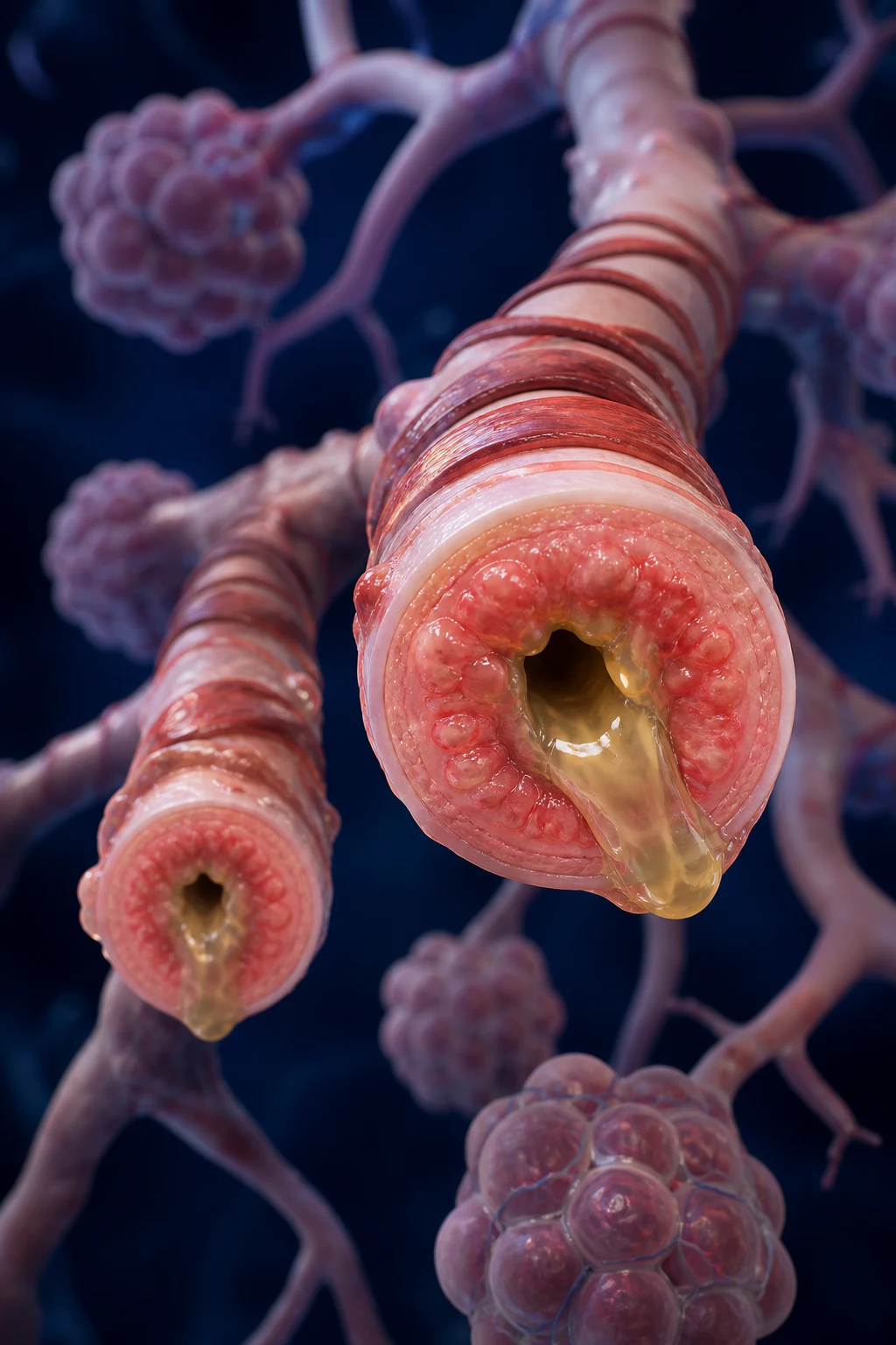
Bronchiolitis is an acute viral lower respiratory tract infection producing inflammation, oedema, epithelial necrosis, and mucus plugging of the small airways (bronchioles) of infants. It is defined clinically — a first or early episode of wheeze and tachypnoea in a child under 2 years, typically preceded by a coryzal prodrome — and it is the most common lower respiratory tract infection in infants, producing a predictable annual winter epidemic that fills paediatric wards and emergency departments.[1]
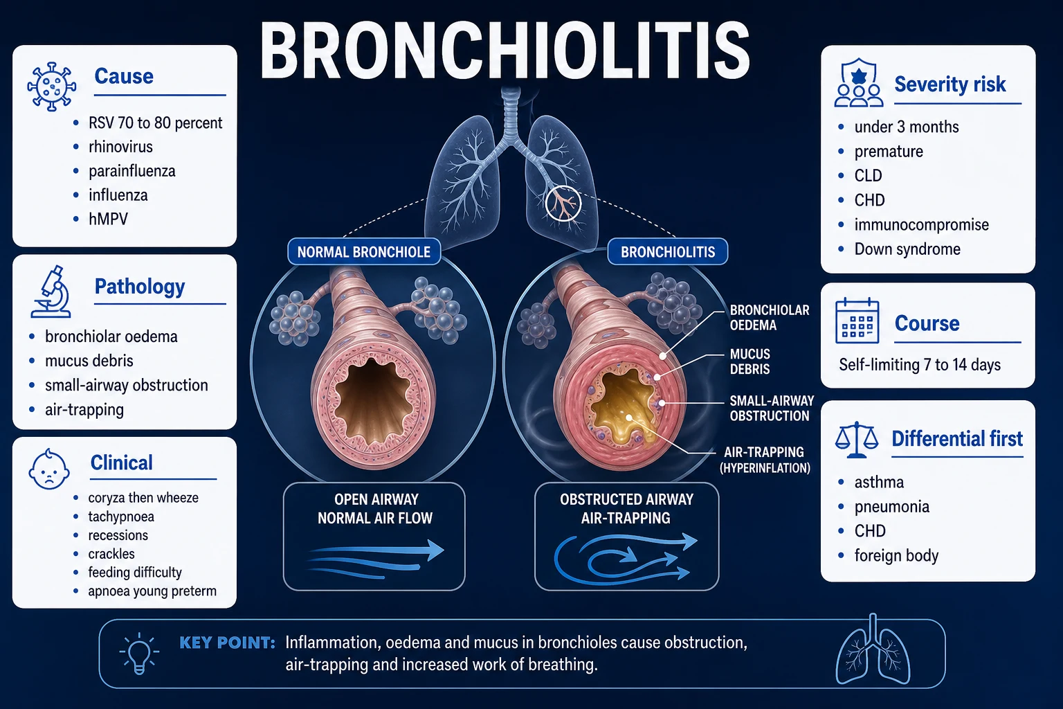
The dominant pathogen is respiratory syncytial virus (RSV), which accounts for 60 to 80 percent of hospitalised cases; rhinovirus, parainfluenza virus, influenza, adenovirus, human metapneumovirus (hMPV), and human bocavirus account for most of the remainder, with co-infections in 20 to 30 percent. Nearly every child has been infected with RSV by the age of 2 years, and reinfection throughout life is the rule. Most episodes are mild and self-limiting, resolving over 7 to 14 days, but bronchiolitis is far from trivial globally: it is among the leading causes of infant hospitalisation in every country, and a major contributor to under-five mortality in low-resource settings.[1][4]
The central clinical skill is to recognise the small minority who will deteriorate — the ex-preterm baby, the infant with congenital heart disease or chronic lung disease, the very young baby who presents with apnoea rather than wheeze — and to apply supportive care that is genuinely evidence-based while resisting the temptation to reach for treatments (bronchodilators, steroids, antibiotics) that trials repeatedly show do not work. The AAP 2014 guideline and the Cochrane reviews converge on the same message: the most powerful intervention in bronchiolitis is usually the decision not to intervene pharmacologically.[1]
Classification
Bronchiolitis is classified by severity, which directly drives the disposition and escalation ladder. There is no single universally adopted severity score analogous to the Westley croup score; the AAP and NICE guidelines both rely on structured clinical assessment across five domains — work of breathing, oxygenation, feeding and hydration, mental state, and the presence of apnoea. A practical bedside scheme divides disease into mild, moderate, and severe.[1]
Mild
- Coryza, cough, mild wheeze, no or minimal recession
- Feeding normally, well hydrated, alert and interactive
- SpO2 over 92 percent in air, no apnoea, RR under age-defined thresholds
- Managed at home with supportive care and safety-net advice
Moderate
- Tachypnoea with visible intercostal and/or subcostal recession, nasal flaring
- Some difficulty feeding, reduced oral intake but maintaining hydration
- SpO2 88 to 92 percent in air, bilateral crackles and wheeze, no apnoea
- Admit for observation, oxygen, and fluids (NG or IV if not feeding); consider escalation if worsening
Severe
- Marked tachypnoea with severe recession, grunting, head bobbing, tracheal tug, accessory muscle use
- SpO2 persistently under 88 percent in air, or cyanosis
- Apnoea (any episode), lethargy, exhaustion, poor perfusion, dehydration
- HDU or PICU: humidified oxygen, HFNC or CPAP, IV fluids, continuous monitoring; intubation for refractory respiratory failure
Severity
Severity
Epidemiology & Risk Factors
Bronchiolitis is ubiquitous in infancy. Annual incidence is 10 to 30 cases per 100 infants under 12 months, with the highest attack rate between 2 and 6 months of age. In temperate climates it produces a sharp winter epidemic (December to March in the northern hemisphere, May to September in the southern), driven by RSV circulation; in tropical and subtropical regions the season is longer and less sharply demarcated, with year-round disease and a rainy-season peak. Globally, RSV alone causes an estimated 33 million acute lower respiratory infections, about 3.2 million hospital admissions, and roughly 60,000 to 120,000 in-hospital deaths in children under 5 each year, with the overwhelming burden in low- and middle-income countries.[4]
Risk factors for severe disease cluster into two groups. First, the host vulnerabilities that impair respiratory reserve or immune defence: prematurity (especially under 29 weeks, or under 32 weeks with chronic lung disease of prematurity), haemodynamically significant congenital heart disease (particularly cyanotic lesions and pulmonary hypertension), chronic lung disease of prematurity (bronchopulmonary dysplasia), immunodeficiency (primary or iatrogenic), neuromuscular disorders (impaired ability to protect the airway and clear secretions), Down syndrome, and congenital airway anomalies. Second, the exposures and social determinants: age under 3 months (the single most powerful predictor of apnoea and admission), lack of breastfeeding, household tobacco-smoke exposure, crowding and daycare attendance, low socioeconomic status, indigenous ethnicity (notably Australian Aboriginal, Native American/Alaska Native, and Canadian First Nations children, who carry several-fold higher hospitalisation rates), and rural or remote location with delayed access to care.[1][2]
Apnoea deserves special emphasis because it changes the clinical picture and the risk profile. It occurs in roughly 1 to 2 percent of all bronchiolitis admissions and in 20 to 25 percent of hospitalised infants under 2 months, and it is most strongly associated with prematurity, young postnatal age, low birthweight, and RSV infection. An apnoeic infant can look deceptively well between events and may have minimal wheeze, so any history of pauses in breathing in a sick infant mandates admission and continuous monitoring.[1][2]
Pathophysiology
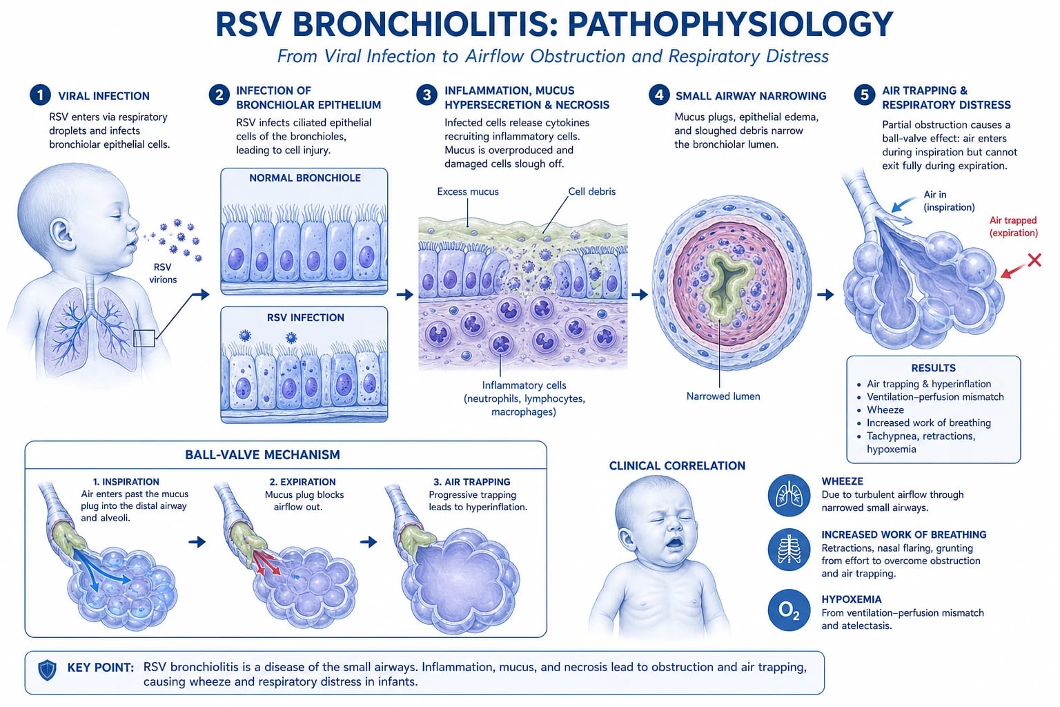
Bronchiolitis begins when a respiratory virus — overwhelmingly RSV — infects the ciliated epithelium of the upper respiratory tract and then descends to the bronchioles (airways under approximately 2 mm in diameter). RSV preferentially binds via its G protein to ciliated cells and replicates, triggering a cascade of epithelial necrosis, submucosal oedema, increased mucus secretion, and a mixed inflammatory infiltrate (lymphocytes, neutrophils, macrophages) that narrows the already small infant bronchiolar lumen.[2]
The crucial pathophysiological point is the disproportionate effect of airway narrowing in infants. Infant bronchioles are tiny to begin with (the small-airway contribution to total airway resistance is far higher in infants than in adults), so a given increment of oedema and mucus produces a much larger rise in resistance than it would in an older child. By the same principle that governs croup — airway resistance is inversely proportional to the fourth power of the radius — modest mucosal thickening multiplies the work of breathing many times over. The obstruction is dynamic and partial: during expiration the positive intrathoracic pressure further compresses the narrowed, floppy bronchioles, producing a ball-valve effect — air enters on inspiration but is trapped on expiration, generating air trapping, hyperinflation, and wheeze. Complete plugging by mucus and cellular debris collapses the downstream alveoli, producing atelectasis.[2]
The gas-exchange consequences are twofold. Continued perfusion of poorly ventilated alveoli produces a ventilation-perfusion (V/Q) mismatch with right-to-left shunting — the principal mechanism of hypoxaemia, which is often disproportionate to the apparent clinical severity. As fatigue mounts and respiratory effort falters, alveolar hypoventilation supervenes and hypercapnia develops, signalling impending respiratory failure. Infants compensate first by increasing respiratory rate and recruiting accessory muscles; decompensation shows as grunting (auto-PEEP to splint alveoli), head bobbing (use of sternocleidomastoid), and ultimately exhaustion with falling rate and effort — a pre-arrest sign.[1][2]
Apnoea in bronchiolitis is incompletely understood but is thought to reflect immaturity of central respiratory control (the preterm brainstem is exquisitely sensitive to hypoxaemia, producing inhibition rather than stimulation of breathing), upper-airway obstruction during sleep, and possibly a direct effect of viral neurotoxicity on the respiratory centre. It is a hallmark of very young and ex-preterm infants rather than a feature of older infants with bronchiolitis.[2]
Clinical Presentation
The classic presentation is a previously well infant aged 2 to 6 months, seen during the winter season, with a 1- to 3-day coryzal prodrome (nasal congestion, clear rhinorrhoea, low-grade fever, occasional sneeze) followed by progressive cough, wheeze, tachypnoea, and difficulty feeding. The tempo is usually over hours to a couple of days, and parents most often report that the baby has "started breathing fast", "is not feeding", or "is making a noise with breathing".[1][2]
Examination findings fall into predictable groups:[1][2]
Upper respiratory
- Coryza, clear or mucopurulent nasal discharge, nasal obstruction (contributes significantly to work of breathing in obligate nose-breathers)
- Mild conjunctivitis, otitis media in 5 to 10 percent
- Low-grade fever (under 39 degrees C); high fever does not exclude bronchiolitis but raises bacterial infection in the differential
Respiratory distress
- Tachypnoea (typically 50 to 70 breaths per minute; over 60 in infants under 2 months, over 50 in 2 to 12 months)
- Nasal flaring, intercostal and subcostal recession, tracheal tug
- Head bobbing (sternocleidomastoid recruitment) in young infants — a sign of significant distress
- Grunting expirations (auto-PEEP), prolonged expiratory phase, use of accessory muscles
- Hyperinflated chest with increased anteroposterior diameter, hyper-resonance to percussion
Auscultation
- Bilateral, widespread, fine end-inspiratory crackles (crepitations) — nearly universal
- High-pitched expiratory wheeze — often widespread; severity of wheeze does not correlate with disease severity
- Reduced air entry in areas of significant atelectasis or mucus plugging
- Asymmetry should prompt consideration of foreign body, lobar pneumonia, or pleural effusion
Systemic / red flags
- Apnoea (witnessed pauses over 15 to 20 seconds, or with desaturation/bradycardia) — may be the sole feature in very young infants
- Cyanosis, pallor, mottling, poor perfusion
- Lethargy, irritability, or reduced responsiveness — signs of hypoxaemia or hypercapnia
- Reduced wet nappies, dry mucous membranes, sunken fontanelle — dehydration
- Hepatomegaly from hyperinflation (not heart failure unless gallop or murmur)
Atypical presentations are important and examiners probe them. The ex-preterm infant under 2 months (corrected age) may present with apnoea and minimal respiratory signs — the wheeze and crackles come later. The infant with congenital heart disease may deteriorate rapidly with shock, hepatomegaly, and a gallop rather than the expected pattern, because the compromised myocardium cannot meet the increased metabolic demand. The immunocompromised infant may shed virus for weeks and present with prolonged or atypical disease. The indigenous or remote infant often presents late and more severely. Recognising these variants is the difference between safe and unsafe practice.[1][2]
Differential Diagnosis
The differential of wheeze and tachypnoea in an infant is broad, and the diagnostic task is to separate common, first-episode viral bronchiolitis from conditions that need fundamentally different management — pneumonia, congenital heart disease with failure, foreign body aspiration, and the recurrent wheezers (asthma, cystic fibrosis, aspiration). The single most useful discriminator is the clinical context: a first episode of wheeze with a coryzal prodrome in winter, in an infant under 2 years, is bronchiolitis until proven otherwise.[1][2]
Bronchiolitis
- First or early episode of wheeze, age under 2 years (peak 2 to 6 months), winter season
- Coryzal prodrome 1 to 3 days, then cough, wheeze, tachypnoea, bilateral crackles
- Low-grade fever, feeding difficulty, poor response to bronchodilator
- Hyperinflation on CXR if performed; clinical diagnosis
Pneumonia (viral or bacterial)
- Fever often higher and more sustained; toxic, ill-appearance more pronounced
- Focal crackles or reduced air entry, localised bronchial breathing; asymmetry on auscultation
- Lobar or patchy consolidation on CXR; raised inflammatory markers in bacterial
- Antibiotics indicated for bacterial pneumonia; viral pneumonia overlaps with bronchiolitis
Asthma / multi-trigger wheeze
- Recurrent wheeze (three or more episodes), personal or family atopy (eczema, food allergy, asthma)
- Older infant or child; rapid and clear response to bronchodilator
- No fever or coryzal prodrome necessarily; triggers include allergens, exercise, cold air
- Trial of bronchodilator justified; inhaled corticosteroids for recurrent disease
Foreign body aspiration
- Sudden onset, witnessed choking, no preceding coryza, no fever initially
- Asymmetric wheeze or reduced air entry; localised air trapping on expiratory CXR or fluoroscopy
- Typical age 1 to 3 years but possible in infants
- Rigid bronchoscopy; do not instrument blindly
Congestive cardiac failure (CHD)
- Recurrent wheeze, poor weight gain, sweating with feeds, tachycardia out of proportion
- Murmur (may be absent), gallop, hepatomegaly, cardiomegaly on CXR
- Differential cyanosis (lower limbs) in some lesions; weak or absent femoral pulses (coarctation)
- Echocardiogram; fluid restriction and diuretics rather than fluids; cardiology referral
Pertussis
- Paroxysmal cough, inspiratory whoop (older infants), post-tussive vomiting, apnoea in young infants
- Coryza prodrome followed by catarrhal stage; lymphocytosis; under- or un-immunised
- Marked lymphocytosis on FBC; PCR on nasopharyngeal aspirate
- Macrolide (azithromycin), admission for infants under 6 months, infection control
Vascular ring / airway anomaly
- Stridor or wheeze from early infancy, positional, feeding-related, recurrent
- Tracheomalacia, laryngomalacia, double aortic arch; no fever, no coryza
- Barium swallow, bronchoscopy, CT/MRI angiography
- ENT and cardiothoracic referral
Gastro-oesophageal reflux / aspiration
- Chronic cough and wheeze, worse after feeds, recurrent, vomiting, failure to thrive
- No fever or coryza; associated apnoea or desaturation in some
- pH-impedance study or milk scan; response to anti-reflux measures
- Not a single acute episode — a recurrent pattern
Cystic fibrosis
- Recurrent wheeze and chest infections, failure to thrive, steatorrhoea, family history
- Persistent despite treatment; salty-tasting skin, meconium ileus history
- Newborn screen positive; sweat chloride over 60 mmol/L
- CF team, chest physiotherapy (here it IS indicated), pancreatic enzymes
The features that should prompt reconsideration of bronchiolitis are: a first wheeze in an infant over 18 to 24 months (think early asthma); asymmetric or focal chest findings (foreign body, pneumonia); a toxic, high-fever child (bacterial infection); failure to respond to appropriate supportive care over 48 to 72 hours; cardiac findings (murmur, hepatomegaly, gallop, differential cyanosis); and recurrent episodes (asthma, CF, immunodeficiency, aspiration, airway anomaly).[1]
Clinical & Bedside Assessment
Bronchiolitis is a clinical diagnosis made at the bedside. The history and a calm, structured observation usually establish both the diagnosis and the severity, and investigations should be minimal — every needle and probe agitates the infant, worsening work of breathing and oxygenation.[1]
History should establish: age (and corrected gestational age if premature), onset and tempo (coryzal prodrome, then worsening over hours to days), the specific respiratory symptoms (cough, wheeze, fast breathing), feeding and hydration (volume of feeds, frequency, wet nappies — fewer than four to five wet nappies in 24 hours suggests dehydration), apnoea or colour change (any witnessed pauses, dusky spells), fever (degree and duration), and the risk profile (prematurity, chronic lung disease, congenital heart disease, immunodeficiency, neuromuscular disease). Immunisation status, household smoke exposure, daycare attendance, and sick contacts complete the picture.[1][2]
Examination is performed calmly, ideally with the infant in the parent's arms and undisturbed. The structured observations are:[1][2]
- Work of breathing — respiratory rate (count for a full minute), depth, recession (intercostal, subcostal, suprasternal), nasal flaring, tracheal tug, head bobbing, grunting, accessory muscle use, and prolonged expiration. Marked recession, grunting, and head bobbing indicate severe disease.
- Oxygenation — SpO2 in air, ideally over 30 to 60 seconds and with the infant settled; note the threshold behaviour (a reading fluctuating around 90 to 92 percent warrants admission and observation).
- Auscultation — bilateral crackles and wheeze confirm lower-respiratory involvement; air entry should be symmetrical (asymmetry is a red flag).
- Hydration and perfusion — mucous membranes, capillary refill, skin temperature, fontanelle, peripheral pulses, hepatomegaly.
- Cardiovascular — heart rate, murmurs (new or unrecognised congenital heart disease), gallop, differential pulses and oxygen saturation (right arm versus lower limbs) to exclude coarctation or duct-dependent lesions.
- Mental state — alert and interactive (reassuring), irritable, lethargic, or floppy (concerning).[1]
The Bronchiolitis Severity Score used in some units (e.g. the Tal or Wang score) quantifies respiratory rate, wheeze, retraction, and overall distress, but no score is universally adopted and none replaces clinical judgement. The practical rule is: any infant with SpO2 persistently under 92 percent in air, any respiratory distress, difficulty feeding, dehydration, or apnoea should be admitted; any infant under 3 months with risk factors or apnoea should be admitted and monitored.[1][2]
Investigations
For typical bronchiolitis, investigations are not needed and should not be performed. They do not change management, they distress the infant, and they generate false-positive leads. The AAP and NICE guidelines explicitly advise against routine chest X-ray, blood tests, or viral testing in a child with a typical presentation.[1]
When investigations are warranted — for severe or atypical disease, an uncertain diagnosis, deterioration despite supportive care, or a complicated course:[1]
- Chest X-ray (if severe, atypical, asymmetric, or poor response): the classic findings are hyperinflation (increased anteroposterior diameter, flattened diaphragms, increased retrosternal air), peribronchial thickening/cuffing, subsegmental atelectasis or patchy infiltrates, and occasionally hyperlucent areas from air trapping. Crucially, the CXR cannot reliably distinguish bronchiolitis from viral pneumonia, and over-reading patchy infiltrates drives unnecessary antibiotic use. A CXR should not be obtained for the sole purpose of "confirming" bronchiolitis.[1]
- Viral testing (nasopharyngeal aspirate or swab for multiplex respiratory PCR): identifies RSV, rhinovirus, influenza, parainfluenza, adenovirus, hMPV, bocavirus, and SARS-CoV-2. It does not change management in the individual infant (treatment is supportive regardless), but it guides infection control, cohorting, isolation, and palivizumab decisions, and may identify influenza (treatable with oseltamivir in high-risk infants).[1]
- Blood tests (FBC, U&E, CRP) only if bacterial co-infection is suspected (high fever, toxic appearance, focal signs, persistent fever, raised inflammatory markers) or for monitoring electrolytes and renal function in the severely ill or dehydrated infant. Bacterial co-infection is uncommon (bacteraemia under 1 to 2 percent, urinary tract infection in 2 to 5 percent) and routine bloods overdiagnose it.[1]
- Capillary or arterial blood gas in severe disease (SpO2 under 90 percent in oxygen, grunting, fatigue, altered consciousness): assesses hypoxaemia, hypercapnia, and acidosis. A rising PaCO2 with respiratory acidosis signals ventilatory failure and the need to escalate to HFNC, CPAP, or intubation. Capillary lactate is a useful bedside marker of severity and tissue perfusion.[1]
- Urinalysis and culture should be considered in febrile infants under 60 days and in those with persistent fever, given the small but real rate of concurrent UTI.[1]
WHEEZE
Management — Resuscitation
Severe bronchiolitis with impending respiratory failure (SpO2 under 88 percent in oxygen, recurrent apnoea, grunting, exhaustion, rising PaCO2, altered consciousness) is a time-critical emergency requiring a calm, escalating bundle of oxygen, ventilatory support, fluids, and monitoring. The aim is to correct hypoxaemia, relieve the work of breathing before fatigue produces hypercapnic failure, treat apnoea, and avoid iatrogenic harm.[1]
The resuscitation bundle, applied without unnecessary intervention, is:[1]
- Assess and secure the airway; keep the infant calm and upright in the parent's arms where possible. Agitation increases oxygen consumption and worsens dynamic airway obstruction; gentle handling and parental presence are therapeutic.
- Supplemental humidified oxygen by nasal cannula or face mask to maintain SpO2 at 92 to 95 percent (NICE); the AAP recommends oxygen for SpO2 under 90 percent persistently, and many units use 92 percent as the operational threshold. Do not chase a normal saturation in an infant with improving work of breathing — once feeding and distress improve, wean and stop.[1]
- Nasal suctioning of visible secretions (gentle, superficial) — nasopharyngeal suction is reserved for infants with significant upper-airway obstruction and is not routinely performed deep. Suctioning briefly improves comfort and feeding in many infants.
- High-flow nasal cannula (HFNC) — humidified, heated, blended oxygen-air at 1 to 2 L/kg/min, is the first-line escalation for moderate-severe bronchiolitis with hypoxia or rising work of breathing not responding to standard oxygen. The PARIS trial (Franklin 2018, NEJM) showed HFNC reduced escalation to CPAP/intubation versus standard oxygen.[8]
- Continuous positive airway pressure (CPAP) at 5 to 8 cmH2O for HFNC failure, refractory hypoxia, or rising hypercapnia. CPAP splints small airways open, reduces work of breathing, and improves gas exchange; it is the preferred non-invasive mode for apnoea and hypercapnia.
- Mechanical ventilation for refractory respiratory failure, recurrent apnoea despite CPAP and caffeine, profound hypercapnia with acidosis, or cardiovascular collapse. Use lung-protective settings (permissive hypercapnia, low tidal volumes) and sedation; pneumothoraces and air leak are recognised complications.[1]
- Fluids — IV or NG isotonic maintenance for any infant unable to maintain hydration. Reduce to 80 percent of standard maintenance in severe disease to avoid fluid overload from increased antidiuretic hormone (SIADH is common in severe bronchiolitis). Monitor electrolytes and sodium.[1]
- Caffeine citrate (loading 20 mg/kg IV/oral, then maintenance 5 mg/kg/day) for apnoea of prematurity or recurrent apnoea in the ex-preterm or young infant; reduces apnoea frequency and the need for ventilation.[1]
Management — Definitive & Stepwise
The cornerstone of bronchiolitis management is supportive care and the judicious avoidance of ineffective therapies. Bronchiolitis is self-limiting, and the role of the clinician is to support the infant through the illness while identifying and treating the few who deteriorate. This principle — doing less, deliberately — is supported by the strongest evidence in paediatric respiratory medicine.[1]
Mild bronchiolitis
- Manage at home; reassure that illness peaks at day 3 to 5 and resolves over 7 to 14 days
- Saline nasal drops and gentle bulb/superficial suction before feeds and sleep
- Small, frequent feeds; continue breastfeeding; antipyretics (paracetamol 15 mg/kg q4-6h or ibuprofen 10 mg/kg q6-8h if over 3 months) for fever and discomfort
- Safety-net advice: return urgently for apnoea, cyanosis, worsening breathing, poor feeding (under half normal), fewer wet nappies, lethargy
Moderate bronchiolitis
- Admit for observation, oxygen (SpO2 under 92 percent), and fluids
- Humidified oxygen via nasal cannula or face mask to target SpO2 92 to 95 percent
- NG feeds (two-thirds to three-quarters maintenance) or IV isotonic fluids if not tolerating oral
- Nasal suctioning; consider HFNC if SpO2 under 92 percent in standard oxygen or work of breathing rising
- Continuous SpO2 monitoring; reassess every 2 to 4 hours
Severe bronchiolitis
- HDU or PICU; humidified oxygen
- HFNC at 1 to 2 L/kg/min as first-line respiratory support; CPAP at 5 to 8 cmH2O if HFNC fails, for apnoea, or for hypercapnia
- IV isotonic fluids at 80 percent maintenance; monitor sodium and electrolytes
- Continuous cardiorespiratory and SpO2 monitoring; capillary gas and lactate
- Intubation and mechanical ventilation for refractory hypoxia or hypercapnia, recurrent apnoea, exhaustion, or cardiovascular collapse; caffeine for apnoea in young/ex-preterm infants
Therapies that do NOT work (and why the gate matters)
The Cochrane reviews and the AAP guideline converge firmly on the following: routine bronchodilators, corticosteroids, antibiotics, and chest physiotherapy are not recommended for typical bronchiolitis. Reaching for them is a marker of discomfort with supportive care, not of evidence-based practice.[1]
- Bronchodilators (salbutamol, adrenaline) — the Cochrane review (Gadomski 2014) of 30 trials in over 1,900 infants found no consistent benefit on oxygen saturation, hospitalisation, or length of stay; adverse effects (tachycardia, tremor, hypokalaemia, agitation) outweigh any small, subjective improvement. A trial of bronchodilator is not routinely recommended; if used in a selected older infant with possible asthma overlap, it should be discontinued if there is no clear, objective response. Nebulised adrenaline may be considered in the emergency department for an infant with marked distress who may be discharged within 24 hours (small short-term benefit), but is not continued as inpatient therapy.[5]
- Corticosteroids (systemic or inhaled) — the Cochrane review (Fernandes 2013) of 17 trials in over 2,500 infants found no benefit on admissions or length of stay. The large combined dexamethasone trials confirmed no effect. Steroids are not recommended for bronchiolitis, even severe disease.[6]
- Antibiotics — bronchiolitis is viral. Bacterial co-infection (bacteraemia, pneumonia, UTI) is uncommon and routine antibiotics are not indicated. Reserve them for suspected bacterial co-infection (high fever, focal signs, raised inflammatory markers, toxic appearance, persistent fever) or an alternative bacterial diagnosis. The presence of purulent nasal discharge alone does not justify antibiotics.[1]
- Chest physiotherapy — the Cochrane review (Roque i Figuls 2016) found no benefit of vibration, percussion, or postural drainage on severity, oxygen saturation, or length of stay, and potential for increased distress. Routine chest physiotherapy is not recommended. (Note: this is not cystic fibrosis — physio is vital in CF but not in bronchiolitis.)[11]
- Nebulised hypertonic saline (3 percent) — the Cochrane review (Zhang 2017) found modest and inconsistent benefit, with effect sizes shrinking when only saline-controlled, high-quality trials were considered; benefit is most plausible in infants with longer hospitalisations (over 3 days) and is minimal in settings with short stays. It is an option in some units for prolonged admission, given with a bronchodilator if the hypertonic saline provokes bronchospasm.[7]
- Ribavirin, nebulised antiviral active against RSV, is not routinely recommended; reserved for the severely immunocompromised (e.g. haematopoietic stem cell transplant recipients) with proven RSV, where it may reduce progression to lower tract disease. Teratogenicity and administration difficulties limit its use.[1]
- Leukotriene receptor antagonists (montelukast), heliox, and surfactant are not recommended outside research for typical bronchiolitis.[1]
PARIS (Franklin 2018, NEJM)
Multicentre randomised controlled trial, 1472 infants under 12 months with bronchiolitis and hypoxia (SpO2 90 to 93 percent)
Population: Moderate-severe bronchiolitis in Australian and New Zealand EDs
Key finding
HFNC reduced the proportion of infants needing escalation to CPAP or intubation (12 percent vs 23 percent, treatment-failure halved) without changing length of stay
Stepwise management summary
Diagnose bronchiolitis clinically (coryza then cough, wheeze, tachypnoea, crackles, age under 2 years).
Grade severity across work of breathing, SpO2, feeding, mental state, and apnoea.
Mild — home: nasal saline, small frequent feeds, antipyretics, written safety-net. NO medications.
Moderate — admit: humidified oxygen to SpO2 92 to 95 percent, NG or IV fluids, nasal suctioning, continuous SpO2 monitoring.
Escalate — HFNC 1 to 2 L/kg/min if SpO2 under 92 percent in standard oxygen or work of breathing rising.
Further escalate — CPAP 5 to 8 cmH2O for HFNC failure, apnoea, or hypercapnia; caffeine for apnoea in young/ex-preterm.
Intubate — refractory hypoxia or hypercapnia, recurrent apnoea, exhaustion, or cardiovascular collapse.
Reconsider the diagnosis if no response or atypical course (bacterial pneumonia, foreign body, CHD).
Discharge when SpO2 over 92 percent in air for a sustained period, feeding well, no distress, parents confident; document the safety-net.
Admission criteria
Indications for hospital admission include: age under 3 months with bronchiolitis (especially ex-preterm); SpO2 persistently under 92 percent in air; moderate or severe respiratory distress; apnoea (any episode); poor feeding or dehydration; toxic appearance or lethargy; significant comorbidity (chronic lung disease, congenital heart disease, immunodeficiency, neuromuscular disease); social concerns (remote residence, parental anxiety, inability to return); and deterioration despite appropriate outpatient care.[1]
Indications for PICU include: SpO2 under 88 to 90 percent despite high-flow oxygen; recurrent or severe apnoea; rising PaCO2 with respiratory acidosis; exhaustion or fatigue; shock, dehydration, or electrolyte disturbance requiring intensive monitoring; and the need for non-invasive or invasive ventilation.[1]
Discharge criteria
Discharge is appropriate when: SpO2 is over 92 percent in air for a sustained period (typically 4 to 8 hours) off all respiratory support; the infant is feeding effectively (at least 75 percent of usual volume) and adequately hydrated; respiratory distress is minimal or absent; there is no apnoea and normal mental state; and parents are confident with a clear written safety-net covering warning signs (apnoea, cyanosis, worsening distress, poor feeding, fewer wet nappies, lethargy) and when to return.[1]
Specific Subtypes & Scenarios
Mild bronchiolitis (the majority) is managed at home with supportive care and a safety-net; the illness peaks at day 3 to 5 and resolves over 7 to 14 days, with the cough and wheeze sometimes persisting for 3 to 4 weeks. Antipyretics for comfort, nasal saline and gentle suction before feeds, and continued breastfeeding are the mainstays.[1]
Bronchiolitis in the infant under 3 months (especially ex-preterm) carries a markedly higher risk of apnoea, admission, and PICU. Admit for observation and continuous monitoring even if the infant looks relatively well; consider caffeine for apnoea; have a low threshold for HFNC or CPAP. Apnoea may persist for several days and often dictates the length of stay.[1]
Bronchiolitis with chronic lung disease of prematurity (bronchopulmonary dysplasia): lower threshold for admission; these infants desaturate quickly and tolerate hypoxia poorly; start oxygen early, monitor closely, and escalate promptly.[1][3]
Bronchiolitis with haemodynamically significant congenital heart disease: high-risk group; admit, monitor in HDU/PICU, give oxygen, but avoid fluid overload (restrict to 75 to 80 percent maintenance, monitor for hepatomegaly and gallop), involve cardiology. These infants are eligible for palivizumab prophylaxis.[3]
Bronchiolitis with immunodeficiency (primary immunodeficiency, transplant recipients, on chemotherapy): prolonged and atypical course; longer viral shedding; broader differential; ribavirin may be considered in severe RSV in stem-cell transplant recipients. Strict isolation and infection-control measures.[1]
Bronchiolitis with neuromuscular disease: weak respiratory muscles and impaired airway protection make these infants high-risk; admit, low threshold for non-invasive ventilation, aggressive airway clearance (here suctioning and occasionally physiotherapy are justified), and early involvement of the neurology/respiratory team.[1]
Apnoea-dominant bronchiolitis: the ex-preterm or very young infant may present with apnoea and minimal respiratory signs. The wheeze and crackles may develop later. Manage with admission, continuous monitoring, caffeine, and early escalation to non-invasive ventilation; intubation if recurrent or severe.[1][2]
Bronchiolitis with respiratory syncytial virus vs rhinovirus: RSV causes the most severe disease and the highest admission rate; rhinovirus-associated bronchiolitis is increasingly recognised and carries a stronger association with subsequent recurrent wheeze and asthma. Human metapneumovirus produces a bronchiolitis clinically indistinguishable from RSV but typically circulates later in the season. Influenza and adenovirus may cause more severe or necrotising disease.[2][4]
Complications & Pitfalls
Complications of bronchiolitis include:[1][2]
- Apnoea — most common in infants under 3 months and ex-preterm; may be the presenting feature; may persist for days; mandate admission and monitoring.
- Respiratory failure — hypoxaemic (V/Q mismatch), hypercapnic (fatigue), or mixed; the feared endpoint prevented by early escalation through oxygen, HFNC, CPAP, and ventilation.
- Dehydration and electrolyte disturbance — from poor oral intake and increased insensible losses; SIADH in severe disease causes hyponatraemia if fluids are not restricted.
- Secondary bacterial infection — pneumonia, otitis media, bacteraemia, UTI; uncommon (under 5 to 10 percent) but recognised; suspected when fever is high or persistent, the infant is toxic, or inflammatory markers are raised.
- Pneumothorax and air leak — rare, usually a complication of positive-pressure ventilation.
- Bronchiolitis obliterans — a rare, severe, irreversible small-airway obliteration most often following adenovirus or Mycoplasma pneumoniae infection; presents with persistent wheeze, crackles, and hyperinflation over weeks to months; high-resolution CT shows mosaic attenuation and air trapping; supportive management, no curative therapy.
- Recurrent wheeze and asthma — infants with severe RSV bronchiolitis (and especially rhinovirus bronchiolitis) have a higher rate of recurrent wheeze and physician-diagnosed asthma in later childhood; the causal relationship (whether the virus causes asthma, or susceptible airways are more affected) remains debated.[2]
- Death — rare in high-income settings (under 0.1 percent of hospitalised) but a major contributor to under-five mortality in low- and middle-income countries.[4]
Pitfalls in management:[1]
- Over-investigation — routine CXR, bloods, and viral swabs do not change management in typical bronchiolitis and generate false positives, overdiagnosis of bacterial infection, and unnecessary antibiotics.
- Over-treatment — bronchodilators, steroids, and antibiotics are entrenched in practice despite clear evidence of no benefit; their routine use is a marker of deviation from guideline-based care.
- Under-recognition of apnoea — the ex-preterm or very young infant may have minimal respiratory signs yet develop life-threatening apnoea; always ask about pauses in breathing and admit the at-risk infant for monitoring.
- Fluid overload — giving full maintenance fluids in severe bronchiolitis risks hyponatraemia (SIADH) and, in infants with congenital heart disease, pulmonary oedema; restrict to 75 to 80 percent maintenance in severe disease and monitor sodium.
- Misdiagnosis — missing bacterial pneumonia (focal signs, toxic), foreign body (sudden onset, asymmetric), congenital heart disease (murmur, hepatomegaly, differential cyanosis), or pertussis (paroxysmal cough, whoop, apnoea, under-immunised).
- Chasing saturations — once the infant is feeding well and the work of breathing has improved, tolerate SpO2 in the low 90s overnight and wean oxygen; prolonged admission purely for "a few points of saturation" is a common cause of unnecessary length of stay.
- Premature discharge of a very young or high-risk infant who subsequently deteriorates with apnoea at home.[1]
Prognosis & Disposition
The prognosis of bronchiolitis is excellent for the great majority. Most infants recover fully within 7 to 14 days, although the cough and wheeze may persist for 3 to 4 weeks. Symptoms typically peak at day 3 to 5 and improve thereafter. Mortality is very low in high-income settings — under 0.1 percent of hospitalised infants — and is largely confined to infants with significant comorbidity (severe congenital heart disease, chronic lung disease, immunodeficiency, neuromuscular disease). In low- and middle-income countries, by contrast, RSV bronchiolitis remains a leading infectious cause of infant death, driven by late presentation, malnutrition, limited oxygen, and lack of PICU access.[1][4]
Recurrence is common: more than half of infants have another wheezing episode within the first two years, and a substantial minority (15 to 30 percent of those hospitalised with RSV) go on to develop recurrent wheeze and physician-diagnosed asthma in childhood — an association strongest with severe RSV and with rhinovirus bronchiolitis, and the subject of ongoing debate about causality versus shared susceptibility.[2]
Disposition is driven by severity, response to treatment, age, comorbidity, and social factors:[1]
Discharge home
- Mild bronchiolitis with SpO2 over 92 percent in air, feeding well, no distress
- No apnoea, no dehydration, normal mental state, age over 3 months without comorbidity
- Confident parents with reliable follow-up and a written safety-net
Admit to ward
- Moderate bronchiolitis needing oxygen, fluids, or observation
- Age under 3 months (especially ex-preterm) or significant comorbidity
- Dehydration or poor feeding; social concerns; failure to improve
Admit to HDU/PICU
- Severe bronchiolitis needing HFNC, CPAP, or ventilation
- Apnoea, hypercapnia, exhaustion, or cardiovascular compromise
- Refractory hypoxia (SpO2 under 88 to 90 percent in high-flow oxygen)
- Need for intensive monitoring or intubation
The safety-net is a non-negotiable part of discharge. Parents must receive written advice to return urgently if the infant has apnoea or colour change (blue or dusky spells), cyanosis, worsening breathing, inability to feed (under half normal intake), fewer wet nappies, lethargy or unresponsiveness, or a high or persistent fever. Follow-up with the general practitioner or paediatrician in 24 to 48 hours is reasonable for moderate cases.[1]
Special Populations
Premature infants (especially under 29 weeks, or under 32 weeks with chronic lung disease) are the highest-risk group: smaller airways, immature respiratory control (predisposing to apnoea), reduced functional reserve, and residual lung disease. Lower threshold for admission, early oxygen, monitoring for apnoea, caffeine, and early escalation. Eligible for palivizumab prophylaxis during RSV season.[3]
Infants with haemodynamically significant congenital heart disease (cyanotic lesions, pulmonary hypertension, large left-to-right shunts) carry high mortality from RSV; admit, monitor in HDU/PICU, give oxygen, restrict fluids, involve cardiology. Eligible for palivizumab.[3]
Immunocompromised infants (primary immunodeficiency, solid-organ or stem-cell transplant, chemotherapy): longer and atypical courses, prolonged viral shedding, broader differentials; consider ribavirin for severe RSV; strict isolation.[1]
Neuromuscular disease (spinal muscular atrophy, congenital myopathy, severe cerebral palsy): impaired cough and airway protection; high-risk; admit, early non-invasive ventilation, aggressive suctioning.[1]
Indigenous and remote populations (Australian Aboriginal, Native American/Alaska Native, Canadian First Nations, Maori): substantially higher hospitalisation and severity rates, driven by crowding, smoke exposure, earlier age of first infection, and access barriers; lower threshold for admission and retrieval.[2]
Infants with Down syndrome: increased risk of severe bronchiolitis, partly from associated congenital heart disease, hypotonia, and immune dysregulation; manage actively.[1]
Pregnant adolescents and adolescent parents: management of the infant is unchanged; ensure adolescent-friendly setting, lactation support, and safeguarding review as appropriate.[1]
Evidence, Guidelines & Regional Differences
The evidence base for bronchiolitis is strong, consistent, and remarkably unified across guidelines in favour of supportive care and against routine pharmacotherapy.[1]
The AAP Clinical Practice Guideline (Ralston 2014) is the international benchmark. It defines bronchiolitis clinically (first episode of wheeze under 2 years with a viral prodrome), recommends against routine radiographic, laboratory, or viral testing in typical cases, and against routine bronchodilators, corticosteroids, antibiotics, and chest physiotherapy. Oxygen is recommended for SpO2 under 90 percent; clinician discretion applies around 90 to 92 percent. Palivizumab is restricted to high-risk groups.[1]
The NICE NG9 guideline (2015, UK) aligns closely with the AAP: clinical diagnosis; no routine CXR, bloods, or viral testing; oxygen for SpO2 under 92 percent; fluids for those not feeding; no bronchodilators, steroids, antibiotics, hypertonic saline, or chest physiotherapy routinely; clear criteria for referral, admission, PICU, and discharge.[1]
Bronchodilators — the Cochrane review (Gadomski 2014) of 30 trials found no clinically meaningful benefit on oxygen saturation, hospitalisation rate, or length of stay, with adverse effects (tachycardia, tremor, hypokalaemia). A clinical trial of a bronchodilator is not routinely recommended.[5]
Corticosteroids — the Cochrane review (Fernandes 2013) found no reduction in admissions or length of stay. Systemic and inhaled corticosteroids are not recommended for bronchiolitis.[6]
Hypertonic saline — the Cochrane review (Zhang 2017) found modest, inconsistent reductions in length of stay, attenuated in saline-controlled and high-quality trials; benefit most plausible for prolonged admissions.[7]
Chest physiotherapy — the Cochrane review (Roque i Figuls 2016) found no benefit. Not recommended.[11]
High-flow nasal cannula — the PARIS trial (Franklin 2018, NEJM) established HFNC as an effective first-line escalation, halving treatment failure versus standard oxygen in 1,472 hypoxic infants. Subsequent evidence supports its safety and efficacy; HFNC has become standard practice in moderate-severe bronchiolitis in most units.[8]
Palivizumab prophylaxis — the IMpact-RSV trial (1998) showed a 55 percent relative reduction in RSV hospitalisation in high-risk preterm infants, establishing palivizumab 15 mg/kg monthly IM during RSV season as the standard of care for high-risk infants. The AAP updated guidance (2014) refined eligibility to focus palivizumab on those most likely to benefit.[10][3]
Nirsevimab — the Griffin 2020 NEJM trial of single-dose nirsevimab (a long-acting anti-RSV F monoclonal antibody with an extended half-life) in preterm infants showed a 70 percent reduction in RSV-related hospitalisation over the 150-day RSV season. Subsequent MELODY-trial evidence extended efficacy to term infants, supporting nirsevimab as a universal, single-dose infant immunisation that is now being adopted in several national immunisation programmes (Spain, France, UK targeted, US via the CDC maternal-and-infant programme).[9]
Australasian practice (Royal Australasian College of Physicians, NHMRC) aligns with AAP and NICE: supportive care, no routine pharmacotherapy, HFNC as first-line escalation (PARIS was an ANZ trial). Palivizumab is funded for high-risk infants under the National Immunisation Programme criteria (preterm under 28 weeks in first season, CLD under 24 months, haemodynamically significant CHD under 12 months, certain immunocompromised). Nirsevimab rollout is being adopted at state level. Indigenous and remote populations warrant lower thresholds for admission and retrieval.[1][8]
The WHO Pocket Book of Hospital Care for Children provides the standard for low-resource settings: supportive care with oxygen (the single most life-saving therapy), hydration, careful feeding, no routine bronchodilators or steroids, and vigilant monitoring for deterioration. In low- and middle-income countries, where RSV mortality is concentrated, the priorities are reliable oxygen delivery (pulse oximetry and oxygen concentrators), breastfeeding support, hand hygiene, and reducing household smoke exposure. Palivizumab is largely impractical (cost, cold chain, monthly injections); nirsevimab's single-dose design makes it the realistic vehicle for population-level RSV prevention in these settings, and Gavi support is being explored. Maternal RSV vaccination offers another scalable strategy.[4]
Controversies and evolving areas include: the exact oxygen saturation threshold for treatment and discharge (90 vs 92 percent); the role of HFNC in mild-moderate disease (risk of over-use and prolonged admission); the inconsistent effect of hypertonic saline; whether nirsevimab and maternal vaccination will displace palivizumab entirely; the strength of the bronchiolitis-asthma link and whether prevention alters long-term wheeze; and the implementation of universal RSV immunisation programmes globally. The strongest evidence in bronchiolitis remains the simplest: oxygen, hydration, monitoring, and the avoidance of ineffective drugs.[1][9]
Prevention
Prevention of RSV bronchiolitis rests on passive immunisation of high-risk infants and general public-health measures.[3]
Palivizumab (Synagis) is a humanised monoclonal antibody against the RSV F (fusion) glycoprotein, given as 15 mg/kg by intramuscular injection monthly during the RSV season (typically 5 monthly doses, November to March in the northern hemisphere). It is prophylactic only, not therapeutic. Eligibility, per the AAP 2014 updated guidance:[3]
- Preterm infants under 29 weeks 0 days gestation in their first RSV season (under 12 months at the start of the season).
- Preterm infants under 32 weeks with chronic lung disease of prematurity (on supplemental oxygen, diuretics, or steroids in the 6 months before season), under 24 months at start of season.
- Haemodynamically significant congenital heart disease under 12 months (cyanotic lesions, pulmonary hypertension, heart failure on medication, or awaiting surgery).
- Severely immunocompromised and certain neuromuscular infants under 24 months in selected regions.
- Down syndrome and cystic fibrosis with lung disease in selected regions.[3]
The IMpact-RSV trial (1998) established efficacy: a 55 percent relative reduction in RSV-related hospitalisation (10.6 percent to 4.8 percent) in high-risk preterm and CLD infants.[10] Palivizumab is not recommended for healthy term infants or for treatment of established disease.
Palivizumab (Synagis)
Dose
15 mg/kg per dose
Nirsevimab (Beyfortus) is a long-acting monoclonal antibody against the RSV F protein with a modified Fc region giving a half-life of over 60 days and protection across the 5-month RSV season from a single intramuscular dose. The Griffin 2020 NEJM trial in preterm infants showed a 70 percent reduction in RSV hospitalisation; subsequent trials (MELODY) extended this to term infants. Recommended by the CDC ACIP for all infants under 8 months in their first RSV season, and for high-risk infants 8 to 19 months entering their second season. Nirsevimab represents a paradigm shift — from selective monthly palivizumab to universal single-dose infant immunisation.[9]
Maternal RSV vaccination (e.g. bivalent prefusion F vaccine, Abrysvo) given in pregnancy (28 to 36 weeks) transfers maternal antibody to the fetus and protects the infant in the first months of life; recommended in several national programmes as an alternative or complement to infant nirsevimab.[1]
General prevention measures include: hand hygiene (the single most effective behavioural measure — alcohol-based hand rubs and soap and water reduce nosocomial RSV spread); breastfeeding (associated with reduced severity and hospitalisation); avoidance of tobacco-smoke exposure; avoiding crowded settings and sick contacts for young infants in RSV season; cohorting and isolation of RSV-positive hospitalised infants; cleaning of toys and surfaces; and excluding healthcare workers with respiratory symptoms from neonatal and infant areas. Hospital infection control (gowns, gloves, masks, single-room or cohort nursing for RSV-positive infants) prevents cross-infection in wards and neonatal units.[1]
Exam Pearls
BRONCHIO
- Bronchiolitis = RSV in an infant under 2 years (peak 2 to 6 months), winter. Nearly all children infected by age 2.[1]
- Clinical diagnosis: coryza 1 to 3 days, then cough + wheeze + tachypnoea + respiratory distress + bilateral crackles. No routine CXR, bloods, or viral testing.[1]
- Treatment is supportive only: oxygen (SpO2 under 92 percent), nasal suctioning, hydration (oral/NG/IV). HFNC then CPAP for severe; intubate for failure.[1][8]
- Bronchodilators, corticosteroids, antibiotics, hypertonic saline, and chest physiotherapy are NOT routinely recommended — multiple Cochrane reviews confirm no benefit.[5][6][7][11]
- Apnoea in an infant under 3 months (especially ex-preterm) may be the presenting feature — admit, monitor, caffeine citrate 20 mg/kg.[2]
- Palivizumab 15 mg/kg monthly IM during RSV season for high-risk infants (preterm under 29 weeks, CLD, haemodynamically significant CHD). Nirsevimab — single dose, all infants.[3][9]
- CXR (if done): hyperinflation, peribronchial thickening, atelectasis — cannot reliably distinguish from viral pneumonia.[1]
- Differential: asthma (recurrent, atopy, older), pneumonia (focal, fever, toxic), CHD (murmur, hepatomegaly, failure), foreign body (sudden, asymmetric), pertussis (paroxysmal cough, whoop).[1]
- Complications: apnoea, respiratory failure, dehydration, secondary bacterial infection, bronchiolitis obliterans (after adenovirus/Mycoplasma), recurrent wheeze/asthma association.[2]
- Self-limiting, 7 to 14 days; cough may last weeks. Discharge when SpO2 over 92 percent in air, feeding well, no distress.[1]
Quick check: which four therapies are explicitly NOT recommended in routine bronchiolitis, and which single non-pharmacological therapy most reduces hospital mortality in low-resource settings?
Exam application bank (NEET-PG / INICET)
One-line answer
Bronchiolitis is an acute viral lower respiratory tract infection of the small airways (bronchioles) in infants, typically under 2 years (peak 2 to 6 months), caused mainly by respiratory syncytial virus (RSV, 70 to 80 percent). Presentation: coryzal prodrome for 1 to 3 days, then worsening cough, wheeze, tachypnoea, and respiratory distress (nasal flaring, recession, grunting, head bobbing), with bilateral crackles and wheeze on auscultation. Most cases are mild and self-limiting. Treatment is supportive: oxygen if SpO2 persistently under 92 percent, nasal suctioning, and hydration (oral, NG, or IV). Bronchodilators, corticosteroids, antibiotics, chest physiotherapy, and routine imaging are NOT recommended. Severe disease may need high-flow nasal cannula, CPAP, or mechanical ventilation. Prevention: palivizumab 15 mg/kg monthly IM for high-risk infants during RSV season; nirsevimab (sin [1]
Worked stems (answer without another resource)
Stem 1 — Classic presentation. Map symptoms to mechanism; name the first investigation and first treatment step with dose/route if drug therapy is standard. [1]
Stem 2 — Unstable / complicated. List red flags that force immediate resuscitation, theatre, ICU, antidote, or reperfusion — and what you do in the first 15 minutes. [1]
Stem 3 — Atypical group. Elderly, pregnancy, child, or immunocompromised: how presentation and thresholds change. [1]
Stem 4 — Differential trap. Name the three closest mimics and one discriminator for each. [1]
Stem 5 — Disposition. Who goes home with safety-netting, who is admitted, who needs HDU/ICU/theatre, and what follow-up is mandatory. [1]
Rapid viva checklist
- Definition + classification
- Pathophysiology chain
- Bedside signs / criteria
- Score with exact components (if any)
- Emergency bundle
- Definitive therapy with doses
- Complications of disease and of treatment
- Special populations
- Guideline/trial name if classic
- Three exam traps
Coverage self-check
If you cannot answer any stem above from this page alone, re-read the matching section — the page is intended to be self-sufficient for final-prof and NEET-PG/INICET questions on Bronchiolitis.
References
- [1]Ralston SL, Lieberthal AS, Meissner HC, Alverson BK, Baley JE, Gadomski AM, et al. Clinical practice guideline: the diagnosis, management, and prevention of bronchiolitis Pediatrics, 2014.PMID 25349312
- [2]Meissner HC Viral Bronchiolitis in Children N Engl J Med, 2016.PMID 26735994
- [3]American Academy of Pediatrics Committee on Infectious Diseases; AAP Bronchiolitis Guidelines Committee Updated guidance for palivizumab prophylaxis among infants and young children at increased risk of hospitalization for respiratory syncytial virus infection Pediatrics, 2014.PMID 25070304
- [4]Shi T, McAllister DA, O'Brien KL, Simoes EAF, Madhi SA, Gessner BD, et al. Global, regional, and national disease burden estimates of acute lower respiratory infections due to respiratory syncytial virus in young children in 2015: a systematic review and modelling study Lancet, 2017.PMID 28689664
- [5]Gadomski AM, Scribani MB Bronchodilators for bronchiolitis Cochrane Database Syst Rev, 2014.PMID 24937099
- [6]Fernandes RM, Bialy LM, Vandermeer B, Tjosvold L, Plint AC, Patel H, et al. Glucocorticoids for acute viral bronchiolitis in infants and young children Cochrane Database Syst Rev, 2013.PMID 23733383
- [7]Zhang L, Mendoza-Sassi RA, Wainwright C, Klassen TP Nebulised hypertonic saline solution for acute bronchiolitis in infants Cochrane Database Syst Rev, 2017.PMID 29265171
- [8]Franklin D, Babl FE, Schlapbach LJ, Oakley E, Craig S, Neutze J, et al. A Randomized Trial of High-Flow Oxygen Therapy in Infants with Bronchiolitis N Engl J Med, 2018.PMID 29562151
- [9]Griffin MP, Yuan Y, Takas T, Domachowske JB, Madhi SA, Manzoni P, et al. Single-Dose Nirsevimab for Prevention of RSV in Preterm Infants N Engl J Med, 2020.PMID 32726528
- [10]The IMpact-RSV Study Group Palivizumab, a humanized respiratory syncytial virus monoclonal antibody, reduces hospitalization from respiratory syncytial virus infection in high-risk infants. The IMpact-RSV Study Group Pediatrics, 1998.PMID 9738173
- [11]Roque i Figuls M, Gine-Garriga M, Granados Rugeles C, Perrotta C, Vilaro J Chest physiotherapy for acute bronchiolitis in paediatric patients between 0 and 24 months old Cochrane Database Syst Rev, 2016.PMID 26833493