Paediatrics · Paediatrics
Congenital Hypothyroidism
Also known as Congenital hypothyroidism · CH · Neonatal hypothyroidism · Cretinism (obsolete)
Congenital hypothyroidism (CH) is thyroid hormone deficiency present at birth, resulting from abnormal thyroid gland development (dysgenesis, about 80%) or inborn errors of hormone biosynthesis (dyshormonogenesis, about 15 to 20%), with a minority due to transient maternal or environmental causes. Affected neonates are usually asymptomatic at birth because maternal thyroxine crosses the placenta; untreated, the disease produces irreversible intellectual disability, short stature and developmental delay, making CH the most common preventable cause of intellectual disability worldwide. Universal newborn screening (TSH on a heel-prick dried blood spot) detects CH before symptoms, and immediate levothyroxine 10 to 15 mcg/kg/day started within the first 2 weeks restores normal cognitive outcome. The cardinal rule is treat first, investigate later — never delay therapy for imaging or genetic tests.
On this page & tools
Your progress
Saved locally on this device.
Exam tags
Red flags
Overview & Definition
Congenital hypothyroidism (CH) is inadequate thyroid-hormone production present from birth, due to abnormal development of the thyroid gland, an inborn error of hormone biosynthesis, or — transiently — maternal or environmental causes.[1]
Thyroid hormone (predominantly T4, peripherally deiodinated to the active T3) is essential for normal brain development, driving myelination, neuronal migration, synaptogenesis and dendritic arborisation, most critically in the first 3 years of life and beginning in utero. A fetus with little or no thyroid function is partially protected by transplacental maternal T4, which is why most neonates with CH look well at birth; once that supply is cut at delivery, the infant's own deficiency is unmasked over weeks, and untreated disease produces irreversible intellectual disability and growth failure. Because the damage is preventable by early replacement, CH is the most common preventable cause of intellectual disability worldwide and is the first condition for which universal newborn screening was introduced (1970s).[2][3]
The historical term "cretinism" (from the French cretin, "Christian" — implying a hapless, innocent creature) is obsolete and offensive; the modern, neutral term is congenital hypothyroidism. The clinical skill is not diagnosing symptomatic CH (which is already too late) but interpreting a positive newborn screen, confirming the diagnosis biochemically, starting levothyroxine immediately, and classifying permanent vs transient disease. [1]
Classification
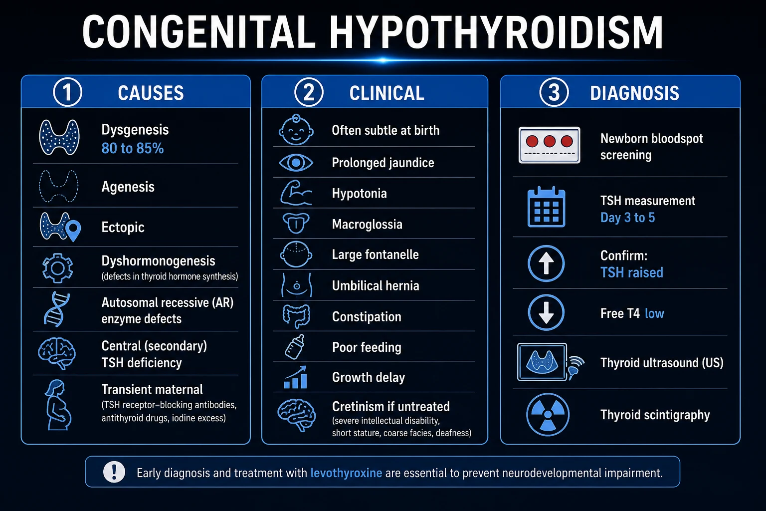
CH is classified along three axes — duration, anatomical level, and aetiology:[1][4]
Primary CH (thyroidal)
- High TSH + low free T4
- Thyroid itself is defective
- Detected by TSH-based newborn screening
- ~95% of all CH
Central CH (secondary/tertiary)
- Low/inappropriately normal TSH + low free T4
- Hypothalamic-pituitary cause (TRH/TSH deficiency)
- NOT detected by TSH-only screening
- Often part of multiple pituitary hormone deficiency
Permanent CH
- Dysgenesis (ectopic, agenesis, hypoplasia) — sporadic
- Dyshormonogenesis — usually autosomal recessive
- Requires lifelong levothyroxine
- Confirmed by re-evaluation at age 3
Transient CH
- Maternal TSH-receptor blocking antibodies (transplacental)
- Maternal carbimazole/PTU
- Iodine excess (contrast, antiseptics) or deficiency
- Hypothyroxinaemia of prematurity
- Resolves by 3 to 6 months; treat meanwhile
By aetiology (high-yield proportions):[1][4]
- Thyroid dysgenesis (~80%) — abnormal gland development. Subtypes: ectopic thyroid (most common — gland arrests along the thyroglossal tract, often sublingual or lingual at the base of the tongue; produces small amounts of hormone, may present later), agenesis (complete absence — most severe, presents earliest with lowest free T4), and hypoplasia (small structurally normal gland in the normal pretracheal position; hemiagenesis in ~5%). The vast majority are sporadic; only about 2% are familial, and the monozygotic-twin discordance rate (~90%) argues strongly against a pure genetic cause in most cases.[4]
- Dyshormonogenesis (~15 to 20%) — inborn errors of one of the six steps of thyroid-hormone biosynthesis; almost all autosomal recessive; classically cause a goitre (TSH-driven gland hypertrophy is intact, the gland is just biochemically blocked). Commonest defect: thyroid peroxidase (TPO) gene (~5% of all CH). Pendred syndrome (SLC26A4/PDS) combines organification defect + sensorineural deafness. Other genes in descending frequency: DUOX2/DUOXA2 (dual oxidase, iodide oxidation — increasingly recognised, often digenic with autosomal-dominant inheritance and incomplete penetrance), thyroglobulin (TG), sodium-iodide symporter SLC5A5/NIS (iodide trapping), DEHAL1/IYD (iodotyrosine deiodinase — iodide recycling failure causes iodine leak and goitre).[4]
- Transient (~10% and rising) — maternal TSH-receptor blocking antibodies, maternal antithyroid drugs, iodine excess (Wolff-Chaikoff) or deficiency, prematurity. Resolves by 3 to 6 months but must be treated meanwhile to protect neurodevelopment.
- Central CH (~1 in 50,000 to 1 in 100,000) — rare, but missed by TSH-only screens (the diagnostic TSH is low or inappropriately normal). Causes: combined pituitary hormone deficiency (POU1F1/PIT1, PROP1, HESX1 — septo-optic dysplasia), isolated TSH-beta subunit defect, TRH receptor defect, or acquired hypothalamic-pituitary disease.[1]
Thyroid embryology in one paragraph (explains dysgenesis anatomy): the medial thyroid anlage forms as an endodermal diverticulum at the foramen caecum (base of tongue, week 4), then descends along the thyroglossal duct in front of the hyoid and larynx to reach its pretracheal position by week 7, where it fuses with the paired ultimobranchial bodies (fourth/fifth pharyngeal pouch) that contribute the parafollicular C-cells. The thyroglossal duct then obliterates (its remnant may persist as a pyramidal lobe or thyroglossal cyst). Any error in this descent produces ectopic thyroid — lingual (at foramen caecum, the commonest ectopic site), sublingual, subhyoid, or intrathoracic. Defects in the transcription factors driving this program — PAX8, NKX2-1/TTF1, FOXE1/TTF2, HHEX, TSHR — account for only ~5 to 10% of dysgenesis (the genes are known, the monogenic yield is low), which is why most dysgenesis is sporadic and recurrence risk in siblings is low (~2%).[4]
Epidemiology & Risk Factors
Incidence is 1 in 2000 to 1 in 4000 live births worldwide, varying by region and screening strategy (incidence is rising over recent decades, attributed to lower TSH cut-offs, increased survival of preterm infants, higher iodine exposure, and more screening of multiples and congenital heart disease).[1]
Congenital Hypothyroidism — by the numbers
Female-to-male ratio ~ 2 : 1 — the reason is incompletely understood (possibly X-linked modifying factors and ascertainment through dysgenesis); the sex skew is most marked for ectopic thyroid. [1]
Risk factors and the cause they favour: [1]
| Risk factor / host | Implication |
|---|---|
| Prematurity / very low birth weight | Hypothyroxinaemia of prematurity; delayed TSH surge; transient CH; some programs repeat screen at 2 to 4 weeks |
| Down syndrome (trisomy 21) | Up to 10 to 30 fold increased risk of CH; also a true differential |
| Congenital heart disease | Iodine-induced transient CH from contrast/antiseptics; also CHD associated with certain dyshormonogenesis syndromes |
| Maternal autoimmune thyroid disease | Transplacental TSH-receptor blocking antibodies cause transient CH; maternal stimulating antibodies cause neonatal Graves (transient) |
| Maternal antithyroid drugs (carbimazole, methimazole, propylthiouracil) | Cross placenta — transient fetal/neonatal hypothyroidism |
| Excess iodine (contrast, povidone-iodine antiseptic, amiodarone) | Wolff-Chaikoff effect — transient hypothyroidism |
| Iodine deficiency (endemic) | Endemic cretinism — combined maternal and fetal deficiency |
| Twins / multiple gestation | Masking by cotwin euthyroid blood (feto-fetal transfusion); repeat screen |
| Family history / consanguinity | Autosomal-recessive dyshormonogenesis (TPO, PDS, NIS, DUOX2) |
Pathophysiology
The hypothalamic-pituitary-thyroid (HPT) axis: the hypothalamus secretes thyrotropin-releasing hormone (TRH), which stimulates the anterior pituitary thyrotrophs to secrete thyroid-stimulating hormone (TSH). TSH binds the TSH receptor (TSHR) on thyroid follicular cells, driving iodide uptake, organification, coupling, and release of T4 (and some T3). T4 and T3 exert negative feedback on both TRH and TSH secretion. Most circulating hormone is protein-bound (to TBG, transthyretin, albumin); only the free fraction is active. Peripheral deiodinases (D1, D2, D3) convert T4 to active T3 (D1, D2) or inactive reverse T3 (D3).[1]
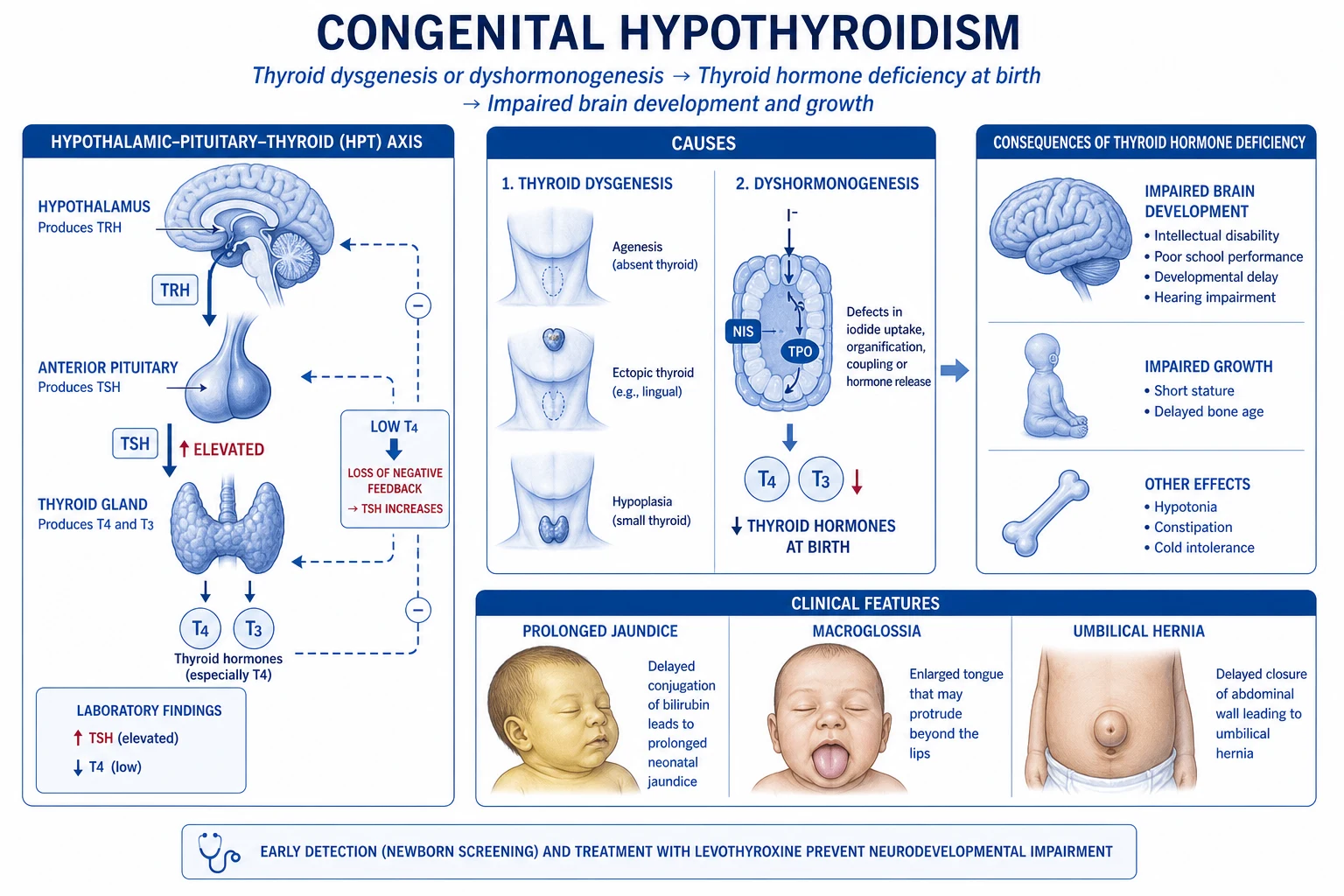
Thyroid hormone and the developing brain. T3 — produced locally by D2 deiodination of T4 in glia and neurons — binds nuclear thyroid-hormone receptors (TRα and TRβ), ligand-regulated transcription factors that heterodimerise with retinoid-X-receptor and bind thyroid-response elements in target genes. TRα dominates in brain, heart and bone; TRβ in liver, cochlea, pituitary and retina (loss-of-function mutations in THRB cause resistance to thyroid hormone, a rare mimic). Activated receptors regulate genes controlling myelin basic protein, proteolipid protein, neurofilaments, microtubule-associated proteins, reelin, and synaptic proteins — driving neuronal migration, dendritic arborisation, axonal myelination, and synaptogenesis. The first 3 years of life (beginning in the third trimester) are the critical window of myelination of the cerebral cortex and cerebellum. Deficiency during this window produces dendritic and axonal hypoplasia, impaired myelination, reduced synapse density, and impaired cochlear development (basis of deafness in Pendred and of sensorineural hearing loss in endemic disease) — the structural correlate of intellectual disability. After about age 3, the brain is largely myelinated and less vulnerable, which is why early replacement is decisive.[1][4]
Two patterns of endemic cretinism (relevant where iodine deficiency persists) illustrate the dual vulnerability of brain and body to thyroid hormone:[6]
- Neurological cretinism — severe maternal + fetal iodine deficiency in early pregnancy (first and second trimester) damages the developing cerebral cortex and cochlea before the fetal thyroid is functional. The child has mental retardation, deaf-mutism, squint, spastic diplegia/gait disorder, and a normal (often euthyroid) thyroid axis at birth — because the late-trimester/postnatal thyroid works normally. This damage is largely irreversible and not corrected by postnatal levothyroxine, which is why maternal iodine sufficiency in pregnancy is the prevention.[6]
- Myxoedematous cretinism — later fetal and postnatal thyroid hormone deficiency produces the classic hypothyroid phenotype: growth retardation, delayed bone age, dry skin, coarse facies, sexual retardation, and low T4 with high TSH — the phenotype closest to the historical "cretin" image. Postnatal levothyroxine partially reverses the somatic features but not the neurological damage already done.[6]
Sporadic CH (dysgenesis/dyshormonogenesis) — the form newborn screening detects — behaves like myxoedematous cretinism pathophysiologically (low thyroid hormone postnatally), but with intact maternal thyroid function the early-pregnancy brain development is preserved, so the neurological window is largely protected if replacement is started early. This is why sporadic CH is the most common preventable cause of intellectual disability: the damage is entirely postnatal and entirely avoidable with timely treatment.[1]
Thyroid dysgenesis results from failure of one or more steps of thyroid embryogenesis (glove-like diverticulum from the foramen caecum at the base of the tongue → descends along the thyroglossal tract → reaches its pretracheal position by week 7, fusing with the ultimobranchial body which contributes parafollicular C-cells). Candidate genes (PAX8, NKX2-1/TTF1, FOXE1/TTF2, TSHR) account for only a small minority of cases, which is why most dysgenesis is sporadic.[4]
Dyshormonogenesis — the biosynthetic steps and their genes:[4]
- Iodide uptake by the sodium-iodide symporter NIS (SLC5A5) — defect causes iodide-trapping failure.
- Iodide efflux to the follicular lumen / H2O2 generation by DUOX2 (DUOXA2) — increasingly recognised cause (often mild, sometimes transient).
- Organification — oxidation of iodide and its coupling to tyrosyl residues on thyroglobulin by thyroid peroxidase (TPO) — commonest dyshormonogenesis; positive perchlorate discharge test.
- Thyroglobulin (TG) synthesis — defect produces goitre with low/absent TG.
- Iodotyrosine deiodinase (DEHAL1/IYD) — failure to recycle iodide from mono- and di-iodotyrosine → iodine wasting, goitre.
- Pendrin (PDS / SCL26A4) — apical iodide efflux at the apical membrane; defect = Pendred syndrome (goitre + sensorineural deafness). [1]
Central (secondary) CH: TRH or TSH deficiency from hypothalamic-pituitary disease — combined pituitary hormone deficiency (POU1F1/PIT1, PROP1, HESX1), isolated TSH-beta subunit defect, or TRH receptor defect. The thyroid gland itself is normal but under-stimulated → low free T4 with low or inappropriately normal TSH → TSH-only newborn screening misses central CH.[1]
Transient CH mechanisms:
- Maternal TSH-receptor blocking antibodies (cross the placenta, block the fetal TSH receptor) — resolves as antibodies clear over 3 to 6 months.
- Maternal antithyroid drugs (carbimazole, methimazole, propylthiouracil) cross the placenta and inhibit fetal thyroid peroxidase — resolves after drug clearance.
- Iodine excess — the Wolff-Chaikoff effect (acute high iodine load transiently inhibits organification); relevant after iodinated contrast, povidone-iodine antiseptics (especially in cardiac surgery), or amiodarone.[7]
- Iodine deficiency — endemic CH (combined maternal + fetal deficiency).
- Hypothyroxinaemia of prematurity — low free T4 with normal TSH, due to HPT-axis immaturity, iodine/nutrient deficiency, and non-thyroidal illness; treatment remains debated.[5]
Clinical Presentation
Most neonates with CH are asymptomatic at birth because maternal T4 crosses the placenta and sustains fetal levels (fetal serum T4 reaches 30 to 60% of maternal by term, enough to mask severe thyroidal deficiency); clinical signs emerge as maternal T4 is metabolised (half-life ~5 days in the newborn) over the first 2 to 6 weeks. The later the diagnosis, the more florid the picture — only ~5 to 10% of screen-detected neonates show overt signs at the 2-week assessment, but almost every symptom-free infant is already biochemically hypothyroid by the time the screen returns.[1][6]
Why maternal T4 only partially protects the neonate: transplacental transfer supplies about 30 to 50 nmol/L of T4 at term, but this is consumed within 1 to 2 weeks of birth; thereafter the infant's own gland must supply all thyroid hormone. A fetus with complete agenesis has the lowest cord-blood T4 and shows signs earliest; a fetus with ectopic tissue or partial dyshormonogenesis retains some endogenous production and may not become clinically hypothyroid for weeks to months, occasionally presenting later in infancy as failure to thrive or developmental delay. This is why relying on clinical signs (rather than screening) detects fewer than 30% of cases in the asymptomatic window — the cardinal justification for universal screening.[3]
Neonatal features (untreated, evolving): [1]
- Prolonged jaundice — usually unconjugated (hypothyroidism reduces hepatic glucuronyl transferase activity, slowing bilirubin conjugation); less commonly conjugated cholestasis.
- Large posterior fontanelle (over 5 mm at term) and widely open sutures — delayed bone maturation; a frequently examiner-asked early sign.
- Macroglossia (large protruding tongue), coarse facies.
- Umbilical hernia, protuberant abdomen, distended abdomen.
- Hypotonia, lethargy, somnolence; poor feeding with weak suck; failure to thrive.
- Hoarse cry (myxoedematous vocal cords).
- Constipation, delayed passage of meconium (over 24 hours).
- Hypothermia, bradycardia, cold, dry, mottled skin; peripheral or generalised oedema (myxoedema).
- Delayed bone age at birth (absent distal femoral epiphysis). [1]
Older infant (diagnosis missed — the classic "cretin" phenotype that screening exists to prevent): coarse facial features (low nasal bridge, puffy eyelids, macroglossia, open mouth with dribbling), large protruding tongue, short stature with markedly delayed bone age (often the presenting complaint), protuberant abdomen with umbilical hernia, dry pale cold skin with carotenaemia and brittle hair, delayed dentition, muscular pseudohypertrophy (Kocher-Debré-Sémélaigne syndrome — rare, rhabdomyosarcoma-like calf hypertrophy in long-standing hypothyroidism), developmental delay (motor more than social initially, then global), intellectual disability (IQ often below 50 if untreated beyond 6 months), delayed puberty, and sensorineural or mixed hearing loss. Constipation may be so severe as to mimic Hirschsprung disease. This entire phenotype is now vanishingly rare where screening is universal.[6]
Atypical / at-risk presentations:
- Preterm / very-low-birth-weight infant — hypothyroxinaemia of prematurity; some programs repeat screening at 2 to 4 weeks.[5]
- Infant of a mother with Graves' disease on carbimazole/PTU — transient CH; also at risk of neonatal hyperthyroidism as stimulating antibodies clear.
- Neonate with congenital heart disease exposed to iodinated contrast or antiseptics — iodine-induced transient CH.[7]
- Twin gestation — masking by cotwin blood; repeat screen.
- Pendred phenotype — goitre (often presenting in childhood) with sensorineural deafness detected on newborn hearing screen.
Differential Diagnosis
A neonate with hypotonia, jaundice, macroglossia or coarse features is not always CH. Distinguish:[6]
- Down syndrome (trisomy 21) — also a true risk factor for CH. Hypotonia, flat facies, upslanting palpebral fissures, single palmar crease, Brushfield spots, sandal-gap. Always check thyroid function in an infant with Down syndrome.
- Beckwith-Wiedemann syndrome — macroglossia, but with macrosomia, omphalocele/umbilical hernia, neonatal hypoglycaemia (hyperinsulinism), ear creases/pits, lateralised overgrowth.
- Mucopolysaccharidoses (e.g. Hurler) — coarse facies, macroglossia, hepatosplenomegaly, older infant presentation; not neonatal.
- Prolonged physiological / breast-milk jaundice (unconjugated) vs cholestasis / biliary atresia (conjugated, pale stools, dark urine).
- Sepsis, congenital infection (TORCH), inborn errors of metabolism presenting with hypotonia and poor feeding.
- Prader-Willi syndrome — neonatal hypotonia, poor feeding, weak suck (later hyperphagia, hypogonadism).
- Zellweger syndrome, Pompe disease — severe neonatal hypotonia.
- Central CH vs primary CH — both are "hypothyroidism" but the TSH distinguishes them (low/inappropriately normal in central; high in primary); TSH-only screening misses central CH. [1]
Always perform venous thyroid function (TSH + free T4) in any neonate with suggestive features — and remember that Down syndrome coexists with CH. [1]
Clinical & Bedside Assessment
When a positive newborn screen returns, the bedside assessment is performed in parallel with venous confirmation and treatment start:[2]
- Growth and feeding — weight, feeding pattern, weight gain, suck strength.
- Anterior examination — facies (coarse?), macroglossia, hoarseness of cry.
- Head — posterior fontanelle (over 5 mm at term is abnormal), anterior fontanelle size, sutures.
- Abdomen — umbilical hernia, distension, constipation.
- Skin — dry, cold, mottled, oedema; jaundice (note type if known).
- Neurology — tone (hypotonia), reflexes (delayed relaxation), alertness.
- Vitals — hypothermia, bradycardia, respiratory depression (suggest severe/late disease — the rare myxoedema coma endpoint).
- Neck — palpate for goitre (suggests dyshormonogenesis or maternal-blocking-antibody effect; its absence favours dysgenesis). [1]
Bone age X-ray (knee or foot at diagnosis) is a marker of in-utero hypothyroid severity — absence of the distal femoral and proximal tibial epiphyses at term implies significant prenatal deficiency and a worse baseline. [1]
Investigations
Tiered: screen → confirm → classify.[1][2]
Tier 1 — Newborn screening (universal):
- Dried capillary blood spot (Guthrie card) TSH taken at day 3 to day 7 (UK day 5; varies by program; earlier in early discharge protocols).
- TSH threshold for recall varies by program and must be age-adjusted (TSH physiologically falls over the first days of life); many programs use 20 to 60 mU/L.[8]
- Primary-TSH screening misses central CH and may miss mild primary CH in preterm infants — some programs use combined T4 + TSH, or repeat the screen at 2 to 4 weeks for preterm / low-birth-weight / ill infants.[5][8]
Tier 2 — Venous confirmation (do not delay treatment):
- Venous TSH (elevated) + free T4 (low) confirms primary CH.
- Low free T4 with low or inappropriately normal TSH = central CH → check other anterior pituitary hormones (cortisol, IGF-1, prolactin), pituitary MRI, and cortisol before starting thyroxine (replacing T4 first in untreated adrenal insufficiency can precipitate adrenal crisis).
- Reference ranges are age-specific — neonatal TSH and T4 are physiologically high. [1]
Tier 3 — Classification of cause (after treatment started):
- Thyroid ultrasound — anatomy, presence and position of the gland; differentiates in-situ vs ectopic vs absent.
- Technetium-99m pertechnetate or iodine-123 scintigraphy — uptake and localisation; no uptake with absent gland or maternal blocking antibodies; high uptake in ectopic or normal position with dyshormonogenesis; goitre with high uptake suggests organification defects.
- Maternal thyroid function and TSH-receptor antibodies — detects transient maternal-antibody CH (especially if maternal history of autoimmune thyroid disease or previous affected infant).
- Serum thyroglobulin — low/absent in agenesis; high in dyshormonogenesis.
- Urinary iodine — iodine status (deficiency or excess).
- Genetic testing — TPO (commonest), SLC26A4/PDS (Pendred), DUOX2/DUOXA2, SLC5A5/NIS, TG, DEHAL1/IYD, and for dysgenesis PAX8, NKX2-1, FOXE1, TSHR — when dyshormonogenesis, goitre, deafness, or syndromic features.
- Perchlorate discharge test (where available) — positive in organification defects (TPO, DUOX2, Pendred).
- Hearing assessment — all neonates (universal hearing screen); mandatory when Pendred suspected.
- Karyotype / chromosomal microarray if syndromic features (e.g. Down syndrome). [1]
Imaging interpretation — pattern recognition at scintigraphy + ultrasound:[1][4]
Agenesis
- No gland on US or scintigraphy
- TSH very high, free T4 very low
- No uptake on pertechnetate scan
- Low/absent serum thyroglobulin
- Most severe; earliest presentation
Ectopic (lingual/sublingual)
- Uptake at base of tongue on scan
- No normal pretracheal gland on US
- Commonest dysgenesis subtype
- Partial function → milder, later presentation
- Female preponderance
Dyshormonogenesis
- Goitre on US (enlarged in-situ gland)
- High uptake in normal position
- High serum thyroglobulin
- Autosomal recessive, often consanguineous
- Positive perchlorate discharge (organification defect)
Transient (maternal TBAb)
- Normal in-situ gland on US
- No uptake on scan (receptor blocked)
- Maternal TSH-receptor blocking antibodies positive
- Resolves by 3 to 6 months
- Mother often has autoimmune thyroid disease
Imaging timing nuance: scintigraphy is most informative within the first 1 to 2 weeks while TSH is still very high (driving uptake); once levothyroxine suppresses TSH, uptake falls and the scan becomes uninterpretable. Ultrasound is operator-dependent but does not depend on TSH — the two are complementary. Never delay treatment to obtain scans.[1]
Management — Resuscitation
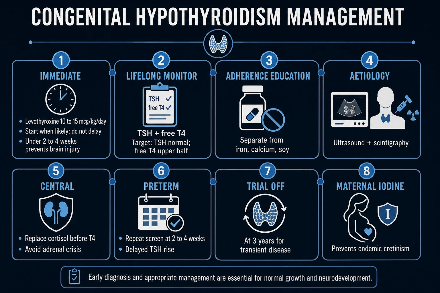
CH is rarely an acute neonatal emergency, but two situations demand urgent action:[1]
- A positive newborn screen — perform venous TSH and free T4 confirmation AND start levothyroxine the same day. Reassure parents; arrange paediatric endocrinology follow-up.
- Late-presenting severe CH with myxoedema coma phenotype (rare in neonates, more often older infants with missed diagnosis) — hypothermia, bradycardia, hypoventilation, obtundation. Manage with:
- Airway/breathing — supportive, warmed humidified oxygen; mechanical ventilation if hypoventilating.
- Warming — active rewarming (radiant warmer, warmed fluids) but avoid rapid rewarming which can precipitate vasodilatory shock.
- Oral/NG levothyroxine 10 to 15 mcg/kg/day (the enteral route works; IV levothyroxine reserved for those unable to absorb or profoundly unstable — 10 to 20 mcg IV bolus then daily maintenance).
- Stress-dose hydrocortisone until coexistent adrenal insufficiency is excluded (central CH commonly coexists with ACTH/cortisol deficiency; replacing T4 first can trigger adrenal crisis).
- Slow warmed IV fluids, glucose, correct hyponatraemia slowly (myxoedema-associated SIADH). [1]
Management — Definitive & Stepwise
Levothyroxine (L-thyroxine, L-T4) is the cornerstone.[1][2]
Drug: levothyroxine sodium (L-T4) [1]
- Dose (initial): 10 to 15 mcg/kg/day (the high end of the range normalises free T4 faster and improves neurodevelopmental outcome; the historical lower doses — 6 to 8 mcg/kg — are no longer recommended for neonates). Typical term neonate: 37.5 to 50 mcg/day.
- Route: oral, once daily. Tablets can be crushed and suspended in a small volume of water/breast milk/formula (not soy formula). IV reserved for inability to absorb or profound instability.
- Timing: in the morning, on an empty stomach, 30 to 60 minutes before a feed (or evening, 3 to 4 hours after the last feed); consistent timing matters.
- Administration pitfalls: absorption reduced by calcium, iron, sucralfate, antacids, proton-pump inhibitors, concentrated soy formula, fibre — separate by at least 4 hours.
- Target biochemistry: free T4 in the upper half of the age-specific reference range within 2 weeks, then maintain; TSH 0.5 to 5.0 mU/L (sustained suppressed TSH below 0.5 risks overtreatment — tachycardia, accelerated bone age, craniosynostosis). [1]
Age-based dosing (maintenance, weight-based titration):[1]
| Age | Typical L-T4 dose |
|---|---|
| Neonate / infant (0 to 12 months) | 10 to 15 mcg/kg/day |
| 1 to 3 years | 4 to 6 mcg/kg/day |
| 3 to 10 years | 3 to 5 mcg/kg/day |
| 10 to 16 years | 2 to 4 mcg/kg/day |
| Adult maintenance | 1.6 mcg/kg/day |
- At 2 weeks and 4 weeks after starting (or any dose change) — TSH and free T4.
- Every 1 to 2 months in the first year.
- Every 2 to 3 months between 1 and 3 years.
- Every 3 to 12 months thereafter (more frequently with growth spurts, dose changes, non-adherence).
- Always recheck 4 weeks after any dose change. [1]
Confirming permanence — trial off treatment at age 3 years:[1]
- At age 3 (after the critical neurodevelopmental window), reduce then stop L-T4 for 4 to 6 weeks and recheck TSH and free T4.
- Permanent CH: TSH rises and free T4 falls → lifelong treatment with structured transition to adult endocrinology.
- Transient CH: values remain normal → no further treatment; reassure.
- Higher baseline TSH, lower starting free T4, ectopic/absent gland, dyshormonogenesis, or positive genetic tests predict permanence and may justify earlier/more confident determination. [1]
Referral and surveillance: paediatric endocrinology, developmental surveillance (motor, language, cognition), hearing and vision, parental education and adherence support, genetic counselling for dyshormonogenesis or syndromic forms. [1]
Specific Subtypes & Scenarios
- Permanent primary CH — dysgenesis. Sporadic, no goitre. Ectopic gland (most common) may produce some hormone and present later; agenesis is the most severe and presents earliest. Treatment is lifelong levothyroxine.[4]
- Permanent primary CH — dyshormonogenesis. Autosomal recessive, goitrous, often consanguineous families. Commonest defect TPO. Pendred syndrome (SLC26A4) = goitre + sensorineural deafness (the deafness is congenital, the goitre develops later). Lifelong levothyroxine; genetic counselling; hearing support.[4]
- Transient CH. Treat with levothyroxine as for permanent CH (the infant's brain does not distinguish); re-evaluate at age 3 (or earlier if clearly transient — e.g. maternal antibodies with normalising labs). Maternal TSH-receptor blocking antibodies classically cause absent uptake on scintigraphy in a normally located gland.[1]
- Central CH. Low free T4 with low or inappropriately normal TSH. Check other pituitary axes (cortisol, GH/IGF-1, gonadotrophins, prolactin); pituitary MRI; ophthalmology for optic nerve hypoplasia (septo-optic dysplasia). Always give stress-dose hydrocortisone before thyroxine until adrenal insufficiency excluded. Dose L-T4 by free T4 (TSH is not a useful marker in central CH). TSH-only newborn screening misses this.[1]
- Hypothyroxinaemia of prematurity. Low free T4 with normal TSH in preterm infants. Treatment is debated — benefit suggested for the most immature (under 28 weeks); routine supplementation not universally recommended; follow local protocol and expert opinion.[5]
- Iodine-induced CH in cardiac neonates. After iodinated contrast for cardiac catheterisation or povidone-iodine antiseptics/dressings — the Wolff-Chaikoff effect causes transient hypothyroidism; consider thyroid surveillance in infants with congenital heart disease undergoing repeated contrast exposure.[7]
- Syndromic dysgenesis. Bamforth-Lazarus (FOXE1) — cleft palate, bifid epiglottis, spiky hair, choanal atresia, CH. Brain-lung-thyroid syndrome (NKX2-1/TTF1) — CH, choreoathetosis, neonatal respiratory distress / interstitial lung disease. PAX8 — CH with renal/urogenital anomalies.[4]
Complications & Pitfalls
Untreated disease (the natural history that screening prevents): irreversible intellectual disability (IQ loss proportional to delay and severity), short stature, delayed bone age, delayed puberty, sensorineural deafness (Pendred), behavioural and neuropsychiatric difficulties, myxoedema.[6]
Overtreatment: tachycardia, irritability, poor weight gain, accelerated bone age (risk of compromised adult height), craniosynostosis (rare). Avoid sustained TSH suppression below 0.5 mU/L. [1]
Undertreatment despite therapy: suboptimal cognitive outcome, growth failure, persistent symptoms — usually from non-adherence, sub-therapeutic dose, or absorption-reducing drug/food interactions. [1]
Classic pitfalls:
- Delaying treatment for imaging or genetics — the cardinal error; treat first, investigate later.
- TSH-only screening missing central CH and mild primary CH in preterm infants.
- Non-adherence / inconsistent administration (giving with milk, calcium, iron, soy).
- Forgetting to exclude adrenal insufficiency before starting T4 in central CH.
- Failing to re-confirm permanence at age 3 — either over-treating a transient case lifelong or, conversely, inappropriately stopping a permanent case.
- Missing CH in a Down syndrome infant — Down syndrome is itself a risk factor. [1]
Prognosis & Disposition
With treatment started within the first 2 weeks and free T4 normalised rapidly, children with CH achieve near-normal IQ and neurodevelopment, attend mainstream school, and live independent lives. Subtle residual deficits (in memory, attention, arithmetic, sensorimotor speed) may persist even with optimal therapy, particularly in those with severe baseline disease (very low starting free T4, absent epiphyseal ossification).[1][6]
Outcome is proportional to:
- Severity at diagnosis (free T4 level, bone age — both markers of prenatal deficiency).
- Age at treatment initiation — earlier (within 2 weeks) is decisively better.
- Starting dose and speed of normalisation — the 10 to 15 mcg/kg/day regimen, normalising free T4 within 2 weeks, improves outcome over older lower-dose regimens.
- Adherence and biochemically optimal maintenance. [1]
Disposition: outpatient under paediatric endocrinology with developmental surveillance; structured transition to adult endocrinology in adolescence/young adulthood.[1]
Prevention
CH prevention operates at three levels — primary (prevent the disease itself in endemic/iodine-deficient populations), secondary (prevent the brain damage by early detection), and tertiary (prevent long-term disability by optimal treatment).[1][2]
Secondary prevention — universal newborn screening (the single most impactful intervention):
- Universal heel-prick TSH screening of every live-born infant, now standard in nearly all high- and middle-income countries, has reduced the incidence of intellectual disability from CH by over 90% in screened populations. Before screening, untreated CH accounted for ~10% of all institutionalised intellectually disabled children; today it is a vanishing cause.
- Timing: day 3 to 7 (UK day 5); repeat at 2 to 4 weeks for preterm, low-birth-weight, ill, or transfused infants.
- Programme quality controls — age-adjusted TSH cut-offs, prompt recall, venous confirmation, and rapid treatment initiation are what make screening effective, not merely performed. [1]
Primary prevention — maternal iodine sufficiency:
- Adequate iodine intake in pregnancy and lactation is essential because the fetus depends entirely on maternal T4 until ~12 weeks' gestation (when the fetal thyroid begins concentrating iodine) and continues to depend on transplacental T4 throughout pregnancy. WHO/UNICEF/ICCIDD recommend 250 mcg iodine/day for pregnant and lactating women (versus 150 mcg/day for non-pregnant adults); universal salt iodisation (20 to 40 ppm at production) is the most cost-effective population strategy.[6]
- In iodine-deficient regions, iodine supplementation of pregnant women (150 mcg/day oral potassium iodide) prevents endemic cretinism (both neurological and myxoedematous forms). Supplementation must start before or in early pregnancy to prevent the irreversible early-pregnancy neurological damage.
Avoiding iatrogenic fetal hypothyroidism:
- Antithyroid drugs in pregnancy — use the lowest effective dose of carbimazole/methimazole or propylthiouracil to keep maternal free T4 in the upper half of the reference range. Propylthiouracil is preferred in the first trimester (lower teratogenicity than carbimazole, which carries aplasia cutis and choanal atresia risk), then switch to carbimazole/methimazole in the second trimester (PTU hepatotoxicity risk). Overtreatment causes transient fetal/neonatal CH.
- Radioactive iodine (I-131) is absolutely contraindicated in pregnancy — it ablates the fetal thyroid (especially after 10 to 12 weeks when the fetal thyroid concentrates iodine), causing permanent primary CH. Pregnancy must be excluded and avoided for 6 months after I-131 therapy.
- Iodine excess avoidance — minimise iodinated contrast and povidone-iodine antiseptics in neonates (especially around cardiac catheterisation and surgery); the immature thyroid is exquisitely sensitive to the Wolff-Chaikoff effect.[7]
- Maternal thyroid screening in pregnancy — women with known autoimmune thyroid disease, previous affected infant, or positive TSH-receptor antibodies should have TFTs in pregnancy and the neonate monitored for transient CH.
Tertiary prevention — optimal treatment and adherence: early high-dose levothyroxine (10 to 15 mcg/kg/day), rapid normalisation of free T4 within 2 weeks, structured monitoring, parental education, and developmental surveillance prevent the residual disability that even treated CH can carry if managed poorly.[1]
Special Populations
- Preterm / very-low-birth-weight infants — delayed TSH surge; hypothyroxinaemia of prematurity; repeat screening at 2 to 4 weeks is recommended by many programs.[5]
- Maternal Graves' disease on antithyroid drugs (carbimazole, methimazole, propylthiouracil) — transient fetal/neonatal CH (drug crosses placenta); monitor neonate for transient CH and for neonatal hyperthyroidism (stimulating antibodies clearing after birth).
- Infants with congenital heart disease exposed to iodinated contrast or povidone-iodine — iodine-induced transient CH; thyroid surveillance.[7]
- Twin / multiple gestation — masking by cotwin euthyroid blood (feto-fetal transfusion); repeat screen.[1]
- Down syndrome — increased risk of CH (and other autoimmune thyroid disease later); screen at birth and at least annually thereafter.
- Pregnancy in a woman with treated CH — increase L-T4 by 25 to 30% immediately on confirmation of pregnancy (demand rises from week 4 to 6); monitor free T4/TSH every 4 weeks in the first half, 6 weekly thereafter; maternal hypothyroidism impairs fetal neurodevelopment. Postpartum return to pre-pregnancy dose.
- Adolescent/transition — structured transfer to adult endocrinology with education on lifelong adherence, pregnancy planning, and the consequences of non-adherence.
Evidence, Guidelines & Regional Differences
Key guideline:[1]
- ESPE/ESE 2020-2021 Consensus (van Trotsenburg et al., Thyroid 2021) — current European standard; recommends universal neonatal screening, immediate levothyroxine at correct dose, frequent monitoring, age-3 re-evaluation, attention to neurodevelopment, and planned transition to adult care.[1]
- ESPE 2014 Consensus (Leger et al., JCEM 2014) — the predecessor; still widely cited.[2]
Screening strategy and TSH cut-offs vary worldwide[8] — most programs are primary-TSH, some use combined T4 + TSH or T4-primary with reflex TSH. TSH cut-offs should be age-adjusted; failure to adjust risks missing mild persistent CH.[8]
[1]Regional deltas (India / ICMR, RPCH): National Newborn Screening expanding; some states (Kerala, Goa, Chandigarh) screen for CH; TSH cut-offs adapted to local reference ranges. Iodine deficiency remains relevant in some regions. Always apply local screening-programme guidance. [1]
Controversies:
- Higher initial L-T4 dose (10 to 15 mcg/kg/day) normalises T4 faster but may transiently suppress TSH; current consensus favours the higher dose for neurodevelopmental benefit.[1]
- Hypothyroxinaemia of prematurity — treatment debated.[5]
- Whether mild persistent TSH elevations missed by screening warrant treatment.[8]
Exam Pearls
CH neonatal features — THE 6 Hs + classic signs
- Most common preventable cause of intellectual disability — screen universally.
- Most common cause of CH: thyroid dysgenesis (about 80%); the commonest subtype is ectopic gland.
- Dyshormonogenesis (about 15 to 20%) is autosomal recessive, usually goitrous; TPO is the commonest gene.
- Pendred syndrome = goitre + sensorineural deafness (SLC26A4).
- TSH on heel-prick (Guthrie card) day 3 to 7; confirm with venous TSH (high) + free T4 (low).
- Levothyroxine 10 to 15 mcg/kg/day, START IMMEDIATELY — treat first, investigate later.
- Critical window: treat within 2 weeks for normal IQ.
- Targets: free T4 upper half of range; TSH 0.5 to 5 mU/L.
- Monitor every 1 to 2 months in year 1; recheck 4 weeks after any dose change.
- Trial off treatment at age 3 to confirm permanent vs transient CH.
- Central CH: low TSH + low free T4 — MISSED by TSH-only screening; give hydrocortisone first until adrenal insufficiency excluded.
- Give L-T4 on empty stomach; separate from iron, calcium, soy, antacids.
- Female : male about 2 : 1.
- Increase L-T4 by 25 to 30% in pregnancy.
- Down syndrome is both a differential AND a true risk factor for CH — screen at birth and annually. [1]
Exam application bank (NEET-PG / INICET)
One-line answer
Congenital hypothyroidism (CH) is thyroid hormone deficiency present at birth, resulting from abnormal thyroid gland development (dysgenesis, about 80%) or inborn errors of hormone biosynthesis (dyshormonogenesis, about 15 to 20%), with a minority due to transient maternal or environmental causes. Affected neonates are usually asymptomatic at birth because maternal thyroxine crosses the placenta; untreated, the disease produces irreversible intellectual disability, short stature and developmental delay, making CH the most common preventable cause of intellectual disability worldwide. Universal newborn screening (TSH on a heel-prick dried blood spot) detects CH before symptoms, and immediate levothyroxine 10 to 15 mcg/kg/day started within the first 2 weeks restores normal cognitive outcome. The cardinal rule is treat first, investigate later — never delay therapy for imaging or genetic t [1]
Worked stems (answer without another resource)
Stem 1 — Classic presentation. Map symptoms to mechanism; name the first investigation and first treatment step with dose/route if drug therapy is standard. [1]
Stem 2 — Unstable / complicated. List red flags that force immediate resuscitation, theatre, ICU, antidote, or reperfusion — and what you do in the first 15 minutes. [1]
Stem 3 — Atypical group. Elderly, pregnancy, child, or immunocompromised: how presentation and thresholds change. [1]
Stem 4 — Differential trap. Name the three closest mimics and one discriminator for each. [1]
Stem 5 — Disposition. Who goes home with safety-netting, who is admitted, who needs HDU/ICU/theatre, and what follow-up is mandatory. [1]
Rapid viva checklist
- Definition + classification
- Pathophysiology chain
- Bedside signs / criteria
- Score with exact components (if any)
- Emergency bundle
- Definitive therapy with doses
- Complications of disease and of treatment
- Special populations
- Guideline/trial name if classic
- Three exam traps
Coverage self-check
If you cannot answer any stem above from this page alone, re-read the matching section — the page is intended to be self-sufficient for final-prof and NEET-PG/INICET questions on Congenital Hypothyroidism.
[1]References
- [1]van Trotsenburg ASP, Stoupa A, Leger J, et al. Congenital Hypothyroidism: A 2020-2021 Consensus Guidelines Update-An ENDO-European Reference Network Initiative Endorsed by the European Society for Pediatric Endocrinology and the European Society for Endocrinology Thyroid, 2021.PMID 33272083
- [2]Leger J, Olivieri A, Donaldson M, et al.; ESPE-PES-SLEP-JSPE-APEG-APPES-ISPAE Congenital Hypothyroidism Consensus Conference Group. European Society for Paediatric Endocrinology consensus guidelines on screening, diagnosis, and management of congenital hypothyroidism J Clin Endocrinol Metab, 2014.PMID 24446653
- [3]Jacob H, Peters C. Screening, diagnosis and management of congenital hypothyroidism: European Society for Paediatric Endocrinology Consensus Guideline Arch Dis Child Educ Pract Ed, 2015.PMID 25776656
- [4]Park SM, Chatterjee VK. Genetics of congenital hypothyroidism J Med Genet, 2005.PMID 15863666
- [5]Klosinska M, Kaczynska A, Ben-Skowronek I. Congenital Hypothyroidism in Preterm Newborns - The Challenges of Diagnostics and Treatment: A Review Front Endocrinol (Lausanne), 2022.PMID 35370986
- [6]Hanley P, Lord K, Bauer AJ. Thyroid Disorders in Children and Adolescents: A Review JAMA Pediatr, 2016.PMID 27571216
- [7]Thaker VV, Leung AM, Braverman LE, Brown RS, Levine B. Iodine-induced hypothyroidism in full-term infants with congenital heart disease: more common than currently appreciated? J Clin Endocrinol Metab, 2014.PMID 25004248
- [8]Kilberg MJ, Rasooly IR, LaFranchi SH, Bauer AJ, Hawkes CP. Newborn Screening in the US May Miss Mild Persistent Hypothyroidism J Pediatr, 2018.PMID 29246344