Paediatrics · Paediatrics
Croup
Croup (acute laryngotracheobronchitis) is the most common cause of acute upper airway obstruction in young children, peaking at 6 months to 3 years, caused mainly by parainfluenza virus type 1 and 2. Presentation: barking seal-like cough, harsh inspiratory stridor, hoarse voice, low-grade fever, worse at night. Most are mild and self-limiting over 3 to 7 days. Severe croup: stridor at rest, marked retractions, cyanosis, altered mental state. Treatment: oral dexamethasone 0.15 to 0.6 mg/kg single dose for all children; nebulised adrenaline 1:1000, 0.5 mL/kg max 5 mL, for moderate to severe with stridor at rest; observe at least 2 to 4 hours after adrenaline for rebound.
On this page & tools
Your progress
Saved locally on this device.
Exam tags
Red flags
Overview & Definition
Croup (acute laryngotracheobronchitis) is an acute viral upper airway infection producing inflammation and oedema of the larynx, trachea, and bronchi, with the dominant obstruction located in the subglottic region (just below the vocal cords, encircled by the cricoid cartilage). It is the most common cause of acute stridor in febrile children between 6 months and 6 years of age, and one of the most frequent reasons for emergency department attendance and hospital admission in early childhood. The clinical syndrome is defined by its triad of barking (seal-like) cough, harsh inspiratory stridor, and hoarse voice, often preceded by 1 to 2 days of coryza and low-grade fever, with symptoms characteristically worse at night.[1][2]
The vast majority of cases are mild and self-limiting, resolving within 3 to 7 days. A small but clinically important fraction progress to moderate or severe disease with significant respiratory compromise, and a tiny minority (under 0.5 percent) require intubation. The disorder sits in a narrow diagnostic space shared with several immediately life-threatening mimics — epiglottitis, bacterial tracheitis, retropharyngeal abscess, and foreign body aspiration — and the central clinical skill is to distinguish the common, benign, self-limiting viral croup from these conditions whose management is fundamentally different and time-critical. The single most important therapeutic advance of the last 30 years has been the recognition that a single dose of oral dexamethasone benefits children with croup of any severity, including those traditionally labelled "mild".[1][3]
Classification
Croup is classified by severity, using either the full Westley croup score (the validated research and clinical tool) or a simplified bedside assessment based on stridor at rest, work of breathing, and oxygenation. Severity directly drives the treatment ladder — the decision to give adrenaline, admit, or discharge hangs on this classification.[1]
Mild (Westley 0 to 2)
- Barking cough, hoarseness, stridor only when agitated or crying
- No retractions, no cyanosis, normal air entry, normal mental state, SpO2 over 92 percent in air
- Treat with single-dose oral dexamethasone 0.15 to 0.6 mg/kg; discharge with safety-net advice
- Over 85 percent of all presentations
Moderate (Westley 3 to 5)
- Stridor at rest or with minimal activity, audible at the bedside
- Mild to moderate sternal/intercostal retractions, mild tachypnoea
- Still feeding, normal colour, SpO2 over 92 percent, normal mental state
- Dexamethasone 0.6 mg/kg PO/IM; observe 2 to 4 hours; consider nebulised adrenaline if not improving; discharge if improved
Severe (Westley 6 to 11)
- Significant stridor at rest, marked retractions (suprasternal, intercostal, subcostal)
- Reduced air entry, prominent use of accessory muscles, nasal flaring, tachypnoea
- Anxious, agitated, or lethargic; SpO2 may dip under 92 percent
- Nebulised adrenaline 1:1000 0.5 mL/kg max 5 mL PLUS dexamethasone 0.6 mg/kg PO/IM/IV; oxygen; admit to HDU/PICU; ENT/anaesthetics on standby
Impending respiratory failure (Westley 12 to 17)
- Cyanosis in air, marked hypoxaemia, reduced level of consciousness, fatigue with falling respiratory effort
- Reduced stridor may paradoxically signal exhaustion and imminent collapse (silent chest)
- Admit PICU immediately, senior anaesthetics and ENT present, prepare for intubation with a tube 0.5 to 1.0 mm smaller than age-predicted

Epidemiology & Risk Factors
Croup is overwhelmingly a disease of early childhood, accounting for around 3 to 5 percent of all children each year and representing one of the leading causes of paediatric emergency attendance and hospital admission. Annual incidence is approximately 6 per 100 children under 6 years in primary care; around 1 to 5 percent of all croup presentations require hospitalisation, and 0.5 to 2 percent of hospitalised children need intensive care. Intubation is required in fewer than 0.5 percent of hospital cases, and death is now rare (under 0.5 percent mortality in intubated children) in countries with modern PICU and safe airway management.[1][2]
The epidemiology follows the circulation of parainfluenza virus type 1, which produces characteristic biennial autumn epidemics in alternate (odd-numbered) years in many temperate countries; parainfluenza type 2 produces smaller, more sporadic outbreaks; parainfluenza type 3 circulates annually and tends to cause bronchiolitis and pneumonia as well as croup at younger ages. Risk factors include age 6 months to 3 years (the age at which the subglottic airway is smallest relative to airflow demand), autumn and winter season, prior episode of croup (recurrence is common), male sex, prematurity, attendance at daycare, and parental smoking. Recurrent croup (more than 2 to 3 episodes in 6 months) or atypical age (under 6 months or over 6 years) should prompt consideration of an underlying anatomical abnormality — subglottic stenosis (post-intubation), subglottic haemangioma, laryngeal web, laryngomalacia — or gastro-oesophageal reflux and airway hyper-reactivity/atopy, and warrants ENT referral for flexible laryngoscopy and bronchoscopy.[1][10]
Pathophysiology
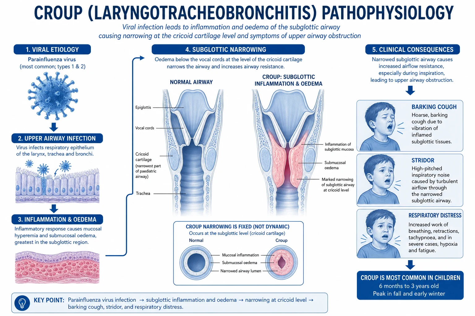
Croup begins when a respiratory virus — overwhelmingly parainfluenza virus type 1 (around 50 to 75 percent of cases), followed by parainfluenza type 2 and 3, influenza A and B, respiratory syncytial virus (RSV), adenovirus, rhinovirus, enterovirus, human metapneumovirus, and occasionally measles in unvaccinated populations — infects the ciliated respiratory epithelium of the nasopharynx and then the larynx, trachea, and bronchi.[1][10]
The resulting mucosal inflammation, vascular congestion, increased mucus production, and fibrinous exudate produce the characteristic subglottic oedema. The cricoid cartilage — the only complete cartilaginous ring of the trachea, lying at the level of the sixth cervical vertebra in adults but more cephalad in infants — cannot expand outward to accommodate the swelling, so the oedema is forced inward into the airway lumen.[11] The subglottic region is also the functionally narrowest segment of the paediatric airway (rather than the glottis, as in adults), so any narrowing here has disproportionate effects.
The clinical consequences are governed by the physics of laminar flow through a tube: by Poiseuille's law, airway resistance is inversely proportional to the fourth power of the radius (resistance proportional to 1 over radius to the fourth power). A small reduction in airway radius therefore produces a large increase in resistance. In a neonate the subglottic lumen is approximately 4 to 5 mm; just 1 mm of circumferential oedema reduces the cross-sectional area by roughly 75 percent and multiplies resistance many-fold. In a toddler the airway is wider but the same principle applies: small changes in oedema, mucous plugging, or agitation produce large swings in work of breathing. This is why croup symptoms fluctuate hour to hour, worsen with crying and agitation (which increase airflow turbulence and dynamic collapse), improve with cool humidified air and corticosteroids (which reduce mucosal oedema), and characteristically peak at night.[1][2]
The barking cough arises from inflammation of the subglottis and vocal cords; the hoarse voice reflects vocal cord involvement; and the stridor is generated by turbulent airflow through the narrowed subglottis. Stridor is inspiratory when obstruction is extrathoracic (at the level of the larynx or subglottis, as in croup) because the negative intrathoracic pressure of inspiration pulls the soft extrathoracic airway inward and narrows it further; biphasic stridor suggests a fixed or more severe obstruction.[1][2]
Clinical Presentation
The classic presentation is a child aged 6 months to 6 years (peak 1 to 2 years) who develops 1 to 2 days of coryza, low-grade fever, and mild cough, followed by the abrupt onset of a barking (seal-like) cough, harsh inspiratory stridor, and hoarse voice, typically worse in the evening and overnight and provoked by crying or distress. The child is usually afebrile or has a low-grade fever (38 to 39 degrees C), looks well between coughing paroxysms, sits upright, and does not drool. The pitch of the cry is often abnormal. Parents frequently describe the cough as sounding "like a seal or a small dog barking" or "as if the child is barking at the moon" — a description so characteristic that a telephone history alone is moderately predictive.[1][2]
Symptoms follow a characteristic diurnal pattern: worse at night, improving in the morning, worsening again the next night, peaking on days 1 to 2 of illness, and resolving over 3 to 7 days. The cough and stridor fluctuate with the child's state — crying, agitation, and distress all worsen obstruction; calm, parental presence, and cool air improve it.[1]
Cardinal features (always present)
- Barking (seal-like) cough — the hallmark, often the first symptom parents recognise
- Harsh inspiratory stridor — initially with crying, may progress to stridor at rest
- Hoarse voice — reflects vocal cord and subglottic involvement
- Low-grade fever, coryza, mild upper respiratory prodrome 1 to 2 days before
Moderate disease
- Stridor at rest (audible without a stethoscope when the child is settled)
- Mild to moderate chest wall retractions (suprasternal, intercostal)
- Mild tachypnoea, normal colour, normal mental state, still feeding
Severe disease (red flags)
- Marked retractions, nasal flaring, prominent accessory muscle use
- Reduced or absent air entry, fatigue, declining respiratory effort
- Cyanosis or SpO2 under 92 percent in air
- Agitation, anxiety, restlessness — or conversely lethargy and reduced consciousness
- Paradoxically quiet stridor in an exhausted child is pre-arrest
The child who looks well and is playful between coughing bouts but produces a seal-like bark on crying has mild croup; the child who sits forward, drools, has no cough, and looks toxic has epiglottitis, not croup. This single clinical distinction is the most important one in paediatric upper airway medicine.[8]
Differential Diagnosis
The differential of stridor and barking cough in a young child is broad, but the immediate clinical task is to separate common, benign, viral croup from the life-threatening mimics that demand a fundamentally different, time-critical response. The cardinal discriminator is toxicity: the child with croup is typically not toxic (or only mildly unwell), whereas the child with epiglottitis, bacterial tracheitis, or a deep neck abscess is toxic, drooling, and rapidly deteriorating.[1][8]
Croup (laryngotracheobronchitis)
- Parainfluenza, age 6 months to 6 years, autumn/winter
- Barking cough, stridor, hoarse voice, low-grade fever, gradual onset over 1 to 2 days
- Child looks well between coughs, sits upright, no drooling, no toxicity
- Steeple sign on AP neck X-ray if performed (clinical diagnosis)
Epiglottitis (supraglottitis)
- Haemophilus influenzae type b (Hib) — now rare with vaccination, but still occurs in unvaccinated and partially vaccinated
- High fever, toxic, drooling, dysphagia, muffled voice, tripod position (sitting forward, mouth open, chin out)
- NO cough, rapid onset over hours, prefers sitting forward
- Thumb sign on lateral neck X-ray; immediate anaesthetics and ENT, do NOT examine throat, secure airway in theatre, IV ceftriaxone
Bacterial tracheitis
- Staphylococcus aureus (most common), also Streptococcus pyogenes, S. pneumoniae, H. influenzae, Moraxella
- Often follows viral croup; high fever, toxic, rapidly progressive, purulent secretions
- Cough may be present but child looks sicker than croup would explain; minimal response to adrenaline
- ICU, broad-spectrum antibiotics (vancomycin or flucloxacillin plus ceftriaxone), prepare for airway — pseudomembranes visible at laryngoscopy
Retropharyngeal / peritonsillar abscess
- Deep neck infection in children 2 to 4 years (retropharyngeal) or older (peritonsillar)
- Fever, neck stiffness, torticollis, dysphagia, muffled voice, refusal to move neck
- Prevertebral soft tissue widening on lateral neck X-ray; CT neck to confirm
- ENT for drainage, IV antibiotics (e.g. ceftriaxone + metronidazole or clindamycin)
Foreign body aspiration
- Sudden onset, witnessed choking, no prodrome, no fever initially
- Asymmetric wheeze or stridor, localised air trapping; age typically 1 to 3 years
- Inspiratory and expiratory CXR may show air trapping or atelectasis
- Rigid bronchoscopy — do not instrument blindly
Angioedema / anaphylaxis
- Acute onset over minutes, urticaria, angioedema of lips/tongue/face, hypotension
- Allergen exposure, prior reactions, ACE inhibitor use (bradykinin-mediated)
- IM adrenaline 0.01 mg/kg (1:1000, 0.01 mL/kg), antihistamines, hydrocortisone, fluids, observe
Congenital / anatomical causes
- Laryngomalacia (congenital stridor from birth, worse supine/feeding/crying, intermittent)
- Subglottic stenosis (post-intubation, persistent stridor, recurrent croup)
- Subglottic haemangioma (infant under 6 months, asymmetric, grows then regresses)
- Vascular ring (e.g. double aortic arch) — stridor with feeding, positional
- Refer to ENT for flexible laryngoscopy and bronchoscopy
The key distinguishing features that rule out uncomplicated croup and demand escalation are: toxicity, high fever (over 39.5 degrees C), drooling, dysphagia, tripod posture, rapid deterioration, no response to adrenaline and dexamethasone, asymmetric chest findings, or a foreign-body history. Any of these should prompt immediate re-evaluation for an alternative diagnosis.[8][12]
Clinical & Bedside Assessment
Croup is a clinical diagnosis made at the bedside. The history and observation alone usually establish the diagnosis; investigations should be minimal and only performed if the diagnosis is uncertain or the child is severely ill, because agitation from any procedure worsens airway obstruction.[1]
History should establish: age, onset and duration (gradual over hours to days is typical; abrupt without prodrome suggests foreign body), preceding coryza or viral illness, the character of the cough (bark, seal-like), stridor (present only with crying or also at rest), fever (degree and duration), feeding and hydration, drooling or dysphagia (which suggest epiglottitis), vaccination history (Hib, measles), immunisation, prior episodes of croup, family history of atopy or airway problems, and any chronic conditions. Trigger factors — crying, agitation, cold air, night-time — should be elicited, as should the response to any treatment already given.[1]
Examination is best performed calmly and at a distance first, allowing the child to remain in the parent's arms. The four cardinal observations are: (1) work of breathing — rate, retractions (suprasternal, intercostal, subcostal), nasal flaring, accessory muscle use, head bobbing in infants; (2) stridor — present at rest, with agitation, or only with crying; (3) oxygen saturation in air; and (4) mental state — alert, anxious, agitated (a sign of hypoxia), or lethargic (a late, ominous sign). The child's position (upright and playing, tripod, or slumped) and skin colour (pink, pale, mottled, or cyanosed) are noted. Auscultation should confirm symmetrical air entry (asymmetry suggests foreign body or pneumonia) and listen for wheeze. Do NOT examine the throat with a tongue depressor if epiglottitis is suspected — this can precipitate complete airway obstruction.[1][8]
The Westley croup score quantifies severity and should be calculated on every child with croup, both to guide treatment and to document change over time:[7]
- Stridor: 0 none, 1 when agitated or active, 2 at rest
- Retractions: 0 none, 1 mild, 2 moderate, 3 severe
- Air entry: 0 normal, 1 mildly decreased, 2 markedly decreased
- Cyanosis: 0 none, 4 with agitation, 5 at rest
- Level of consciousness: 0 normal, 5 disoriented[7][1]
The total (0 to 17) is banded as mild (0 to 2), moderate (3 to 5), severe (6 to 11), and impending respiratory failure (12 to 17).[7][1]
Investigations
Most croup is a clinical diagnosis, and no investigations are needed for a typical mild or moderate case in an age-appropriate child with a classic history and response to treatment. The principle is first do no harm: agitating a child with croup (by venepuncture, X-ray, or laryngoscopy) worsens the obstruction, so any test should have a clear clinical purpose.[1]
When investigations are warranted — for severe or atypical disease, an uncertain diagnosis, failure to respond to treatment, or recurrence:[1]
- Neck X-ray (anteroposterior and lateral) only if the diagnosis is uncertain or to exclude an alternative. The classic finding is the steeple sign (also called the pencil tip sign) on the AP view: a smooth, symmetric, tapered narrowing of the subglottic air column, often several centimetres below the vocal cords, replacing the normal "shoulders" of the subglottis. The lateral view excludes retropharyngeal abscess (prevertebral soft tissue widening greater than 7 mm at C2 or 14 mm at C6 in young children) and epiglottitis (thickened, rounded thumb sign of the epiglottis and swelling of the aryepiglottic folds). The steeple sign is neither sensitive nor specific — it is absent in up to half of confirmed croup and can be seen in other causes of subglottic narrowing (subglottic stenosis, haemangioma) — so it is a confirmatory, not diagnostic, sign.[1]
- Viral PCR (multiplex respiratory panel or nasopharyngeal aspirate) may identify parainfluenza, RSV, influenza, or other viruses; it does not change management of an individual child but is useful for cohorting in hospital and surveillance.
- Bloods (FBC, CRP, electrolytes, venous blood gas) are reserved for severe disease, toxic appearance, or suspected bacterial superinfection.
- Capillary or arterial blood gas for impending respiratory failure (looking for hypoxaemia, hypercapnia, and acidosis).
- Laryngoscopy / bronchoscopy is not routine in croup; it is reserved for atypical presentations, recurrent or persistent croup, suspected foreign body, or failure to respond to appropriate therapy, and should be performed by an experienced ENT or anaesthetic operator with a secured airway plan, often in theatre.[1]
STRIDOR
Management — Resuscitation
Severe croup (Westley 6 or above, or any child with stridor at rest, marked retractions, cyanosis, altered consciousness, or SpO2 under 92 percent in air) is a time-critical airway emergency. The aim is to relieve obstruction, reduce oedema, and avoid agitation, which alone can precipitate complete obstruction.[1]
The resuscitation bundle, applied calmly and without unnecessary intervention, is:[1][6]
- Keep the child calm and in the parent's arms — agitation and crying increase airflow turbulence and dynamic airway collapse, worsening obstruction. Avoid painful procedures, IV cannulation if possible (use oral or inhaled routes), and unnecessary investigations. Parental presence is therapeutic.
- Humidified oxygen only if SpO2 is under 92 percent in air, delivered by mask held close to (not forced onto) the face. The aim is to maintain SpO2 over 92 percent; hypoxaemia indicates significant disease or an alternative diagnosis.
- Nebulised adrenaline 1:1000 (L-epinephrine or racemic), 0.5 mL/kg (maximum 5 mL), given via a nebuliser with oxygen, repeated every 15 to 30 minutes as needed. Onset is within 10 to 30 minutes; effect lasts 1 to 2 hours; rebound can occur after the drug wears off, so the child must be observed for at least 2 to 4 hours after the last dose. L-epinephrine (1:1000) and racemic epinephrine (2.25 percent) are clinically equivalent.[6][7]
- Dexamethasone 0.6 mg/kg PO/IM/IV (maximum 16 mg) as a single dose, given early — onset is slower (4 to 6 hours) but sustained.
- Continuous monitoring — pulse oximetry, ECG, respiratory rate, and bedside observation by a nurse or parent.
- Early involvement of senior anaesthetics, PICU, and ENT for any child who is deteriorating, has repeated adrenaline requirements, fatigues, becomes cyanosed, or whose level of consciousness drops — these are markers of impending respiratory failure requiring intubation (using an endotracheal tube 0.5 to 1.0 mm smaller than the age-predicted size, because of subglottic narrowing) by the most experienced operator available, with a low threshold for surgical airway if intubation fails.[1]
Heliox (a helium-oxygen mixture, usually 70 to 30) may be used as a temporising measure in severe croup refractory to adrenaline and dexamethasone while awaiting definitive airway management — its lower density reduces turbulent flow through the narrowed airway — but it is not a substitute for definitive care and evidence is limited.[1]
Suspected epiglottitis (high fever, drooling, tripod, toxic, no cough): this is a separate emergency. Do NOT examine the throat, do NOT lie the child flat, do NOT attempt IV cannulation that distresses the child, and do NOT delay transfer. Call anaesthetics and ENT immediately, take the child to theatre for awake fibreoptic intubation or surgical airway by the most experienced operator, give IV ceftriaxone 75 to 100 mg/kg (max 2 g) once the airway is secured.[8]
Suspected bacterial tracheitis (toxic, high fever, purulent secretions, rapid deterioration, poor response to adrenaline): admit to PICU, give broad-spectrum IV antibiotics (e.g. vancomycin 15 mg/kg or flucloxacillin 50 mg/kg plus ceftriaxone 75 mg/kg to cover Staph aureus, Streptococcus, and H. influenzae), prepare for early airway intervention.[12]
Management — Definitive & Stepwise
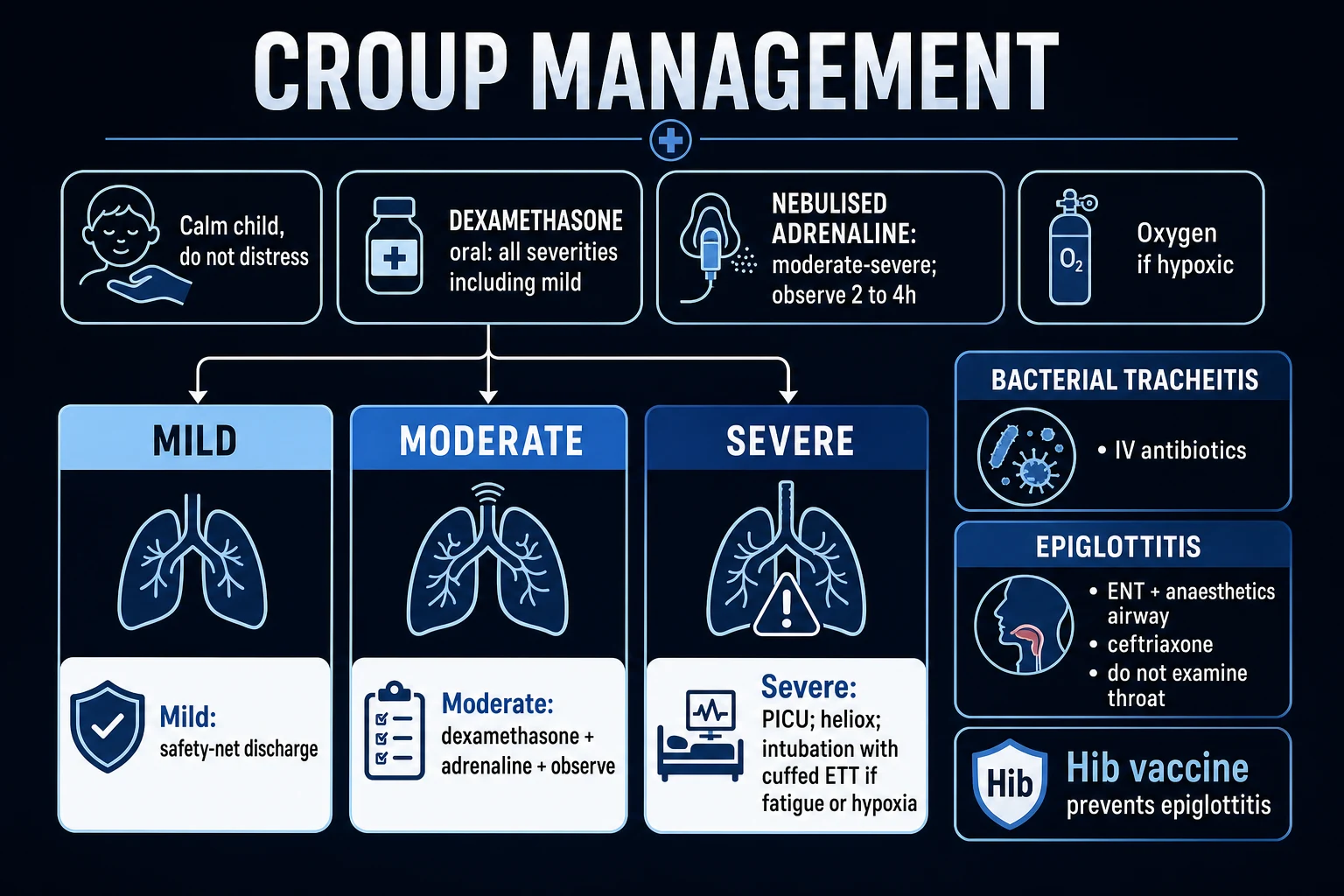
The cornerstone of croup management is corticosteroids for every child with croup, regardless of severity. This is supported by multiple Cochrane reviews and large randomised trials showing that a single dose of dexamethasone reduces symptom severity, shortens hospital stay, reduces the need for adrenaline, and reduces return visits — and that the benefit extends even to children traditionally classified as "mild".[3][4]
Mild croup (Westley 0 to 2)
- Single dose of oral dexamethasone 0.15 to 0.6 mg/kg (0.15 mg/kg is as effective as 0.6 mg/kg for mild disease; many units use 0.6 mg/kg for simplicity)
- Alternative if vomiting or unable to take oral: nebulised budesonide 2 mg single dose, or IM dexamethasone 0.6 mg/kg
- Discharge home with clear safety-net advice; no adrenaline; no observation required beyond standard clinical review
- Paracetamol 15 mg/kg q4-6h or ibuprofen 10 mg/kg q6-8h for fever and discomfort; encourage oral fluids; cool humidified air (no evidence base but commonly advised)
Moderate croup (Westley 3 to 5)
- Single dose of oral or IM dexamethasone 0.6 mg/kg (max 16 mg)
- Observe in ED for 2 to 4 hours; repeat assessment and Westley score
- Nebulised adrenaline 1:1000 0.5 mL/kg max 5 mL if stridor at rest persists or worsens
- Discharge once no stridor at rest, no cyanosis, normal mental state, SpO2 over 92 percent in air, tolerating oral fluids — and at least 2 to 4 hours after any adrenaline
- Admit if no improvement, recurrent need for adrenaline, young age, dehydration, or social concerns
Severe croup (Westley 6 to 11)
- Nebulised adrenaline 1:1000 0.5 mL/kg max 5 mL immediately, repeat every 15 to 30 min as needed
- Dexamethasone 0.6 mg/kg PO/IM/IV single dose
- Humidified oxygen if SpO2 under 92 percent; keep calm in parent's arms
- Admit to HDU or PICU; continuous monitoring
- Senior anaesthetics and ENT informed; prepare for intubation if fatiguing, cyanosed, or reduced consciousness
Impending respiratory failure (Westley 12 to 17)
- PICU admission, senior anaesthetics and ENT present at bedside
- Continue adrenaline and dexamethasone; add Heliox as temporiser if available
- Intubate (ETT 0.5 to 1.0 mm smaller than predicted) before cardiovascular collapse
- Mechanical ventilation with sedation and paralysis; ENT for surgical airway if intubation fails
Specific drugs
Dexamethasone is the corticosteroid of choice: a single oral dose has high bioavailability, a long biological half-life (36 to 72 hours, so a single dose covers the natural course), and a wealth of trial evidence. The dose range 0.15 to 0.6 mg/kg (maximum 16 mg) is supported by randomised trials and Cochrane review; 0.6 mg/kg is the most widely used single dose in many units because it works across the full severity spectrum, but 0.15 mg/kg is non-inferior in mild croup.[3][4]
Nebulised budesonide 2 mg is an evidence-based alternative for the child who vomits or cannot take oral medication; randomised trials show it is equivalent to oral or IM dexamethasone.[5]
Prednisolone (1 to 2 mg/kg) has historically been used in some regions but has weaker evidence than dexamethasone, a shorter half-life (requiring multi-day courses), and is now generally superseded; some units still use it where dexamethasone is unavailable.[3]
Nebulised adrenaline (epinephrine) 1:1000 — L-epinephrine (the simpler, cheaper, more widely available form) at 0.5 mL/kg up to a maximum of 5 mL, or racemic epinephrine (2.25 percent solution) at 0.5 mL/kg up to a maximum of 5 mL, are clinically equivalent. Onset is 10 to 30 minutes; duration of effect is 1 to 2 hours; rebound worsening can occur after the drug wears off, so observation of at least 2 to 4 hours after the last dose is mandatory before discharge. Adrenaline works by alpha-1-mediated vasoconstriction of the subglottic mucosa, rapidly reducing oedema.[6][7]
Antibiotics are NOT indicated in uncomplicated viral croup. They are reserved for suspected bacterial superinfection (bacterial tracheitis, pneumonia, otitis media) or an alternative bacterial diagnosis.[1]
Cough suppressants, decongestants, and antihistamines have no role and may cause sedation, which is dangerous in a child with airway obstruction.[1]
Stepwise management summary
- Diagnose clinically — barking cough, stridor, hoarse voice, age 6 months to 6 years.
- Assess severity — Westley score, vital signs, mental state, SpO2.
- Give dexamethasone 0.15 to 0.6 mg/kg PO/IM/IV to every child with croup (single dose).
- Add nebulised adrenaline 0.5 mL/kg max 5 mL for moderate to severe (stridor at rest).
- Give humidified oxygen only if SpO2 under 92 percent.
- Keep the child calm in the parent's arms; avoid unnecessary procedures.
- Observe at least 2 to 4 hours after adrenaline for rebound.
- Discharge when discharge criteria met, with safety-net advice.
- Admit for severe, recurrent, atypical, refractory, or socially complex cases.
- Investigate atypical or recurrent croup with ENT referral and endoscopy.[1]
Specific Subtypes & Scenarios
Spasmodic croup (also called "recurrent allergic croup" or "spasmodic laryngitis") describes recurrent, brief (hours) nocturnal episodes of barking cough and stridor in a child who is well between episodes, often atopic (eczema, asthma, family history of atopy), and without fever or viral prodrome. The aetiology is thought to be allergic or viral-triggered laryngeal oedema rather than the classic viral laryngotracheobronchitis. Each episode responds to cool air and a single dose of dexamethasone; recurrent episodes warrant ENT evaluation for an underlying anatomical cause and consideration of reflux and atopy management.[1][10]
Recurrent croup (more than 2 to 3 episodes in 6 months, or any recurrence with atypical features) should trigger a search for an underlying cause: subglottic stenosis (post-intubation or congenital), subglottic haemangioma, laryngeal web, laryngomalacia, laryngeal cleft, gastro-oesophageal reflux, airway hyper-reactivity, or immunodeficiency. Referral to paediatric ENT for flexible laryngoscopy and bronchoscopy is indicated, and aerodigestive evaluation (including triple endoscopy in selected centres) may identify surgically correctable lesions.[10]
Atypical age — croup under 6 months or over 6 years is unusual and should prompt the same workup as recurrent croup; congenital airway anomalies (laryngomalacia, subglottic haemangioma, vascular ring) and acquired causes (subglottic stenosis, foreign body) dominate.[10]
Refractory croup — failure to respond to adrenaline and dexamethasone within 2 to 4 hours, or deterioration despite treatment, is a red flag: reconsider the diagnosis (epiglottitis, bacterial tracheitis, retropharyngeal abscess, foreign body, anatomical lesion), escalate to PICU, and involve senior anaesthetics and ENT.[1][12]
Croup in a child with an artificial airway history (prematurity, prior intubation, tracheostomy) raises the likelihood of subglottic stenosis and warrants lower threshold for admission and ENT input.[1]
Measles croup — in unvaccinated children, measles can cause severe, necrotising laryngotracheobronchitis requiring PICU and ENT; consider in areas with falling vaccination coverage, alongside the classic coryza, conjunctivitis, Koplik spots, and descending rash.[1]
Complications & Pitfalls
Complications are uncommon but potentially life-threatening:[1][12]
- Respiratory failure — the feared endpoint of progressive obstruction; prevented by early recognition, dexamethasone, and judicious adrenaline.
- Hypoxic neurological injury — rare, in children who arrest or have prolonged severe hypoxaemia.
- Bacterial superinfection — secondary bacterial tracheitis (Staph aureus, Streptococcus), pneumonia, or otitis media may complicate viral croup; suspect with new or persistent fever, toxicity, or purulent secretions.
- Pneumomediastinum and pneumothorax — from the high intrathoracic pressures generated by severe coughing or positive-pressure ventilation; usually self-limiting but pneumothorax may require drainage.
- Dehydration — from reduced oral intake due to increased work of breathing; assess hydration and treat with oral, NG, or IV fluids.
- Pulmonary oedema — after relief of severe upper airway obstruction (negative-pressure pulmonary oedema); recognise and treat with oxygen and diuretics if needed.
- Recurrence — common in atopic children; consider underlying cause if frequent.[1][12]
- Mistaking epiglottitis or bacterial tracheitis for croup — the most dangerous error. Any toxic, drooling, tripod, or rapidly deteriorating child needs an alternative diagnosis considered and escalation, not "more croup treatment".
- Delaying corticosteroids — give dexamethasone to every child with croup, even mild; the benefit is established.
- Over-reliance on the steeple sign — it is neither sensitive nor specific; do not require it to make the diagnosis.
- Discharging too soon after adrenaline — rebound occurs; observe at least 2 to 4 hours.
- Agitating the child — crying and struggling worsen obstruction; keep calm, avoid unnecessary procedures, and treat in the parent's arms.
- Using the wrong endotracheal tube — intubating a child with subglottic oedema with an age-predicted tube size risks trauma; use a tube 0.5 to 1.0 mm smaller.
- Missing recurrent croup — investigate recurrent or atypical presentations for underlying anatomical causes.[1][8]
Prognosis & Disposition
The prognosis of croup is excellent with appropriate treatment. Most children recover fully within 3 to 7 days, with symptoms peaking on days 1 to 2 and improving thereafter. Hospitalisation is needed in 1 to 5 percent of presentations, intensive care in 0.5 to 2 percent of hospitalised cases, and intubation in fewer than 0.5 percent. Mortality is now very low in countries with modern PICU care — historically under 0.5 percent of intubated children — and is essentially confined to children with significant comorbidity, delayed presentation, or alternative diagnoses (epiglottitis, bacterial tracheitis) mistaken for croup.[1]
Disposition is determined by severity, response to treatment, age, comorbidity, and social factors:[1]
Discharge home
- Mild croup with no stridor at rest after dexamethasone
- Moderate croup that has improved after dexamethasone (and adrenaline if given), observed at least 2 to 4 h after adrenaline
- SpO2 over 92 percent in air, no cyanosis, normal mental state
- Tolerating oral fluids, parents confident, reliable follow-up
- Clear safety-net advice: return if stridor at rest, marked recession, cyanosis, drooling, fatigue, poor feeding
Admit to ward
- Moderate croup not improved after dexamethasone and observation
- Persistent stridor at rest requiring repeated adrenaline
- Age under 6 months, significant comorbidity, dehydration, poor social circumstances
- Need for ongoing observation or parental anxiety
Admit to HDU/PICU
- Severe croup (Westley 6 or above)
- Persistent or recurrent need for adrenaline
- Fatigue, cyanosis, altered consciousness
- Refractory disease or suspected alternative diagnosis (epiglottitis, bacterial tracheitis)
- Post-intubation management
Discharge criteria are: no stridor at rest, no significant retractions, normal air entry, normal mental state, SpO2 over 92 percent in room air, tolerating oral fluids, and at least 2 to 4 hours since the last dose of adrenaline (to detect rebound). Parents must be given a clear written safety-net covering warning signs and when to return.[1]
Special Populations
Infants under 6 months: croup is uncommon in this age group and an atypical presentation should prompt consideration of congenital airway anomalies (laryngomalacia, subglottic haemangioma, laryngeal web, vascular ring), germ-layer defects, or subglottic stenosis from prior intubation. Admit for observation, oxygen, and ENT referral; have a lower threshold for airway intervention.[1]
Older children (over 6 years): croup is uncommon and should raise suspicion of anatomical causes (subglottic stenosis, laryngeal polyps), foreign body, malignancy, or bacterial tracheitis. Investigate and involve ENT.[1]
Children with asthma or atopy: more likely to have recurrent croup and spasmodic features; co-treatment with bronchodilators if wheeze is present; the principles of croup management (dexamethasone, adrenaline) are unchanged.[10]
Immunocompromised children: longer and atypical viral courses, broader differentials (CMV, fungal, mycobacterial), higher risk of secondary infection; admit and involve microbiology and immunology.[1]
Children with chronic lung disease, congenital heart disease, or neuromuscular disorders: lower threshold for admission and HDU; these children tolerate respiratory compromise poorly.[1]
Pregnant adolescent or adolescent mother: management is the same; ensure adolescent-friendly setting, contraception and safeguarding review as appropriate.[1]
Evidence, Guidelines & Regional Differences
The evidence base for croup management is strong and remarkably consistent across guidelines.[3]
Corticosteroids: the Cochrane review (Russell 2011, updated from 2004) of 38 randomised trials in over 4,500 children confirmed that a single dose of corticosteroids (dexamethasone or budesonide) reduces the Westley score at 6 and 12 hours, reduces the need for adrenaline, shortens hospital stay by around 12 hours, and reduces return visits and readmissions.[3] The landmark Bjornson 2004 NEJM trial showed that a single oral dose of dexamethasone 0.6 mg/kg benefits even children with mild croup, reducing return visits and improving outcomes — establishing the principle that corticosteroids are for every child with croup, not just severe cases.[4]
Nebulised budesonide 2 mg was shown by Johnson 1998 NEJM to be equivalent to IM dexamethasone for moderately severe croup, providing an inhaled alternative.[5]
Nebulised adrenaline: the Cochrane review (Bjornson 2013) confirmed that nebulised adrenaline produces rapid (within 30 minutes) symptomatic improvement in moderate to severe croup compared with placebo, but the effect is short-lived (1 to 2 hours), with rebound after the drug wears off — hence the need for observation.[6] The original Westley 1978 study established racemic epinephrine (and the Westley score itself) for croup.[7]
Humidified air / mist therapy: the Cochrane review (Moore 2011, later withdrawn for updates) found no evidence of benefit for humidified or cool mist therapy in croup; many units still use it for comfort but it should not be presented as evidence-based.[9]
Antibiotics: no role in uncomplicated viral croup.[1]
Australasian practice (reflected in guidelines from the Royal Australasian College of Physicians and the Paediatric Research in Emergency Departments International Collaborative) is similar: oral dexamethasone 0.15 to 0.6 mg/kg single dose for all severities; nebulised 1:1000 adrenaline 0.5 mL/kg max 5 mL for moderate to severe; heliox in selected PICUs for refractory disease.[1]
In India, South Asia, and many low- and middle-income settings, the principles are identical but access to adrenaline, nebulisers, and PICU may be limited; measles croup remains a consideration where vaccination coverage has fallen. The Indian Academy of Pediatrics guidance aligns with international standards: dexamethasone for all, adrenaline for moderate to severe, oxygen if hypoxic, antibiotics only for bacterial superinfection.[1]
Controversies include the optimal dexamethasone dose (0.15 vs 0.6 mg/kg — both effective, 0.6 more widely used for simplicity and across severity), the role of heliox and mixed helium-oxygen therapy (limited evidence, used in refractory disease), the duration of observation after adrenaline (2 to 4 hours is conventional but variable), and whether a second dose of dexamethasone benefits the rare child with persistent symptoms (limited evidence; some units give a second dose at 24 hours). The role of prophylactic corticosteroids for recurrent or spasmodic croup is unproven.[1]
Exam Pearls
BARKING
- Croup = parainfluenza virus type 1, age 6 months to 6 years, barking cough + inspiratory stridor + hoarse voice.
- Dexamethasone 0.15 to 0.6 mg/kg PO/IM/IV single dose for every child with croup — including mild.
- Nebulised adrenaline 1:1000, 0.5 mL/kg max 5 mL for moderate to severe (stridor at rest); onset 10 to 30 min, duration 1 to 2 h, observe at least 2 to 4 hours for rebound.
- Westley croup score: stridor 0/1/2, retractions 0/1/2/3, air entry 0/1/2, cyanosis 0/4/5, consciousness 0/5; max 17; mild 0 to 2, moderate 3 to 5, severe 6 to 11, impending failure 12 to 17.
- Steeple sign (pencil tip) on AP neck X-ray — narrowed subglottic air column; confirmatory but neither sensitive nor specific.
- Cricoid is the only complete cartilaginous ring and the narrowest part of the paediatric airway — oedema is forced inward, and resistance is proportional to 1 over radius to the fourth power (Poiseuille).
- Epiglottitis: high fever, drooling, tripod, toxic, NO cough — Hib, do NOT examine throat, anaesthetics and ENT, secure airway in theatre, IV ceftriaxone.
- Bacterial tracheitis: Staph aureus, toxic, purulent secretions, rapid deterioration, poor response to adrenaline — PICU, broad antibiotics, prepare for airway.
- Spasmodic croup: atopic child, no fever, recurrent nocturnal episodes, well between attacks — cool air and a single dose of dexamethasone.
- Recurrent or atypical croup (under 6 months, over 6 years, more than 2 to 3 episodes): ENT referral, flexible laryngoscopy and bronchoscopy for anatomical cause (subglottic stenosis, haemangioma, web).
- Humidified air / mist: no evidence of benefit. Antibiotics: no role in uncomplicated viral croup.
- Croup is self-limiting over 3 to 7 days; most children can be discharged with safety-net advice.
- Intubate with an ETT 0.5 to 1.0 mm smaller than age-predicted for subglottic oedema.[1]
Exam application bank (NEET-PG / INICET)
One-line answer
Croup (acute laryngotracheobronchitis) is the most common cause of acute upper airway obstruction in young children, peaking at 6 months to 3 years, caused mainly by parainfluenza virus type 1 and 2. Presentation: barking seal-like cough, harsh inspiratory stridor, hoarse voice, low-grade fever, worse at night. Most are mild and self-limiting over 3 to 7 days. Severe croup: stridor at rest, marked retractions, cyanosis, altered mental state. Treatment: oral dexamethasone 0.15 to 0.6 mg/kg single dose for all children; nebulised adrenaline 1:1000, 0.5 mL/kg max 5 mL, for moderate to severe with stridor at rest; observe at least 2 to 4 hours after adrenaline for rebound. [1]
Worked stems (answer without another resource)
Stem 1 — Classic presentation. Map symptoms to mechanism; name the first investigation and first treatment step with dose/route if drug therapy is standard. [1]
Stem 2 — Unstable / complicated. List red flags that force immediate resuscitation, theatre, ICU, antidote, or reperfusion — and what you do in the first 15 minutes. [1]
Stem 3 — Atypical group. Elderly, pregnancy, child, or immunocompromised: how presentation and thresholds change. [1]
Stem 4 — Differential trap. Name the three closest mimics and one discriminator for each. [1]
Stem 5 — Disposition. Who goes home with safety-netting, who is admitted, who needs HDU/ICU/theatre, and what follow-up is mandatory. [1]
Rapid viva checklist
- Definition + classification
- Pathophysiology chain
- Bedside signs / criteria
- Score with exact components (if any)
- Emergency bundle
- Definitive therapy with doses
- Complications of disease and of treatment
- Special populations
- Guideline/trial name if classic
- Three exam traps
Coverage self-check
If you cannot answer any stem above from this page alone, re-read the matching section — the page is intended to be self-sufficient for final-prof and NEET-PG/INICET questions on Croup.
References
- [1]Bjornson CL, Johnson DW Croup Lancet, 2008.PMID 18295000
- [2]Cherry JD Clinical practice. Croup N Engl J Med, 2008.PMID 18216359
- [3]Russell KF, Liang Y, O'Gorman K, Johnson DW, Klassen TP Glucocorticoids for croup Cochrane Database Syst Rev, 2011.PMID 21249651
- [4]Bjornson CL, Klassen TP, Williamson J, et al A randomized trial of a single dose of oral dexamethasone for mild croup N Engl J Med, 2004.PMID 15385657
- [5]Johnson DW, Jacobson S, Edney PC, Duffy P, Thompson T A comparison of nebulized budesonide, intramuscular dexamethasone, and placebo for moderately severe croup N Engl J Med, 1998.PMID 9709042
- [6]Bjornson C, Russell KF, Vandermeer B, Durec T, Klassen TP, Johnson DW Nebulized epinephrine for croup in children Cochrane Database Syst Rev, 2013.PMID 24114291
- [7]Westley CR, Cotton EK, Brooks JG Nebulized racemic epinephrine by IPPB for the treatment of croup: a double-blind study Am J Dis Child, 1978.PMID 347921
- [8]Tibballs J, Watson T Symptoms and signs differentiating croup and epiglottitis J Paediatr Child Health, 2011.PMID 21091577
- [9]Moore M, Little P WITHDRAWN: Humidified air inhalation for treating croup Cochrane Database Syst Rev, 2011.PMID 21678339
- [10]Wall SR, Wat D, Spiller OB, et al The viral aetiology of croup and recurrent croup Arch Dis Child, 2009.PMID 18801765
- [11]Kwon JH, Kim YH, Han DH, et al Analysis of the functionally-narrowest portion of the pediatric upper airway in sedated children Medicine (Baltimore), 2018.PMID 29979422
- [12]Hopkins A, Lahiri T, Salerno R, Heath B Changing epidemiology of life-threatening upper airway infections: the reemergence of bacterial tracheitis Pediatrics, 2006.PMID 17015531
- [13]Johnson DW Croup BMJ Clin Evid, 2014.PMID 25263284