Paediatrics · Paediatrics
Developmental Dysplasia of the Hip
Also known as DDH · Congenital dislocation of the hip · CDH · Congenital hip dysplasia · Acetabular dysplasia
Developmental dysplasia of the hip (DDH) is a spectrum of hip joint abnormalities in the infant and child ranging from a lax, dislocatable hip to a frankly dislocated hip with a shallow (dysplastic) acetabulum. Formerly called congenital dislocation of the hip (CDH); renamed because the disorder can develop after birth. Risk factors: breech presentation, female sex, firstborn, oligohydramnios, family history, foot deformity. Newborn: Barlow test (dislocatable) and Ortolani test (reducible clunk). Older infant: limited abduction, asymmetric skin folds, Galeazzi sign. Walker: Trendelenburg gait, leg-length discrepancy. Image with ultrasound under 4 to 6 months, plain X-ray over 4 to 6 months. Treat by age: Pavlik harness 0 to 6 months; closed reduction and spica cast 6 to 18 months; open reduction over 18 months; osteotomy for residual dysplasia. Feared complication: avascular necrosis of the femoral head.
On this page & tools
Your progress
Saved locally on this device.
Exam tags
Red flags
Overview & Definition
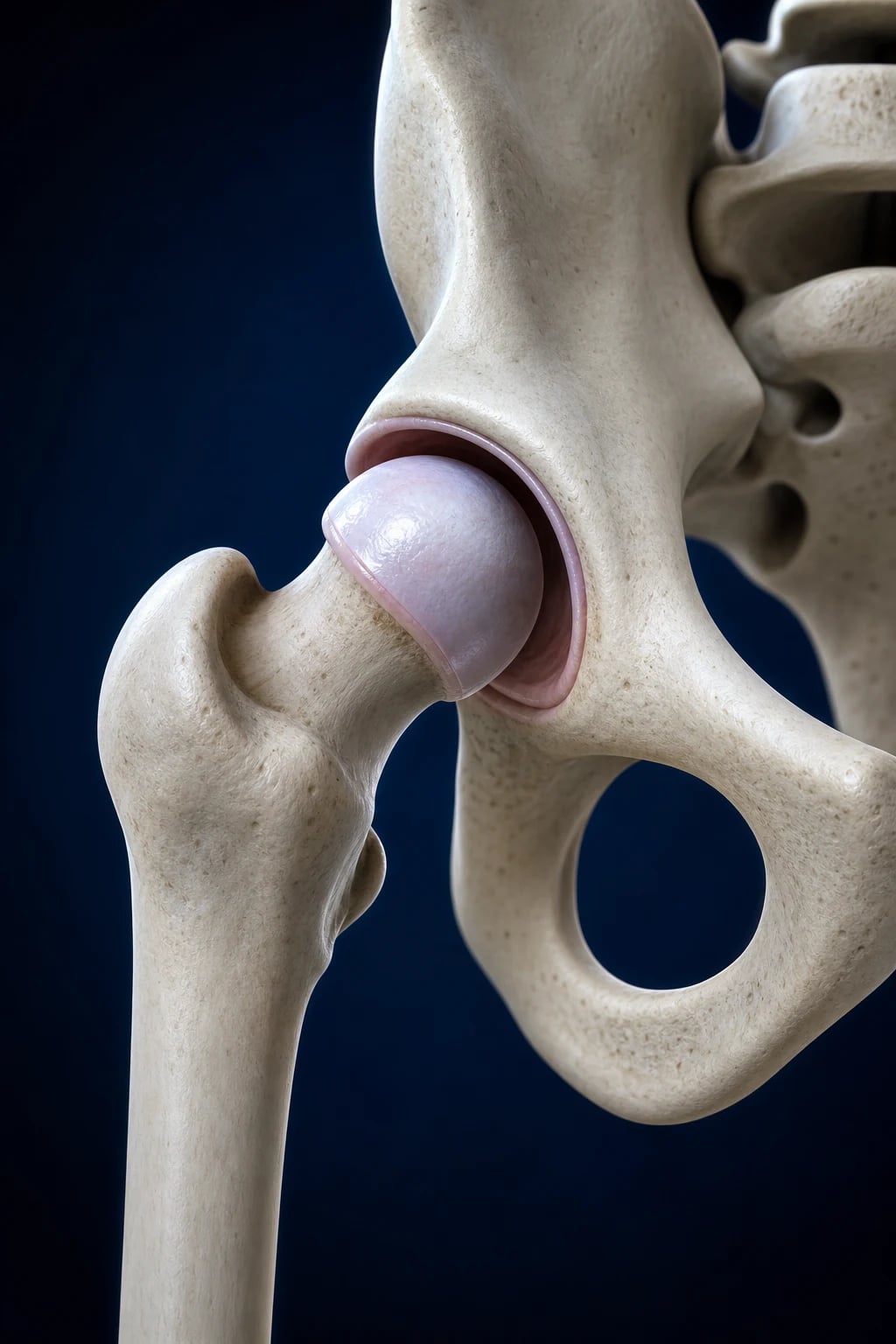
Developmental dysplasia of the hip (DDH) is the modern, preferred term for a spectrum of hip joint abnormalities in the developing child in which the proximal femur and acetabulum grow abnormally and out of proportion to one another, ranging from a hip that is merely lax and dislocatable to one that is completely and irreducibly dislocated with a shallow, dysplastic acetabulum.[3]
The older term congenital dislocation of the hip (CDH) has been abandoned because it implied the hip was dislocated at birth. The current term recognises that the disorder is developmental — a hip may be normal or only mildly lax at birth yet dislocate over the first weeks to months of life as the lax capsule and shallow socket fail to keep the femoral head reduced. About one in five cases that are eventually diagnosed are not detectable on the newborn examination.[3]
The unifying pathological principle is lack of concentric reduction: the femoral head must sit centrally in the acetabulum to stimulate normal, deep acetabular growth (and to grow a normal femoral neck). Once the head is displaced — even intermittently — acetabular dysplasia (shallow socket), femoral-head deformity, and capsular stretching develop in a self-perpetuating cycle that ends in early secondary osteoarthritis by the third to fifth decade if untreated.[3][4]
Why DDH matters clinically: it is one of the few conditions in paediatrics where early, simple, non-operative treatment (a Pavlik harness worn for a few weeks in a newborn) reliably prevents a lifetime of disability, while late presentation needs major surgery and is associated with permanent joint damage. The skill the examiner is testing is detection — knowing the risk factors, performing Barlow/Ortolani correctly, choosing the right imaging modality by age, and escalating through the age-based management ladder.[1]
Classification
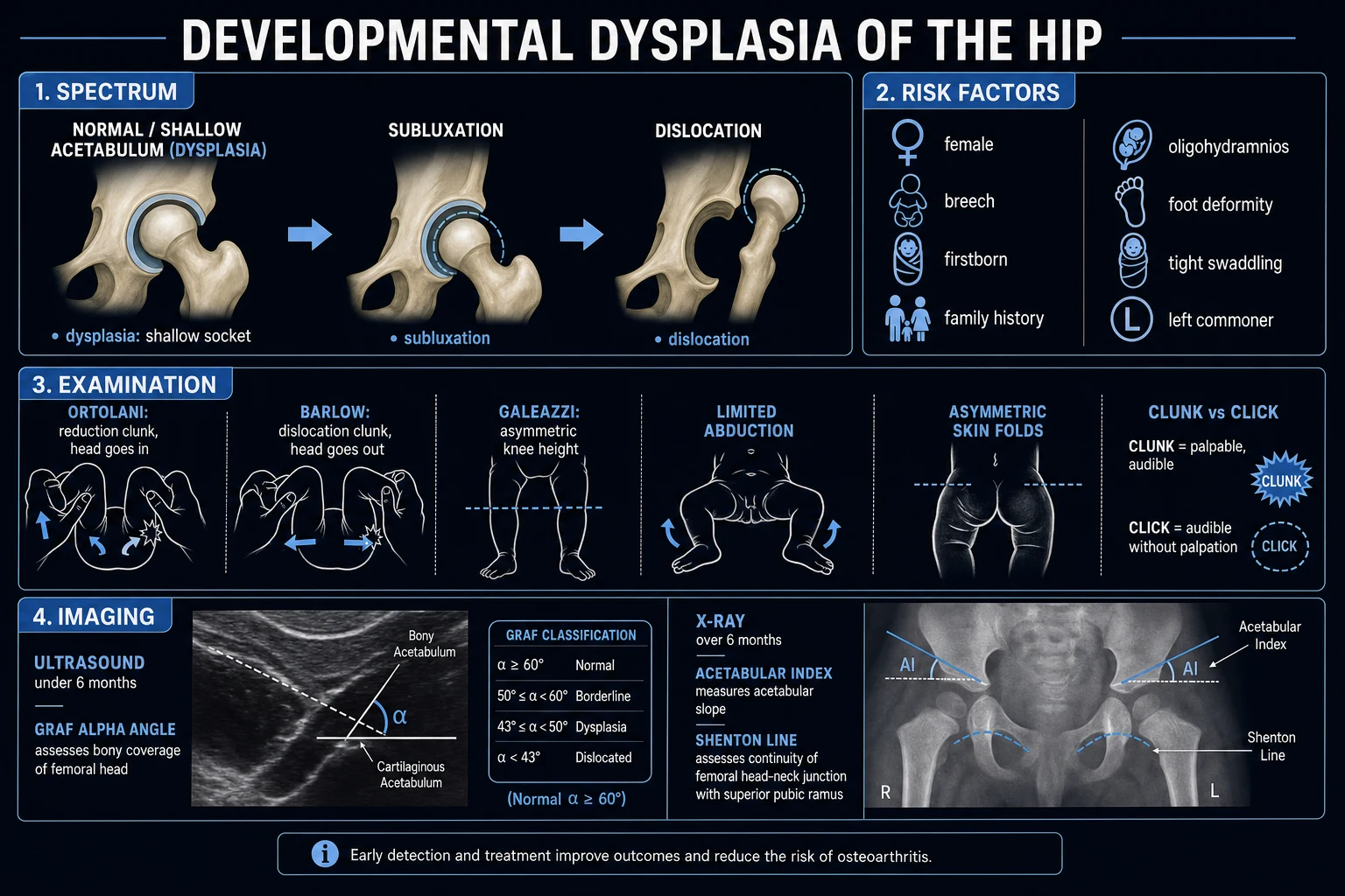
DDH is classified in three complementary ways: by clinical instability (the bedside finding), by radiographic/Graf ultrasound type (the morphological severity), and by timing of presentation (which dictates treatment). All three are examinable. [1]
Clinical (instability)
bedside
- **Dislocatable** hip (positive **Barlow**) — head sits in the socket but can be pushed out
- **Dislocated but reducible** (positive **Ortolani**) — head is out but can be lifted back in (the classic clunk)
- **Dislocated and irreducible** — head is out and cannot be reduced; Ortolani is negative (teratologic / late DDH)
- **Subluxated** — head partially in contact with the acetabulum, lies between reduced and dislocated
Graf ultrasound
morphology
- **Type I** — mature normal hip (**alpha angle over 60 degrees**)
- **Type IIa** — physiologically immature under 3 months (alpha 50 to 59 degrees), normal for age
- **Type IIc / D** — critical, unstable (alpha 43 to 49 degrees)
- **Type III** — subluxated (alpha under 43 degrees, cartilaginous roof displaced upwards)
- **Type IV** — dislocated (cartilaginous roof displaced downwards)
By timing
drives treatment
- **Early / newborn DDH** — detected in first weeks; treated with **Pavlik harness**
- **Late-presenting DDH** — diagnosed after walking age; needs reduction ± osteotomy
- **Teratologic DDH** — dislocated in utero, fixed at birth; associated with neuromuscular disorders, arthrogryposis, Larsen syndrome
- **Acquired / post-natal DDH** — develops after a normal newborn examination; argues for repeated screening
Important distinctions:
- Obligate vs positional dislocation. A hip that dislocates only in certain positions (e.g. adduction) and reduces spontaneously is positional — common in the newborn and may resolve with observation. A hip that must be dislocated in every resting position is obligate and needs urgent treatment.
- Teratologic vs typical DDH. Teratologic dislocations occur before birth (in utero), are fixed, are often bilateral, and are associated with genetic / neuromuscular syndromes (arthrogryposis, Larsen syndrome, myelodysplasia, Ehlers-Danlos). Typical DDH develops around or after birth, is usually unilateral, and occurs in an otherwise normal infant.[4]
Epidemiology & Risk Factors
Incidence: true dislocation occurs in roughly 1 to 2 per 1000 live births; if all instability (including mild laxity that resolves spontaneously) is included, the figure rises to 1 to 2 per 100 newborns. Bilateral in about 1 in 5 of true dislocations. The left hip is affected three times more often than the right (because the commonest intra-uterine position is left occiput anterior, in which the left hip lies adducted against the maternal sacrum).[3]
Female sex: roughly 4 to 8 times the risk of males — the female pelvis is wider and more lax (maternal relaxin effect is greater on female foetal ligaments). About 80% of dislocated hips are in girls.[3]
DDH risk factors — word 4F + BOLSTER
4F-BOLST
4 to 8x risk; 80% of cases
tight primigravid uterus restricts movement
positive in 12% (genetic component)
12 to 25% of breech babies have DDH — hips flexed and knees extended in utero
single biggest modifiable risk factor — universal US screening recommended
reduced intra-uterine space crowds the hips
left hip 3x more common (LOA position)
hips held extended and adducted postnatally — cultural risk
metatarsus adductus, calcaneovalgus, clubfoot — same intra-uterine crowding
Mechanism of breech risk. In the frank (extended) breech position the foetal hips are flexed and the knees extended, so the femoral head rests against a shallow, posteriorly inclined acetabulum. After delivery the extended knees suddenly allow the hips to extend and adduct, leveraging the head out of the socket. Breech DDH is more often bilateral and teratologic in character. Foot deformities (metatarsus adductus, congenital talipes equinovarus / clubfoot) and congenital muscular torticollis are independent risk factors because they share the same underlying cause — intra-uterine packaging / crowding ("packaging disorders"). The presence of any one of these mandates a careful hip examination.[3]
Postnatal risk — swaddling. Tight swaddling with the hips extended and adducted (the traditional "straight-leg" wrap) is a major and modifiable risk in cultures where it is practised. Safe swaddling leaves the hips flexed and abducted (the "frog-leg" position); parent education reduces late DDH.[3]
Genetics. A positive family history raises the risk approximately 6-fold. Inheritance is polygenic; siblings of an affected child and children of an affected parent should be screened with extra care. [1]
Pathophysiology
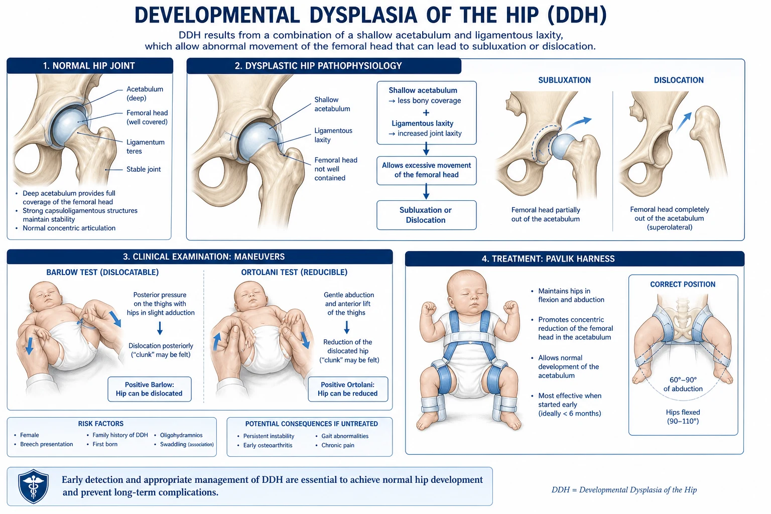
Normal hip development requires the femoral head and acetabulum to grow together under the stimulus of concentric contact — the head acts as a template that deepens the socket, and the socket keeps the head spherical. This is the principle of Heuter-Volkmann and Wolff laws applied to the immature joint: bone and cartilage remodel along the lines of mechanical force.[3]
The cascade proceeds in five linked steps:[3][4]
- Ligamentous and capsular laxity. The newborn hip joint is normally loose — the capsule is elastic and the ligamentum teres, transverse acetabular ligament, and iliofemoral ligament are lax. This is exaggerated in girls (greater relaxin sensitivity) and in those with connective-tissue disorders.
- Shallow, more vertical acetabulum. The neonatal acetabulum is largely cartilaginous and slopes more vertically than the adult version; if the femoral head is not pressed against it concentrically, it does not deepen normally.
- Subluxation and dislocation. In the adducted, extended position the head is levered posteriorly and superiorly over the acetabular rim. At first this is reducible (Ortolani positive); with time the soft tissues adapt to the dislocated position and the hip becomes irreducible (Ortolani negative).
- Soft-tissue and bony adaptation. The joint capsule stretches and develops an hour-glass constriction; the iliopsoas tendon tightens across the anterior rim (the pectineo-capsular tightness); the acetabular labrum inverts (forming the limbus) and a ridge of hypertrophied cartilage forms on the rim (neolimbus) which can block closed reduction. The transverse acetabular ligament is pulled superiorly and across the socket, narrowing the inferior outlet. The adductor muscles shorten. The acetabulum becomes dysplastic (shallow, oval, oblique) and the femoral head flattens (coxa magna) and the femoral neck retroverts.
- Secondary osteoarthritis. An unreduced hip develops early, severe, secondary OA of the joint by the third to fifth decade; the contralateral "normal" hip is also at risk because of altered pelvic mechanics and leg-length discrepancy. In adults a missed unilateral DDH is the commonest cause of secondary OA of the hip in young women. [1]
Why the left side is commonest. In the left occiput anterior (LOA) lie — the most common foetal position — the left hip lies against the maternal sacrum and is held adducted; the right hip is freer. Hence the left hip is the more frequently affected.[3]
Clinical Presentation
Presentation is age-dependent — the signs change as the child grows and the soft tissues stiffen. The aim of screening is to detect DDH in the newborn, when treatment is simplest.[1]
Newborn (birth to 3 months)
The classical signs are the dynamic instability tests, performed with the baby relaxed and supine on a firm surface: [1]
- Ortolani test (reduction / reducibility test). With the infant supine, the examiner flexes the hips and knees to 90 degrees and grasps each knee with the middle finger on the greater trochanter and the thumb on the inner thigh. The hips are gently abducted while the middle finger lifts the greater trochanter anteriorly. A positive Ortolani = a palpable (and sometimes audible) "clunk" as the dislocated femoral head reduces back into the acetabulum. This is the most reliable newborn sign of a dislocated, reducible hip.
- Barlow test (dislocation / dislocatability test). From the same starting position, with the hip adducted, the examiner applies gentle posterior pressure on the flexed knee while pressing the femoral head posteriorly with the thumb. A positive Barlow = the femoral head slides out of the acetabulum over the posterior rim (the hip is dislocatable). The pressure is then released and the hip reduced — this is the opposite movement to Ortolani. [1]
Mnemonic: Barlow = Ballot out (dislocatable); Ortolani = "O"nly in (reducible). A clunk on abduction (Ortolani) is the reducible hip; a clunk on adduction (Barlow) is the dislocatable hip.[1]
Older infant (3 to 12 months)
By this age the soft tissues have tightened and the hip is usually irreducible, so Ortolani/Barlow become negative — the examiner feels only limited abduction. The signs shift to the consequences of a chronically dislocated hip: [1]
- Limited abduction — the most reliable sign in this age group. Examine both hips together: the affected hip fails to abduct fully (often to less than 60 to 70 degrees) because the adductors and iliopsoas are tight. Asymmetry of abduction is highly suggestive when unilateral; bilateral limitation must be assessed against normal ranges.
- Asymmetric gluteal and thigh skin folds. Best seen from behind with the baby prone. The affected side shows extra or higher thigh folds (because the dislocated femur is shortened) and an asymmetric gluteal cleft. Sensitive but not specific — many normal infants have asymmetric folds.
- Galeazzi / Allis sign (apparent femoral shortening). With the baby supine and hips and knees flexed to 90 degrees and feet flat on the couch, the knees are at different heights — the knee on the affected (dislocated) side sits lower because the femoral head has migrated superiorly, shortening the effective thigh. Bilateral dislocation gives a falsely negative (symmetrical) Galeazzi — both knees are low.
- Klisic test. Place the thumb on the anterior superior iliac spine and the middle (or ring) finger on the greater trochanter; draw an imaginary line between them. In a dislocated hip, the line points towards the umbilicus or above (the trochanter is high); in a normal hip the line points to the groin. Useful in chubby infants.
- Telescoping or piston mobility — the head can be moved up and down within the lax capsule. [1]
Walking-age child (over 12 months)
The child walkers late (often over 15 months) and presents with: [1]
- Painless limp — the single most useful pointer. DDH is painless (unlike septic arthritis, Perthes, SCFE).
- Trendelenburg gait — the pelvis drops on the contralateral side during stance phase on the affected leg because the abductors (gluteus medius/minimus) are unable to stabilise the pelvis from their shortened, disadvantaged position (the head is no longer a stable fulcrum). On standing on the affected leg the opposite pelvis falls — positive Trendelenburg sign.
- Leg-length discrepancy (LLD) — the affected leg is shorter (apparent shortening from the displaced head).
- Hyperlordosis and a waddling gait in bilateral cases (the child rocks from side to side — "sailor's gait").
- Bilateral dislocation causes a symmetrical waddle with hyperlordosis — easily missed because Galeazzi is symmetric and abduction is symmetrically limited; clinical suspicion must come from gait and the waddling, broad-based stance. [1]
DDH — key numbers
Atypical presentations
- Bilateral dislocation — easy to miss because Galeazzi is symmetric and abduction is symmetrically limited; presents as a waddling, broad-based gait with lumbar hyperlordosis and delayed walking.
- Late presentation in adolescence / adulthood — secondary osteoarthritis of the hip in a young woman; the dysplastic acetabulum and shallow socket are seen on pelvic X-ray done for hip pain. A previously undiagnosed DDH is the commonest cause of secondary OA in young women.
- Teratologic / syndromic DDH — fixed dislocation at birth, often bilateral, with arthrogryposis, Larsen syndrome, myelodysplasia, Ehlers-Danlos, Down syndrome. The hip is part of a wider disorder; treatment must address the underlying condition.
- Neuromuscular hip dysplasia (cerebral palsy, myelomeningocele) — progressive, late, often painless; treated differently (adductor releases, varus derotational osteotomy). [1]
Differential Diagnosis
A limping or short-limbed infant is not always DDH. The following must be considered and distinguished: [1]
Septic arthritis of the hip
do not miss
- **Painful, febrile, unwell child** who refuses to weight-bear or move the hip (pseudoparalysis)
- DDH is **painless** with full (or only abduction-limited) range; septic is **hot, tender, held flexed and abducted**
- Raised **CRP, ESR, WCC**; urgent ultrasound shows effusion; aspirate before treating
- **Emergency** — IV antibiotics, washout; not harness
Transient (toxic) synovitis
commonest limp
- **Afebrile or low-grade fever, recent viral illness**, well child, painful hip
- Self-limiting in 1 to 2 weeks; ultrasound may show small effusion, **normal bloods**
- Diagnosis of exclusion — must rule out septic arthritis (Kocher criteria)
Perthes disease (Legg-Calvé-Perthes)
age 4 to 8
- Idiopathic **avascular necrosis of the capital femoral epiphysis** in a boy aged **4 to 8**
- Insidious painless limp, **decreased abduction and internal rotation**
- X-ray shows **subchondral fracture → fragmentation → reossification**; treated by containment
Slipped capital femoral epiphysis (SCFE)
adolescent
- Adolescent (often **obese, hypothyroid or with growth-hormone excess**)
- **Externally rotated, antalgic gait**; pain referred to knee
- X-ray: **Klein line does not intersect the femoral head** on AP; **physeal slip** on frog lateral
- Emergency pinning; DO NOT reduce chronic slip
Congenital coxa vara
rare
- **Neck-shaft angle under 110 degrees** (developmental, sometimes post-Perthes or trauma)
- Painless limp and shortening like DDH, but hip is **stable** (Barlow/Ortolani negative, full abduction)
- X-ray: vertical physis, **triangular metaphyseal fragment** (Fairbank triangle)
Neuromuscular dislocation
spastic
- **Cerebral palsy (spastic diplegia), myelomeningocele, polio** — gradual dislocation from muscle imbalance (adductors/iliopsoas overpower abductors)
- **Painless** but with abnormal tone, reflexes, delayed motor milestones
- Treatment is of the underlying tone problem, not primarily the hip
Proximal focal femoral deficiency (PFFD)
congenital
- Congenital shortening of the proximal femur with a **variable absent femoral head/neck**
- Marked LLD at birth, **stable but short** thigh; X-ray diagnostic
The distinguishing triad for DDH is: (1) painless, (2) stable bar to dislocation is broken (positive Barlow/Ortolani, or limited abduction in the older infant), (3) otherwise well child with normal development and bloods. Pain, fever, or raised inflammatory markers shift the diagnosis away from DDH. [1]
Clinical & Bedside Assessment
A structured hip examination should be performed at every well-baby check from birth until the child is walking independently, because some cases develop after a normal newborn examination.[1]
Examination technique — general principles:
- Examine on a firm, flat surface with the infant calm and relaxed (examine before undressing fully or after a feed). A crying, tense baby gives false-negative tests.
- Expose both hips fully and examine both together for symmetry.
- Inspect for skin folds (thigh and gluteal), leg length (Galeazzi), and pelvic level.
- Move through the dynamic tests in order: abduction, Ortolani, Barlow, Galeazzi, Klisic. [1]
The dynamic instability tests (newborn, under 3 months)
- Ortolani — described above; the reduction test. The clunk of reduction is best felt with the middle finger on the greater trochanter.
- Barlow — described above; the dislocation / dislocatability test (the provocation test).
- Piston / telescope test — apply longitudinal traction; a dislocated hip shows excessive up-and-down play. [1]
Pitfalls:
- A soft "click" (high-pitched, easily reproduced, with no clunk) is common and benign in the newborn — it usually arises from the iliopsoas tendon snapping over the iliopectineal eminence or from knee/shoulder ligaments. A clunk (low-pitched, felt as the head passing over the rim) is abnormal. Click ≠ clunk.
- Examine repeatedly — a hip that was normal at birth may become abnormal, and a lax hip may tighten up. [1]
Static signs (older infant, 3 to 12 months)
- Limited abduction (under 60 to 70 degrees on the affected side) — the most reliable sign once Ortolani/Barlow have become negative.
- Galeazzi / Allis sign — apparent shortening; knee sits lower on the affected side with hips and knees flexed to 90 degrees and feet flat.
- Asymmetric skin folds — extra thigh fold, asymmetric gluteal cleft.
- Klisic test — line from ASIS to greater trochanter points to umbilicus or higher. [1]
Gait (walking child, over 12 months)
- Trendelenburg gait (pelvis drops on the contralateral side in stance phase) and positive Trendelenburg sign when standing on the affected leg.
- Leg-length discrepancy and an antalgic but painless limp.
- Bilateral — waddling "sailor's" gait, lumbar hyperlordosis, delayed walking. [1]
Investigations
Imaging choice is age-dependent and is the most frequently examined investigation point. The principle: below 4 to 6 months the femoral head is cartilaginous and invisible on X-ray, so ultrasound is the modality of choice; above 4 to 6 months the femoral head ossifies and plain X-rays become useful.[2]
Ultrasound (under 4 to 6 months)
- First-line in infants under 4 to 6 months because the cartilaginous femoral head and acetabular labrum are directly visualised.
- The standard coronal neutral view (Graf technique) shows the iliac wing, bony acetabular roof, labrum, and femoral head.
- Graf classification (alpha and beta angles):
- alpha angle — inclination of the bony acetabular roof to the vertical iliac line. Normal over 60 degrees.
- beta angle — inclination of the cartilaginous roof to the vertical iliac line. Normal under 55 degrees.
- Graf types: I (normal, alpha over 60), IIa (immature, alpha 50 to 59, under 3 months, normal for age), IIb (immature, over 3 months, abnormal), IIc (critical, alpha 43 to 49, head still in), D (decentering, alpha 43 to 49 with subluxation), III (subluxated, alpha under 43, cartilage pushed up), IV (dislocated, cartilage pushed down).
- Dynamic ultrasound (Harcke technique) assesses stability by stressing the hip under the probe (a sonographic Barlow) — combines morphology and stability.[2]
Plain radiographs (over 4 to 6 months)
Once the ossific nucleus of the femoral head appears (around 4 to 6 months), an AP pelvis X-ray is the modality of choice. The classical lines and measurements are reproduced in full because they are high-yield exam material:[3]
[1]- Hilgenreiner line (Y-Y line) — a horizontal line drawn through the triradiate cartilages (the Y-cartilages) of both acetabula. It is the reference line for all other measurements.
- Perkins line (Ombredanné line) — a vertical line drawn perpendicular to Hilgenreiner through the lateral edge of the acetabular roof. The acetabulum is thus divided into four quadrants. The normal femoral head lies in the inner-lower quadrant. In DDH the head is displaced superiorly and laterally (outer-upper quadrant).
- Acetabular index (acetabular angle) — the angle between Hilgenreiner line and a line drawn from the triradiate cartilage to the lateral edge of the acetabular roof. Normal values: under 30 degrees at 1 year (newborn 25 to 30 degrees, decreasing with age; over 30 degrees is dysplastic; over 40 degrees is severely dysplastic).[3]
- Shenton's line (Shenton's arch) — a smooth, imaginary curvilinear line drawn along the inferior margin of the femoral neck and continued along the superior margin of the obturator foramen. In a normal hip it forms a smooth arc. In DDH the arc is broken because the femoral neck is displaced superiorly.
- Centre-edge angle of Wiberg — angle between a vertical line through the centre of the femoral head and a line from that centre to the lateral edge of the acetabulum. Used in older children and adults; normal over 25 degrees (under 20 degrees is dysplasia).
- Smith-Petersen (iliac) line, Klein line, IMF line, teardrop distance — additional measurements used in specific situations.
- Delayed / asymmetric appearance of the ossific nucleus on the affected side is an early X-ray clue.
MRI is occasionally used pre-operatively to assess reduction and the labrum, but is not routine. CT is used post-operatively to confirm reduction within a spica cast. [1]
When to image — a clinical rule
| Age | Modality | Reason |
|---|---|---|
| Birth to 6 months | Ultrasound (Graf + dynamic) | Femoral head is cartilage; X-ray useless |
| Over 6 months | AP pelvis X-ray (Hilgenreiner, Perkins, acetabular index, Shenton's line) | Ossific nucleus visible; lines measurable |
| Risk factor, normal exam | Selective US at 4 to 6 weeks (UK NIPE) | Avoids over-treatment of resolving laxity |
| Post-reduction in spica | CT (or MRI) | Confirm concentric reduction |
Management — Resuscitation
DDH is not an acute resuscitation emergency. There is no time-critical bundle. The resuscitative principle is early detection — every well-baby check (newborn, 6 to 8 week, 6 to 9 month, and pre-walking) must include a hip examination with Barlow and Ortolani, and infants with risk factors must be referred for ultrasound even if the examination is normal.[1]
The only "urgent" pathway is a positive Ortolani in the newborn (a frankly dislocated, reducible hip), which warrants prompt referral to a paediatric orthopaedic service for ultrasound and Pavlik harness fitting, ideally within 2 weeks — delay allows the soft tissues to stiffen and the head to become irreducible. [1]
Management — Definitive & Stepwise
Treatment is age-based, with the goal of achieving and maintaining concentric, stable reduction so that the acetabulum and femoral head remodel normally. The earlier the treatment, the simpler, the higher the success rate, and the lower the complication rate.[1][4]
Stage 1 — Newborn / infant under 6 months: Pavlik harness
- Device: a dynamic flexion-abduction orthosis (the Pavlik harness) that holds the hips flexed to about 100 degrees and abducted 30 to 50 degrees while allowing movement within the safe zone (the human or frog position). It does not rigidly fix the hip.
- Mechanism: flexion-abduction relaxes the iliopsoas and adductors, points the femoral head at the acetabulum, and lets the head reduce and stay reduced while the capsule tightens and the acetabulum deepens. The harness relies on the Ortolani principle — reduction by abduction and anterior lift.
- Fit and monitoring: worn 23 hours per day (off only for bathing). Ultrasonographic confirmation of reduction within 1 to 3 weeks is mandatory — if the harness has not reduced the hip by 3 to 4 weeks, it should be discontinued (prolonged ineffective harnessing erodes the acetabulum posteriorly and is a recognised cause of Pavlik-harness-induced dysplasia).
- Duration: typically 6 to 12 weeks depending on initial severity and rate of maturation on ultrasound, with weaning once the hip is stable and the acetabulum has matured (Graf type I).
- Success rate: 85 to 95% in hips reduced within the first 6 months of life; falls sharply thereafter.[1]
- Complications of the harness itself: avascular necrosis of the femoral head (1 to 2%, from excessive abduction forcing the head hard against the rim and compromising the medial circumflex femoral artery), femoral nerve palsy (transient, from hyperflexion — reduce the flexion if the quadriceps become inactive), skin breakdown, and failure of reduction (inferior dislocation — the head points inferiorly under the transverse ligament rather than into the socket).
- Alternatives: the Von Rosen splint (a rigid aluminium splint) and various abduction braces are alternatives, but the Pavlik harness is preferred because it allows dynamic movement and a lower AVN rate. Double / triple nappies are not recommended as a treatment — they do not reliably hold the hip reduced and delay definitive treatment.
Stage 2 — Infant 6 to 18 months: closed reduction and spica cast
- Indication: late diagnosis, or failure of the Pavlik harness to reduce the hip within 3 to 4 weeks.
- Procedure: under general anaesthesia, gentle closed reduction is performed (the Ortolani manoeuvre); a percutaneous adductor longus tenotomy is usually added to relax the adductors and reduce the pressure on the femoral head (reduces AVN risk). An arthrogram (contrast injected into the joint) confirms concentric reduction and identifies interposed soft tissues (labrum, pulvinar, transverse ligament).
- Position: the hip is held in the "human position" (flexion about 100 degrees, abduction 30 to 45 degrees — moderate, not forced wide abduction, which causes AVN). A hip spica cast is applied for about 12 weeks (often changed at 6 weeks to maintain fit).
- Post-reduction imaging: CT or MRI through the cast confirms concentric reduction; if reduction is inadequate, proceed to open reduction.
- Failure of closed reduction (interposed limbus, pulvinar, tight iliopsoas) → open reduction. [1]
Stage 3 — Toddler over 18 months: open reduction ± femoral osteotomy
- By this age the soft-tissue adaptations (hour-glass capsule, inverted limbus, hypertrophied pulvinar, tight iliopsoas) usually prevent closed reduction.
- Open reduction via an anterolateral (Smith-Petersen / bikini) or medial (Ludloff / Ferguson) approach: the capsule is opened, the pulvinar (fibro-fatty tissue in the depth of the acetabulum) is excised, the transverse acetabular ligament is divided if it constricts the inferior outlet, the inverted limbus is everted, the iliopsoas tendon is released from the lesser trochanter or inside the pelvis, and a capsulorrhaphy tightens the capsule. The head is reduced and a spica cast applied for 6 to 12 weeks.
- Femoral shortening / derotational osteotomy is usually added in the over-2 to 3-year-old to bring the femoral head down to the level of the acetabulum without tension (the head has migrated proximally and the soft tissues have shortened). A proximal femoral varus derotational osteotomy (VDRO) corrects excessive anteversion.
- Pelvic osteotomy (Salter innominate, Pemberton, Dega) is added if the acetabulum is dysplastic and does not cover the head adequately. [1]
Stage 4 — Older child (over 3 to 4 years) and residual dysplasia: osteotomies
Once the child is over 3 to 4 years at presentation, the acetabular dysplasia is usually too severe to remodel after reduction alone, and a redirectional or reshaping pelvic osteotomy is needed to deepen and reorient the socket: [1]
- Salter innominate osteotomy — a single cut through the ilium above the acetabulum; the acetabular fragment is rotated forward and outward (redirection) and held with a wedge of bone and pins. Used in children up to about 6 to 8 years; corrects anterior and lateral deficiency.
- Pemberton acetabuloplasty — a pericapsular osteotomy hinged on the triradiate cartilage that reshapes (deepens) the acetabulum; useful for a shallow, dysplastic socket in children whose triradiate cartilage is still open.
- Dega osteotomy — a partial (incomplete) iliac osteotomy, hinged posteriorly, opening anteriorly — used particularly in neuromuscular dysplasia and posterior deficiency.
- Chiari medial displacement osteotomy — a salvage procedure; the ilium is cut above the acetabulum and the distal fragment (with the hip) is displaced medially under the iliac wing to provide a new, deeper shelf. Used when the head cannot be concentrically reduced (salvage).
- Ganz (Bernese) periacetabular osteotomy (PAO) — performed in adolescents and young adults with residual dysplasia but a congruent joint; multiple cuts free the acetabulum from the pelvis and reorient it. Preserves the posterior column and allows early mobilisation. The modern standard for symptomatic acetabular dysplasia in the skeletally mature.
- Shelf (arthroplasty) procedures — historically used to extend the acetabular roof; largely superseded. [1]
Femoral-sided procedures: proximal femoral varus derotational osteotomy (VDRO) corrects coxa valga and excessive anteversion and is often combined with a pelvic osteotomy in older children. [1]
Stage 5 — Adult / secondary osteoarthritis
Untreated or under-treated DDH presents in the adult as secondary osteoarthritis of the hip. Early dysplasia without arthritis may be treated by a PAO; established arthritis needs a total hip replacement — which is technically demanding because of the shallow socket, altered anteversion, leg-length discrepancy, and possible prior surgery. [1]
Post-treatment principles and physiotherapy
After any operative treatment, physiotherapy restores abduction, range of motion, and (in the older child) gait. Long-term follow-up is mandatory: serial radiographs to confirm acetabular and femoral remodelling, to detect residual dysplasia or avascular necrosis, and to plan secondary procedures. [1]
Specific Subtypes & Scenarios
- Breech DDH. Universal ultrasound screening is recommended for infants delivered breech (especially frank breech), regardless of clinical findings. Often bilateral and may be teratologic in pattern. A higher threshold of suspicion and earlier imaging than for non-breech infants.[2]
- Bilateral DDH. Easily missed — Galeazzi is symmetric (both knees low), abduction is symmetrically limited, and the limp is masked by a waddling, broad-based gait. Often presents as delayed walking with lumbar hyperlordosis. Requires active radiographic screening in any child with risk factors and delayed walking.[4]
- Teratologic / syndromic DDH (arthrogryposis, Larsen, Ehlers-Danlos, myelodysplasia, Down syndrome). Fixed dislocation at birth, often bilateral and stiff; the Pavlik harness usually fails and open reduction is needed early. The hip is one feature of a systemic disorder; management is multidisciplinary.
- Neuromuscular hip dysplasia (cerebral palsy — spastic diplegia / quadriplegia; myelomeningocele). Progressive, late, often bilateral, driven by muscle imbalance (spastic adductors and iliopsoas overpower weak abductors). Treated by adductor and iliopsoas releases plus varus derotational femoral osteotomy and, often, pelvic osteotomy. The goal is a painless, seated, hygienic hip (sitting tolerance) rather than a normal gait.
- Late presentation after walking. In cultures with traditional tight swaddling or where newborn screening is incomplete, children still present at 2 to 5 years with a painless limp. Treatment is open reduction + femoral shortening + pelvic osteotomy; the result is poorer than in early-treated cases and the AVN risk is higher.
- Residual acetabular dysplasia after successful reduction. Some hips reduce but the acetabulum fails to remodel. Serial radiographs track the acetabular index and centre-edge angle; if dysplasia persists beyond about 3 to 4 years of age, a pelvic osteotomy is indicated to prevent secondary OA.
Complications & Pitfalls
Avascular necrosis (AVN) of the femoral head
most feared
- The **single most feared complication** of DDH treatment and of untreated dislocation
- Mechanism: **forced abduction / extreme flexion** (Pavlik or spica) compresses or stretches the **medial circumflex femoral artery** and the lateral epiphyseal vessels
- Risk minimised by avoiding forced abduction, using the **human position** (moderate abduction), and performing **adductor tenotomy** before spica
- Consequences: **coxa magna** (enlarged head), **coxa breva** (short neck), **premature physeal closure** with leg-length discrepancy, **coxa plana**, early OA
- Reported in **1 to 2%** of Pavlik-treated hips and up to **10 to 15%** of closed/open reductions in older infants
Failure of reduction / redislocation
incomplete or lost
- Pavlik fails in **5 to 15%** — must detect by ultrasound by 3 to 4 weeks and abandon the harness
- Closed reduction fails when **soft tissues** (limbus, pulvinar, tight iliopsoas) interpose — arthrogram reveals this
- Redislocation after open reduction from inadequate capsulorrhaphy or excessive femoral anteversion
Residual acetabular / femoral dysplasia
remodelling failure
- Acetabulum fails to deepen normally despite a reduced head — tracked on serial X-rays
- Femoral neck **retroversion** and **coxa valga** persist
- Needs **pelvic and/or femoral osteotomy** if it does not resolve by 3 to 4 years of age
Leg-length discrepancy
growth disturbance
- From AVN-induced premature physeal closure, or persistent femoral shortening
- May need **epiphysiodesis** of the contralateral leg or limb-lengthening procedures later
Joint stiffness / redislocation
functional loss
- Prolonged spica immobilisation → stiffness and muscle wasting; physiotherapy essential
- **Posterior dislocation** can recur if the head is not held concentrically
Early secondary osteoarthritis
long-term
- The end-result of an unreduced or under-treated hip — **by the third to fifth decade**
- **Commonest cause of secondary OA of the hip in young women**
- May need **PAO** (early) or **total hip replacement** (late) — technically demanding
Femoral / pudendal nerve palsy
harness-related
- Transient palsy from excessive flexion (femoral nerve, quadriceps) or compression in spica (pudendal / peroneal)
- Reduce the flexion / re-pad the cast; usually resolves within weeks
- Persistent palsy = remove the cast
Classic pitfalls:
- Treating a benign click as DDH (over-diagnosis, unnecessary harness) — distinguish the benign click from the pathological clunk.
- Missing bilateral DDH because Galeazzi and abduction are symmetric — suspect when there is delayed walking with hyperlordosis.
- Failing to image the breech infant with risk factors even when the examination is normal — a normal newborn exam does not exclude DDH.
- Allowing prolonged ineffective Pavlik harnessing (more than 3 to 4 weeks without reduction) — causes posterior acetabular erosion and inferior dislocation.
- Forcing the hip into wide abduction in a spica (instead of the moderate "human position") — high AVN risk.
- Delaying closed or open reduction past the optimal age window — higher AVN, poorer remodelling, residual dysplasia.
- Using double or triple nappies as definitive treatment — they do not reliably reduce the hip and waste the early window.
- Forgetting that DDH is painless — a painful hip with fever is septic arthritis, an emergency, and harnessing is contraindicated. [1]
Prognosis & Disposition
Prognosis is age-at-diagnosis-dependent — the earlier the treatment, the better the outcome:[1]
- Detected and treated in the newborn period with a Pavlik harness — near-normal hip in over 90% of cases; the acetabulum remodels fully, AVN is rare (1 to 2%), and long-term function is excellent.
- Detected in early infancy (under 6 months) — Pavlik harness success 85 to 95%.
- Detected between 6 and 18 months — closed or open reduction; AVN risk 5 to 15%; good outcome achievable but residual dysplasia is common and may need secondary osteotomy.
- Detected after 18 months / walking age — open reduction + femoral ± pelvic osteotomy; AVN risk 10 to 30%; long-term OA risk substantial even with technically successful reduction.
- Untreated into adulthood — near-universal early secondary osteoarthritis of the hip by the third to fifth decade. [1]
Disposition. Every infant with a positive Ortolani or Barlow, with a risk factor (breech, family history, foot deformity, torticollis, oligohydramnios), or with late signs (limited abduction, Galeazzi positive, asymmetric folds) should be referred to paediatric orthopaedics. Newborns with a positive Ortolani are often started in a Pavlik harness directly; isolated risk factors with a normal exam warrant selective ultrasound at 4 to 6 weeks (NIPE in the UK). Long-term follow-up to skeletal maturity is necessary to detect residual dysplasia and AVN. [1]
Special Populations
- The breech infant. Universal ultrasound at 4 to 6 weeks is recommended (NICE / NIPE / AAP) regardless of clinical findings, because the breech lie is the single biggest modifiable risk. Many of these hips are normal on ultrasound, but the few that are not benefit from early harness.
- Firstborns and infants of mothers with oligohydramnios. Treated as standard risk factors — careful clinical exam and selective ultrasound.
- Infants with a first-degree family history of DDH. Selective ultrasound screening.
- Infants with associated packaging disorders (torticollis, metatarsus adductus, congenital talipes equinovarus, calcaneovalgus foot). All such infants need a careful hip exam and selective ultrasound.
- Neuromuscular populations (cerebral palsy, myelomeningocele, arthrogryposis). Surveillance radiographs throughout childhood; the natural history is progressive dysplasia, so early adductor / iliopsoas releases and osteotomies are often needed.
- The adolescent / young adult with residual acetabular dysplasia. Symptomatic dysplasia (groin pain, limp) with a centre-edge angle under 20 degrees warrants consideration of a Ganz periacetabular osteotomy before arthritis develops.
- Pregnancy and cultural practices. Education on safe swaddling (hips flexed and abducted, not extended and adducted) is a public-health intervention that reduces late DDH in communities practising tight swaddling. [1]
Evidence, Guidelines & Regional Differences
Key controversies and evidence points:[2][3]
- Universal versus selective ultrasound screening. Universal newborn ultrasound screening detects more dysplasia but also over-diagnoses clinically insignificant lax hips that would resolve spontaneously, exposing infants to unnecessary treatment and AVN risk. Most European (including NICE / NIPE), UK, and North American (AAP) guidelines therefore recommend selective ultrasound for infants with risk factors (breech, family history) or an abnormal clinical exam, rather than universal screening. Several countries (notably Austria and Germany) have adopted universal screening and report a fall in late-presenting cases; the debate continues.
- Pavlik harness success rate: 85 to 95% when initiated in the first 6 months; falls to under 50% after 6 months. The harness must be ultrasound-confirmed to have achieved reduction by 3 to 4 weeks; prolonged ineffective harnessing causes Pavlik-harness-induced dysplasia.[1]
- AVN prevention. Avoiding forced wide abduction, using the human position, performing adductor tenotomy before spica, and not waiting too long before reducing — these reduce the AVN rate. The medial circumflex femoral artery is the watershed supply of the femoral head.
- The role of double / triple nappies has been discredited — they do not reliably hold the hip reduced and are no longer recommended.
Landmark themes in DDH: the shift from CDH to DDH (recognising postnatal development), the move away from rigid splints to the dynamic Pavlik harness, the recognition of safe swaddling as a public-health measure, and the rise of the periacetabular osteotomy (Ganz) for adult dysplasia. The single biggest evidence-based message: early detection by repeated clinical examination, with selective ultrasound for at-risk infants, prevents late presentation and its lifelong consequences. [1]
Exam Pearls
- Spectrum from a lax hip to a dislocated hip; the head must be held concentrically for the socket to grow deep.[3]
- Risk factors: breech, female, firstborn, family history, oligohydramnios, foot deformity (metatarsus adductus, clubfoot), torticollis, tight swaddling.[3]
- Left side 3x commoner; girls 4 to 8x.[3]
- Newborn: Barlow = dislocatable (push posteriorly); Ortolani = reducible (lift anteriorly, feel the clunk).[1]
- Click is benign; clunk is pathological.
- Older infant: limited abduction, asymmetric skin folds, Galeazzi (knee lower on affected side).[1]
- Walker: painless limp, Trendelenburg gait, leg-length discrepancy; bilateral = waddling, hyperlordosis, delayed walking.
- Imaging: ultrasound under 4 to 6 months (cartilaginous head); X-ray over 4 to 6 months.[2]
- Graf alpha over 60 degrees is normal; under 43 is subluxated/dislocated.
- X-ray lines: Hilgenreiner (Y-Y horizontal), Perkins (vertical), Shenton's arc (broken in DDH), acetabular index (under 30 degrees normal at 1 year).
- Femoral head normally lies in the inner-lower quadrant defined by Hilgenreiner + Perkins.
- Treatment by age: Pavlik harness 0 to 6 months (85 to 95% success); closed reduction + adductor tenotomy + spica 6 to 18 months; open reduction over 18 months; pelvic osteotomy for residual dysplasia.[4]
- Pavlik harness: flexion 100 degrees, abduction 30 to 50 degrees, 23 hours/day; check reduction by ultrasound at 1 to 3 weeks; abandon if unreduced by 3 to 4 weeks.
- Most feared complication: avascular necrosis of the femoral head (medial circumflex femoral artery); avoid forced abduction.
- Double / triple nappies are NOT a treatment.
- DDH is painless — a febrile, painful hip is septic arthritis, NOT DDH.
- Breech baby: universal ultrasound screening.
- Untreated → early secondary osteoarthritis of the hip by the third to fifth decade; commonest cause of secondary OA in young women.
- Pelvic osteotomies (high-yield): Salter (redirectional, under 8), Pemberton (reshaping), Dega (opening, neuromuscular), Chiari (medial displacement, salvage), Ganz PAO (adolescent / adult dysplasia).
Exam application bank (NEET-PG / INICET)
One-line answer
Developmental dysplasia of the hip (DDH) is a spectrum of hip joint abnormalities in the infant and child ranging from a lax, dislocatable hip to a frankly dislocated hip with a shallow (dysplastic) acetabulum. Formerly called congenital dislocation of the hip (CDH); renamed because the disorder can develop after birth. Risk factors: breech presentation, female sex, firstborn, oligohydramnios, family history, foot deformity. Newborn: Barlow test (dislocatable) and Ortolani test (reducible clunk). Older infant: limited abduction, asymmetric skin folds, Galeazzi sign. Walker: Trendelenburg gait, leg-length discrepancy. Image with ultrasound under 4 to 6 months, plain X-ray over 4 to 6 months. Treat by age: Pavlik harness 0 to 6 months; closed reduction and spica cast 6 to 18 months; open reduction over 18 months; osteotomy for residual dysplasia. Feared complication: avascular necrosis o
Worked stems (answer without another resource)
Stem 1 — Classic presentation. Map symptoms to mechanism; name the first investigation and first treatment step with dose/route if drug therapy is standard. [1]
Stem 2 — Unstable / complicated. List red flags that force immediate resuscitation, theatre, ICU, antidote, or reperfusion — and what you do in the first 15 minutes. [1]
Stem 3 — Atypical group. Elderly, pregnancy, child, or immunocompromised: how presentation and thresholds change. [1]
Stem 4 — Differential trap. Name the three closest mimics and one discriminator for each. [1]
Stem 5 — Disposition. Who goes home with safety-netting, who is admitted, who needs HDU/ICU/theatre, and what follow-up is mandatory. [1]
Rapid viva checklist
- Definition + classification
- Pathophysiology chain
- Bedside signs / criteria
- Score with exact components (if any)
- Emergency bundle
- Definitive therapy with doses
- Complications of disease and of treatment
- Special populations
- Guideline/trial name if classic
- Three exam traps
Coverage self-check
If you cannot answer any stem above from this page alone, re-read the matching section — the page is intended to be self-sufficient for final-prof and NEET-PG/INICET questions on Developmental Dysplasia of the Hip.
References
- [1]Hart ES, Grottkau BE, Rebello GN, et al. Developmental dysplasia of the hip: an update on diagnosis and management from birth to 6 months Curr Opin Pediatr, 2018.PMID 29194074
- [2]Imrie M, Scott V, Stearns P, et al. Ultrasonography in the Diagnosis and Management of Developmental Dysplasia of the Hip JBJS Rev, 2019.PMID 31880623
- [3]Kotlarsky P, Sailesh S, Castelein RM, et al. Developmental dysplasia of the hip: What has changed in the last 20 years? World J Orthop, 2015.PMID 26716085
- [4]Young JR, Varnell CD, Gurd AR, et al. Team Approach: Developmental Dysplasia of the Hip JBJS Rev, 2020.PMID 32890048