Paediatrics · Paediatrics
Down Syndrome
Also known as Trisomy 21 · Down syndrome · Down's syndrome · Mongolism (obsolete, do not use)
Down syndrome is the commonest autosomal chromosomal abnormality in liveborn infants, caused by a full or partial extra copy of chromosome 21 (trisomy 21). It produces a characteristic facial phenotype, intellectual disability, and a cluster of associated conditions — congenital heart disease (AVSD), duodenal atresia, hypothyroidism, leukaemia, and atlantoaxial instability. Three cytogenetic forms: non-disjunction (95%), Robertsonian translocation (4%), and mosaicism (1%). Maternal age is the dominant risk factor. Diagnose with karyotype; screen antenatally with combined first-trimester screen and cell-free DNA (NIPT). Management is lifelong, multidisciplinary, anticipatory (AAP 2022 schedule) — early intervention, cardiac surveillance, thyroid and FBC monitoring, atlantoaxial precautions. Life expectancy now 50 to 60 years.
On this page & tools
Your progress
Saved locally on this device.
Practise this topic
Exam tags
Red flags
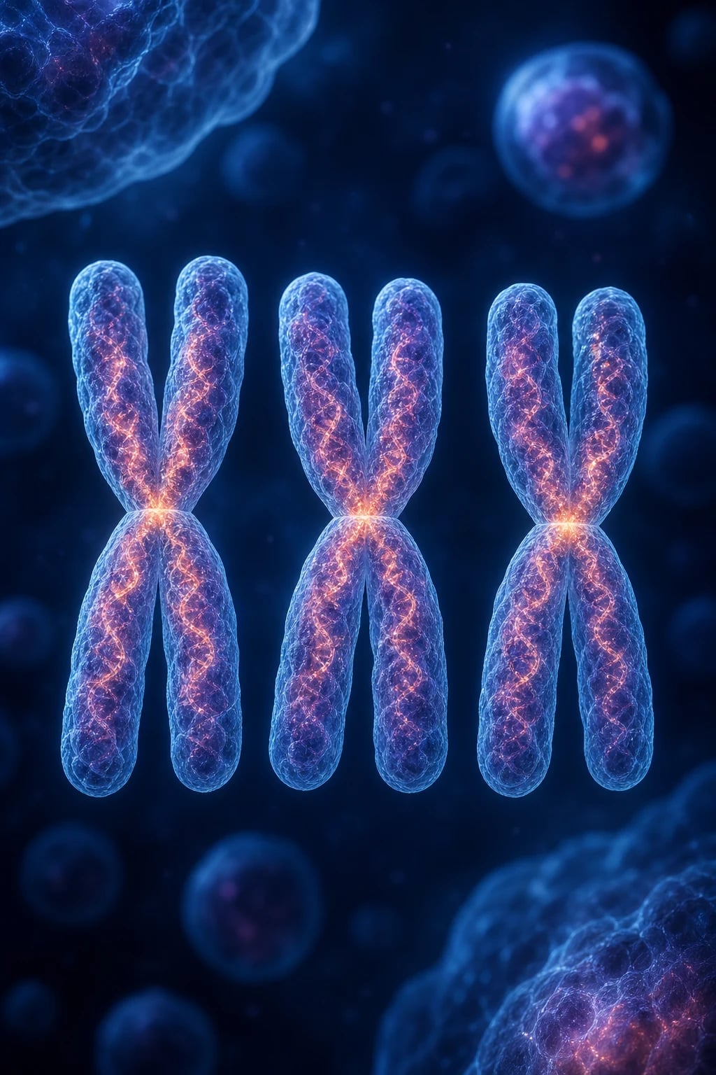
Overview & Definition
Down syndrome is a chromosomal disorder caused by a full or partial extra copy of chromosome 21 (trisomy 21), producing a recognisable constellation of dysmorphic features, intellectual disability, and associated medical conditions. It is the commonest autosomal chromosomal abnormality in liveborn infants and the single commonest genetic cause of intellectual disability worldwide.[1]
The condition is named after the British physician John Langdon Down, who in 1866 described the clinical phenotype and (incorrectly) classified it by ethnicity as "Mongolian idiocy" — the obsolete term mongolism is now considered offensive and is not used. The chromosomal basis — a third copy of chromosome 21 — was discovered by the French paediatrician and geneticist Jérôme Lejeune in 1959, a landmark that established Down syndrome as the first human condition shown to be caused by a chromosomal aberration and launched clinical cytogenetics. [1]
Clinically, Down syndrome matters for four reasons: (1) it is common, so every clinician will encounter it; (2) it carries a predictable, screenable complication profile (cardiac, thyroid, haematological, atlantoaxial) that, if anticipated, transforms prognosis; (3) the cytogenetic subtype changes genetic counselling — a translocation may signal a carrier parent with a high recurrence risk; and (4) it is the template for understanding gene-dosage disorders and the Down syndrome critical region. [1]
Classification
Down syndrome is classified by cytogenetic mechanism, because the mechanism determines recurrence risk and genetic counselling:[1][2]
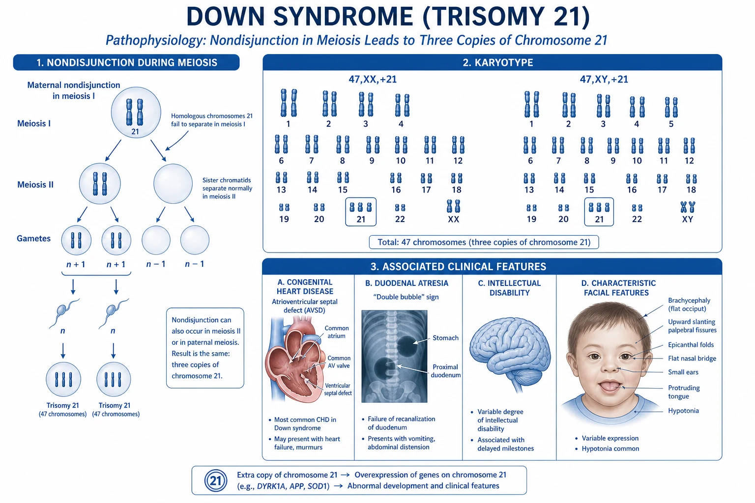
Free trisomy 21 (non-disjunction) — 95%
- Karyotype: 47,XX,+21 or 47,XY,+21 (47 chromosomes, three free copies of 21)
- Cause: failure of chromosome 21 to separate during meiosis — usually maternal meiosis I (~75% maternal meiosis I, 25% meiosis II; only ~5 to 10% paternal origin)
- Sporadic event; not inherited; risk rises with maternal age due to cohesin deterioration in oocytes arrested since fetal life
- Recurrence risk about 1% (slightly higher if the mother is young — implies a predisposition to non-disjunction)
Robertsonian translocation — 3 to 4%
- Karyotype: 46 chromosomes total (one 21 is translocated onto another acrocentric chromosome), but with extra 21q long-arm material
- Most common: t(14;21) — long arm of 21 fused with chromosome 14; also t(21;21) and rarely t(21;13/15/22)
- About 25% are inherited from a balanced-translocation carrier parent; 75% are de novo
- Recurrence risk depends on the carrier: mother with t(14;21) about 10 to 15%; father with t(14;21) under 1%; t(21;21) carrier up to 100% — therefore parental karyotype is mandatory when a translocation is found
Mosaicism — 1 to 2%
- Karyotype: two cell lines — 46 (normal) and 47,+21 (trisomic) — from post-zygotic (mitotic) non-disjunction
- Phenotype is typically milder: fewer dysmorphic features, higher IQ (sometimes borderline-normal), better motor and language milestones — proportional to the fraction and tissue distribution of trisomic cells
- Can be missed on peripheral blood karyotype if the trisomic line is scarce; may require a second tissue (skin fibroblasts) if clinical suspicion is high and blood is normal
Partial trisomy 21 (rare)
- A segment of 21q is duplicated without full trisomy
- Phenotype varies with the duplicated region — typically the Down syndrome critical region around 21q22
- Diagnosed by chromosomal microarray or FISH
Down syndrome — key numbers
Epidemiology & Risk Factors
The overall incidence of Down syndrome is approximately 1 in 700 live births (1.4 per 1000).[4][5] There is no racial, ethnic, or socioeconomic predilection, and males and females are affected equally.
The single dominant risk factor is maternal age, which applies specifically to the non-disjunction form:[4]
| Maternal age | Approximate risk of Down syndrome |
|---|---|
| Under 30 | 1 in 1000 to 1500 |
| 35 | 1 in 350 |
| 40 | 1 in 100 |
| 45 | 1 in 30 |
The age effect reflects cohesin protein deterioration in oocytes, which are arrested in prophase I of meiosis since fetal life. As a woman ages, the cohesin rings that hold homologous chromosomes together degrade, increasing the chance of non-disjunction — overwhelmingly during maternal meiosis I. Paternal age over 50 contributes a smaller additional risk; the paternal origin of the extra chromosome 21 accounts for only about 5 to 10% of non-disjunction cases. [1]
Recurrence risk is the counselling question that determines the work-up of a future pregnancy: [1]
- Free trisomy 21 (non-disjunction): recurrence risk about 1% — higher than the population baseline, implying some parents carry a predisposition to non-disjunction. The younger the mother at the first affected child, the higher the recurrence risk.
- Robertsonian translocation: depends entirely on the carrier. Mother with a balanced t(14;21): about 10 to 15% recurrence. Father with a balanced t(14;21): under 1%. A t(21;21) carrier: recurrence up to 100% (every conceptus is affected or non-viable). This is why both parents must be karyotyped when a child has a translocation Down syndrome.
- Mosaicism: recurrence risk is low (about 1%), unless a parent has gonadal mosaicism. [1]
A paradox of modern epidemiology: although the live-birth prevalence of Down syndrome has fallen in countries with widespread prenatal screening and access to termination, the absolute number of affected conceptuses is rising because of the secular trend toward later childbearing.[4][5]
Pathophysiology
The core defect in Down syndrome is gene-dosage imbalance: the presence of three copies of chromosome 21 leads to overexpression of dosage-sensitive genes. Chromosome 21 is the smallest human autosome (about 48 million base pairs, encoding roughly 225 protein-coding genes), so the disorder is a model of how modest gene-dosage excess can produce a multisystem phenotype. [1]
The Down syndrome critical region (DSCR) is a segment of 21q22 whose triplication reproduces many of the major features (craniofacial, cardiac, and some cognitive). Current evidence favours a multigene dosage model — no single gene accounts for the whole syndrome, but a cluster of dosage-sensitive genes acting in concert produces the phenotype. The key candidate genes and their effects:[1]
- DYRK1A (dual-specificity tyrosine-phosphorylation-regulated kinase 1A) — overexpression impairs neuronal proliferation and synaptic plasticity, contributing to intellectual disability, microcephaly, and Alzheimer-type neurodegeneration.
- APP (amyloid precursor protein) — overexpressed, drives amyloid-beta plaque deposition, explaining the near-universal Alzheimer-type neuropathology by the 40s and clinical dementia in most by the 60s.
- RCAN1 (regulator of calcineurin 1, formerly DSCR1) — affects cardiac and neural development; contributes to the AVSD phenotype.
- SOD1 (superoxide dismutase 1) — overexpression increases oxidative stress, implicated in premature ageing and immune dysfunction.
- CBS (cystathionine beta-synthase) — perturbs folate and homocysteine metabolism.
- ETS2, ERG — transcription factors involved in haematopoiesis and cardiac development.
- JAK2 — a haematopoietic signalling gene relevant to the leukaemic predisposition.
- IFNAR1, IFNAR2, IFNGR2 — interferon receptor genes; their triplication produces interferon hyperresponsiveness, contributing to the autoimmune and leukaemic diathesis. [1]
Cytogenetic mechanisms in detail: [1]
Non-disjunction (95%): during meiosis, the two chromosome 21 homologues fail to separate. The error occurs most often in maternal meiosis I (~75% of cases) — the oocyte retains both chromosome 21s, and upon fertilisation by a normal sperm the zygote carries three copies. The failure is linked to age-dependent degradation of meiotic cohesin (REC8) in oocytes that have been arrested in prophase I since before birth. The result is a free trisomy: 47,XX,+21 or 47,XY,+21. [1]
Robertsonian translocation (3 to 4%): the long arm of an acrocentric chromosome 21 fuses with another acrocentric chromosome (14, 21, 13, 15, or 22) at or near the centromere, with loss of the short arms. The carrier has 46 chromosomes but carries the full chromosome 21 long-arm material plus the translocated piece — phenotypically normal (balanced carrier). When gametogenesis produces an unbalanced gamete carrying both the normal 21 and the translocated 21q, the resulting child has effectively three copies of 21q despite having only 46 chromosomes. The commonest is t(14;21); a t(21;21) carrier produces only unbalanced gametes. [1]
Mosaicism (1 to 2%): non-disjunction occurs after fertilisation during a mitotic division of the early embryo, generating two cell lines — one normal (46) and one trisomic (47,+21). The clinical phenotype depends on what fraction of cells are trisomic and which tissues they populate; high trisomic fractions reproduce the full phenotype, low fractions produce a subtle or even subclinical picture that may escape diagnosis until adulthood. [1]
Haematological mechanism (examiner favourite): trisomy 21 cooperates with acquired GATA1 mutations in fetal haematopoietic cells. GATA1 is on the X chromosome; the mutation produces a short, dominant-negative GATA1s isoform. In trisomy 21 fetal liver haematopoiesis, this drives transient abnormal myelopoiesis (TAM) in about 10% of neonates, and is the obligatory first hit for acute megakaryoblastic leukaemia (AMKL), which is 500 times more common in Down syndrome.[6]
Clinical Presentation
The clinical phenotype is recognised at birth by the experienced examiner, but is confirmed only by karyotype — never by clinical impression alone. [1]
Craniofacial:
- Brachycephaly (short, broad head) and a flat facial profile
- Upslanting palpebral fissures (the namesake "mongoloid" slant — up at the lateral canthi)
- Epicanthal folds (skin folds over the inner canthi)
- Flat nasal bridge
- Protruding / spade-like tongue (relative macroglossia — the oral cavity is small, so a normal-sized tongue protrudes)
- Small, low-set ears with an overfolded helix
- Short neck with excess nuchal skin (a remnant of the increased nuchal translucency seen antenatally) [1]
Eyes:
- Brushfield spots — white-to-grey speckles arranged around the periphery of the iris (present in 50 to 85% of light-eyed infants; a classic buzzword)
- Refractive errors (myopia, hypermetropia), strabismus, nystagmus
- Nasolacrimal duct obstruction (congenital dacryocystocoele, epiphora)
- Congenital cataracts (check the red reflex at birth) [1]
Hands and feet:
- Single transverse palmar crease (simian crease) — in about 45% of children with Down syndrome (but also present in 1 to 2% of the normal population, so it is a marker, not a diagnosis)
- Short broad hands (brachydactyly)
- Clinodactyly of the 5th finger — inward curving due to a hypoplastic middle phalanx
- Wide sandal gap — a gap between the 1st and 2nd toes, with a corresponding plantar crease [1]
Musculoskeletal and growth:
- Neonatal hypotonia ("floppy baby") — one of the earliest clinical clues, present from birth
- Hyperflexible joints and ligamentous laxity
- Short stature — birth length and adult height are below the 3rd centile (plot on Down syndrome-specific growth charts, not standard charts) [1]
Neurodevelopment:
- Global developmental delay — motor milestones delayed (sitting ~11 months, walking ~21 months), speech delayed (expressive worse than receptive)
- Intellectual disability — typically mild to moderate, IQ range 25 to 75, median about 50; mosaic cases often higher
- Behavioural phenotype — affectionate, sociable, good imitation and visual memory; also stubbornness, short attention, and a higher rate of autism spectrum disorder (up to 18%) and ADHD [1]
Cardiac (40 to 60%): most are asymptomatic at birth; a murmur, cyanosis, differential saturations, or heart failure (poor feeding, sweating with feeds, tachypnoea, hepatomegaly) may be the presenting clue. AVSD is the most characteristic lesion; also VSD, ASD, PDA, Tetralogy of Fallot. See Complications. [1]
Gastrointestinal: failure to thrive, feeding difficulty, bilious vomiting (duodenal atresia), failure to pass meconium (Hirschsprung), chronic diarrhoea and failure to thrive (coeliac disease), constipation, gastro-oesophageal reflux. [1]
Endocrine: features of hypothyroidism — lethargy, constipation, dry skin, poor growth, prolonged jaundice. [1]
Atypical presentations (examiner trap):
- Mosaicism may present with subtle features and mild learning difficulty that is only diagnosed in later childhood or adulthood — keep Down syndrome on the differential of unexplained intellectual disability at any age.
- Adults present with early-onset Alzheimer dementia (memory loss, personality change, functional decline in the 40s to 50s).
- Translocation cases are phenotypically indistinguishable from non-disjunction — the distinction is cytogenetic, not clinical.
- A neonate with minimal dysmorphism but a complex cardiac lesion (AVSD) should prompt a karyotype. [1]
Differential Diagnosis
A handful of conditions share hypotonia, dysmorphism, or developmental delay and must be distinguished from Down syndrome — always by karyotype, which is the single decisive test. [1]
Congenital hypothyroidism
- Shares: hypotonia, macroglossia, delayed milestones, constipation, coarse features, prolonged jaundice
- Distinguishes: no dysmorphic hand features (no single palmar crease, no clinodactyly), no upslanting fissures
- Diagnosis: high TSH, low free T4 on newborn blood spot screen; normal karyotype; responds to levothyroxine
Edwards syndrome (trisomy 18)
- Shares: cardiac defects, intellectual disability
- Distinguishes: small-for-gestational-age, clenched fists with overlapping index over 3rd finger, rocker-bottom feet, prominent occiput, low-set ears, short sternum
- Much more lethal (median survival weeks); diagnosis by karyotype 47,+18
Patau syndrome (trisomy 13)
- Shares: cardiac defects, intellectual disability, hypotonia
- Distinguishes: cleft lip/palate, polydactyly (postaxial), holoprosencephaly, microphthalmia, cutis aplasia of scalp
- Very lethal (median survival days); diagnosis by karyotype 47,+13
Noonan syndrome
- Shares: short stature, congenital heart disease, dysmorphism
- Distinguishes: downslanting (not upslanting) palpebral fissures, webbed neck, pectus, pulmonary stenosis (not AVSD), autosomal dominant (PTPN11 etc.), normal karyotype
Fragile X syndrome
- Shares: intellectual disability, hypotonia
- Distinguishes: long face, large ears, macro-orchidism after puberty, autistic features, joint laxity; the commonest INHERITED cause of intellectual disability in boys
- Diagnosis: FMR1 CGG-repeat expansion (DNA test); normal karyotype
Zellweger syndrome (peroxisomal)
- Shares: severe hypotonia, flat face, high forehead
- Distinguishes: hepatomegaly, seizures, calcific stippling of patellae, elevated very-long-chain fatty acids; lethal in infancy
Beckwith-Wiedemann syndrome
- Shares: macroglossia, hypotonia
- Distinguishes: macrosomia (not short stature), omphalocele/exomphalos, visceromegaly, neonatal hypoglycaemia (hyperinsulinism); opposite growth pattern to Down syndrome
The decisive point: a child with suggestive features must always have a karyotype — clinical diagnosis alone is wrong in up to a third of cases and critically misses translocation (with its recurrence-risk implications for the parents).[2]
Clinical & Bedside Assessment
Antenatal screening (the first opportunity to detect):[1][4]
- Combined first-trimester screen (11 to 14 weeks): raised nuchal translucency (NT), raised beta-hCG, and low PAPP-A — detects about 80 to 85% of cases.
- Quad screen (15 to 22 weeks): low alpha-fetoprotein, low unconjugated oestriol, raised inhibin-A, raised beta-hCG.
- Cell-free DNA / non-invasive prenatal testing (NIPT): analyses fetal DNA fragments in maternal plasma from 10 weeks; sensitivity over 99% and false-positive about 0.1% for trisomy 21 — the most accurate screen but still a screen (not diagnostic), and is confirmed by invasive testing.
- Definitive prenatal diagnosis: chorionic villus sampling (CVS, 10 to 13 weeks) or amniocentesis (15 to 20 weeks) with karyotype or QF-PCR.
Postnatal clinical assessment:
- Full dysmorphology examination: craniofacial, eyes (red reflex, Brushfield spots), hands, feet, and skin.
- Growth plotted on Down syndrome-specific growth charts.
- Cardiac: auscultation, four-limb blood pressures, pre- and post-ductal saturations.
- Abdomen: distension, absence of meconium passage, scaphoid abdomen (duodenal atresia may distend the upper abdomen).
- Neurology: tone (neonatal hypotonia), Moro reflex, hip examination (developmental dysplasia of the hip is commoner). [1]
Named bedside signs / manoeuvres:
- Nuchal translucency ultrasound (antenatal) — the single best first-trimester marker.
- Red reflex (ophthalmoscope) — to detect congenital cataract.
- Allis / Galeazzi sign and hip Ortolani-Barlow — to screen for DDH.
- Rectal examination / digital rectal examination — to exclude imperforate anus; contrast study if Hirschsprung suspected. [1]
Investigations
The investigative strategy has two layers: confirm the diagnosis (karyotype) and screen for the predictable complications (the anticipatory care bundle).[1]
1. Confirm the diagnosis — KARYOTYPE (definitive):
- Conventional karyotype is the gold standard because it distinguishes non-disjunction (47,+21) from translocation (46 with extra 21q) and mosaicism. This distinction is mandatory for genetic counselling.
- Chromosomal microarray (CMA) is more sensitive for copy-number changes across the genome but may miss a balanced Robertsonian translocation — so when the working diagnosis is Down syndrome, request a karyotype (not just microarray).[1]
- Fluorescence in situ hybridisation (FISH) for chromosome 21 gives a rapid result (within 24 to 48 hours) but does not define the mechanism — karyotype must follow.
- Quantitative fluorescent PCR (QF-PCR) — rapid, used antenatally.
2. Screen for the predictable complications (anticipatory bundle): [1]
| Investigation | Timing | Rationale |
|---|---|---|
| Echocardiogram | Before discharge, or by 2 weeks of age, regardless of symptoms | 40 to 60% have CHD; AVSD may be silent early; pulmonary hypertension develops if untreated |
| Thyroid function (TSH, free T4) | Newborn blood spot; repeat at 6 and 12 months, then annually | 15% have congenital or acquired hypothyroidism |
| Full blood count | At birth; monitor periodically | Detect transient abnormal myelopoiesis (TAM) and leukaemia |
| Audiology | Newborn hearing screen; repeat at 6 months and annually | Conductive loss from otitis media with effusion is common |
| Vision / ophthalmology | Red reflex at birth; ophthalmology by 6 months; then annually | Cataracts, refractive errors, strabismus |
| Polysomnography (sleep study) | By 4 years, and whenever snoring or sleep-disordered breathing is reported | Obstructive sleep apnoea in ~60% |
| Coeliac serology (tTG-IgA + total IgA) | Annually from 2 to 3 years, and if symptomatic | Higher coeliac risk |
| Cervical spine imaging | Only if symptomatic (neck pain, torticollis, gait change, neurological signs) — measure atlanto-dens interval (over 5 mm abnormal) | Routine screening radiographs are NOT recommended by AAP 2022 (a major change from older guidance)[1] |
| Growth | Every visit, on DS-specific charts | Short stature, failure to thrive |
3. Targeted investigations for the presenting problem:
- Bilious vomiting + double bubble on AXR → duodenal atresia; surgical referral.
- Failure to pass meconium → contrast enema and rectal biopsy for Hirschsprung.
- Cytopenia / blast cells on film → haematology referral; bone marrow if leukaemia suspected. [1]
Management — Resuscitation
Most Down syndrome neonates are stable at birth, but a small subset present with surgical or cardiac emergencies that need immediate action: [1]
- Bilious vomiting (suspected duodenal atresia): pass a large-bore nasogastric tube (decompression), establish intravenous access, give maintenance IV fluids (correct dehydration and electrolytes), keep nil by mouth, and make an urgent surgical referral. Bilious vomiting in a neonate is a surgical emergency until proven otherwise.
- Cyanosis / differential saturations / shock (duct-dependent cardiac lesion): start prostaglandin E1 (alprostadil) 0.01 to 0.05 mcg/kg/min IV to keep the ductus arteriosus open, oxygen as needed, and transfer to a cardiac centre. Do not give 100% oxygen blindly in a duct-dependent lesion — it can close the duct.
- Airway difficulty: the combination of macroglossia and hypotonia can obstruct the airway and complicate bag-mask ventilation; use an oropharyngeal airway, nurse in a neutral/sniffing position, and have a low threshold for senior airway support.
- Failure to pass meconium with abdominal distension (Hirschsprung): rectal washout/examination, rehydrate, and refer to paediatric surgery for biopsy; beware enterocolitis (a life-threatening complication).
- Suspected sepsis / leukaemia in a neonate with TAM: cultures, antibiotics, and supportive care per local protocols. [1]
Management — Definitive & Stepwise
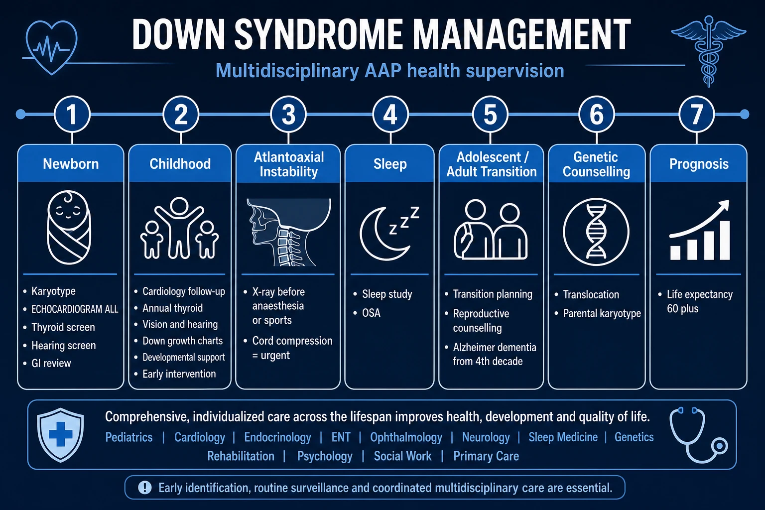
There is no cure for Down syndrome. Definitive management is lifelong, multidisciplinary, and anticipatory, anchored to the AAP 2022 Health Supervision schedule.[1] The goal is to prevent, detect early, and treat the predictable complications, and to maximise development and quality of life.
1. Early intervention (birth to 3 years) — the highest-yield investment:
- Physiotherapy for hypotonia and motor delay.
- Speech and language therapy — expressive language is the most delayed domain; sign language / augmentative communication bridges the gap.
- Occupational therapy for fine motor and Activities of Daily Living skills.
- Developmental stimulation and structured play.
- Evidence: early intervention measurably improves functional outcomes, IQ, and social participation. [1]
2. Cardiac management:
- Prophylaxis against infective endocarditis only for high-risk cardiac lesions (unrepaired cyanotic CHD, prosthetic material, previous IE) — per current guidelines, routine antibiotic prophylaxis is not needed for most repaired lesions.
- Surgical repair: AVSD typically at 3 to 6 months (before irreversible pulmonary vascular disease); VSD, ASD, PDA, and Tetralogy repaired per cardiology timing.
- Pulmonary hypertension surveillance — the reason early cardiac surgery is essential. [1]
3. Endocrine management:
- Levothyroxine for hypothyroidism — paediatric weight-based dosing: 10 to 15 mcg/kg/day in infants, 5 to 6 mcg/kg/day in children, titrate to TSH within reference range and free T4 normal; lifelong, with annual monitoring.
- Type 1 diabetes managed per standard paediatric protocols. [1]
4. Gastrointestinal management:
- Duodenal atresia: duodenoduodenostomy or duodenojejunostomy.
- Hirschsprung disease: pull-through procedure after stabilisation.
- Gastro-oesophageal reflux: head-up positioning, feed thickeners, proton-pump inhibitor (e.g. omeprazole 0.7 to 1.4 mg/kg/day); Nissen fundoplication if refractory.
- Coeliac disease: strict lifelong gluten-free diet; dietetic support. [1]
5. ENT / audiology / vision:
- Otitis media with effusion ("glue ear"): grommets (ventilation tubes) for persistent effusion with hearing loss.
- Hearing aids for sensorineural or persistent conductive loss.
- Surgical correction of strabismus and cataracts. [1]
6. Haematology-oncology management:
- Transient abnormal myelopoiesis (TAM): most cases resolve spontaneously by 3 months; low-dose cytarabine is reserved for life-threatening features (hepatomegaly with organ dysfunction, hydrops, bleeding). Monitor FBC.
- Leukaemia (ALL or AMKL): treat with standard paediatric chemotherapy protocols, modified for the increased chemo-sensitivity and toxicity in Down syndrome — DS-ALL and DS-AMKL patients receive reduced-intensity regimens and have better leukaemia-free survival for AMKL but higher treatment-related mortality, hence the dose adjustments.[6]
7. Atlantoaxial instability precautions:
- Counsel to avoid high-risk activities: trampolining, diving, contact/collision sports, gymnastics (tumbling), and cervical manipulation.
- Refer for surgical (C1-C2) fusion if symptomatic (neurological signs) or if the atlanto-dens interval exceeds 5 mm.
- Note again: routine screening cervical radiographs are no longer recommended (AAP 2022).[1]
8. Sleep-disordered breathing:
- Adenotonsillectomy for obstructive sleep apnoea (OSA in ~60%).
- CPAP if OSA persists post-operatively. [1]
9. Behavioural and psychiatric:
- Screen for and treat autism, ADHD, anxiety, depression (especially in adolescents and adults).
- Behavioural and psychological support; educational input. [1]
10. Education and social participation:
- Individualised Education Plan (IEP); mainstream schooling with support where possible.
- Supported employment and independent/semi-independent living in adulthood. [1]
Lifelong anticipatory surveillance schedule (AAP 2022, summarised):[1]
| Age | Key actions |
|---|---|
| Newborn | Karyotype; echo by 2 weeks; thyroid; FBC; newborn hearing and vision screen |
| Infancy (0 to 1 yr) | Early intervention referral; thyroid at 6 and 12 months; ophthalmology by 6 months; audiology at 6 months |
| Early childhood (1 to 5 yr) | Annual thyroid, vision, audiology; sleep study by 4 yr; coeliac serology from 2 to 3 yr; atlantoaxial counselling |
| School age (5 to 13 yr) | Annual thyroid, vision, audiology, FBC, coeliac screen; monitor development and behaviour |
| Adolescence (13 to 21 yr) | Transition to adult care; reproductive counselling; mental health; sleep; atlantoaxial counselling |
| Adulthood | Annual thyroid, FBC, audiology, vision; dementia surveillance from 40s; lifelong cardiac and BMI monitoring |
Specific Subtypes & Scenarios
Free trisomy 21 (non-disjunction, 95%): the textbook phenotype; sporadic; recurrence about 1%; no further parental testing needed beyond counselling. [1]
Robertsonian translocation (3 to 4%): phenotypically identical to non-disjunction, so the distinction is purely cytogenetic. The clinical action triggered by finding a translocation is to karyotype both parents and counsel on recurrence (mother t(14;21) ~10 to 15%; father t(14;21) under 1%; t(21;21) carrier up to 100%). Offer preimplantation genetic diagnosis (PGD) and prenatal diagnosis (CVS/amniocentesis) in future pregnancies. [1]
Mosaicism (1 to 2%): typically a milder phenotype — fewer dysmorphic features, higher IQ, better milestones — proportional to the fraction and tissue distribution of trisomic cells. Mosaicism can be missed on peripheral blood (if the trisomic clone is small or tissue-limited); if clinical suspicion is high and the blood karyotype is normal, consider skin fibroblast karyotype. Recurrence risk is low. [1]
Twinning (examiner pearl): in monozygotic twins, if one is affected, both are (the non-disjunction event occurred before twinning). In dizygotic twins, only one twin is typically affected — a classic demonstration that Down syndrome is genetic, not environmental. [1]
Neonate with Down syndrome + duodenal atresia: recognise the double bubble on AXR; this is the classic syndromic association (about 30% of duodenal atresia cases have Down syndrome). Resuscitate and refer for surgery. [1]
Adult with Down syndrome and cognitive decline: suspect Alzheimer disease — amyloid deposition begins in the 30s to 40s (APP gene triplication); clinical dementia appears in 50 to 70% by age 60. Use baseline cognitive testing and a collateral history; exclude reversible causes (hypothyroidism, depression, OSA, sensory deprivation). [1]
Pregnancy in a woman with Down syndrome: female fertility is usually preserved; there is a 30 to 50% chance the child will have Down syndrome, plus higher risks of miscarriage and prematurity; high-risk obstetric care with genetic counselling is essential. Males with Down syndrome are almost always infertile. [1]
Complications & Pitfalls
Cardiac (40 to 60%): AVSD (the most specific and most strongly associated), VSD, ASD, PDA, Tetralogy of Fallot. Untreated defects lead to pulmonary hypertension (Eisenmenger) and heart failure; the rationale for early echocardiography and early surgical repair is to prevent irreversible pulmonary vascular disease. Infective endocarditis prophylaxis applies to high-risk lesions only. [1]
Gastrointestinal: duodenal atresia (double bubble), Hirschsprung disease, imperforate anus, tracheo-oesophageal fistula, coeliac disease, gastro-oesophageal reflux, chronic constipation. The combination of Down syndrome + a GI atresia is a classic exam stem. [1]
Endocrine: hypothyroidism (~15%, congenital and acquired), type 1 diabetes, short stature, and infertility (males nearly always; females usually fertile). [1]
Haematology-oncology: transient abnormal myelopoiesis (TAM) in ~10% of neonates; leukaemia with a 10 to 20-fold increased risk — acute lymphoblastic leukaemia (ALL) (the commonest) and acute megakaryoblastic leukaemia (AMKL, FAB M7) which is 500 times more common in Down syndrome; immune dysfunction with increased susceptibility to infection.[6]
Neurological: atlantoaxial instability (C1-C2 subluxation from ligamentous laxity), seizures (commoner than in the general population), and early-onset Alzheimer disease (amyloid plaques universal by 40s; dementia in 50 to 70% by 60s). [1]
Respiratory: obstructive sleep apnoea (~60%, from macroglossia, adenotonsillar hypertrophy, and hypotonia); recurrent upper respiratory tract infections and otitis media. [1]
Sensory: conductive and sensorineural hearing loss, refractive error, strabismus, cataracts. [1]
Psychiatric: autism spectrum disorder (up to 18%), ADHD, depression, anxiety, obsessive-compulsive features. [1]
Pitfalls (examiner favourites):
- Assuming a clinical diagnosis is enough — it misses translocation (and recurrence risk) in up to a third of cases; always confirm with karyotype.
- Forgetting the annual thyroid and FBC surveillance — hypothyroidism and leukaemia are silent until late.
- Missing leukaemia in a child with unexplained cytopenia, hepatosplenomegaly, or petechiae — the threshold for a FBC and film is low.
- Missing coeliac disease in a child failing to thrive or with chronic diarrhoea.
- Requesting routine cervical spine X-rays in asymptomatic children — no longer recommended (AAP 2022); investigate only if symptomatic.
- Failing to counsel about atlantoaxial precautions before sports participation.
- Not plotting growth on Down syndrome-specific charts — standard charts falsely label children as failing to thrive.
- Overlooking reproductive counselling in adolescent and adult females. [1]
Prognosis & Disposition
Life expectancy has risen dramatically — from under 10 years in the 1960s to 50 to 60 years today — driven by early cardiac surgery, antibiotics for infection, thyroid replacement, and structured anticipatory care.[1][3]
Intellectual outcome is variable: IQ typically 25 to 75 (mild to moderate intellectual disability), with mosaic cases often higher. Most children achieve functional literacy and many progress to supported employment and semi-independent living as adults. [1]
Fertility: females are usually fertile (30 to 50% chance of an affected child); males are almost always infertile. [1]
Neurodegeneration: Alzheimer-type pathology is near-universal neuropathologically by 40 to 50 years, with clinical dementia in 50 to 70% by age 60 — driven by APP gene triplication and amyloid-beta overproduction. [1]
Disposition: the modern approach is home with community multidisciplinary support, not institutional care. Discharge planning connects the family to early intervention, a named paediatrician, cardiology, audiology, ophthalmology, and a Down syndrome support organisation. [1]
Special Populations
Advanced maternal age (over 35): offer combined first-trimester screening and NIPT; offer invasive diagnostic testing (CVS/amniocentesis) with full non-directive counselling, including the option of termination. Screening should be offered to all pregnant women regardless of age (universal screening is now standard; age alone does not determine who is offered testing).[1]
Known translocation carrier or previous affected child: parental karyotype and genetic counselling; offer PGD and prenatal diagnosis in every future pregnancy. Recurrence risk depends on the carrier and the translocation (see Classification). [1]
Adolescents and adults: reproductive counselling (contraception for females; discussion of fertility and recurrence), transition to adult services, mental health screening (depression, anxiety), and dementia surveillance from the 40s. [1]
Anaesthesia and surgery (examiner trap): Down syndrome patients have specific anaesthetic risks: atlantoaxial instability (care with intubation/extubation; consider cervical immobilisation and awake fibreoptic intubation if symptomatic), macroglossia and a small airway (difficult intubation), bradycardia and arrhythmia risk, increased sensitivity to anaesthetic agents (reduced MAC requirement; beware of over-sedation and prolonged recovery), and subacute bacterial endocarditis prophylaxis for high-risk cardiac lesions. A thorough pre-anaesthetic assessment of the cervical spine and cardiac status is essential. [1]
Pregnancy in a woman with Down syndrome: high-risk obstetric management; 30 to 50% chance of an affected child; increased risks of miscarriage, prematurity, and cardiac decompensation. [1]
Immunocompromise: relative immune dysfunction (interferon hyperresponsiveness, mild immunodeficiency) increases susceptibility to respiratory and skin infections and underlies part of the leukaemic predisposition. [1]
Evidence, Guidelines & Regional Differences
Global benchmark — AAP 2022 (Bull MJ, et al., Pediatrics): the Health Supervision for Children and Adolescents With Down Syndrome guideline is the international reference anticipatory-care schedule, superseding the 2011 version.[1][2] The headline change in 2022 was the withdrawal of routine cervical spine screening radiographs — replaced by symptom-based assessment and activity counselling — because asymptomatic atlanto-dens interval widening does not reliably predict neurological injury.
Antenatal screening: combined first-trimester screening and NIPT have transformed detection (sensitivity over 99% for NIPT), with a fall in live-birth prevalence in screened populations despite rising maternal age.[4][5]
Leukaemia treatment: modified, reduced-intensity protocols for myeloid leukaemia of Down syndrome (ML-DS) exploit the increased chemosensitivity of DS blasts; outcomes for AMKL are now better in Down syndrome than in non-Down syndrome AMKL, a rare example of the syndrome conferring a treatment advantage.[6]
UK: NICE and NHS-aligned antenatal screening pathways; NIPT (as the "combined test" or "cell-free DNA test") is offered within the NHS fetal anomaly screening programme. Termination of affected pregnancies is legally available within the gestational limits, which is a recognised area of ethical and political debate. [1]
India: combined first-trimester screening and NIPT are available in urban centres but access and uptake vary widely; termination is legal under the Medical Termination of Pregnancy (MTP) Act within gestational limits, with specific provisions for fetal abnormality. Anticipatory care follows AAP principles but is limited by access to specialist multidisciplinary services in rural areas. [1]
Controversies: (1) the ethics of termination of Down syndrome pregnancies (a live debate in disability rights and law); (2) inclusion in mainstream versus special education; (3) growth hormone therapy for short stature (evidence weak; not routinely recommended); (4) the use of "Down syndrome-specific" vitamin/antioxidant supplements (no evidence of benefit).
Exam Pearls
[1]Down syndrome associations — HEART FLAPS
HEART FLAPS
15% — congenital and acquired; screen at birth, 6 and 12 months, then annually
Type 1 diabetes; short stature; infertility in males
C1-C2; avoid contact sports; image and fuse if symptomatic
Obstructive sleep apnoea (~60%); recurrent otitis media
Annual TSH/fT4 surveillance (reinforces H)
Upslanting fissures, epicanthal folds, flat face, protruding tongue, Brushfield spots
ALL and AMKL — 10 to 20x risk; AMKL is 500x; TAM in 10% of neonates
Most characteristic cardiac lesion — echo by 2 weeks
Single transverse (simian) crease in ~45%; clinodactyly of 5th finger; sandal gap
Duodenal atresia (double bubble), Hirschsprung, imperforate anus, TEF, coeliac
- Trisomy 21 — the commonest chromosomal disorder and the commonest genetic cause of intellectual disability.
- Three cytogenetic types and their buzzwords: non-disjunction (95%, 47,+21, sporadic, maternal age), Robertsonian translocation (4%, t(14;21), karyotype the parents for recurrence), mosaicism (1%, milder phenotype).
- Classic phenotype buzzwords: upslanting palpebral fissures, epicanthal folds, flat facial profile, protruding tongue, single transverse palmar crease, clinodactyly, sandal gap, Brushfield spots.
- AVSD is the cardiac lesion most associated with Down syndrome (and most AVSD cases have Down syndrome).
- Duodenal atresia = double bubble on AXR (stomach bubble + dilated proximal duodenum, no distal gas).
- Leukaemia: ALL and AMKL (FAB M7, megakaryoblastic) — the 500x association; transient abnormal myelopoiesis in 10% of neonates.
- Atlantoaxial instability — counsel against contact sports; investigate if symptomatic; routine X-rays no longer recommended (AAP 2022).
- Lejeune (1959) discovered trisomy 21; Langdon Down (1866) first described the clinical syndrome.
- APP gene on chromosome 21 explains early Alzheimer-type dementia (amyloid by 40s).
- Mosaicism = milder phenotype; translocation = check parents; non-disjunction = maternal age. [1]
Exam application bank (NEET-PG / INICET)
One-line answer
Down syndrome is the commonest autosomal chromosomal abnormality in liveborn infants, caused by a full or partial extra copy of chromosome 21 (trisomy 21). It produces a characteristic facial phenotype, intellectual disability, and a cluster of associated conditions — congenital heart disease (AVSD), duodenal atresia, hypothyroidism, leukaemia, and atlantoaxial instability. Three cytogenetic forms: non-disjunction (95%), Robertsonian translocation (4%), and mosaicism (1%). Maternal age is the dominant risk factor. Diagnose with karyotype; screen antenatally with combined first-trimester screen and cell-free DNA (NIPT). Management is lifelong, multidisciplinary, anticipatory (AAP 2022 schedule) — early intervention, cardiac surveillance, thyroid and FBC monitoring, atlantoaxial precautions. Life expectancy now 50 to 60 years.
Worked stems (answer without another resource)
Stem 1 — Classic presentation. Map symptoms to mechanism; name the first investigation and first treatment step with dose/route if drug therapy is standard. [1]
Stem 2 — Unstable / complicated. List red flags that force immediate resuscitation, theatre, ICU, antidote, or reperfusion — and what you do in the first 15 minutes. [1]
Stem 3 — Atypical group. Elderly, pregnancy, child, or immunocompromised: how presentation and thresholds change. [1]
Stem 4 — Differential trap. Name the three closest mimics and one discriminator for each. [1]
Stem 5 — Disposition. Who goes home with safety-netting, who is admitted, who needs HDU/ICU/theatre, and what follow-up is mandatory. [1]
Rapid viva checklist
- Definition + classification
- Pathophysiology chain
- Bedside signs / criteria
- Score with exact components (if any)
- Emergency bundle
- Definitive therapy with doses
- Complications of disease and of treatment
- Special populations
- Guideline/trial name if classic
- Three exam traps
Coverage self-check
If you cannot answer any stem above from this page alone, re-read the matching section — the page is intended to be self-sufficient for final-prof and NEET-PG/INICET questions on Down Syndrome.
[1] [1]References
- [1]Bull MJ, Trotter T, Santoro SL, et al. Health Supervision for Children and Adolescents With Down Syndrome Pediatrics, 2022.PMID 35490285
- [2]Bull MJ, Committee on Genetics. Health supervision for children with Down syndrome Pediatrics, 2011.PMID 21788214
- [3]Williams K, Minkovitz CS, Grason H, et al. Disparities in Health Supervision for Children With Down Syndrome Clin Pediatr (Phila), 2017.PMID 28135877
- [4]Wu J, Morris JK. Trends in maternal age distribution and the live birth prevalence of Down's syndrome in England and Wales: 1938-2010 Eur J Hum Genet, 2013.PMID 23361224
- [5]Loane M, Morris JK, Addor MC, et al. Twenty-year trends in the prevalence of Down syndrome and other trisomies in Europe: impact of maternal age and prenatal screening Eur J Hum Genet, 2013.PMID 22713804
- [6]Sappok T, Krieg S, Rosenbusch ML, et al. Cancer Frequencies and Screening in Individuals With Down Syndrome: A Comprehensive Nationwide Cross-Sectional Analysis JCO Glob Oncol, 2026.PMID 42341246