Paediatrics · Paediatrics
Henoch-Schonlein Purpura (IgA Vasculitis)
Also known as Henoch-Schonlein purpura · HSP · IgA vasculitis · IgAV · Anaphylactoid purpura · Purpura rheumatica
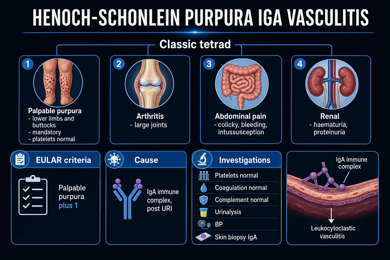
Henoch-Schonlein purpura (HSP), now officially renamed IgA vasculitis (IgAV) by the Chapel Hill Consensus Conference, is an immune-complex-mediated small-vessel (leukocytoclastic) vasculitis characterised by dominant IgA1 immune-complex deposition in the skin, gastrointestinal tract, joints and glomeruli. It is the most common vasculitis of childhood. Classically presents after a recent upper respiratory infection with the tetrad of palpable purpura (lower limbs/buttocks, mandatory), arthritis/arthralgia (knees, ankles), colicky abdominal pain, and renal involvement (haematuria/proteinuria). Platelets are NORMAL (key distinction from ITP). Diagnosis is clinical using EULAR/PRINTO/PRES 2010 or 1990 ACR criteria. Management is supportive in most cases (rest, hydration, analgesia, NSAIDs for arthritis); corticosteroids for severe gastrointestinal or renal involvement. Crucially, steroids do NOT prevent nephritis (Jauhola, 2011). Long-term outcome is excellent in children but renal involvement must be monitored for 6 to 12 months as nephritis can develop late, after the rash has resolved; ~1 to 2% progress to end-stage renal disease.
On this page & tools
Your progress
Saved locally on this device.
Exam tags
Red flags
Overview & Definition
Henoch-Schonlein purpura (HSP) — renamed IgA vasculitis (IgAV) at the 2012 Chapel Hill Consensus Conference on the Nomenclature of Systemic Vasculitides — is an immune-complex-mediated small-vessel vasculitis characterised by dominant IgA1 deposition within the walls of small blood vessels (capillaries, venules and arterioles) of the skin, gastrointestinal tract, joints and kidneys.[1][2]
It is the most common vasculitis of childhood, accounting for approximately half of all childhood vasculitides. It is overwhelmingly a clinical diagnosis: the combination of palpable purpura in dependent areas with arthritis, abdominal pain or renal involvement in a child (often after a viral upper respiratory infection) is so characteristic that laboratory confirmation is frequently unnecessary. The challenge for the clinician is not making the diagnosis but recognising and managing its complications — most importantly intussusception (which in HSP is atypically ileo-ileal, not ileo-colic) and HSP nephritis, which can appear weeks to months after the rash and is the single most important determinant of long-term prognosis.[7]
The single highest-yield distinction in a child with purpura is the platelet count: in HSP the platelets are normal (the rash is a vasculitic purpura), whereas in immune thrombocytopenia (ITP) the platelets are low. Confusing the two — or worse, missing meningococcaemia by attributing a rapidly progressive purpuric rash to HSP — is a classic and serious error.[1]
Classification
HSP is classified primarily by the organ systems involved and, when nephritis is present, by renal histology.[7]
By clinical phenotype (organ dominance): [1]
- Cutaneous-dominant (simple HSP) — palpable purpura ± mild arthritis; no significant GI or renal disease; excellent prognosis; self-limiting.
- GI-dominant — prominent abdominal pain, vomiting, GI bleeding; risk of intussusception and protein-losing enteropathy.
- Renal-dominant (HSP nephritis) — haematuria/proteinuria, nephritic or nephrotic syndrome, hypertension; the major determinant of long-term outcome.
- Joint-dominant — arthritis/arthralgia is the prominent feature; usually fleeting and non-erosive. [1]
By age of onset: [1]
- Childhood HSP — the prototype (see Epidemiology); benign course in most.
- Adult HSP — rarer (about one-quarter the incidence) but more severe, with a higher burden of renal and gastrointestinal disease and a worse prognosis.[6]
HSP nephritis — International Study of Kidney Disease in Children (ISKDC) histological classification (used to grade renal biopsy findings and guide immunosuppression):[7]
- Grade I — minimal glomerular changes.
- Grade II — mesangial proliferation only.
- Grade III — focal/mesangial proliferation with fewer than 50% crescents.
- Grade IV — mesangial proliferation with 50 to 75% crescents.
- Grade V — mesangial proliferation with more than 75% crescents.
- Grade VI — membranoproliferative-like changes. [1]
Higher ISKDC grades (especially IV to VI, i.e. crescents in more than half of glomeruli) carry the worst renal prognosis and trigger aggressive immunosuppression. [1]
Epidemiology & Risk Factors
HSP is the most common vasculitis of childhood, with an annual incidence of approximately 10 to 20 cases per 100,000 children — roughly three to four times the adult incidence.[1][2]
Age and sex: [1]
- Peak age 4 to 6 years; most cases occur between 3 and 15 years. It is rare under 2 years and uncommon over 20 (the ACR criteria use "age under 20 at onset" as a discriminating feature).
- Boys are affected more than girls, ratio approximately 1.2 to 2:1.
- Seasonal clustering in autumn, winter and spring — mirroring preceding viral respiratory infections. [1]
Triggers (an identifiable antecedent is found in approximately half of cases, 1 to 3 weeks before onset):[1]
- Upper respiratory tract infections (most common — ~50% of cases), particularly with Group A Streptococcus pyogenes (elevated ASO titres are often found).
- Other infections — mycoplasma, parvovirus B19, Epstein-Barr virus, hepatitis B and C, HIV.
- Drugs — beta-lactam antibiotics (penicillins, cephalosporins), NSAIDs, ACE inhibitors, quinolones.
- Vaccinations — measles-mumps-rubella (MMR), influenza, hepatitis B, COVID-19 (rare, controversial association).
- Insect bites, foods, environmental/temperature exposure. [1]
Host and ethnic factors: incidence and severity vary by ethnicity — higher rates reported in children of Asian descent; family history of IgA nephropathy or autoimmune disease may be relevant. Adults developing HSP are more likely to have severe and persistent disease.[6]
Pathophysiology
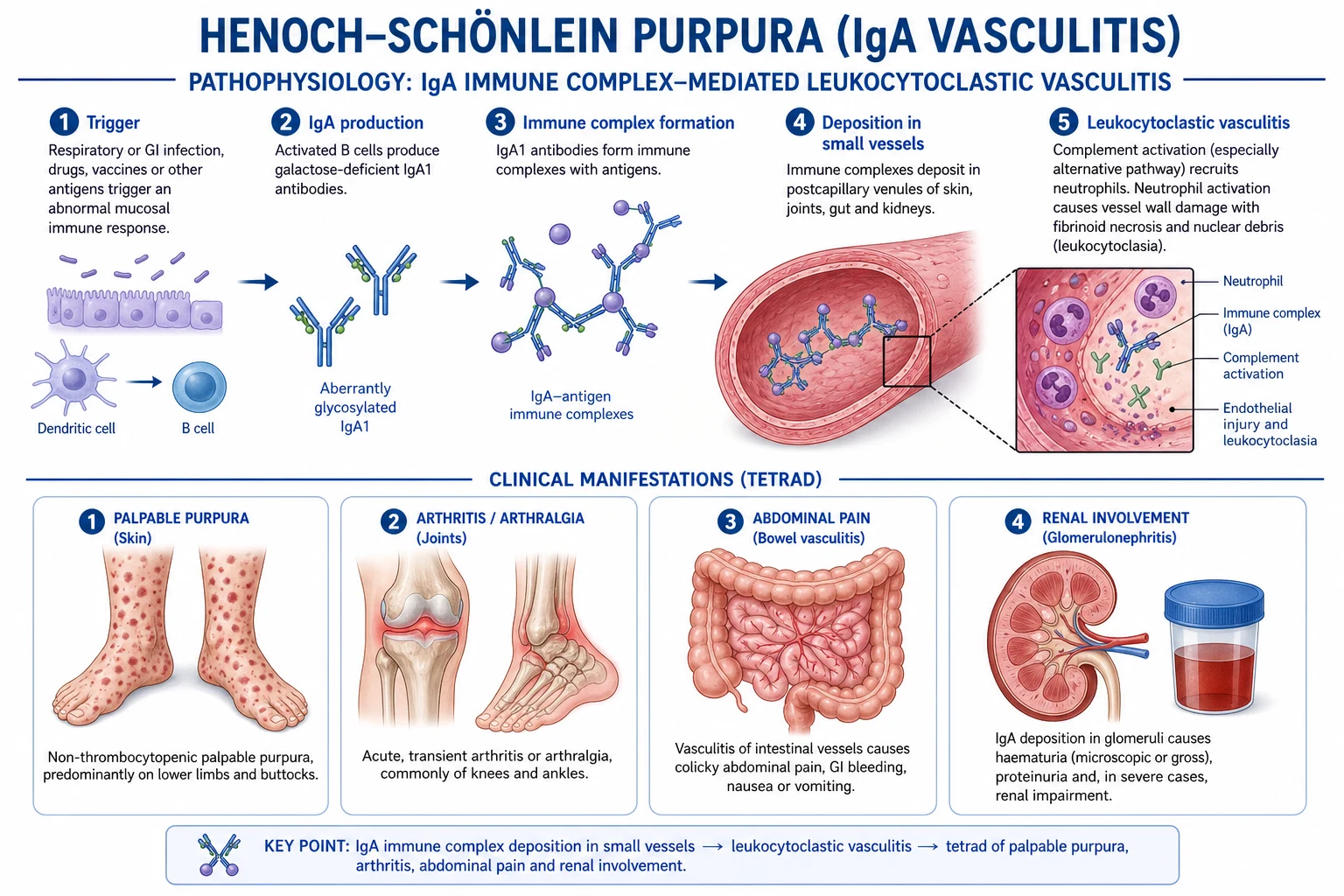
HSP is a small-vessel leukocytoclastic vasculitis driven by galactose-deficient IgA1 (Gd-IgA1) immune complexes. The mechanism is now understood to be identical to that of IgA nephropathy (Berger disease) — HSP and IgA nephropathy are considered two ends of the same disease spectrum, with HSP representing the systemic form (skin, gut, joints, kidney) and IgA nephropathy the renal-limited form.[7]
The molecular cascade (step by step): [1]
- Aberrant O-glycosylation of IgA1. Serum IgA1 molecules normally carry O-linked sugars (galactose and sialic acid) on their hinge-region serine/threonine residues. In genetically susceptible individuals, these hinge-region glycans are galactose-deficient (Gd-IgA1).
- Autoantibody formation. The abnormal Gd-IgA1 is recognised as foreign, stimulating production of IgG (and IgA) anti-glycan autoantibodies directed against the exposed N-acetylgalactosamine residues.
- Circulating immune-complex formation. The Gd-IgA1 autoantibodies bind their antigen to form large circulating immune complexes.
- Deposition. These immune complexes are deposited in the mesangium of the glomerulus and in the walls of small vessels (dermal capillaries and venules, gut submucosa, synovium, testis) — the organs affected clinically.
- Complement activation. Deposited IgA activates the alternative and lectin complement pathways (IgA does NOT activate the classical pathway). This generates C3a, C5a and the membrane-attack complex (C5b-9).
- Neutrophil chemotaxis and inflammation. Complement-derived chemotaxins recruit neutrophils, which degranulate and release proteases and reactive oxygen species, producing fibrinoid necrosis of the vessel wall and nuclear debris ("nuclear dust") — the histological hallmark of leukocytoclastic vasculitis.
- Clinical consequences — vessel-wall inflammation and rupture cause the characteristic palpable purpura (extravasation from dermal venules), GI oedema and bleeding, periarticular swelling, and a proliferative/crescentic glomerulonephritis.[2][7]
Vessel size: HSP predominantly affects small vessels (capillaries, venules, arterioles), which places it — alongside IgA nephropathy, cutaneous leukocytoclastic angiitis and cryoglobulinaemic vasculitis — in the immune-complex small-vessel vasculitis group of the Chapel Hill nomenclature.[1]
Why HSP and IgA nephropathy are one disease: identical Gd-IgA1 biology; identical mesangial IgA deposition on renal biopsy. The difference is purely the distribution of deposition (systemic in HSP, renal-only in Berger disease) and the presence of extrarenal features.[7]
Clinical Presentation
HSP typically presents acutely in a previously well child, often 1 to 3 weeks after an upper respiratory tract infection. The features of the classic tetrad may appear in any order, though palpable purpura is usually first; when GI symptoms or arthritis precede the rash by days, the diagnosis can be challenging.[1]
1. Skin — palpable purpura (the mandatory, defining feature). [1]
- Palpable, non-blanching purpura and petechiae, symmetrical, with a predominantly dependent distribution — lower limbs, buttocks, and extensor surfaces (it is unusual on the trunk or face, though in infants the face and ears may be involved).
- The lesion is palpable (you can feel it) because it is an inflammatory vasculitic infiltrate, not a flat bleed. This is a key bedside discriminator from ITP, where purpura is typically non-palpable.
- Lesions evolve in crops, may become bullous, vesicular, or necrotic in severe cases, and can leave residual hyperpigmentation. [1]
2. Joints — arthritis / arthralgia (~75%). [1]
- Periarticular swelling and tenderness, typically of the knees and ankles (also wrists and elbows; rarely small joints).
- It is periarticular oedema rather than true intra-articular synovitis — hence non-erosive and leaves no deformity.
- Usually fleeting, may migrate, and is often the most distressing symptom for the child. It typically resolves within days without sequelae. [1]
3. Gastrointestinal (~50 to 75%). [1]
- Colicky periumbilical abdominal pain, nausea, vomiting, and GI bleeding (occult blood in stool, melaena, or occasionally haematemesis).
- The pain may be severe and precede the rash — leading to diagnostic confusion with appendicitis or other surgical pathology.
- Intussusception complicates up to ~5% of cases; in HSP it is characteristically ileo-ileal (unlike idiopathic ileo-colic intussusception), making it difficult to reduce by enema and often requiring surgery.
- Other GI complications: protein-losing enteropathy (hypoalbuminaemia), bowel ischaemia or perforation (rare). [1]
4. Renal — HSP nephritis (~30 to 50%). [1]
- Microscopic or macroscopic haematuria, proteinuria, oedema, hypertension, and rarely acute kidney injury.
- Renal involvement usually appears within days to weeks of the rash, but it can develop weeks to months later — the reason prolonged surveillance of urinalysis and blood pressure is mandatory.
- Most cases are mild (isolated haematuria/proteinuria); a minority develop nephritic or nephrotic syndrome, hypertension, or rapidly progressive glomerulonephritis. [1]
5. Other features: [1]
- Scrotal involvement in boys — swelling, pain and tenderness of the scrotum/testes that can mimic testicular torsion; the vasculitis affects the spermatic cord vessels.
- Central nervous system (rare) — headache, irritability, seizures, posterior reversible encephalopathy syndrome (PRES), intracranial bleed.
- Pulmonary (rare) — interstitial pneumonitis, pulmonary haemorrhage.
- Pancreatitis, parotitis, carditis (very rare). [1]
Atypical presentations (examiner favourites): [1]
- Rash preceded by abdominal pain or arthritis — the diagnosis is not considered until the purpura appears.
- Isolated abdominal pain with guaiac-positive stool in a child — consider HSP even before the rash.
- Adults — present with more severe and persistent renal and GI disease, a higher relapse rate, and a worse prognosis.[6]
- Immunocompromised host — atypical or severe course.
HSP tetrad — the four pillars
- PALPABLE PURPURA — lower limbs/buttocks, symmetrical, dependent, MANDATORY defining feature
- ARTHRITIS/ARTHRALGIA — knees/ankles, periarticular, non-erosive, fleeting
- ABDOMINAL PAIN — colicky periumbilical; risk of intussusception (ILEO-ILEAL)
- RENAL — haematuria/proteinuria; may develop LATE; main determinant of prognosis
The pivotal discriminator: HSP vs ITP
- HSP: purpura is PALPABLE, on dependent areas, with arthritis/abdominal/renal — PLATELETS NORMAL
- ITP: purpura is NON-palpable, scattered, isolated — PLATELETS LOW (under 100 x10^9/L)
- Both: otherwise well child, no sepsis — but the platelet count decides the answer
Atypical presentations to expect
- Abdominal pain or arthritis BEFORE the rash — diagnosis only made when purpura appears
- Adult HSP — more severe renal/GI, worse prognosis, lower threshold to biopsy and treat
- Scrotal swelling in a boy — exclude testicular torsion (urgent Doppler ultrasound)
- Late-onset nephritis weeks-months after the rash — follow up urinalysis and BP for 6-12 months
Differential Diagnosis
A child with purpura is a high-stakes differential. The first question is always "is this child sick?" — rapidly progressive purpura with fever and shock is meningococcaemia until proven otherwise and is a medical emergency.[1]
Important differentials and their distinguishing features: [1]
- Meningococcal septicaemia / DIC — the must-not-miss diagnosis. The child is acutely ill, febrile, tachycardic, and shocked; purpura is rapidly progressive, not confined to the lower limbs, may appear anywhere. Treat as an emergency: IV ceftriaxone, sepsis-six bundle, admit to PICU. Never attribute a sick child's purpura to HSP.
- Immune thrombocytopenia (ITP) — otherwise well child, isolated non-palpable petechiae/purpura, platelets under 100 x10^9/L (often under 20), normal coagulation, no arthritis/abdominal/renal. Blood film excludes leukaemia.
- Systemic lupus erythematosus (SLE) — usually an older peri-pubertal girl; malar rash, photosensitivity, oral ulcers, cytopenias, positive ANA and anti-dsDNA, low C3/C4; vasculitic rash may coexist.
- Hypersensitivity / leukocytoclastic vasculitis — drug-related (antibiotics, NSAIDs), infection-related; urticarial component, drug history, distribution may differ.
- Kawasaki disease — under 5 years; prolonged fever over 5 days, conjunctival injection, strawberry tongue, cracked lips, polymorphous rash, cervical lymphadenopathy, oedema/peeling of hands and feet; risk of coronary artery aneurysms. Treated with IVIG and aspirin — a vasculitis of medium vessels.
- Septicaemia / endocarditis — ill child, heart murmur (endocarditis), splinter haemorrhages, positive blood cultures.
- Thrombotic microangiopathy (HUS/TTP) — microangiopathic haemolytic anaemia (schistocytes on film), thrombocytopenia, acute kidney injury; classically after bloody diarrhoea (typical HUS — shiga toxin).
- Arthritis mimics — septic arthritis (single hot joint, fever, unwell), reactive arthritis, juvenile idiopathic arthritis (JIA, chronic over 6 weeks).
- Abdominal pain mimics — appendicitis, intussusception (HSP causes it!), mesenteric adenitis, peptic ulcer, testicular torsion (HSP scrotal involvement), urinary tract infection.
- Clotting disorders — haemophilia, von Willebrand disease (bleeding pattern differs, abnormal coagulation), disseminated intravascular coagulation. [1]
Distinguishing framework: distribution and palpability of the rash, platelet count and coagulation, blood pressure and urinalysis, fever/sepsis picture, and age/sex/serology (ANA, dsDNA, ANCA, complement) resolve most cases.[1]
Clinical & Bedside Assessment
A focused general and systemic examination is the cornerstone of diagnosis and severity assessment.[1]
General and vital signs: [1]
- Temperature, heart rate, respiratory rate, capillary refill — assess for sepsis / shock (a sick child mandates meningococcaemia workup, not HSP).
- Blood pressure — interpret against age- and height-based centiles; hypertension signals renal involvement. BP must be measured at every visit.
- Hydration, nutritional state, weight (baseline for drug dosing and to detect fluid retention). [1]
Skin (the defining examination): [1]
- Confirm the purpura is palpable (run a finger over it — you can feel the infiltrate) and non-blanching (perform the glass/diascopy test — press a glass slide over the lesion; it does not fade).
- Document distribution (dependent/extensor — lower limbs, buttocks), symmetry, extent, and any bullae, necrosis or ulceration. [1]
Joints: [1]
- Inspect and palpate knees, ankles, wrists, elbows for swelling, warmth, tenderness, and range of motion.
- Note that swelling is typically periarticular (oedema around the joint) rather than intra-articular effusion. [1]
Abdomen: [1]
- Auscultate bowel sounds; palpate for tenderness (typically periumbilical), guarding, rigidity, rebound (peritonism signals surgical pathology).
- Palpate for a mass — a sausage-shaped right-upper-quadrant mass and red-currant-jelly stool suggest intussusception.
- Perform a digital rectal examination if GI bleeding is suspected, and check stool for occult blood. [1]
Genital examination (boys): [1]
- Examine the scrotum and testes — swelling, tenderness, and lie of the testes. Tender swollen scrotum in a boy with HSP must be evaluated for testicular torsion (urgent Doppler ultrasound / surgical review).[1]
Renal assessment: [1]
- Look for peripheral or periorbital oedema, jugular venous pressure, BP, and perform bedside urinalysis (dipstick) for blood and protein at every visit. [1]
Neurological (if symptomatic): conscious level, focal deficits, signs of raised intracranial pressure. [1]
Investigations
HSP is a clinical diagnosis — extensive investigation is unnecessary in a typical case. The role of investigations is to (a) support the diagnosis in atypical cases, (b) exclude mimics, (c) assess organ involvement and severity, and (d) establish a baseline for monitoring.[1][4]
Diagnostic criteria — reproduced verbatim
EULAR/PRINTO/PRES 2010 classification criteria (Ozen 2010)[4] — the current standard for childhood HSP:
Mandatory: Purpura or petechiae with lower-limb predominance (not related to thrombocytopenia) PLUS at least 1 of:
- Diffuse abdominal pain (any pattern)
- Histopathology showing predominant IgA deposition (leukocytoclastic vasculitis or proliferative glomerulonephritis)
- Arthritis or arthralgia of any joint
- Renal involvement — any haematuria and/or proteinuria [1]
Sensitivity ~100%, specificity ~87%.[4]
1990 American College of Rheumatology (ACR) criteria (Mills 1990)[3] — 2 or more of:
- Age under 20 years at onset.
- Palpable purpura (not related to thrombocytopenia).
- Acute abdominal pain, diffuse, with melaena or haematemesis.
- Biopsy showing granulocytes in the walls of small arterioles and/or venules.
- Arthritis or arthralgia of any joint. [1]
First-line investigations (typical case)
- Full blood count — platelets NORMAL (key discriminator from ITP); may show mild leukocytosis, eosinophilia, or anaemia (GI bleeding).
- Coagulation screen — PT, INR, APTT normal (excludes DIC and clotting disorders in atypical presentations).
- CRP and ESR — mildly raised; not diagnostic but excludes nothing.
- Urea, electrolytes, creatinine — to detect renal involvement; baseline for monitoring.
- Serum albumin — low in nephrotic-range proteinuria or protein-losing enteropathy.
- Urinalysis (dipstick) + urine albumin-to-creatinine ratio (ACR) — for blood and protein; repeat at every visit and at follow-up. The single most important monitoring test.
- Blood pressure — against age/height centile, at every visit.
- Stool — occult/guaiac blood if GI symptoms; FOB.
- ASO titre / anti-DNase B — if recent streptococcal infection suspected.
- ANA, anti-dsDNA, ANCA, complement (C3, C4) — only in atypical or persistent cases to exclude SLE and other vasculitides; C3 may be normal or low, C4 usually normal. [1]
Imaging
- Abdominal ultrasound — if severe or colicky abdominal pain, mass, or GI bleeding — to exclude intussusception (typical target/donut/pseudokidney sign on longitudinal/transverse views; in HSP usually ileo-ileal).
- Renal ultrasound — if acute kidney injury, hypertension, or to exclude other renal pathology.
- Scrotal Doppler ultrasound — in a boy with scrotal pain/swelling to exclude testicular torsion (absent/decreased flow). [1]
Tissue biopsy (atypical, severe, or renal-predominant disease)
- Skin biopsy of a fresh lesion — histology shows leukocytoclastic vasculitis; immunofluorescence shows IgA deposition in the small-vessel walls (diagnostic).
- Renal biopsy — indications: nephrotic-range proteinuria, nephritic syndrome, acute kidney injury, hypertension, or persistent proteinuria. Histology shows mesangial proliferation and crescents (graded by ISKDC I to VI); immunofluorescence shows mesangial IgA deposition. Findings guide the intensity of immunosuppression.[7]
HSP — high-yield numbers
Management — Resuscitation
Most children with HSP have mild, self-limiting disease requiring no resuscitation and managed as an outpatient. The resuscitation mindset is for complications:[1]
- Severe abdominal pain / suspected intussusception — admit, nil by mouth, IV fluids, analgesia, and urgent abdominal ultrasound. If intussusception is confirmed, surgical review: HSP intussusception is typically ileo-ileal and not amenable to pneumatic/hydrostatic enema reduction (which works for ileo-colic) — often requires surgery.
- Significant GI bleeding — IV access, FBC, group and save, crossmatch, fluid resuscitation; transfuse for haemodynamic compromise; exclude coagulopathy.
- Acute nephritic syndrome / hypertensive emergency — admit, BP control (calcium-channel blocker e.g. amlodipine 0.1 to 0.3 mg/kg/day, or nifedipine), fluid balance, nephrology input, treat hyperkalaemia/acidosis, look for and treat pulmonary oedema.
- Acute kidney injury — IV fluids (carefully), correct electrolytes, nephrology referral; severe disease may require renal replacement therapy.
- Scrotal swelling / suspected testicular torsion — urgent scrotal Doppler ultrasound; if torsion cannot be excluded, urgent surgical exploration (do not delay). [1]
Management — Definitive & Stepwise
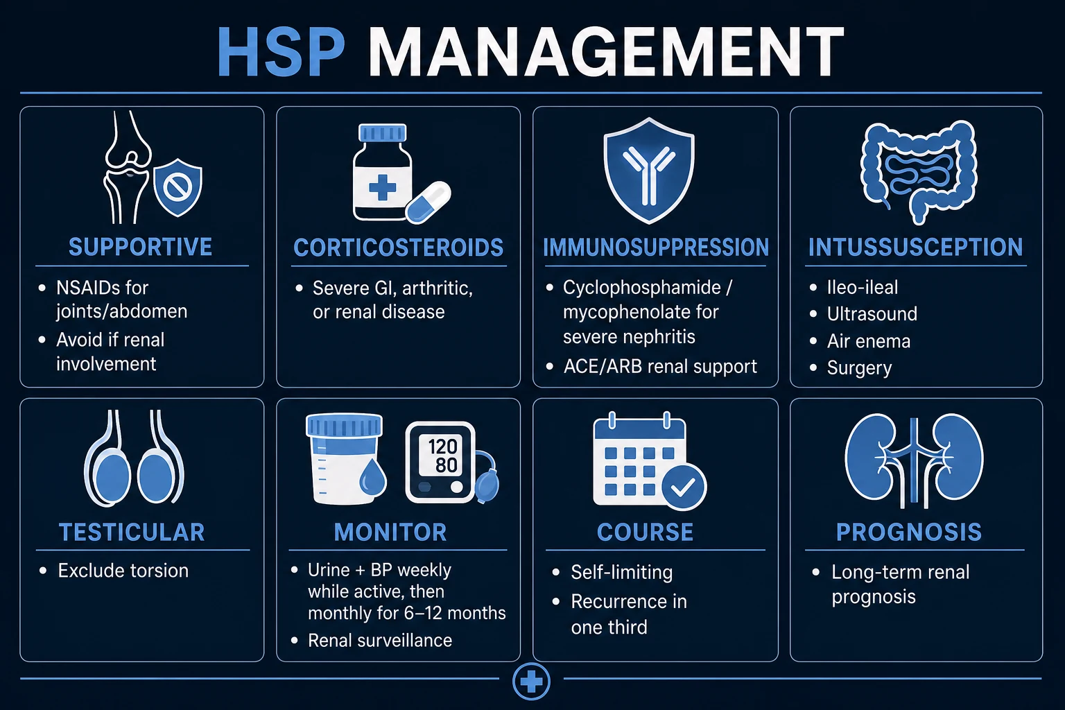
The cornerstone is supportive care; specific anti-inflammatory treatment is reserved for severe organ involvement.[1][5]
Step 1 — Supportive care (most patients)
- Rest during the active phase, especially when arthritis or abdominal pain is severe.
- Adequate hydration and a normal diet (NPO only if intussusception/ileus suspected).
- Simple analgesia / antipyretics — paracetamol 10 to 15 mg/kg every 4 to 6 hours (max 60 mg/kg/day or 2 g/day, whichever lower). [1]
Step 2 — Symptom-directed therapy
- Arthritis / arthralgia — NSAIDs are first-line and very effective:
- Ibuprofen 5 to 10 mg/kg TDS (max 30 mg/kg/day or 2.4 g/day).
- Naproxen 5 mg/kg BD (max 1 g/day) is an alternative.
- CRITICAL caveat: AVOID NSAIDs if there is significant renal involvement (rising creatinine, heavy proteinuria, AKI, hypertension) — they reduce renal perfusion and can precipitate AKI. Use paracetamol or short-course steroids instead.[1]
- Abdominal pain — analgesia (paracetamol; avoid opioids if possible to avoid masking surgical pathology); consider short-course corticosteroids if severe (see below).
Step 3 — Corticosteroids (selected indications)
Corticosteroids reduce the duration and severity of GI and joint symptoms and are used for active, severe disease — but, crucially, they do NOT prevent nephritis and are not given routinely to all patients.[5]
Indications for corticosteroids: [1]
- Severe GI involvement — intense abdominal pain, GI bleeding, protein-losing enteropathy not responding to supportive care.
- Significant renal involvement — nephrotic-range proteinuria, nephritic syndrome, declining renal function, hypertension (with nephrology input, often combined with other immunosuppressants).
- Severe arthritis unresponsive to NSAIDs.
- Marked systemic symptoms — persistent fever, malaise, significant systemic inflammation.
- Severe scrotal involvement. [1]
Corticosteroid regimens: [1]
- Oral prednisolone 1 to 2 mg/kg/day (max 60 to 80 mg/day) for 1 to 2 weeks, then taper over a further 2 to 4 weeks.
- Severe disease (e.g. severe GI bleeding, crescentic glomerulonephritis): IV methylprednisolone pulses 10 to 30 mg/kg/day (max 1 g) for 3 consecutive days, followed by oral prednisolone and taper. [1]
Step 4 — HSP nephritis (ISKDC III to VI / severe disease)
Severe renal disease requires combined immunosuppression under specialist (paediatric nephrology) guidance — evidence base is limited to small trials and observational data:[7]
- Prednisolone as above PLUS
- Cyclophosphamide 2 mg/kg/day oral (or IV pulses) — historically first-line for crescentic disease; now often used for the most severe / rapidly progressive cases.
- Azathioprine 1 to 2 mg/kg/day — for less severe disease, or as a steroid-sparing maintenance agent.
- Mycophenolate mofetil (MMF) — increasingly used; 600 mg/m^2 BD.
- Calcineurin inhibitors — ciclosporin 4 to 5 mg/kg/day or tacrolimus 0.1 mg/kg/day.
- Rituximab 375 mg/m^2 per dose (B-cell depletion) — emerging role in refractory disease.
- Plasma exchange — considered in rapidly progressive crescentic GN with renal failure.
- Renin-angiotensin system blockade for persistent proteinuria — ACE inhibitor (enalapril 0.1 mg/kg/day, titrate) or ARB (losartan); renoprotective and reduces proteinuria. Monitor potassium and creatinine. [1]
Step 5 — Surgical complications
- Intussusception in HSP is ileo-ileal (unlike idiopathic ileo-colic); enema reduction usually fails — most need surgical reduction/resection.
- Testicular torsion — urgent surgical exploration if Doppler cannot exclude. [1]
Follow-up and surveillance — critical
Because HSP nephritis can develop late, weeks to months after the rash resolves, every child with HSP must have scheduled monitoring of blood pressure and urinalysis:[5]
- During active disease: BP and urinalysis weekly (or at each clinical contact).
- After resolution: BP and urinalysis monthly for 6 months, then every 3 to 6 months up to 12 months (longer if there has been any renal involvement).
- Escalation triggers — rising creatinine, new or heavy proteinuria, nephritic or nephrotic syndrome, hypertension, persistent GI bleeding, suspected intussusception, CNS symptoms: prompt re-evaluation and (where indicated) renal biopsy and intensified immunosuppression. [1]
Management summary ladder
| Step | Setting | Therapy | Trigger |
|---|---|---|---|
| 1 | Outpatient | Rest, hydration, paracetamol | Typical mild HSP |
| 2 | Outpatient | Add NSAIDs (ibuprofen/naproxen) | Arthritis/arthralgia, NORMAL renal function |
| 3 | Outpatient/ward | Oral prednisolone 1 to 2 mg/kg/day | Severe GI/joint/systemic symptoms |
| 4 | Inpatient | IV methylprednisolone pulses + nephrology | Severe GI bleed, crescentic GN, AKI |
| 5 | Specialist | Add cyclophosphamide/MMF/calcineurin/rituximab | ISKDC III to VI, refractory disease |
| 6 | Surgery | Surgical reduction/exploration | Intussusception (ileo-ileal), torsion |
Specific Subtypes & Scenarios
- Cutaneous-dominant / simple HSP — the commonest phenotype; palpable purpura ± mild arthritis; self-limiting in 4 to 6 weeks; supportive care only; full recovery.
- GI-dominant HSP — severe colicky pain, vomiting, GI bleeding; risk of intussusception (ileo-ileal) and protein-losing enteropathy; may need admission, NPO, IV fluids, analgesia, steroids for severe pain/bleeding, surgery for intussusception.
- HSP nephritis — defined by haematuria/proteinuria; severity graded histologically by ISKDC I to VI; grades IV to VI (crescents in more than 50% of glomeruli) have the worst prognosis and warrant aggressive immunosuppression (steroid + cyclophosphamide/MMF/calcineurin/rituximab) and ACE inhibition.[7]
- Adult HSP — rarer but more severe and persistent; higher renal involvement (more crescentic disease), more GI complications, higher relapse rate, worse long-term renal outcome; lower threshold to biopsy and treat aggressively.[6]
- Recurrent HSP — relapses in about one-third of patients, usually within the first year; most commonly cutaneous (rash alone) and milder than the initial episode; treat each relapse on its merits; recurrence is commoner in younger children.
- Scrotal HSP — painful scrotal swelling in boys; must exclude testicular torsion (urgent Doppler); if vasculitis alone, treated supportively ± short-course steroids.
Complications & Pitfalls
Gastrointestinal: [1]
- Intussusception — classically ileo-ileal in HSP (unlike idiopathic ileo-colic); presents with colicky pain, palpable mass, red-currant-jelly stool; usually needs surgery (enema reduction generally fails).
- GI haemorrhage, protein-losing enteropathy (hypoalbuminaemia, oedema), bowel ischaemia/perforation (rare). [1]
Renal (the major determinant of long-term prognosis): [1]
- IgA nephritis — microscopic or macroscopic haematuria, proteinuria, hypertension.
- Nephritic or nephrotic syndrome.
- Rapidly progressive crescentic glomerulonephritis (RPGN) — crescents on biopsy, rapid decline in GFR; aggressive immunosuppression required.
- Progression to chronic kidney disease and, in ~1 to 2%, end-stage renal disease (higher in adults and crescentic disease).[7]
Surgical: [1]
- Testicular torsion (scrotal HSP); rare orchitis. [1]
Neurological (rare): headache, seizures, posterior reversible encephalopathy syndrome (PRES), intracranial bleed. [1]
Skin: necrosis, ulceration, scarring (rare in children). [1]
Recurrence — about one-third of patients relapse, usually mild and cutaneous. [1]
Classic pitfalls (high-yield for exams):[1]
- Missing meningococcaemia by attributing a sick child's rapidly progressive purpura to HSP.
- Missing intussusception (or treating it as non-specific pain).
- Missing late-onset nephritis by not following up urinalysis and BP for 6 to 12 months.
- Giving steroids routinely "to prevent nephritis" — they do NOT (Jauhola 2011); reserve for active severe disease.
- Missing testicular torsion in a boy with scrotal pain.
- Underestimating HSP in adults (more severe disease, worse prognosis).
- Using NSAIDs in the presence of renal involvement — risk precipitating AKI.
- Confusing HSP with ITP — check the platelet count. [1]
Prognosis & Disposition
Overall prognosis is excellent in children — most cases resolve spontaneously within 4 to 6 weeks without sequelae.[1]
- Recurrence in approximately one-third, usually within the first year and most commonly cutaneous.
- Long-term outcome is determined by the kidneys. Approximately 1 to 2% progress to end-stage renal disease; the rate is higher in adults and in those with crescentic (ISKDC IV to VI) disease, nephrotic-range proteinuria, hypertension, or renal impairment at onset.[6][7]
Predictors of poor renal outcome: nephrotic-range proteinuria, reduced GFR at onset, hypertension, biopsy showing crescents in more than 50% of glomeruli (ISKDC IV to VI), older age, adult onset. [1]
Follow-up: blood pressure and urinalysis for at least 6 to 12 months after onset (longer if any renal involvement has occurred), to detect late-onset nephritis. Patients with persistent proteinuria require long-term nephrology follow-up. [1]
Disposition: [1]
- Mild disease (typical) — outpatient management with close follow-up and safety-net advice to return if severe abdominal pain, swollen scrotum, dark urine, reduced urine output, or persistent fever.
- Severe GI disease, intussusception, significant renal involvement, hypertension, AKI, scrotal swelling, or systemic toxicity — admit for investigation, treatment and monitoring. [1]
Special Populations
- Children — weight-based dosing for ALL drugs (prednisolone 1 to 2 mg/kg, ibuprofen 5 to 10 mg/kg TDS, enalapril 0.1 mg/kg/day); blood pressure interpreted against age- and height-based centiles; most cases benign.
- Adults with HSP — rarer but more severe and persistent; higher renal involvement, more crescentic disease, more GI complications, higher relapse rate, worse long-term renal prognosis; lower threshold to biopsy and treat aggressively with steroids ± immunosuppression.[6]
- Pregnancy — HSP is uncommon in pregnancy; pre-existing HSP nephritis or chronic kidney disease warrants nephrology/obstetric medicine counselling regarding hypertensive and renal risk in pregnancy. ACE inhibitors and ARBs are CONTRAINDICATED in pregnancy (fetopathy) — use labetalol, nifedipine or methyldopa for hypertension. NSAIDs are avoided, especially after 20 weeks.
- Anticoagulated / bleeding-risk patients — avoid NSAIDs; balance the bleeding risk of steroids against GI haemorrhage; titrate treatment intensity.
- Immunocompromised host — atypical or more severe course; lower threshold to biopsy and to use aggressive immunosuppression; exclude mimics (drug-induced vasculitis, infection).
- Established renal disease — ACE inhibitor or ARB first-line for proteinuria; monitor potassium and creatinine; refer to nephrology; counsel on long-term risk of CKD/ESRD and pregnancy implications.
Evidence, Guidelines & Regional Differences
Classification criteria (both reproduced above in Investigations): [1]
- 1990 ACR criteria (Mills, PMID 2202307)[3] — 2 of 5: age under 20, palpable purpura, acute abdominal pain, biopsy of small vessels, arthritis/arthralgia. Designed for adults and children but less accurate in children.
- EULAR/PRINTO/PRES 2010 criteria (Ozen, PMID 20388738)[4] — the current paediatric standard: palpable purpura (mandatory, lower-limb predominant) PLUS at least 1 of diffuse abdominal pain, IgA on biopsy, arthritis/arthralgia, renal involvement. Sensitivity ~100%, specificity ~87%.
Key treatment evidence: [1]
- Jauhola et al. (Archives of Disease in Childhood, 2010; PMID 21169167)[5] — a landmark Finnish prospective study of over 200 children demonstrating that early prednisolone did NOT prevent the development of HSP nephritis. This is practice-changing: steroids are not given prophylactically to all patients, only for active severe GI/renal/systemic disease. (A related multicentre trial by the same group reported in the NEJM reached the same conclusion.)
- Audemard-Verger et al. (Autoimmunity Reviews, 2015; PMID 25911648)[6] — review of adult HSP: more severe renal/GI phenotype, worse prognosis, more aggressive management warranted.
- Davin and Coppo (Nature Reviews Nephrology, 2014; PMID 21144113)[7] — authoritative review of HSP nephritis and its shared biology with IgA nephropathy (Gd-IgA1).
Cochrane / KDIGO: high-quality randomised evidence for immunosuppression in HSP nephritis is limited; recommendations are largely based on observational data and small trials. Active research areas include rituximab, mycophenolate mofetil, plasmapheresis and tonsillectomy for refractory or severe renal disease. [1]
[1] [1]Exam Pearls
The HSP tetrad — P A R A
PARA
Lower limbs/buttocks, symmetrical, dependent, MANDATORY defining feature. Platelets NORMAL.
Colicky periumbilical; risk of INTUSSUSCEPTION (ILEO-ILEAL, not ileo-colic).
Haematuria/proteinuria; may develop LATE (weeks-months); main determinant of prognosis.
Knees/ankles, periarticular, non-erosive, fleeting; NSAIDs first-line if renal-normal.
- HSP is the MOST COMMON VASCULITIS in children; a small-vessel leukocytoclastic vasculitis with IgA immune-complex deposition.[1]
- The tetrad — Palpable purpura (mandatory) + Abdominal pain + Renal + Arthritis.
- PLATELETS ARE NORMAL — the single highest-yield discriminator from ITP (platelets low).
- Follows a URTI 1 to 3 weeks earlier in ~50%.
- Intussusception in HSP is ILEO-ILEAL (not ileo-colic) — usually needs surgery, NOT enema reduction.
- Steroids do NOT prevent nephritis (Jauhola 2011)[5] — give for active severe GI/renal/systemic disease, not prophylactically.
- Renal involvement may develop LATE — monitor urinalysis and BP for 6 to 12 months.
- HSP and IgA nephropathy (Berger disease) are the SAME disease (galactose-deficient IgA1) — HSP is the systemic form.[7]
- Adult HSP = more severe renal/GI disease and worse prognosis.[6]
- Biopsy (skin/kidney) shows leukocytoclastic vasculitis with IgA on immunofluorescence.
- NSAIDs for arthritis but AVOID if renal involvement (precipitates AKI).
- Scrotal swelling in a boy with HSP — exclude testicular torsion (urgent Doppler ultrasound).
- Do NOT miss MENINGOCOCCAEMIA — rapidly progressive purpura, ill child, not confined to the legs; treat as emergency (IV ceftriaxone, sepsis bundle).
- EULAR/PRINTO/PRES 2010: purpura (mandatory) + 1 of abdominal pain, IgA on biopsy, arthritis, renal. Sensitivity ~100%.[4]
- ISKDC IV to VI (crescents in over 50% of glomeruli) = poor renal prognosis -> aggressive immunosuppression.
Exam application bank (NEET-PG / INICET)
One-line answer
Henoch-Schonlein purpura (HSP), now officially renamed IgA vasculitis (IgAV) by the Chapel Hill Consensus Conference, is an immune-complex-mediated small-vessel (leukocytoclastic) vasculitis characterised by dominant IgA1 immune-complex deposition in the skin, gastrointestinal tract, joints and glomeruli. It is the most common vasculitis of childhood. Classically presents after a recent upper respiratory infection with the tetrad of palpable purpura (lower limbs/buttocks, mandatory), arthritis/arthralgia (knees, ankles), colicky abdominal pain, and renal involvement (haematuria/proteinuria). Platelets are NORMAL (key distinction from ITP). Diagnosis is clinical using EULAR/PRINTO/PRES 2010 or 1990 ACR criteria. Management is supportive in most cases (rest, hydration, analgesia, NSAIDs for arthritis); corticosteroids for severe gastrointestinal or renal involvement. Crucially, steroids do
Worked stems (answer without another resource)
Stem 1 — Classic presentation. Map symptoms to mechanism; name the first investigation and first treatment step with dose/route if drug therapy is standard. [1]
Stem 2 — Unstable / complicated. List red flags that force immediate resuscitation, theatre, ICU, antidote, or reperfusion — and what you do in the first 15 minutes. [1]
Stem 3 — Atypical group. Elderly, pregnancy, child, or immunocompromised: how presentation and thresholds change. [1]
Stem 4 — Differential trap. Name the three closest mimics and one discriminator for each. [1]
Stem 5 — Disposition. Who goes home with safety-netting, who is admitted, who needs HDU/ICU/theatre, and what follow-up is mandatory. [1]
Rapid viva checklist
- Definition + classification
- Pathophysiology chain
- Bedside signs / criteria
- Score with exact components (if any)
- Emergency bundle
- Definitive therapy with doses
- Complications of disease and of treatment
- Special populations
- Guideline/trial name if classic
- Three exam traps
Coverage self-check
If you cannot answer any stem above from this page alone, re-read the matching section — the page is intended to be self-sufficient for final-prof and NEET-PG/INICET questions on Henoch-Schonlein Purpura (IgA Vasculitis).
References
- [1]Gardner-Medwin JM, Dolezalova P, Cummins C, et al. Henoch-Schönlein Purpura (IgA Vasculitis): Rapid Evidence Review Am Fam Physician, 2020.PMID 32803924
- [2]Yang YH, Yu HH, Chiang BL. A Review of IgA Vasculitis (Henoch-Schönlein Purpura) Past, Present, and Future Med Sci Monit, 2024.PMID 38281080
- [3]Mills JA, Michel BA, Bloch DA, et al. The American College of Rheumatology 1990 criteria for the classification of Churg-Strauss syndrome (allergic granulomatosis and angiitis) Arthritis Rheum, 1990.PMID 2202307
- [4]Ozen S, Pistorio A, Iusan SM, et al. EULAR/PRINTO/PRES criteria for Henoch-Schönlein purpura, childhood polyarteritis nodosa, childhood Wegener granulomatosis and childhood Takayasu arteritis: Ankara 2008. Part I: Overall methodology and clinical characterisation Ann Rheum Dis, 2010.PMID 20388738
- [5]Jauhola O, Ronkainen J, Koskimies O, et al. Missing data approaches in eHealth research: simulation study and a tutorial for nonmathematically inclined researchers J Med Internet Res, 2010.PMID 21169167
- [6]Audemard-Verger E, Pillebout E, Guillevin L, et al. Cardiac endothelium-myocyte interaction: clinical opportunities for new heart failure therapies regardless of ejection fraction Eur Heart J, 2015.PMID 25911648
- [7]Davin JC, Coppo R. Quantitative determination of van Gogh's painting grounds using SEM/EDX Microsc Microanal, 2011.PMID 21144113