Paediatrics · Paediatrics
Kawasaki Disease
Also known as Mucocutaneous lymph node syndrome · Kawasaki syndrome
Kawasaki disease is an acute, self-limited, medium-vessel vasculitis of childhood of unknown cause that preferentially involves the coronary arteries. It is the leading cause of acquired heart disease in children in developed nations. Diagnosis is clinical: fever for at least five days PLUS four of five principal features (bilateral non-purulent conjunctivitis, oral changes, polymorphous rash, extremity changes, cervical lymphadenopathy); an incomplete form (two to three features) is common in young infants and carries a higher coronary aneurysm risk. Cornerstone treatment is intravenous immunoglobulin 2 g/kg as a single infusion over 10 to 12 hours PLUS aspirin (high-dose anti-inflammatory then low-dose anti-platelet) within the first 10 days of illness, which reduces coronary artery aneurysm risk from roughly 25 per cent to 3 to 5 per cent. Echocardiography is mandatory at baseline, one to two weeks, and four to six weeks. Intravenous immunoglobulin resistance occurs in 10 to 20 per cent; repeat immunoglobulin, infliximab, corticosteroids, anakinra or cyclosporine are used in refractory disease.
On this page & tools
Your progress
Saved locally on this device.
Exam tags
Red flags
Overview & Definition
Kawasaki disease (KD), originally described by Tomisaku Kawasaki in 1967 as mucocutaneous lymph node syndrome, is an acute, self-limited febrile vasculitis of childhood that predominantly affects medium-sized muscular arteries, with a striking predilection for the coronary arteries. It is defined clinically by a characteristic febrile mucocutaneous illness of defined duration and is now recognised as the single leading cause of acquired heart disease in children in developed countries — surpassing rheumatic heart disease wherever intravenous immunoglobulin (IVIG) has not supplanted it.[1]
Three properties make KD a high-yield and clinically dangerous disease. First, the diagnosis is entirely clinical — there is no confirmatory laboratory test, so pattern recognition is essential. Second, the coronary arteries are uniquely vulnerable: inflammation of the vasa vasorum and media weakens the arterial wall, producing aneurysms that may thrombose, rupture, or persist for life. Third, timely treatment within the first 10 days of fever with IVIG dramatically changes outcome, reducing the coronary aneurysm rate from roughly 25 per cent to 3 to 5 per cent — one of the most cost-effective interventions in all of paediatrics.[3][1]
The clinical skill in KD is not the textbook case (fever plus five features is unmistakable) but three subtler judgements: (a) recognising incomplete KD in an irritable febrile infant with only two or three features; (b) acting fast — the IVIG clock starts at the first day of fever, not the day of diagnosis; and (c) escalating treatment in the 10 to 20 per cent who are IVIG-resistant. Each is examined directly in NEET-PG, INICET, USMLE and PLAB.[2][8]
Classification
KD is classified by how completely the clinical criteria are met and, separately, by coronary artery involvement (expressed as a Z-score). The distinction between complete and incomplete KD is the most examinable and clinically consequential classification in the disease. [1]
Complete KD
classic form, ~80 per cent
- Fever for at least 5 days PLUS at least 4 of 5 principal clinical features
- All 5 features may be present
- Standard threshold for IVIG and aspirin within 10 days
- Coronary aneurysm risk ~3 to 5 per cent if treated early
Incomplete KD
atypical form, ~20 per cent and rising
- Fever for at least 5 days PLUS 2 to 3 principal features
- Most common in infants under 6 months AND older children over 5 years
- HIGHER aneurysm risk if diagnosis is delayed
- Use supplemental laboratory criteria + echocardiography; lower threshold to treat with IVIG
- Same treatment as complete KD once suspected
IVIG-resistant KD
10 to 20 per cent
- Persistent or recrudescent fever 36 hours to 7 days after the first IVIG infusion
- Higher aneurysm risk than responders
- Escalate: repeat IVIG, infliximab, corticosteroids, anakinra, cyclosporine
KD shock syndrome
~5 to 7 per cent
- Systolic blood pressure below the 5th percentile for age OR signs of poor perfusion
- Driven by myocarditis and capillary leak
- Fluid-sparing resuscitation plus vasopressors; consider adjunctive steroids with IVIG
Coronary artery classification (AHA 2017, by internal lumen dimension Z-score) — reproduced verbatim, because the Z-score band dictates anticoagulation intensity and duration of follow-up:[1]
Coronary artery Z-score classification (AHA 2017)
The Z-score is the number of standard deviations a measured coronary internal diameter lies above the population mean for body surface area; it normalises dimensions to the child's size so a 3 mm left main coronary artery means very different things in a 3 kg neonate and a 16 kg toddler.[1]
Epidemiology & Risk Factors
KD is a disease of young children: 80 to 85 per cent of cases occur in children under five years of age, with a peak incidence between six months and two years. It is rare under three months (passive maternal antibody is partly protective) and uncommon after five years, but cases do occur in older children and even adults — and these atypical age groups are precisely the ones most often missed.[1][8]
There is a striking ethnic gradient: incidence is highest in children of East Asian ancestry (Japan ~ 300 per 100,000 children under five; Korea and Taiwan similarly high), intermediate in children of Asian ancestry living in Western countries, and lowest in White populations (United States ~ 25 per 100,000, United Kingdom ~ 15 per 100,000). There is a slight male predominance (1.4 to 1.6:1), and boys have a higher coronary aneurysm risk. Seasonal clustering is typical — winter and spring peaks in temperate climates, summer peaks in some Asian series — suggesting an infectious trigger in a genetically susceptible host.[1][2]
Risk factors for coronary aneurysm (the outcome that matters and that examiners probe):[1][7]
- Age under one year (and especially under six months) — highest absolute risk.
- Age over five years — atypical, late diagnosis.
- Male sex.
- Delay in IVIG beyond day 10 of fever (or no IVIG at all).
- Baseline coronary abnormality (Z-score elevation) on the first echocardiogram.
- Markers of intense inflammation: high C-reactive protein (CRP), high erythrocyte sedimentation rate (ESR), low albumin, low haemoglobin for age, high neutrophil count, elevated alanine aminotransferase (ALT), and thrombocytosis over 450,000 per cubic millimetre in the second week.
- IVIG resistance (persistent fever).
- Kobayashi score at least 5 (a Japanese-derived predictor of IVIG non-response; less sensitive in non-Asian populations). [1]
Recurrence occurs in about 3 per cent of cases, and a family history of KD modestly increases risk, reinforcing a genetic contribution (polymorphisms in ITPKC, FCGR2A, CD40 and the CASP3 locus).[8]
Pathophysiology
KD is an immune-mediated necrotising vasculitis of medium-sized muscular arteries. The cause is unknown but believed to be an aberrant immune response to one or more infectious triggers in a genetically susceptible child — supported by the age distribution (almost no neonates, rare adults), seasonal and geographic clustering, epidemic waves, and the self-limited nature. Candidate triggers (none proven) include superantigens, viruses, and intracellular pathogens; SARS-CoV-2 produced a distinct paediatric inflammatory multisystem syndrome (PIMS-TS / MIS-C) that overlaps KD immunologically.[1][10]
Immunopathology. Activation of innate immunity (TLR signalling) drives a cytokine cascade — interleukin-1, interleukin-6, interleukin-17 and tumour necrosis factor-alpha (TNF-alpha) — that recruits activated CD8-positive T cells and macrophages into the coronary arterial wall. The result is transmural inflammation of the vasa vasorum and media, destruction of the internal elastic lamina, and smooth-muscle-cell apoptosis. As the inflamed wall weakens, arterial dilation and then aneurysm follow; superimposed platelet activation and a hypercoagulable state (thrombocytosis, high fibrinogen) predispose to thrombotic occlusion and myocardial infarction. The interleukin-1 axis in particular explains why anakinra (an interleukin-1 receptor antagonist) can rescue refractory cases.[2][10]
Histopathology progresses through three phases, which mirror the clinical course and are classic viva material:[1][10]
- Acute phase (weeks 1 to 2): perivasculitis and intimal polymorphonuclear infiltrate of the small vasa vasorum; microvascular panarteritis of medium arteries. Clinically: high fever, irritable child, mucocutaneous features, myocarditis (often subclinical, with tachycardia out of proportion to fever), pericardial effusion.
- Subacute phase (weeks 2 to 4): aneurysm formation peaks; inflammatory infiltrate shifts to lymphocytes and plasma cells; thrombosis may complicate aneurysms. Clinically: fever settles, desquamation begins, coronary aneurysms are detectable on echocardiography, thrombocytosis is maximal.
- Convalescent phase (weeks 4 to 8): inflammation resolves; aneurysms may regress (small aneurysm) or organise, stenose and calcify (medium and giant aneurysm). Myofibroblastic proliferation and intimal thickening set the stage for lifelong accelerated atherosclerosis and stenosis. [1]
Clinical Presentation
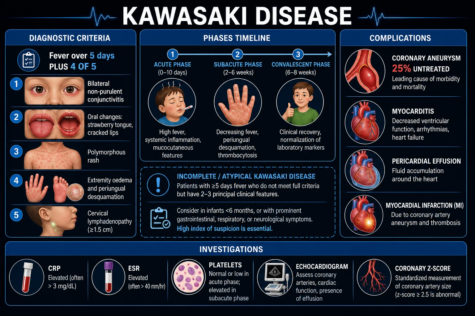
The hallmark is fever for at least five days (classically high, over 39 degrees Celsius, remittent and unresponsive to antibiotics) accompanied by bilateral, non-purulent mucocutaneous features that appear over several days rather than all at once. The child is typically irritable — more so than the degree of fever would explain. The five principal clinical features (need four, plus fever, for complete KD) are reproduced verbatim below.[1]
The five principal clinical features (AHA 2017)
Five principal features — COREL
COREL
bilateral, bulbar, non-exudative (no pus), with limbal sparing; usually painless, no significant photophobia
red cracked bleeding lips, strawberry tongue (red, swollen papillae over a white coating then beefy red), diffuse oropharyngeal injection — WITHOUT ulceration or exudate (exudate suggests adenovirus or streptococcus)
polymorphous (morbilliform, maculopapular, scarlatiniform or erythema-multiforme-like), NOT vesicular or bullous; trunk and extremities; often accentuated in the perineum (which desquamates early)
acute: erythema and indurative oedema of palms and soles (child refuses to bear weight); subacute: periungual (fingertip) desquamation in weeks 2 to 3; convalescent: transverse grooves across nails (Beau lines) months later
cervical, at least 1.5 cm in diameter, usually unilateral, non-purulent; the LEAST common of the five features and the one most easily missed
Other common but non-diagnostic features (helpful when the picture is incomplete): anterior uveitis (often detected only on slit lamp in the first week), sterile pyuria (urethritis), arthritis (especially knees, ankles, hips), abdominal pain, diarrhoea and vomiting, hydropic gallbladder, painless BCG-vaccination-site erythema/induration (a useful sign in BCG-vaccinated Indian and Japanese infants), aseptic meningitis, pneumonitis, and irritability out of proportion to illness. Recovery of fingertip desquamation in week two is so characteristic that a febrile illness followed by peeling fingertips is KD until proven otherwise.[1][8]
Kawasaki disease — the numbers that decide answers
Atypical presentations (the cases examiners love): in infants under six months, the full criteria are rarely met — presentation may be sepsis-like, with fever and irritability alone, sometimes meningitic or with diarrhoea as the dominant symptom; aneurysm risk is highest in this group, so a low threshold for echocardiography and IVIG is mandatory.[5] In older children (over five years) and adults, rash and mucous membrane changes are often blunted while myocarditis, arthralgia and abdominal pain dominate. KD shock syndrome presents as toxic shock (tachycardia, hypotension, cool peripheries, altered mentation) and is easily mistaken for septic shock.[2]
Differential Diagnosis
KD has no confirmatory test, so the differential is the diagnostic exercise. The list below pairs each mimicker with the distinguishing feature that excludes KD — memorise the discriminator, not just the name.[1][8]
- Scarlet fever (Streptococcus pyogenes): tonsillar exudate, positive throat swab / rapid antigen, Pastia lines in flexures, sandpaper-textured rash, prompt response to penicillin, no conjunctivitis or extremity oedema. Distinguishing feature: exudative pharyngitis and response to antibiotics.
- Measles: Koplik spots on buccal mucosa (pathognomonic), cough, coryza, conjunctivitis (the 3 Cs), rash spreads head to toe, unvaccinated child; prodromal high fever then rash as fever peaks (KD: fever persists with rash). Distinguishing feature: Koplik spots and head-to-toes spread.
- Staphylococcal scalded skin syndrome: tender, flaccid bullae and superficial skin peeling, perioral fissuring with radial crusting, caused by exfoliative toxin; no coronary involvement; Nikolsky positive. Distinguishing feature: superficial bullous peeling and Nikolsky sign.
- Stevens-Johnson syndrome / toxic epidermal necrolysis: target lesions, mucosal ulceration at two or more sites, skin pain, blistering, drug exposure; conjunctivitis is purulent/exudative. Distinguishing feature: mucosal ulceration and target lesions, not strawberry tongue.
- Drug reaction (DRESS, serum sickness-like): exposure history, eosinophilia (DRESS), no thrombocytosis trend, no peeling fingertips. Distinguishing feature: temporal link to a drug.
- Juvenile idiopathic arthritis (systemic-onset, Still disease): quotidian (spiking) fever for at least six weeks, salmon-pink evanescent rash, arthritis (may appear late), high ferritin; no conjunctivitis or strawberry tongue. Distinguishing feature: quotidian fever for weeks and arthritis.
- Adenovirus and other viral exanthems: exudative conjunctivitis and pharyngitis, short fever course, no peeling; resolves without specific therapy. Distinguishing feature: purulent conjunctivitis and short fever.
- Toxic shock syndrome: hypotension, renal or hepatic involvement, desquamation late; staphylococcal or streptococcal source; no coronary aneurysm. Distinguishing feature: early hypotension and multi-organ failure (though KD shock syndrome overlaps — see below).
- Leptospirosis and rickettsial disease (scrub typhus): eschar (scrub typhus), conjunctival suffusion, jaundice and renal failure, serology positive. Distinguishing feature: eschar and serology. [1]
Practical rule: a child with fever for at least five days, raised inflammatory markers, and any one feature of KD deserves echocardiography and paediatric review — over-diagnosis (and treating a viral exanthem with IVIG) is far less dangerous than missing KD and inflicting a lifelong coronary aneurysm.[1]
Clinical & Bedside Assessment
Begin with ABCDE and a focused cardiac assessment. The acutely irritable febrile child can decompensate from myocarditis or KD shock syndrome even before the full rash appears. [1]
Vital signs and general appearance. Confirm fever for at least five days (plot the temperature chart from the onset). Tachycardia disproportionate to fever or dehydration suggests myocarditis; a gallop rhythm, hepatomegaly, or cool peripheries with hypotension defines KD shock syndrome. Measure blood pressure in all four limbs (a systolic gradient suggests aortic or subclavian involvement, though rare) and continuous SpO2. [1]
Head and neck. Look for bilateral bulbar conjunctival injection with limbal sparing (sparing of the limbal ring around the iris is classic and opposite to adenovirus, which affects the tarsal conjunctiva with purulent discharge). Inspect the lips (red, dry, cracked, bleeding), tongue (white coating with red swollen papillae = white strawberry tongue; later beefy red with prominent papillae = red strawberry tongue), and oropharynx (diffuse injection, no ulceration, no exudate — exudate points away from KD). Palpate cervical nodes (a node of at least 1.5 cm, usually unilateral, is the least common principal feature). In BCG-vaccinated children, inspect the BCG site for erythema, induration or pus — a useful culture-specific sign.[1][8]
Skin. Characterise the polymorphous rash (vesicles or bullae exclude KD), and examine the perineum (early accentuation and peeling is characteristic, and easy to miss). Examine palms and soles for indurative oedema and erythema (the child often refuses to walk or be touched). In week two, look for periungual desquamation; months later, Beau lines. [1]
Cardiac. Auscultate for tachycardia, gallop (S3), muffling of heart sounds (pericardial effusion), and a new murmur (mitral or aortic regurgitation from valvulitis, or a flow murmur from anaemia). A pericardial rub suggests pericarditis. [1]
Abdomen, joints and neurology. Palpate for hepatomegaly, gallbladder hydrops (right upper quadrant mass) and arthritis (especially large joints). Briefly assess meningeal signs and conscious level (aseptic meningitis is a recognised feature). Examine the urine (sterile pyuria = urethritis). [1]
Time the illness. The single most important bedside number is day of illness — day one is the first day of fever. The IVIG window closes at day ten, so a child seen on day seven with two features has far less margin than one seen on day three. [1]
Investigations
There is no diagnostic test for KD — the diagnosis is clinical, and investigations serve three purposes: (a) to support the diagnosis in incomplete KD (AHA supplemental laboratory criteria); (b) to exclude mimickers; and (c) to detect and stratify coronary involvement (the outcome that defines prognosis and management).[1]
First-line bloods in every suspected case: [1]
- Full blood count: leukocytosis with neutrophilia, normocytic normochromic anaemia for age; platelet count rises progressively and often exceeds 450,000 per cubic millimetre by the second week (thrombocytosis is a near-cardinal feature and a thrombosis risk).
- CRP and ESR: both markedly elevated (CRP often over 3 mg/dL; ESR often over 40 mm/h) and track the response to IVIG.
- Liver function tests: low albumin (under 3.0 g/dL), raised ALT (over 50 U/L), occasionally mild bilirubin and a hydropic gallbladder on ultrasound.
- Serum sodium: hyponatraemia (sodium under 135 mmol/L) is common and is a Kobayashi score component.
- Coagulation and fibrinogen: mildly deranged, fibrinogen high — part of the prothrombotic state.
- Blood cultures and throat swab: usually sterile — but mandatory to exclude sepsis, streptococcal and staphylococcal disease (anti-streptolysin-O titre, anti-DNase B).
- Viral studies and serology (EBV, adenovirus, measles, enterovirus, leptospirosis, scrub typhus) as the differential demands. [1]
Urine: sterile pyuria (leukocytes with negative culture) reflects urethritis — a recognised supportive feature. [1]
Electrocardiogram (ECG): look for arrhythmias (sinus tachycardia out of proportion to fever), prolonged PR or QT interval, low voltages (pericardial effusion), ST-T wave changes, and signs of ischaemia or myocarditis (non-specific, but baseline ECG is essential and serial ECGs track recovery). [1]
AHA 2017 supplemental laboratory criteria for incomplete KD
A child with fever for at least five days and two to three principal features is evaluated for incomplete KD. If CRP is at least 3 mg/dL and/or ESR is at least 40 mm/h, and three or more of the following supplemental criteria are present, treat as KD; if fewer than three, perform echocardiography and treat if coronary abnormalities are found:[1]
- Albumin under 3.0 g/dL
- Anaemia for age
- ALT elevation
- Platelet count at least 450,000 per cubic millimetre (usually after day 7)
- White blood cell count at least 15,000 per cubic millimetre
- White blood cells in urine at least 10 per high-power field [1]
A positive echocardiogram (any coronary Z-score at least 2.5, or perivascular brightness, or other features listed below) at any time confirms KD and mandates IVIG regardless of laboratory count. [1]
Echocardiography — the decisive investigation
Echocardiography is mandatory in every confirmed or suspected case of KD. It is not optional, and a normal first scan does not exclude later aneurysm formation, so serial imaging is the rule.[1]
Timing of echocardiography (AHA 2017): [1]
- Baseline (at diagnosis) — to detect established coronary abnormalities, myocarditis, pericardial effusion, valvular regurgitation.
- One to two weeks after treatment — to detect developing aneurysms (the peak of aneurysm formation is weeks 2 to 4).
- Four to six weeks after onset of illness — to define the maximal coronary status and stratify long-term risk. Additional scans (e.g. at six to twelve months) are required for any child with abnormalities. [1]
What the echocardiogram reports (per coronary segment): the internal lumen diameter as a Z-score for body surface area, the presence of aneurysm (saccular or fusiform), thrombus, perivascular brightness, decreased left ventricular function, pericardial effusion, and valvular regurgitation. Use the Z-score (not the raw millimetre value) to classify involvement.[1]
Coronary Z-score classification (AHA 2017, reproduced verbatim)
The Z-score is the number of standard deviations above the mean internal lumen diameter for body surface area. Memorise the bands — they drive antiplatelet versus anticoagulant therapy and the duration of follow-up:[1]
| Z-score band | AHA 2017 classification | Implication |
|---|---|---|
| Z under 2 | No involvement | normal |
| Z 2 to under 2.5 (OR a fall in Z of at least 1 on follow-up from an initially normal value) | Dilation only | transient; usually regresses |
| Z at least 2.5 to under 5 | Small aneurysm | often regresses; aspirin |
| Z at least 5 to under 10 (and absolute dimension under 8 mm) | Medium aneurysm | thrombosis risk; aspirin, consider anticoagulant |
| Z at least 10 OR absolute dimension at least 8 mm | Large or giant aneurysm | highest thrombosis and infarction risk; aspirin plus anticoagulant (warfarin or direct oral); lifelong cardiology |
Other imaging is reserved for selected patients: cardiac magnetic resonance imaging or computed tomography coronary angiography in older children with persistent giant aneurysms to define the coronary tree, and coronary angiography before any intervention. [1]
Management — Resuscitation
KD is rarely a collapse-resuscitation disease, but two scenarios demand time-critical resuscitation: myocarditis with heart failure or shock, and KD shock syndrome (KDSS). Recognise them by tachycardia disproportionate to fever, gallop rhythm, cool peripheries, weak pulses, prolonged capillary refill, oliguria, and altered mentation.[2]
[1]Resuscitation bundle (suspected KDSS or myocarditis): [1]
- ABCDE, high-flow oxygen if hypoxic or shocked, continuous SpO2, ECG and (in severe cases) non-invasive or invasive ventilation.
- Two reliable intravenous accesses, blood cultures, venous gas and lactate, FBC, CRP, ESR, electrolytes, LFTs, coagulation, troponin and NT-proBNP.
- Cautious fluid resuscitation — 10 mL/kg boluses to a maximum of 20 to 30 mL/kg in the first hour, titrated to perfusion; stop and escalate to vasopressors if no response or if hepatomegaly, gallop or rising respiratory distress develops.
- Vasopressors and inotropes: noradrenaline first-line for vasoplegic shock; adrenaline or dobutamine for low-output myocarditis; milrinone for myocarditis with high systemic vascular resistance (caution with hypotension).
- Echocardiography urgently to assess ventricular function, pericardial effusion and coronary arteries; cardiology and paediatric intensive care involvement from the outset.
- IVIG 2 g/kg as a single infusion over 10 to 12 hours (do NOT delay for blood tests once KD is suspected); in shock, discuss adjunctive corticosteroids (e.g. methylprednisolone 2 mg/kg/day or pulsed 20 to 30 mg/kg/day for three days) with the treating team.
- Aspirin once swallowed or via nasogastric tube — high-dose 30 to 50 mg/kg/day (see Definitive Management). [1]
Time-critical pearl: the IVIG clock is day one of fever, not the day of admission. A child seen on day nine of fever with shock still benefits from IVIG, and treatment beyond day ten is still indicated if active inflammation or coronary abnormalities persist — the ten-day threshold is a risk-reduction window, never a contraindication.[1]
Management — Definitive & Stepwise
Goal: extinguish systemic and coronary inflammation within the first ten days of fever to prevent aneurysm formation, then prevent thrombosis while the artery heals. Definitive management is IVIG plus aspirin, with escalation pathways for IVIG-resistant disease and intensification for high-risk patients at baseline.[1]
Step 1 — Intravenous immunoglobulin (IVIG), the cornerstone
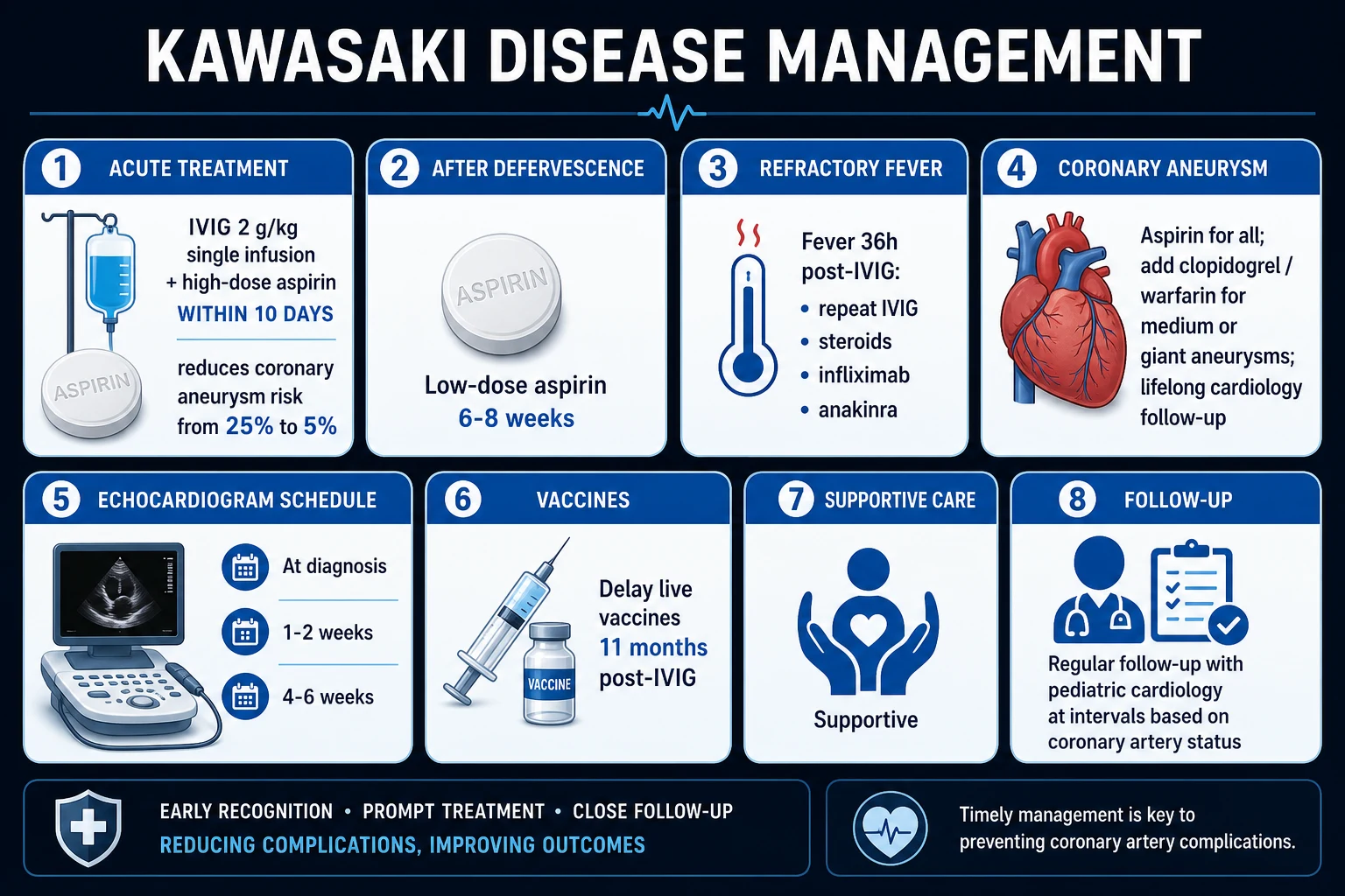
- Agent and dose: IVIG 2 g/kg as a SINGLE intravenous infusion over 10 to 12 hours (not divided over days). The single-infusion regimen was established by Newburger et al. (1991, NEJM), who showed a 2 g/kg single dose was superior to four daily 400 mg/kg doses for preventing coronary aneurysms.[4]
- Timing: within the first ten days of illness (from day one of fever). The original 1986 trial (Newburger et al.) demonstrated that IVIG plus aspirin reduced coronary aneurysms from about 20 per cent to 4 per cent; the 1991 trial refined the regimen.[3][4]
- Beyond day ten: still treat if fever or active inflammation persists, or if coronary abnormalities are present — the benefit persists, and untreated patients have a 25 per cent aneurysm risk.[1]
- Pre-treatment checks: haemoglobin (risk of haemolysis, especially in non-O blood groups), IgA levels if known IgA deficiency (anaphylaxis risk, although rare with modern preparations), renal function, and ensure adequate venous access and observation (rate-related reactions: headache, chills, fever; slow the infusion). Delay live vaccines (MMR, varicella) for eleven months after IVIG because passive antibody blunts vaccine take.[1]
Step 2 — Aspirin
- High-dose (anti-inflammatory) phase: aspirin 30 to 50 mg/kg/day in four divided doses during the acute febrile phase (some Asian regimens use 80 to 100 mg/kg/day; AHA accepts 30 to 50 mg/kg/day).[1]
- Low-dose (anti-platelet) phase: once the child is afebrile for 48 to 72 hours, switch to aspirin 3 to 5 mg/kg once daily.[1]
- Duration: six to eight weeks in patients with no coronary abnormalities (continued until inflammation has fully resolved and the convalescent echocardiogram is normal); indefinitely in patients with persistent coronary abnormalities (aneurysm).[1]
- Reye syndrome caution — during influenza and varicella season, consider annual influenza vaccination and temporary substitution of dipyridamole or clopidogrel for aspirin if the child develops influenza or chickenpox.[1]
Step 3 — Echocardiographic surveillance
- Baseline at diagnosis, repeat at one to two weeks, and again at four to six weeks (more frequently if abnormalities). The convalescent scan stratifies long-term risk (Z-score band).[1]
Step 4 — Define IVIG resistance and escalate
IVIG-resistant (non-responder) KD is defined as persistent or recrudescent fever (at least 38 degrees Celsius) at 36 hours to seven days after completion of the first IVIG infusion; it occurs in 10 to 20 per cent of patients and carries a higher coronary aneurysm risk.[1][9]
IVIG-resistant Kawasaki disease — escalation ladder
Adjunctive primary therapy for high-risk patients (AHA 2024): in children with a high predicted risk of IVIG resistance (e.g. Kobayashi score at least 5 in validated populations) or with coronary aneurysm already present at baseline, the 2024 AHA update endorses adding corticosteroids to the first IVIG dose to intensify primary therapy, reducing aneurysm progression.[2][9]
Kobayashi score (predicts IVIG resistance)
A validated seven-variable score from Japan (Kobayashi et al., 2006); a score of at least 5 identifies IVIG non-responders with high sensitivity in Japanese cohorts (lower in non-Asian populations, so use cautiously outside East Asia).[7]
Kobayashi score components
Step 5 — Long-term anti-thrombotic strategy by Z-score band
- No involvement / dilation only: aspirin 3 to 5 mg/kg/day for 6 to 8 weeks, then stop if echocardiogram normal.[1]
- Small aneurysm: aspirin until regression; counsel on aspirin safety.
- Medium aneurysm: aspirin plus low-dose antiplatelet or anticoagulant in selected patients (e.g. add clopidogrel); avoid contact sports.
- Giant aneurysm (Z at least 10 or at least 8 mm): aspirin plus therapeutic anticoagulation with warfarin or a direct oral anticoagulant, lifelong cardiology follow-up, and counselling on myocardial infarction risk and restriction of contact sports.[1]
Specific Subtypes & Scenarios
Incomplete Kawasaki disease
Definition: fever for at least five days plus two or three principal clinical features (not four). It accounts for an increasing proportion of cases (over 20 per cent in some Western series) and is most common in infants under six months and children over five years — exactly the groups at highest risk of aneurysm. Apply the AHA supplemental laboratory criteria (above) and perform echocardiography; treat with IVIG and aspirin once the suspicion is supported.[1][5] Coronary aneurysm risk in incomplete KD exceeds that of complete KD, almost entirely because of diagnostic delay.[1]
IVIG-resistant Kawasaki disease
Persistent or recrudescent fever 36 hours to 7 days after the first IVIG dose. Manage with the escalation ladder above; repeat echocardiography at each step because these children are at markedly higher aneurysm risk.[6][9]
Kawasaki disease shock syndrome (KDSS)
Defined by systolic blood pressure below the 5th percentile for age OR signs of poor perfusion (cold extremities, weak pulses, prolonged capillary refill, oliguria, altered mentation) in a child meeting KD criteria. Mechanism: myocarditis with low output PLUS systemic capillary leak, mimicking septic shock. Management is fluid-sparing resuscitation, early vasopressors and inotropes, IVIG and adjunctive corticosteroids, with a low threshold for paediatric intensive care. KDSS is a strong independent predictor of IVIG resistance and coronary aneurysm.[2][10]
Kawasaki disease with coronary artery aneurysm at diagnosis
When an aneurysm is already present on the baseline echocardiogram (especially a medium or giant aneurysm), intensify primary therapy: IVIG plus adjunctive corticosteroids (and consider a second immunomodulator), with serial echocardiography every 48 to 72 hours in the acute phase to monitor for thrombosis and progression.[9]
Recurrent Kawasaki disease
Recurs in about 3 per cent, usually within the first year; treat each episode as a new case with IVIG and aspirin, and screen carefully for coronary sequelae, as recurrence increases aneurysm risk.[1]
MIS-C / PIMS-TS (SARS-CoV-2-associated)
A separate but overlapping syndrome: older median age (around 8 years), prominent gastrointestinal and cardiac features, frequent left ventricular dysfunction, positive SARS-CoV-2 serology or exposure, and a cytokine profile rich in interleukin-1 and interleukin-6. Treated with IVIG, corticosteroids and, in severe cases, anakinra or other biologics; coronary involvement is less frequent and often milder than in classical KD but must be sought.[2]
Complications & Pitfalls
Cardiovascular (the complications that define KD): [1]
- Coronary artery aneurysm — the cardinal complication: about 25 per cent untreated, 3 to 5 per cent with IVIG within 10 days. Small aneurysms often regress; medium and giant aneurysms (Z at least 10 or at least 8 mm) rarely regress and carry the highest thrombosis, stenosis, rupture and infarction risk.[3][1]
- Myocarditis — common and often subclinical in the acute phase; may progress to acute heart failure or cardiogenic shock (KDSS).
- Pericarditis with effusion (occasionally tamponade).
- Valvular regurgitation (mitral more than aortic) from valvulitis.
- Arrhythmias — sinus tachycardia out of proportion to fever is an early myocarditis sign; ventricular ectopy may herald ischaemia.
- Coronary thrombosis and myocardial infarction — the principal cause of death; giant aneurysms are the substrate.
- Long-term sequelae — even after regression, the affected segment develops intimal thickening and accelerated atherosclerosis, so young adults with prior KD may present with premature ischaemic heart disease decades later.[1]
Non-cardiac: aseptic meningitis, sterile pyuria and urethritis, hydropic gallbladder (right upper quadrant pain), arthritis, anterior uveitis, pneumonitis, diarrhoea, macrophage activation syndrome (MAS) (a life-threatening cytokine storm with fever, splenomegaly, hyperferritinaemia, cytopenias and hypofibrinogenaemia).[8]
Classic pitfalls
- Waiting for the full five features before acting — incomplete KD is common and dangerous; treat on suspicion with supportive labs and echo.
- Mis-timing the IVIG window — the clock starts at day one of fever, not the day of admission; never withhold IVIG because "it is day 11" if inflammation persists.[1]
- Stopping aspirin too early — switch to low-dose at 48 to 72 hours afebrile but continue 6 to 8 weeks (or indefinitely if aneurysm persists).
- Misreading the Z-score — a "3 mm coronary artery" is meaningless without a Z-score for body surface area; always classify by Z-score band.
- Forgetting to defer live vaccines for eleven months after IVIG.
- Missing KD shock syndrome by attributing tachycardia and hypotension to "dehydration" and flooding with fluid — this worsens myocarditis-induced pulmonary oedema.
- Reassuring a parent on a normal baseline echocardiogram — aneurysms form predominantly in weeks 2 to 4, so the serial scans are the safety net, not the baseline alone.
Prognosis & Disposition
With IVIG within the first ten days of fever, prognosis is excellent: about 95 per cent recover fully, coronary aneurysm risk falls to 3 to 5 per cent, and mortality is 0.1 to 0.2 per cent — almost always from thrombotic occlusion of a giant aneurysm causing myocardial infarction in the subacute or convalescent phase (peak mortality at 15 to 45 days after onset).[1][3]
Aneurysm outcome by Z-score band: small aneurysms (Z 2.5 to under 5) regress in the majority within one to two years; medium aneurysms (Z 5 to under 10) partially regress or stenose; giant aneurysms (Z at least 10 or at least 8 mm) rarely regress, often thrombose or stenose, and require lifelong cardiology follow-up.[1]
Disposition. All patients with confirmed or strongly suspected KD should be admitted for IVIG and aspirin under paediatric supervision, with echocardiography and (where indicated) paediatric cardiology input. Discharge once afebrile for at least 24 to 48 hours, with a clear echocardiography schedule (one to two weeks, four to six weeks), aspirin safety-netting, vaccine deferral advice, and a low threshold to re-present for recurrent fever, chest pain, pallor, or breathlessness.[1]
Long-term risk stratification (AHA 2017/2024) places every KD survivor into one of five risk levels by maximal Z-score; level 5 (giant aneurysm) requires lifelong antiplatelet plus anticoagulant therapy, annual cardiology review, stress imaging, and counselling on ischaemic heart disease.[1][2]
Special Populations
- Infants under six months — incomplete presentation is the rule (fever and irritability with one or two features), the aneurysm risk is highest (often over 30 per cent), and the diagnosis is most often missed. Lower threshold for echocardiography and IVIG; some centres perform KD screening on any infant with unexplained fever for at least five days plus raised inflammatory markers.[5][1]
- Older children (over five years) and adults — fewer mucocutaneous features, more arthralgia, abdominal pain and cardiac presentation (myocarditis, arrhythmia, even infarction); a high index of suspicion is essential, and aneurysm risk remains significant.
- IVIG-resistant patients (10 to 20 per cent) — higher aneurysm risk; manage with the escalation ladder and intensive echocardiographic surveillance.[6][9]
- Children with baseline coronary aneurysm — intensify primary therapy (IVIG plus corticosteroids, consider anakinra or infliximab); serial echocardiography every 48 to 72 hours.[9]
- Children with giant aneurysm — lifelong antiplatelet plus anticoagulant, annual cardiology review, restricted physical activity, counselling on myocardial infarction symptoms, and cardiology-led pregnancy and surgical planning in later life.[1]
- Resource-limited settings (relevant to India / South Asia) — IVIG is expensive and often intermittently available; where IVIG cannot be obtained, high-dose aspirin plus corticosteroids is the pragmatic fallback, and advocacy for IVIG access is part of management. Always weigh BCG status, scrub typhus and leptospirosis in the differential.[2]
- Reye syndrome and influenza — during influenza season, prioritise influenza vaccination and substitute a non-aspirin antiplatelet agent during active influenza or varicella infection.[1]
- Incidence is rising and under-diagnosis is common; BCG-site inflammation is a useful regional sign.
- Differential must include scrub typhus (eschar) and leptospirosis (icterus, renal involvement), both endemic.
- IVIG supply and cost are practical barriers — a written management plan should anticipate stock-outs (high-dose aspirin plus corticosteroids as a stop-gap).
- Kobayashi score was validated in Japanese cohorts and performs poorly in South Asian children; do not withhold IVIG on the basis of a low score.
Evidence, Guidelines & Regional Differences
Landmark trials and guidelines (every one named is examinable): [1]
- Newburger et al., 1986 (NEJM, PMID 2426590): the pivotal trial showing IVIG plus aspirin reduced coronary aneurysms from about 20 per cent to 4 per cent versus aspirin alone — the foundation of modern management.[3]
- Newburger et al., 1991 (NEJM, PMID 1709446): established that a single 2 g/kg IVIG infusion over 10 to 12 hours is superior to four daily 400 mg/kg doses, with fewer coronary abnormalities and faster defervescence.[4]
- Kobayashi et al., 2006 (Circulation, PMID 16735679): the risk score for IVIG unresponsiveness, enabling primary adjunctive therapy in high-risk Japanese patients (less reliable in non-Asian populations).[7]
- Burns et al., 2005 (Journal of Pediatrics, PMID 15870671): infliximab (anti-TNF-alpha) for refractory KD, with rapid defervescence and good short-term coronary outcomes.[6]
- McCrindle et al., 2017 (Circulation, PMID 28356445): the AHA scientific statement — the current cornerstone guideline, defining complete and incomplete KD criteria, the supplemental laboratory algorithm, the Z-score coronary classification, IVIG and aspirin regimens, the IVIG-resistance ladder, and long-term risk stratification.[1]
- Jone et al., 2024 (Circulation, PMID 39534969): the AHA update, endorsing adjunctive corticosteroids with the first IVIG dose in high-risk patients and clarifying anakinra and other biologic roles in refractory disease.[2]
- Dionne et al., 2019 (Pediatrics, PMID 31048414): treatment intensification in patients with coronary aneurysm at diagnosis.[9]
Regional differences: [1]
- Japan / JCS guidelines — use the Kobayashi score routinely to guide primary adjunctive steroids; the standard IVIG dose is identical (2 g/kg single).
- United States / AHA — relies on clinical and laboratory features (the supplemental criteria) rather than the Kobayashi score; emphasises Z-score-based long-term stratification.
- United Kingdom / NICE and RCPCH — similar to AHA; KD is a notifiable consideration under PIMS-TS surveillance.
- India / ICMR and IAP —KD should be considered in any child with fever for at least five days unresponsive to antibiotics; IVIG access varies, and scrub typhus, leptospirosis and measles must be actively excluded. The Indian Academy of Paediatrics endorses IVIG 2 g/kg plus aspirin within 10 days where available.[2]
Controversies: the relative roles of infliximab versus corticosteroids versus anakinra in refractory disease are still being refined; the value of the Kobayashi score outside Japan is debated; and whether MIS-C represents a distinct entity or a SARS-CoV-2-triggered variant of KD remains under study.[2]
Exam Pearls
- Fever for at least five days PLUS four of five features = complete Kawasaki disease (AHA 2017).[1]
- COREL mnemonic: Conjunctivitis (bilateral, non-purulent, limbal sparing), Oral changes (strawberry tongue, cracked lips), Rash (polymorphous, not vesicular), Extremity changes (acute oedema then periungual desquamation), Lymphadenopathy (cervical, at least 1.5 cm, usually unilateral — the least common feature).[1]
- Leading cause of ACQUIRED heart disease in children in developed countries (coronary aneurysm).[1]
- IVIG 2 g/kg single infusion over 10 to 12 hours PLUS aspirin within the first 10 days of fever; reduces coronary aneurysm risk from about 25 per cent to 3 to 5 per cent.[3][4]
- Aspirin: high-dose 30 to 50 mg/kg/day (anti-inflammatory) then low-dose 3 to 5 mg/kg/day (anti-platelet) for 6 to 8 weeks, or indefinitely if aneurysm persists.[1]
- Echocardiography at baseline, 1 to 2 weeks, and 4 to 6 weeks.[1]
- Coronary Z-score bands (AHA 2017): dilation only Z 2 to under 2.5; small Z 2.5 to under 5; medium Z 5 to under 10 (and under 8 mm); giant Z at least 10 OR at least 8 mm.[1]
- IVIG-resistant (10 to 20 per cent): persistent or recrudescent fever 36 hours to 7 days after the first IVIG dose — repeat IVIG, infliximab 5 mg/kg, corticosteroids, anakinra, cyclosporine.[6][9]
- Incomplete KD: fever for at least five days plus 2 to 3 features — common in infants under 6 months, higher aneurysm risk; use supplemental labs + echo; lower threshold to treat.[1][5]
- Strawberry tongue = red, swollen papillae; periungual desquamation in the subacute phase is pathognomonic in context.
- BCG-site erythema/induration is a useful sign in vaccinated infants.
- Defer live vaccines (MMR, varicella) for eleven months after IVIG.[1]
- KD shock syndrome: hypotension from myocarditis plus capillary leak — fluid-sparing resuscitation, vasopressors, IVIG plus steroids.[2]
- Mortality 0.1 to 0.2 per cent, almost always from thrombotic myocardial infarction in a giant aneurysm (peak 15 to 45 days after onset).[1]
Exam application bank (NEET-PG / INICET)
One-line answer
Kawasaki disease is an acute, self-limited, medium-vessel vasculitis of childhood of unknown cause that preferentially involves the coronary arteries. It is the leading cause of acquired heart disease in children in developed nations. Diagnosis is clinical: fever for at least five days PLUS four of five principal features (bilateral non-purulent conjunctivitis, oral changes, polymorphous rash, extremity changes, cervical lymphadenopathy); an incomplete form (two to three features) is common in young infants and carries a higher coronary aneurysm risk. Cornerstone treatment is intravenous immunoglobulin 2 g/kg as a single infusion over 10 to 12 hours PLUS aspirin (high-dose anti-inflammatory then low-dose anti-platelet) within the first 10 days of illness, which reduces coronary artery aneurysm risk from roughly 25 per cent to 3 to 5 per cent. Echocardiography is mandatory at baseline, on [1]
Worked stems (answer without another resource)
Stem 1 — Classic presentation. Map symptoms to mechanism; name the first investigation and first treatment step with dose/route if drug therapy is standard. [1]
Stem 2 — Unstable / complicated. List red flags that force immediate resuscitation, theatre, ICU, antidote, or reperfusion — and what you do in the first 15 minutes. [1]
Stem 3 — Atypical group. Elderly, pregnancy, child, or immunocompromised: how presentation and thresholds change. [1]
Stem 4 — Differential trap. Name the three closest mimics and one discriminator for each. [1]
Stem 5 — Disposition. Who goes home with safety-netting, who is admitted, who needs HDU/ICU/theatre, and what follow-up is mandatory. [1]
Rapid viva checklist
- Definition + classification
- Pathophysiology chain
- Bedside signs / criteria
- Score with exact components (if any)
- Emergency bundle
- Definitive therapy with doses
- Complications of disease and of treatment
- Special populations
- Guideline/trial name if classic
- Three exam traps
Coverage self-check
If you cannot answer any stem above from this page alone, re-read the matching section — the page is intended to be self-sufficient for final-prof and NEET-PG/INICET questions on Kawasaki Disease.
References
- [1]McCrindle BW, Rowley AH, Newburger JW, et al. Diagnosis, Treatment, and Long-Term Management of Kawasaki Disease: A Scientific Statement for Health Professionals From the American Heart Association Circulation, 2017.PMID 28356445
- [2]Jone PN, Daniels S, Jacques K, et al. Update on Diagnosis and Management of Kawasaki Disease: A Scientific Statement From the American Heart Association Circulation, 2024.PMID 39534969
- [3]Newburger JW, Takahashi M, Burns JC, et al. The treatment of Kawasaki syndrome with intravenous gamma globulin N Engl J Med, 1986.PMID 2426590
- [4]Newburger JW, Takahashi M, Beiser AS, et al. A single intravenous infusion of gamma globulin as compared with four infusions in the treatment of acute Kawasaki syndrome N Engl J Med, 1991.PMID 1709446
- [5]Burns JC, Wiggins JW Jr, Toews WH, et al. Clinical spectrum of Kawasaki disease in infants younger than 6 months of age J Pediatr, 1986.PMID 3772656
- [6]Burns JC, Mason WH, Hauger SB, et al. Infliximab treatment for refractory Kawasaki syndrome J Pediatr, 2005.PMID 15870671
- [7]Kobayashi T, Inoue Y, Takeuchi K, et al. Prediction of intravenous immunoglobulin unresponsiveness in patients with Kawasaki disease Circulation, 2006.PMID 16735679
- [8]Son MBF, Newburger JW. Kawasaki Disease Pediatr Rev, 2018.PMID 29437127
- [9]Dionne A, Burns JC, Dahdah N, et al. Treatment Intensification in Patients With Kawasaki Disease and Coronary Aneurysm at Diagnosis Pediatrics, 2019.PMID 31048414
- [10]Soni PR, Gottesman E, Binstadt BA, et al. A Comprehensive Update on Kawasaki Disease Vasculitis and Myocarditis Curr Rheumatol Rep, 2020.PMID 32020498