Paediatrics · Paediatrics
Neonatal Jaundice
Also known as Neonatal hyperbilirubinaemia · Physiological jaundice · Pathological jaundice · Kernicterus · Bilirubin encephalopathy · ABO incompatibility · Rh haemolytic disease of the newborn · Biliary atresia · Breast milk jaundice
Neonatal jaundice = yellow skin and sclera from elevated bilirubin in the first 28 days of life, visible when total serum bilirubin (TSB) exceeds 5 to 7 mg per dL (85 to 120 micromol per L). Jaundice within the FIRST 24 HOURS is always PATHOLOGICAL and demands urgent investigation for haemolysis and sepsis. Unconjugated (indirect) hyperbilirubinaemia (about 85%) is fat-soluble, crosses the immature blood-brain barrier, and causes kernicterus (acute bilirubin encephalopathy with lethargy, high-pitched cry, opisthotonus; chronic choreoathetoid cerebral palsy, sensorineural hearing loss). Conjugated (direct) hyperbilirubinaemia (about 15%) is water-soluble, does not cause kernicterus, but signals hepatobiliary disease — most importantly biliary atresia (pale stool, dark urine, conjugated jaundice) needing Kasai portoenterostomy before 60 days. Treatment is phototherapy (blue light converts unconjugated bilirubin to water-soluble photoisomers) and exchange transfusion for dangerous levels. Thresholds are age-, gestation- and risk-factor-specific on the Bhutani nomogram.
On this page & tools
Your progress
Saved locally on this device.
Exam tags
Red flags
Overview & Definition
Neonatal jaundice (neonatal hyperbilirubinaemia) is the yellow discolouration of the skin, sclera and mucous membranes caused by an elevated serum bilirubin in the first 28 days of life. Clinical jaundice becomes visible when the total serum bilirubin (TSB) exceeds about 5 to 7 mg per dL (85 to 120 micromol per L); below this the infant looks normal. It is one of the most common conditions of the newborn — roughly 6 in 10 term and 8 in 10 preterm infants become visibly jaundiced — and it is the single most common reason a newborn is readmitted to hospital in the first week of life.[1]
The whole clinical problem turns on two distinctions that must be made on every jaundiced baby. First, when did it appear? Jaundice within the first 24 hours of life is always pathological — never physiological — and must be investigated as a matter of urgency, because early jaundice is almost always haemolytic and rises fast. Second, what type of bilirubin is it? Bilirubin circulates in two forms: unconjugated (indirect), which is fat-soluble, binds albumin loosely, and — when it accumulates — crosses the immature neonatal blood-brain barrier to stain and damage the basal ganglia and brainstem nuclei, causing kernicterus; and conjugated (direct), which is water-soluble, excreted in bile and urine, and never causes kernicterus but is always a marker of hepatobiliary disease. About 85% of neonatal jaundice is unconjugated and about 15% is conjugated.[1]
The clinical task is therefore narrow and orderly: confirm jaundice with a serum bilirubin; decide whether it is physiological or pathological by timing, level, rate of rise and type; plot it on the Bhutani hour-specific nomogram; treat the dangerous unconjugated form with phototherapy or exchange transfusion to prevent kernicterus, which is permanent and entirely preventable; and exclude biliary atresia in every conjugated case because its surgical cure (the Kasai portoenterostomy) works only if performed early.[1][6]
Classification

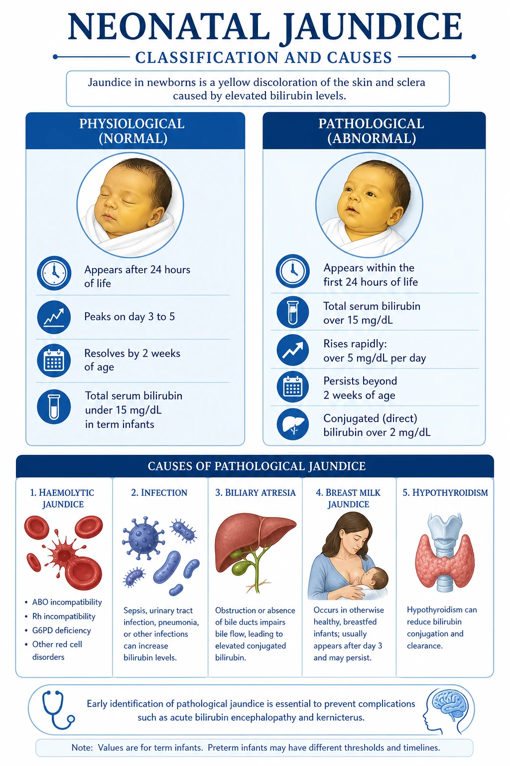
Neonatal jaundice is classified along two independent axes. The biochemical axis splits it into unconjugated and conjugated hyperbilirubinaemia, which tells you the mechanism and the danger. The behavioural axis splits it into physiological and pathological, which tells you whether to reassure the parents or to investigate urgently.[1]
Physiological
benign adaptation
- Appears **AFTER 24 hours** of age (usually day 2 to 3)
- Peaks at **day 3 to 5** (term) or **day 5 to 7** (preterm)
- TSB under **12 to 15 mg per dL** (205 to 256 micromol per L) in a term infant
- Rises less than **5 mg per dL per day**
- Resolves by **14 days** (term) or **21 days** (preterm)
- Caused by normal neonatal physiology: high red-cell load, immature glucuronyl transferase, increased enterohepatic circulation
- Always unconjugated; the baby is otherwise well and feeding
Pathological
investigate urgently
- Appears **WITHIN the first 24 hours** (always pathological)
- TSB **over 15 mg per dL** in a term infant or **over 12 mg per dL** in a preterm infant
- Rises **more than 5 mg per dL per day** (more than 0.2 to 0.3 mg per dL per hour)
- Persists **beyond 14 days** (term) or **21 days** (preterm) — prolonged jaundice
- **Conjugated (direct) fraction over 2 mg per dL** or over 20% of total
- Causes: haemolysis (ABO, Rh, G6PD), sepsis, biliary atresia, TORCH, metabolic disease, hypothyroidism
- The baby may be unwell — lethargy, poor feeding, fever or hypothermia, dark urine, pale stool
By bilirubin type
mechanism and danger
- **Unconjugated (indirect, about 85%):** physiological, prematurity, ABO or Rh haemolysis, G6PD deficiency, breast milk jaundice, cephalohaematoma, Crigler-Najjar, Gilbert, hypothyroidism
- **Conjugated (direct, about 15%):** biliary atresia, neonatal hepatitis, choledochal cyst, TPN cholestasis, TORCH infections, alpha-1-antitrypsin deficiency, galactosaemia, tyrosinaemia
- **Unconjugated = kernicterus risk** (fat-soluble, crosses BBB). **Conjugated = no kernicterus** but always means liver or biliary disease
The single rule that protects every jaundiced newborn is that physiological jaundice is a diagnosis of exclusion: it can be made only when the timing, level, rate of rise, duration and bilirubin fraction all fit, and the baby is clinically well. Anything else is pathological and must be investigated.[1]
Epidemiology & Risk Factors
Neonatal jaundice — the numbers that frame the problem
Neonatal jaundice is near-universal. Visible jaundice occurs in about 60% of term and 80% of preterm infants, and severe hyperbilirubinaemia (TSB over 25 mg per dL) develops in roughly 1 to 2% of term infants in high-income settings. It is the most common cause of neonatal readmission in the first week. Kernicterus is now rare in countries with routine anti-D prophylaxis, blood-group screening and phototherapy — incidence about 0.4 to 2 per 100,000 live births — but it remains a leading, and preventable, cause of brain injury in settings without these safeguards.[1][4]
The risk factors for significant hyperbilirubinaemia cluster around four themes. Prematurity and low gestational age lower the threshold for both visible jaundice and treatment, because the preterm liver conjugates bilirubin even less efficiently and the preterm blood-brain barrier is more permeable. Haemolysis — ABO or Rh incompatibility, glucose-6-phosphate dehydrogenase (G6PD) deficiency, hereditary spherocytosis — multiplies the bilirubin load several-fold and is the dominant cause of dangerous early jaundice. Feeding and weight loss: exclusive breastfeeding, suboptimal intake, delayed meconium passage and excessive weight loss all increase enterohepatic reabsorption of bilirubin (the so-called breastfeeding jaundice of the first week). Clinical context adds instrumental or traumatic delivery, cephalohaematoma or extensive bruising (a large enclosed blood load resorbs into bilirubin), a sibling who needed phototherapy, East Asian ethnicity, and maternal diabetes. A haemolysing baby with a raised reticulocyte count, falling haemoglobin or a positive direct antiglobulin test (DAT) is on a different trajectory from a well, feeding, term infant, and the nomogram must be read with that risk in mind.[1]
India and South Asia: neonatal jaundice remains a major cause of morbidity. G6PD deficiency is common in certain communities and is a leading cause of severe hyperbilirubinemic kernicterus; many regions now screen at-risk populations.[8] Cultural practices — offering water, glucose or honey instead of frequent breastfeeding — worsen dehydration and enterohepatic circulation. Home deliveries without bilirubin monitoring delay detection of severe jaundice until kernicterus has already occurred. Biliary atresia frequently presents late, with the Kasai performed beyond 60 days, which halves its success.[6] Access to reliable phototherapy in rural areas is uneven, and exchange transfusion may be delayed by lack of blood. Newborn screening (TSH, G6PD) is expanding but coverage is variable. The WHO and the Indian Academy of Pediatrics promote universal pre-discharge bilirubin assessment, G6PD screening in high-prevalence zones, and phototherapy at the primary-care level to close this gap.[8]
Pathophysiology
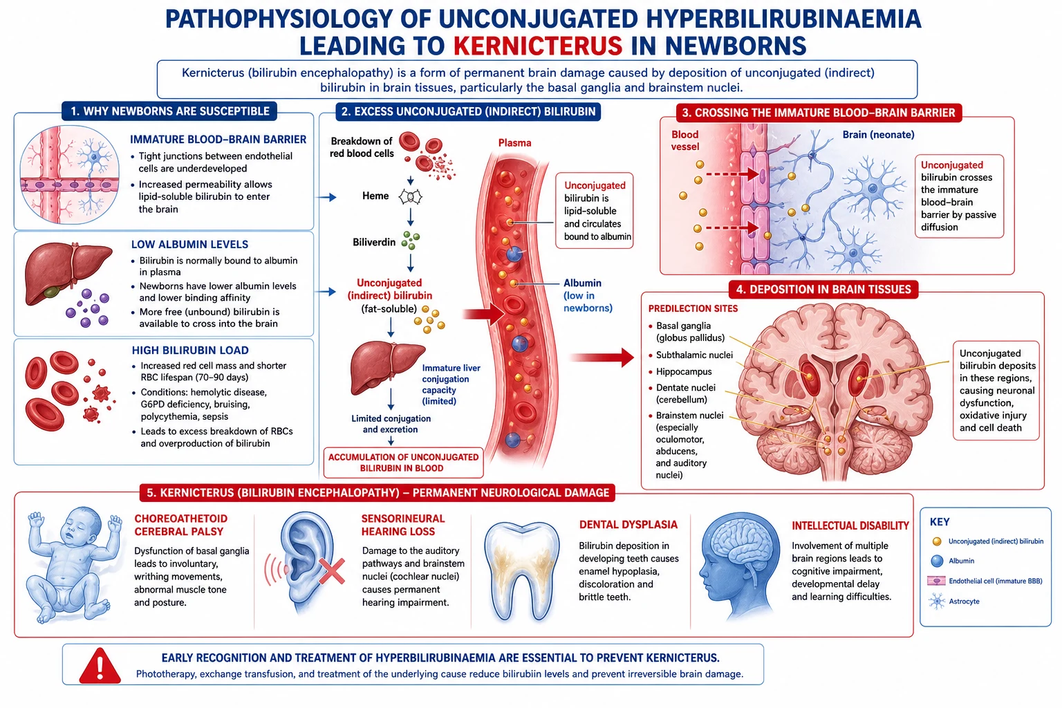
Understanding neonatal jaundice means tracing one molecule — bilirubin — from its origin to its danger. Bilirubin is the end-product of haem catabolism. When senescent red blood cells are broken down (chiefly in the spleen), their haemoglobin is split; the haem moiety is cleaved by haem oxygenase to biliverdin, which is in turn reduced by biliverdin reductase to unconjugated bilirubin. This unconjugated bilirubin is lipid-soluble, only sparingly soluble in water, and circulates in plasma tightly bound to albumin. It is taken up by the liver, where the microsomal enzyme uridine diphosphate glucuronyl transferase (UGT1A1) conjugates it with one or two molecules of glucuronic acid, producing conjugated (direct) bilirubin, which is water-soluble. Conjugated bilirubin is actively secreted across the canalicular membrane into bile, flows into the gut, and is converted by gut bacteria into urobilinogen and then stercobilin (the brown pigment of stool). A small fraction is reabsorbed and re-excreted by the liver — the enterohepatic circulation.[1]
The neonatal liver-gut system is, by design, several steps behind an adult's, and this immaturity is the basis of physiological jaundice. Three mechanisms combine:[1]
- Increased bilirubin load. Neonates are born with a larger red-cell mass (haematocrit 50 to 60%) and a shorter red-cell lifespan — about 70 to 90 days versus 120 days in adults — so they generate roughly two to three times the bilirubin load per kilogram.
- Immature hepatic conjugation. UGT1A1 activity at birth is only about 1% of adult activity and does not reach adult levels until roughly 6 to 12 weeks of age. The conjugating bottleneck is therefore most severe in the first days of life.
- Increased enterohepatic circulation. The neonatal gut is sterile at birth and slow to transit; meconium holds bilirubin; and beta-glucuronidase present in breast milk and the immature gut deconjugates bilirubin in the intestinal lumen, regenerating lipid-soluble unconjugated bilirubin that is reabsorbed into the portal circulation and recycled to the liver. Delayed passage of meconium, suboptimal intake and dehydration all amplify this loop.[1]
When production outstrips conjugation, unconjugated bilirubin accumulates. Most of it is bound to albumin and harmless; the danger is the small free (unbound) fraction, which is lipid-soluble and crosses cell membranes. Two further neonatal factors worsen the danger: low serum albumin (less binding capacity) and an immature blood-brain barrier with increased permeability. When free unconjugated bilirubin crosses the blood-brain barrier it is taken up by neurons and deposits preferentially in the basal ganglia (especially the globus pallidus and subthalamic nucleus), the brainstem auditory nuclei, the oculomotor nuclei, the hippocampus and the cerebellum. This deposition causes kernicterus — literally "yellow nuclei" — bilirubin-induced neuronal injury.[1][7]
The clinical corollary is decisive: only unconjugated bilirubin causes kernicterus. Conjugated bilirubin is water-soluble, cannot cross the barrier, and is excreted harmlessly into bile and urine (which it turns dark). This is why a raised conjugated fraction, although always pathological, is dangerous for a different reason — it signals biliary obstruction that, if not relieved, leads to cirrhosis and liver failure, not to brain injury. Prematurity adds a special hazard: preterm infants develop kernicterus at lower TSB levels than term infants — the so-called low-bilirubin kernicterus — because their blood-brain barrier is more permeable and their albumin lower, so a "safe" level on a term nomogram may still injure a 30-week infant.[7]
Clinical Presentation
The visible sign of neonatal jaundice is yellow discolouration of the skin and sclera that advances in a characteristic cephalocaudal (head-to-toe) direction, a progression formalised by Kramer's rule. Because jaundice is visible only when TSB exceeds about 5 to 7 mg per dL, and because visual estimation is unreliable (it over-reads in bright light and under-reads in dark skin), Kramer's rule is a screening tool only: a serum bilirubin must always confirm it.[1]
[1]A well baby with physiological jaundice looks otherwise normal — alert, feeding, afebrile, passing yellow stools and clear urine. The features that change the picture from "reassure" to "investigate urgently" are the tempo, the type of jaundice, and the general condition:[1]
- Early onset (within 24 h): jaundice in the first day is pathological until proven otherwise; think ABO or Rh haemolysis, G6PD deficiency, or sepsis.[1]
- A sick infant: lethargy, poor feeding, hypotonia, irritability, fever or hypothermia, grunting or apnoea — these suggest sepsis or acute bilirubin encephalopathy and are emergencies.[1]
- Signs of acute kernicterus (bilirubin encephalopathy): in escalating order, lethargy, poor suck, hypotonia, a high-pitched cry, irritability, then hypertonia with back arching (opisthotonus), seizures, apnoea and ultimately coma. A baby showing these signs needs an immediate exchange transfusion, not further investigation.[1][7]
- Conjugated jaundice clues: pale, clay-coloured (acholic) stools (no stercobilin reaches the gut — the single most important sign of biliary atresia), dark yellow-brown urine that stains the nappy (bilirubinuria), and hepatomegaly with a firm liver edge. A persistently jaundiced infant with pale stools must be referred urgently for surgical exclusion of biliary atresia.[6]
- Haemolysis clues: pallor (anaemia), splenomegaly or hepatomegaly, tachypnoea or heart failure from severe anaemia, and a history of a previous affected sibling or maternal blood-group incompatibility.[1]
Differential Diagnosis
The differential diagnosis follows the bilirubin type. Each cause has a distinguishing feature, and the bedside task is to match the clinical picture to one of them.[1]
| Cause | Bilirubin type | Key distinguishing feature |
|---|---|---|
| Physiological jaundice | Unconjugated | After 24 h, peaks day 3 to 5, under 15 mg per dL, resolves by 2 weeks; baby well |
| Breast milk jaundice | Unconjugated | Late onset (after day 7), persists for weeks, baby thriving; diagnosis of exclusion; resolves if breastfeeding paused 24 to 48 h (rarely needed) |
| Breastfeeding jaundice | Unconjugated | Early (day 3 to 5), inadequate intake, weight loss over 10%, dry stools; corrects with feeding support |
| ABO incompatibility | Unconjugated | Mother O, baby A or B; Coombs (DAT) positive; first pregnancy can be affected; mild to moderate |
| Rh (D) incompatibility | Unconjugated | Mother Rh-negative, baby Rh-positive; severe, often antenatal hydrops; Coombs strongly positive; preventable with anti-D |
| G6PD deficiency | Unconjugated | X-linked recessive (males), Mediterranean/Asian/African ancestry; triggered by drugs, fava beans, infection; Heinz bodies, bite cells; Coombs negative |
| Hereditary spherocytosis | Unconjugated | Autosomal dominant; spherocytes on film, raised osmotic fragility, Coombs negative; family history, splenomegaly |
| Cephalohaematoma or bruising | Unconjugated | Resorbing enclosed blood; rises day 2 to 4; instrumental delivery or birth trauma |
| Sepsis | Either | Unwell infant, fever or hypothermia, poor feeding, raised CRP, positive cultures; worsens jaundice by multiple mechanisms |
| Hypothyroidism | Either (often mixed) | Raised TSH on newborn screen; prolonged jaundice, constipation, hypotonia, large fontanelle, umbilical hernia |
| Crigler-Najjar syndrome type I | Unconjugated | Rare AR; absent UGT1A1; severe unconjugated jaundice from day 1 to 2; kernicterus risk; needs lifelong phototherapy or transplant |
| Crigler-Najjar type II (Arias) | Unconjugated | Partial UGT deficiency; milder; responds to phenobarbital |
| Gilbert syndrome | Unconjugated | Common, benign; reduced UGT; mild; contributes to prolonged jaundice |
| Biliary atresia | Conjugated | Term baby, 2 to 6 weeks old; pale stools, dark urine, hepatomegaly; Kasai before 60 days; progresses to cirrhosis |
| Neonatal hepatitis | Conjugated | Viral (TORCH), idiopathic; hepatocellular injury; stool may retain colour |
| Choledochal cyst | Conjugated | Cystic dilation of bile duct; palpable abdominal mass, intermittent jaundice |
| TPN cholestasis | Conjugated | Preterm infants on prolonged total parenteral nutrition; resolves slowly on enteral feeding |
| TORCH infections | Conjugated (often mixed) | Cytomegalovirus, toxoplasma, rubella, syphilis, herpes; petechiae, hepatosplenomegaly, thrombocytopenia, IUGR |
| Alpha-1-antitrypsin deficiency | Conjugated | Autosomal recessive (PiZZ); cholestasis then cirrhosis; emphysema in adults |
| Galactosaemia | Conjugated | Reducing substances in urine, cataracts, liver failure, E. coli sepsis; eliminate lactose |
| Tyrosinaemia | Conjugated | Liver failure, cabbage-like odour; treat with nitisinone |
A practical first split at the bedside: if the Coombs test is positive the cause is immune haemolysis (ABO or Rh); if Coombs negative but haemolysing think G6PD, hereditary spherocytosis or other non-immune haemolysis; if conjugated, refer for surgical exclusion of biliary atresia regardless of how well the baby looks.[1][6]
Clinical & Bedside Assessment
The bedside assessment of a jaundiced newborn has one purpose: to decide whether this baby is safe or needs investigation and treatment now. Three questions frame the encounter.[1]
1. How old is the baby in hours? Bilirubin thresholds are age-in-hours specific — a TSB of 12 mg per dL is normal at 96 hours but dangerous at 12 hours. Always express age in hours, not days, when reading the nomogram.[2]
2. How does the baby look? A full set of observations and a focused examination: temperature, activity, suck, tone, cry; weight and the percentage lost since birth; feeding pattern and stool/urine output; anterior fontanelle (dehydration); liver and spleen size; skin for bruising, cephalohaematoma or petechiae; any focus of infection. Lethargy, poor feeding, high-pitched cry or abnormal tone shifts the assessment immediately to "acute bilirubin encephalopathy until proven otherwise."[1]
3. What are the risk factors? Gestational age; maternal and baby blood group; a previous sibling with jaundice, anaemia or hydrops; exclusive breastfeeding and intake; instrumental delivery, bruising or cephalohaematoma; ethnic ancestry (East Asian, Mediterranean, African — G6PD); maternal diabetes; a family history of haemolysis or inherited red-cell disorders.[1]
The five questions that decide whether to treat
The Bhutani nomogram is the central interpretive tool: it plots TSB against age in hours for term and near-term infants and assigns a percentile risk — low (under 40th), intermediate (40th to 95th) or high (over 95th) — for developing significant hyperbilirubinaemia. A predischarge bilirubin in the high-risk zone predicts later treatment with high sensitivity, which is why universal predischarge bilirubin screening is now recommended.[2]
Investigations
Investigation is targeted to the clinical picture. Every jaundiced newborn needs a total and fractionated serum bilirubin; the rest follows the timing, level and bilirubin type.[1]
| Test | Why | When |
|---|---|---|
| Total serum bilirubin (TSB) with direct/indirect fractions | Confirms jaundice, defines type and severity; plot on Bhutani nomogram | Every jaundiced baby; transcutaneous bilirubin (TcB) acceptable for screening, confirm with TSB if high or preterm |
| Maternal and baby blood group + Rh | Identifies ABO and Rh incompatibility | Jaundice in first 24 h, or any haemolysis pattern |
| Direct antiglobulin test (DAT / direct Coombs) | Antibodies coating baby's red cells = immune haemolysis (ABO, Rh) | Suspected haemolysis; positive in Rh and some ABO |
| Full blood count + reticulocytes + blood film | Anaemia and raised reticulocytes = haemolysis; spherocytes, bite cells, Heinz bodies | Any haemolysis picture |
| G6PD assay | X-linked haemolysis; do during acute episode and repeat when stable | Male, Mediterranean/Asian/African, family history, unexplained haemolysis |
| Sepsis screen (blood culture, CRP, urine culture, LP if indicated) | Sepsis worsens and mimics jaundice | Unwell infant, premature, prolonged rupture of membranes |
| Thyroid function (TSH, free T4) | Congenital hypothyroidism (newborn screen) | Prolonged jaundice |
| Liver function tests (ALT, AST, ALP, GGT, albumin) | Hepatocellular vs cholestatic pattern; GGT very high in biliary atresia | Conjugated hyperbilirubinaemia |
| Hepatobiliary ultrasound | Triangular cord sign, small/absent or abnormal gallbladder in biliary atresia; choledochal cyst | Conjugated jaundice, pale stools |
| HIDA (hepatobiliary scintigraphy) | No bowel excretion of radiotracer = biliary atresia (does not distinguish from severe hepatitis) | Suspected biliary atresia (often after ultrasound) |
| Liver biopsy / intraoperative cholangiography | Definitive diagnosis of biliary atresia at laparotomy | When imaging equivocal; cholangiography is the gold standard |
| TORCH, metabolic (galactosaemia, alpha-1-AT, tyrosinaemia) screen | Infective and metabolic conjugated jaundice | Conjugated jaundice not fitting biliary atresia |
A raised direct (conjugated) bilirubin — defined as over 1 mg per dL if total under 5, or over 20% of total, or over 2 mg per dL absolute — is never physiological and mandates urgent hepatobiliary evaluation to exclude biliary atresia, because the Kasai operation's success falls sharply after 60 days of age.[1][6]
Management — Resuscitation
Resuscitation in neonatal jaundice means one thing: recognising and treating the bilirubin level that is about to cause brain injury, before it does. The dangerous phenotype is the rapidly rising unconjugated bilirubin from haemolysis, and the single emergency is acute bilirubin encephalopathy, where every hour of delay in lowering the bilirubin adds permanent neurological damage.[1]
The resuscitation sequence for a dangerously jaundiced infant is:[1]
- Assess airway, breathing, circulation — a baby with encephalopathy may have apnoea and need respiratory support.
- Take blood for TSB with fractions, blood group, Coombs, FBC, reticulocytes, crossmatch, G6PD, sepsis screen, and venous blood gas and glucose.
- Start intensive (double or triple) phototherapy immediately — every minute under blue light converts bilirubin to water-soluble photoisomers.[3]
- Correct dehydration and hypoglycaemia, support feeding; maintain temperature (phototherapy overheads warm, naked baby loses heat).
- If the bilirubin is at or above the exchange transfusion threshold, or there are signs of encephalopathy — proceed to exchange transfusion without delay.[1]
- In immune haemolysis, give intravenous immunoglobulin (IVIG) to interrupt haemolysis while arranging exchange.[5]
Management — Definitive & Stepwise
Definitive management is driven entirely by the bilirubin level read against the age-, gestation- and risk-specific threshold. Three interventions — phototherapy, intravenous immunoglobulin and exchange transfusion — form a ladder, with phenobarbital and feeding support as adjuncts and the underlying cause treated in parallel.[1]
Phototherapy — first-line treatment
Phototherapy is the single most important treatment of unconjugated hyperbilirubinaemia. Blue light in the 425 to 475 nm absorption band of bilirubin converts unconjugated bilirubin in the skin into water-soluble photoisomers — chiefly lumirubin and 4Z,15E-bilirubin — that are excreted in bile and urine without needing hepatic conjugation. The effect is photochemical, not thermal, and is proportional to the irradiance and the surface area of skin exposed.[3][9]
Phototherapy done correctly
1 — Confirm the indication
Plot TSB on the Bhutani nomogram against age in hours, gestation and risk factors. Start phototherapy when the level is **at or above the phototherapy threshold** for that baby (AAP 2022 graphs; NICE threshold graphs). Never start for "borderline" without checking the nomogram — overtreatment separates mother and baby unnecessarily.
2 — Maximise exposure
Place the baby in a **bassinet under the light source**, naked except for a **small nappy** and **eye shields** (to protect the retina). Use **intensive phototherapy**: two or more light sources from above and below (Bilibed, fibre-optic blanket under the back), or a single high-intensity LED unit delivering at least **30 microwatts per square centimetre per nanometre**.
3 — Protect the eyes and monitor
**Eye shields must be in place** whenever the light is on. Monitor **temperature** (risk of overheating or hypothermia in a naked infant), **hydration** (insensible water loss rises), **feeding and weight**, and **stool/urine output**. Continue breastfeeding; supplement with expressed milk or formula only if intake is inadequate.
4 — Re-measure bilirubin
Check TSB at **4 to 6 hours** after starting, then every **6 to 12 hours** depending on the level and trajectory. A rise despite intensive phototherapy signals ongoing haemolysis — consider IVIG (immune) or exchange transfusion.
5 — Stop when safe
Discontinue when TSB is **at least 1 to 2 mg per dL below the threshold** and the trajectory is downward. Re-check 12 to 24 hours after stopping, as rebound can occur, especially in haemolysis and prematurity.
Contraindications and cautions: avoid phototherapy when the direct (conjugated) bilirubin is very high (over 4 to 5 mg per dL) because photoisomers can transiently worsen the bronze baby syndrome — a dark grey-brown discolouration of skin, serum and urine that resolves slowly; conjugated jaundice needs hepatobiliary investigation, not light. Watch for temperature instability, fluid loss and interruption of breastfeeding.[3]
Exchange transfusion — for severe or escalating disease
Exchange transfusion is the treatment of severe hyperbilirubinaemia, of bilirubin that rises despite intensive phototherapy, and of acute bilirubin encephalopathy. It removes bilirubin and circulating antibodies and corrects anaemia in one procedure.[1]
Exchange transfusion
Dose
A **double-volume exchange** (160 mL per kg for a term infant) of **CMV-negative, crossmatch-compatible, irradiated, leucodepleted, fresh (under 7 days) red cells** reconstituted to a haematocrit of about 40 to 50%. Performed in **aliquots of 5 to 10 mL per kg**, drawn out and replaced over 30 to 90 minutes.
Intravenous immunoglobulin (IVIG) — for immune haemolysis
Intravenous immunoglobulin (IVIG)
Dose
**0.5 to 1 g per kg** over 2 hours, repeat once after 12 hours if the bilirubin continues to rise (some protocols use a single 1 g per kg dose).
Phenobarbital — enzyme induction
Phenobarbital
Dose
**5 mg per kg per day** for **3 to 5 days** (some regimens use 3 to 5 mg per kg once daily).
Hydration, feeding and adjuncts
Adequate hydration and feeding are not optional. Dehydration and suboptimal intake increase enterohepatic circulation; correcting them lowers bilirubin. Continue breastfeeding throughout phototherapy; supplement with expressed breast milk or formula only when intake is inadequate. Routine IV fluids are not needed unless the baby is dehydrated or unable to feed. There is no role for albumin infusions, glycerine suppositories, or herbal remedies as routine treatment; aggressive feeding support is the evidence-based adjunct.[1]
Treat the underlying cause
The definitive therapy is the one that stops bilirubin generation or obstruction: anti-D immunoprophylaxis for Rh-negative mothers prevents sensitisation and the next affected pregnancy; intrauterine transfusion rescues the severely hydropic fetus; antibiotics treat sepsis; thyroxine treats congenital hypothyroidism; eliminating galactose treats galactosaemia; and the Kasai portoenterostomy restores bile flow in biliary atresia. Without addressing the cause, phototherapy is only a holding measure.[6]
Specific Subtypes & Scenarios
Biliary atresia — the conjugated jaundice not to miss
Biliary atresia is a progressive fibro-obliterative cholangiopathy of unknown cause in which the extrahepatic (and eventually intrahepatic) bile ducts are destroyed, producing complete obstruction. A term, previously well infant develops conjugated jaundice, pale (acholic) stools, dark urine and hepatomegaly between 2 and 6 weeks of age. The diagnosis rests on hepatobiliary ultrasound (abnormal or absent gallbladder, triangular cord sign), HIDA scan (no bowel excretion), and liver biopsy (bile duct proliferation, portal fibrosis), confirmed by intraoperative cholangiography. The definitive treatment is the Kasai portoenterostomy, in which the atretic ducts are excised and a Roux loop of jejunum is anastomosed to the porta hepatis to restore bile drainage. The single most important prognostic factor is age at surgery: success is highest when the Kasai is performed before 30 to 60 days of age, and falls steadily thereafter. Even with a successful Kasai, many children develop biliary cirrhosis and ultimately need liver transplantation; biliary atresia remains the commonest indication for paediatric liver transplant worldwide.[6]
Haemolytic disease of the newborn — ABO and Rh
ABO incompatibility is the commonest immune haemolysis: a group O mother has naturally occurring anti-A and anti-B IgG that cross the placenta and haemolyse the red cells of a group A or B baby. It is usually mild to moderate, can affect the first pregnancy, and produces a positive DAT with spherocytes on the film. Rh (D) incompatibility requires prior sensitisation: a Rh-negative mother is exposed to Rh-positive red cells (at delivery, miscarriage, antepartum bleed or transfusion) and mounts anti-D IgG that, in a subsequent Rh-positive pregnancy, crosses the placenta and causes severe haemolysis ranging through anaemia, compensatory extramedullary haematopoiesis (hepatosplenomegaly), hydrops fetalis and stillbirth. The DAT is strongly positive. Anti-D immunoprophylaxis (anti-D immunoglobulin to the Rh-negative mother at 28 weeks and within 72 hours of delivery, and after any sensitising event) has reduced Rh disease dramatically where it is used. Intrauterine transfusion is reserved for severe mid-trimester anaemia detected by middle cerebral artery peak systolic velocity Doppler.[1]
G6PD deficiency — a South Asian and Mediterranean hazard
Glucose-6-phosphate dehydrogenase deficiency is the commonest enzymopathy of red cells, inherited as an X-linked recessive trait, so it most often affects males. The enzyme normally regenerates NADPH, which keeps glutathione reduced and protects red cells from oxidative damage. When it is deficient, an oxidative trigger — a drug (sulphonamides, primaquine, nitrofurantoin, nalidixic acid, methylene blue, aspirin in high dose), fava beans, henna, or an infection — causes haemoglobin to denature and precipitate as Heinz bodies, the cells are removed by the spleen, and acute haemolysis follows with a sudden spike in unconjugated bilirubin. The Coombs test is negative (this is non-immune haemolysis), the blood film shows bite cells and Heinz bodies, and the assay confirms low enzyme activity. Kernicterus from G6PD haemolysis remains a significant problem in South Asia and the Mediterranean, where routine G6PD screening of at-risk newborns is advocated to prevent it. Management is avoidance of triggers, phototherapy and exchange transfusion for dangerous levels.[8]
Breast milk jaundice versus breastfeeding jaundice
These two common entities are easily confused but distinct. Breastfeeding jaundice (suboptimal intake jaundice) occurs in the first week, peaks with the weight loss, and is driven by inadequate intake, dehydration, delayed meconium passage and increased enterohepatic circulation; it is corrected by feeding support (more frequent feeds, lactation review, supplementation if needed), not by stopping breastfeeding. Breast milk jaundice is a late, benign, prolonged unconjugated jaundice of the thriving breastfed infant, beginning after the first week and persisting for weeks to a couple of months; it is thought to relate to factors in breast milk that inhibit conjugation or increase enterohepatic reabsorption. It is a diagnosis of exclusion after haemolysis, hypothyroidism, infection and conjugated causes are ruled out; it needs no treatment, and breastfeeding should continue. A useful (but rarely needed) confirmatory test is a transient fall in bilirubin when breastfeeding is paused for 24 to 48 hours.[1]
Prolonged jaundice — the framework
Jaundice persisting beyond 14 days in a term infant or 21 days in a preterm infant is prolonged jaundice and must be investigated rather than dismissed. The first split is unconjugated versus conjugated. Prolonged unconjugated causes include breast milk jaundice, hypothyroidism, G6PD deficiency, haemolysis, sepsis or UTI, and rare inherited disorders (Crigler-Najjar, Gilbert). Prolonged conjugated jaundice is always pathological and demands urgent hepatobiliary evaluation for biliary atresia (every day counts toward the Kasai deadline), neonatal hepatitis, TORCH infection, TPN cholestasis, alpha-1-antitrypsin deficiency, galactosaemia and metabolic disease. The most dangerous error in paediatrics is to reassure a parent about "breast milk jaundice" in a 4-week-old with pale stools who in fact has biliary atresia.[6]
Complications & Pitfalls
The headline complication is kernicterus (bilirubin encephalopathy), and its staging is worth memorising because it dictates how hard to push treatment.[1][7]
| Phase | Timing | Features | Reversibility |
|---|---|---|---|
| Early acute (phase 1) | First 1 to 2 days | Lethargy, poor suck, hypotonia, diminished Moro, mild high-pitched cry | Reversible with urgent exchange transfusion |
| Intermediate acute (phase 2) | Days 3 to 5 | High-pitched cry, irritability, hypertonia, back-arching (opisthotonus), fever, seizures | Partially reversible; act immediately |
| Advanced acute (phase 3) | After day 5 | Apnoea, coma, seizures, deep stupor, often death | Poorly reversible |
| Chronic kernicterus | Months to years | Choreoathetoid cerebral palsy, sensorineural hearing loss (often the first and most common deficit), gaze palsy (Parinaud syndrome / sunset sign), dental enamel dysplasia (green teeth), intellectual disability | Permanent — the goal of all treatment is to prevent this |
Other complications and pitfalls deserve specific mention:[1]
- Bronze baby syndrome: a dark grey-brown discolouration of skin, serum and urine seen in infants with high conjugated bilirubin given phototherapy; the photoisomers of conjugated bilirubin are brown. It is cosmetically alarming but usually benign and resolves as the liver recovers; it should prompt review of the conjugated fraction rather than discontinuation of phototherapy for unconjugated disease.[3]
- Apnoea of acute bilirubin encephalopathy: apnoea is a recognised feature of severe bilirubin neurotoxicity and may be the presenting sign; a jaundiced baby with apnoea needs an immediate exchange transfusion, not a sleep study.[7]
- Low-bilirubin kernicterus in preterm infants: the most treacherous pitfall. Preterm infants develop kernicterus at TSB levels considered "safe" in term infants, because their blood-brain barrier is more permeable and albumin lower. The threshold graph must be read for the correct gestation, and unwell preterm infants treated at lower levels.[7]
- Rebound after phototherapy: stopping phototherapy too early leads to a rebound, especially in haemolysis and prematurity; always re-check 12 to 24 hours after stopping.[3]
- Mislabelling pathological jaundice "physiological": the cardinal error. Jaundice in the first 24 hours, a rapidly rising bilirubin, a conjugated fraction, or a sick infant is never physiological.[1]
- Missed biliary atresia: reassuring "breast milk jaundice" in a 4-week-old with pale stools. Check the stool colour and the conjugated fraction in every prolonged jaundice.[6]
- Over-reliance on visual estimation: Kramer's rule under-reads in dark skin and in bright phototherapy light; always measure serum bilirubin.[1]
- Failing to read the nomogram by hours: a bilirubin of 13 mg per dL is normal at 96 hours but pathological at 12 hours.[2]
Prognosis & Disposition
Physiological jaundice resolves completely and has an excellent prognosis. Treated haemolytic jaundice that is brought below the encephalopathy threshold has a good prognosis, provided kernicterus has not developed. Once chronic kernicterus is established the damage is permanent: choreoathetoid cerebral palsy, sensorineural hearing loss (the most common and often the earliest detected by newborn hearing screening), dental dysplasia, gaze palsy and variable cognitive impairment, with life-long disability. Biliary atresia prognosis is dominated by the age at Kasai: successful bile drainage is achieved in roughly 50 to 70% when the Kasai is performed before 60 days and falls steeply thereafter; many children still progress to cirrhosis and need liver transplantation, which is curative.[6]
Disposition follows the bilirubin trajectory. Below the phototherapy threshold and clinically well: outpatient with a follow-up bilirubin at the appropriate interval and feeding support. At or above the phototherapy threshold: inpatient phototherapy; discharge when the level is 1 to 2 mg per dL below threshold and falling, with a planned re-check. At or above the exchange threshold, rising despite intensive phototherapy, or any sign of encephalopathy: neonatal unit, exchange transfusion, and senior paediatric and haematology input. Any conjugated hyperbilirubinaemia: paediatric surgical and hepatology referral for exclusion of biliary atresia, as a time-critical pathway. The safety-net for every discharged jaundiced newborn is a written warning to parents to return urgently if the baby is lethargic, not feeding, has a high-pitched cry, dark urine or pale stools.[1]
Special Populations
Preterm and low-birth-weight infants carry the highest risk of both severe hyperbilirubinaemia and kernicterus. They have lower UGT activity, lower albumin and a more permeable blood-brain barrier, and they tolerate high bilirubin worse. Treatment thresholds are lower for lower gestational ages, read directly from the AAP 2022 or NICE threshold graphs; the concept of a single "safe level" does not apply. Low-bilirubin kernicterus — brain injury at TSB levels below the classical exchange threshold — is a recognised entity in this group and is the principal reason the preterm threshold was not raised in the 2022 AAP revision.[7]
Late-preterm infants (34 to 36 weeks) are a particular trap: they look like term babies but behave like preterms, with slower maturation of conjugation, poorer feeding, greater weight loss and higher readmission rates. They are treated by the threshold graph for their gestation, not their appearance.[1]
G6PD-deficient infants need trigger avoidance (no fava beans, no henna, careful drug selection), parental education, and a low threshold for treatment of any acute haemolytic episode; in endemic regions they may justify targeted newborn screening.[8]
Infants of diabetic mothers have a higher rate of polycythaemia and jaundice; check haematocrit and treat polycythaemia if symptomatic.[1]
Exclusively breastfed infants have a higher baseline jaundice; the task is to support feeding, recognise suboptimal intake jaundice, and not abandon breastfeeding.[1]
Surgical and hepatobiliary infants (biliary atresia, choledochal cyst) are managed on a time-critical pathway: any conjugated jaundice in a baby under 8 weeks is a surgical referral, full stop.[6]
Evidence, Guidelines & Regional Differences
The two dominant guidelines are the American Academy of Pediatrics 2022 revision (PMID 35927462) and NICE Clinical Guideline CG98 (UK, updated 2024). They agree on the principles — universal pre-discharge bilirubin assessment, hour-specific nomogram interpretation, phototherapy then exchange as a ladder, and aggressive treatment of haemolysis — but differ in detail.[1]
The AAP 2022 revision (replacing the 2004 guideline) was a landmark. It raised the phototherapy thresholds slightly (judged safe after population modelling), introduced an explicit "escalation of care" framework for severely elevated bilirubin (the level at which one adds IVIG and prepares exchange), and — importantly — removed race-based threshold adjustment, instead using the baby's own risk factors (gestation, albumin, haemolysis, illness). Thresholds are presented as graphs by age in hours and gestational age. As an approximation for a 40-week, low-risk infant, the phototherapy threshold rises from about 12 mg per dL at 24 hours to about 17 to 18 mg per dL at 72 to 96 hours, and the exchange transfusion threshold from about 18 mg per dL at 24 hours to about 25 to 27 mg per dL at 96 hours; these are lower for lower gestational ages and for any risk factor (haemolysis, illness, low albumin).[1]
The NICE CG98 guideline (UK) expresses thresholds in micromol per L and provides separate threshold graphs by gestational age (under 38 weeks, 38 weeks or more) and by the baby's age in hours after birth. As an approximation for a 38-week-or-more infant, the phototherapy threshold rises from about 200 micromol per L (about 12 mg per dL) at 24 hours to about 300 micromol per L (about 17.5 mg per dL) at 96 hours, and the exchange transfusion threshold from about 340 micromol per L (about 20 mg per dL) at 24 hours to about 430 micromol per L (about 25 mg per dL) at 96 hours; again, lower for preterm infants.[1]
The conversion between units — 1 mg per dL equals 17.1 micromol per L — is a frequent exam point; learn it. Both guidelines stress that the nomogram must be read with the risk profile of the baby: a haemolysing, preterm, ill infant is treated at a lower level than a well, term, low-risk infant at the same age.[1]
Australia / New Zealand: practice broadly follows the AAP and NICE principles, with local hospital thresholds; universal bilirubin screening at 24 to 48 hours; strong focus on G6PD screening in high-risk populations (Mediterranean, Middle Eastern, South-East Asian, African); established biliary atresia surgical pathways at paediatric liver centres.[8]
Controversies persist: the ideal threshold for phototherapy in preterm infants (the 2022 AAP revision did not raise these as it did for term, fearing low-bilirubin kernicterus); the role of IVIG (Cochrane evidence is low-certainty); the duration of phototherapy after which rebound is unlikely; and the utility of unbound (free) bilirubin as a more direct measure of neurotoxicity than total bilirubin. The Bhutani nomogram (1999) remains the foundational interpretive tool, validated in healthy term and near-term infants; it is less directly applicable to sick or preterm babies, in whom the threshold graphs govern.[2][4][5]
Exam Pearls
Causes of PATHOLOGICAL neonatal jaundice — JAUNDICE
JAUNDICE
ALWAYS pathological — haemolysis until proven otherwise
immune haemolysis — Coombs (DAT) positive
sepsis, TORCH, UTI — unwell infant
conjugated jaundice, pale stools
non-immune haemolysis from oxidative triggers
early onset, weight loss, dehydration
prolonged jaundice, raised TSH on screen
galactosaemia, tyrosinaemia, alpha-1-antitrypsin
Self-test: a baby at 18 hours has a TSB of 14 mg per dL, all unconjugated. What do you do?
This is pathological jaundice — jaundice within the first 24 hours is always pathological, and 14 mg per dL at 18 hours is far above the 95th percentile for age. The most likely cause is immune haemolysis (ABO or Rh) given the early, rapid rise. Management: start intensive phototherapy immediately; take blood for fractionated bilirubin, blood group (mother and baby), Coombs (DAT), FBC, reticulocytes, film, G6PD and sepsis screen; plot on the Bhutani nomogram; assess hydration and feeding; and have a low threshold for IVIG (if Coombs positive and rising) and exchange transfusion (if the level reaches the exchange threshold or signs of encephalopathy appear). The cause of the next pregnancy is prevented by anti-D if Rh incompatibility.[1][2]
Exam application bank (NEET-PG / INICET)
One-line answer
Neonatal jaundice = yellow skin and sclera from elevated bilirubin in the first 28 days of life, visible when total serum bilirubin (TSB) exceeds 5 to 7 mg per dL (85 to 120 micromol per L). Jaundice within the FIRST 24 HOURS is always PATHOLOGICAL and demands urgent investigation for haemolysis and sepsis. Unconjugated (indirect) hyperbilirubinaemia (about 85%) is fat-soluble, crosses the immature blood-brain barrier, and causes kernicterus (acute bilirubin encephalopathy with lethargy, high-pitched cry, opisthotonus; chronic choreoathetoid cerebral palsy, sensorineural hearing loss). Conjugated (direct) hyperbilirubinaemia (about 15%) is water-soluble, does not cause kernicterus, but signals hepatobiliary disease — most importantly biliary atresia (pale stool, dark urine, conjugated jaundice) needing Kasai portoenterostomy before 60 days. Treatment is phototherapy (blue light converts [1]
Worked stems (answer without another resource)
Stem 1 — Classic presentation. Map symptoms to mechanism; name the first investigation and first treatment step with dose/route if drug therapy is standard. [1]
Stem 2 — Unstable / complicated. List red flags that force immediate resuscitation, theatre, ICU, antidote, or reperfusion — and what you do in the first 15 minutes. [1]
Stem 3 — Atypical group. Elderly, pregnancy, child, or immunocompromised: how presentation and thresholds change. [1]
Stem 4 — Differential trap. Name the three closest mimics and one discriminator for each. [1]
Stem 5 — Disposition. Who goes home with safety-netting, who is admitted, who needs HDU/ICU/theatre, and what follow-up is mandatory. [1]
Rapid viva checklist
- Definition + classification
- Pathophysiology chain
- Bedside signs / criteria
- Score with exact components (if any)
- Emergency bundle
- Definitive therapy with doses
- Complications of disease and of treatment
- Special populations
- Guideline/trial name if classic
- Three exam traps
Coverage self-check
If you cannot answer any stem above from this page alone, re-read the matching section — the page is intended to be self-sufficient for final-prof and NEET-PG/INICET questions on Neonatal Jaundice.
References
- [1]Kemper AR, Newman TB, Slaughter JL, et al. (American Academy of Pediatrics Subcommittee on Hyperbilirubinemia). Clinical Practice Guideline Revision: Management of Hyperbilirubinemia in the Newborn Infant 35 or More Weeks of Gestation Pediatrics, 2022.PMID 35927462
- [2]Bhutani VK, Johnson L, Sivieri EM. Predictive ability of a predischarge hour-specific serum bilirubin for subsequent significant hyperbilirubinemia in healthy term and near-term newborns Pediatrics, 1999.PMID 9917432
- [3]Maisels MJ, McDonagh AF. Phototherapy for neonatal jaundice N Engl J Med, 2008.PMID 18305267
- [4]Watchko JF. Vigintiphobia revisited Pediatrics, 2005.PMID 15930239
- [5]Zwiers C, van der Bom JG, Giers G, et al. Immunoglobulin for alloimmune hemolytic disease in neonates Cochrane Database Syst Rev, 2018.PMID 29551014
- [6]Tyraskis A, Ismail K, Davenport M, et al. Steroids after the Kasai procedure for biliary atresia: the effect of age at Kasai portoenterostomy Pediatr Surg Int, 2016.PMID 26590818
- [7]Watchko JF. The enigma of low bilirubin kernicterus in premature infants: why does it still occur, and is it preventable? Semin Perinatol, 2014.PMID 25267279
- [8]Arain YH, Iqbal SM, Majeed I, et al. Prevention of Kernicterus in South Asia: role of neonatal G6PD deficiency and its identification Indian J Pediatr, 2014.PMID 24763814
- [9]Hansen TWR, Maisels MJ, Ebbesen F, et al. Sixty years of phototherapy for neonatal jaundice - from serendipitous observation to standardized treatment and rescue for millions J Perinatol, 2020.PMID 31420582