Paediatrics · Paediatrics
Seizures in Children
Also known as Seizures in Children
Seizures affect 5% of children (at least one seizure); epilepsy affects 1%. Febrile seizures are most common (2-5% of children aged 6 months to 5 years). ILAE classification 2017: focal, generalised, unknown onset. Key syndromes: childhood absence (3Hz spike-wave, ethosuximide), juvenile myoclonic (valproate), infantile spasms (ACTH/vigabatrin). Status epilepticus: over 5 min — lorazepam → levetiracetam → phenytoin.
On this page & tools
Your progress
Saved locally on this device.
Practise this topic
Exam tags
Red flags
Overview
A seizure is the transient occurrence of signs and/or symptoms due to abnormal excessive or synchronous neuronal activity in the brain. Epilepsy is a chronic disorder of the brain defined by ANY ONE of the following (ILAE 2014): (1) at least two unprovoked seizures occurring more than 24 hours apart; (2) one unprovoked seizure with a probability of further seizures of at least 60 percent over the next 10 years; or (3) diagnosis of an epilepsy syndrome. [1]
Seizures in children span an enormous clinical spectrum — from a single self-limiting febrile convulsion in a toddler to drug-resistant developmental epilepsy syndromes and time-critical status epilepticus. Approximately 5 percent of children will have at least one afebrile seizure by age 16, while 1 percent have active epilepsy. Roughly 2 to 5 percent of children aged 6 months to 5 years experience a febrile seizure, making this the single most common seizure scenario an MBBS graduate will encounter. Distinguishing provoked from unprovoked seizures, benign from progressive syndromes, and epileptic from non-epileptic events is a core paediatric neurology competency. [1]
Definitions and Classification Terminology
Mastering the vocabulary is non-negotiable for the viva: [1]
- Provoked (acute symptomatic) seizure — occurring in close temporal relationship to a transient CNS insult (fever, trauma, electrolyte disturbance, hypoglycaemia, drug withdrawal, CNS infection). Risk of recurrence is lower; usually does NOT establish an epilepsy diagnosis.
- Unprovoked seizure — occurs without an identifiable proximate trigger. Two or more (more than 24 hours apart) define epilepsy.
- Focal seizure — origin within networks limited to one hemisphere.
- Generalised seizure — origin within and rapidly engaging bilaterally distributed networks.
- Aura — a focal seizure manifesting as subjective sensory/psychic phenomena; it is itself a seizure, NOT a warning.
- Postictal state — transient cognitive, behavioural, or motor disturbance after a seizure (Todd paresis, confusion, sleep, headache).
- Epileptic encephalopathy — the epileptic activity itself contributes to cognitive/behavioural impairment beyond what the underlying lesion causes (e.g., West, Lennox-Gastaut, Landau-Kleffner).
- Seizure threshold — the susceptibility of the brain to generate seizures; influenced by genetics, sleep, fever, metabolic state, and drugs. [1]
Pathophysiology of Seizures
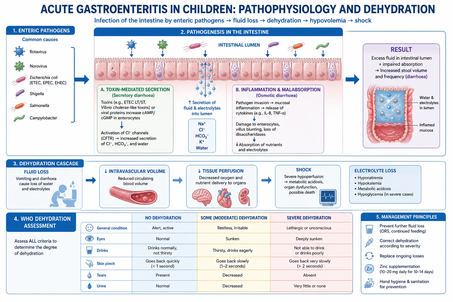
A seizure results from an imbalance between excitation (predominantly glutamatergic via NMDA, AMPA, and kainate receptors) and inhibition (predominantly GABAergic via GABA-A fast inhibition and GABA-B slow inhibition). The cellular events include: [1]
- Paroxysmal depolarising shift (PDS) — the intracellular hallmark of an epileptiform neuron; a large calcium-mediated depolarisation that triggers a burst of sodium action potentials, followed by a long after-hyperpolarisation.
- Synchronisation — gap junctions, recurrent excitatory collaterals, and loss of surround inhibition allow a focus to recruit neighbouring neurons.
- Propagation — spread through cortical and thalamocortical networks. The thalamocortical loop is central to generalised spike-wave discharges (typical absence): reciprocal connections between thalamic relay nuclei (GABAergic reticular nucleus) and cortical pyramidal cells generate the 3 Hz oscillation via T-type calcium channels (this is why ethosuximide, a T-type calcium channel blocker, is specific for absence).
- Termination — active inhibition (GABA release, neuronal Na-K ATPase, glial uptake), membrane pump fatigue, and endogenous anticonvulsant neuropeptides. Failure of these mechanisms produces status epilepticus. [1]
Genetic basis: Many childhood epilepsies are channelopathies — mutations in ion channel genes. Examples include SCN1A (Dravet — loss of function; paradoxically worsened by sodium channel blockers), SCN2A/SCN8A (benign familial neonatal-infantile seizures), KCNQ2/3 (benign familial neonatal seizures), GABA-A receptor subunit genes GABRA1, GABRG2 (febrile seizures plus, absence), SLC2A1 (GLUT1 deficiency — ketogenic diet responsive), CHRNA4 (autosomal dominant nocturnal frontal lobe epilepsy), and CDKL5, ARX, STXBP1 (early infantile epileptic encephalopathies). [1]
Structural causes include malformations of cortical development (focal cortical dysplasia, polymicrogyria, heterotopia), tumours (dysembryoplastic neuroepithelial tumour DNET, ganglioglioma), mesial temporal sclerosis (the commonest cause of focal epilepsy in adolescents/adults, often following prolonged febrile seizures in childhood), stroke, and traumatic brain injury. [1]
ILAE 2017 Classification of Seizures
The International League Against Epilepsy (ILAE) 2017 classification is the framework examiners expect. Seizures are first categorised by onset, then by awareness (for focal), and by motor vs non-motor features. The 2017 revision replaced older terms: "partial" became "focal", "simple partial" became "focal aware", "complex partial" became "focal impaired-awareness", and "secondarily generalised" became "focal to bilateral tonic-clonic". [1]
Step 1 — Onset
| Category | Definition |
|---|---|
| Focal onset | Originates within networks limited to ONE hemisphere; may spread |
| Generalised onset | Engages bilaterally distributed networks from onset |
| Unknown onset | Onset cannot be determined (e.g., nocturnal, unwitnessed) |
Step 2 — Focal onset subclassified by awareness and features
- Focal aware (formerly simple partial) — consciousness preserved; the patient is fully responsive during the event. Duration typically under 60 seconds.
- Focal impaired-awareness (formerly complex partial) — consciousness impaired; patient may stare, automate, be amnesic for the event. Duration typically 30 seconds to 2 minutes, often with postictal confusion.
- With motor onset: automatisms (lip-smacking, hand fumbling, picking at clothes — classical of temporal lobe), clonic, hyperkinetic, autonomic, tonic, atonic, versive (forced head/eye turn — frontal).
- With non-motor onset: sensory (somatosensory, olfactory, visual, auditory, gustatory — olfactory hallucinations are classical of mesial temporal), autonomic, cognitive, emotional (fear, deja vu, epigastric rising).
- Evolution to focal to bilateral tonic-clonic — the focal phase may evolve to a convulsion. [1]
Step 3 — Generalised onset
Motor:
- Generalised tonic-clonic (GTC) — tonic stiffening (10 to 20 seconds) then rhythmic clonic jerking (30 to 60 seconds); cyanosis, tongue-biting, urinary incontinence, postictal drowsiness.
- Clonic — rhythmic jerking without preceding tonic phase.
- Tonic — sustained bilateral stiffening.
- Myoclonic — brief, shock-like bilateral jerks.
- Atonic / drop attacks — sudden loss of postural tone; child drops "like a puppet with cut strings"; prone to facial/head injury; classical of Lennox-Gastaut and Doose syndrome.
- Epileptic spasms — the seizure type of West syndrome; may be classified as unknown onset if laterality unclear. [1]
Non-motor (absence):
- Typical absence — sudden brief (5 to 10 second) arrest of activity, blank stare, no postictal confusion; 3 Hz spike-wave on EEG; classical of CAE.
- Atypical absence — onset/offset less abrupt, longer duration, more pronounced tone changes; 1.5 to 2.5 Hz slow spike-wave; seen in Lennox-Gastaut.
- Myoclonic absence — absence with rhythmic myoclonic jerks.
- Eyelid myoclonia — jerking of eyelids with or without absence; hallmark of Jeavons syndrome (eyelid myoclonia with absence), photosensitive. [1]
Step 4 — Unknown onset
- Motor: tonic-clonic, epileptic spasms.
- Non-motor: autonomic, behaviour arrest.
- Unclassified. [1]
Step 5 — Aetiology (SIGMIU)
Every epilepsy should be assigned a likely cause under the six ILAE aetiology categories — examiners love this framework: [1]
| Category | Examples |
|---|---|
| Structural | Focal cortical dysplasia, mesial temporal sclerosis, tumour (DNET, ganglioglioma), stroke, tuberous sclerosis, Sturge-Weber |
| Genetic | Dravet (SCN1A), benign familial neonatal seizures (KCNQ2), JME (polygenic), CAE (GABA receptor), GEFS+ |
| Infectious | Neurocysticercosis, bacterial meningitis sequelae, viral encephalitis, cerebral malaria, tuberculosis |
| Metabolic | Hypoglycaemia, hypocalcaemia, hyponatraemia, pyridoxine dependency, GLUT1 deficiency, mitochondrial disorders |
| Immune | Anti-NMDA receptor encephalitis, anti-LGI1, anti-GAD, Rasmussen encephalitis |
| Unknown | Cause not yet identified |
SIGMIU
Febrile Seizures
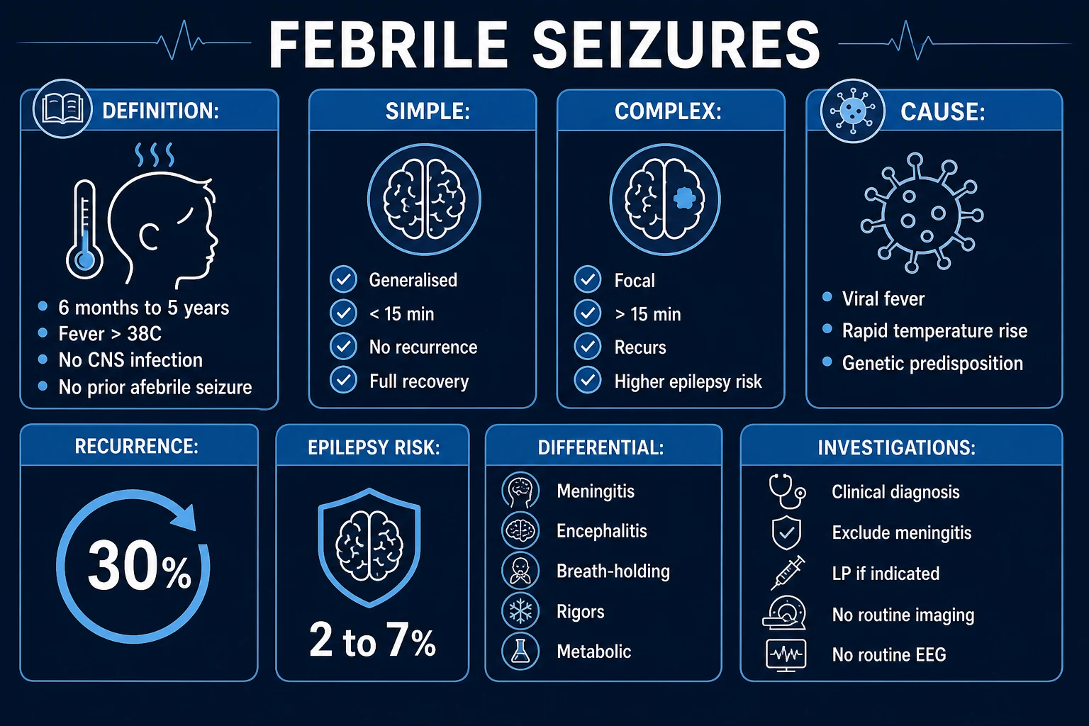
A febrile seizure is a seizure occurring in a child aged 6 months to 60 months (5 years), associated with a temperature of 38 degrees C or higher, without evidence of CNS infection, metabolic abnormality, or history of prior afebrile seizures. They are the commonest seizure type of early childhood, affecting 2 to 5 percent of children. Peak incidence is at 18 months; boys slightly predominate. [1]
Pathophysiology: An immature brain with developing myelination and GABAergic inhibition is uniquely susceptible to the convulsant effect of rapidly rising fever, independent of absolute temperature. Genetic predisposition is strong (autosomal dominant with incomplete penetrance; GEFS+ spectrum; SCN1A in some). The fever trigger classically is a viral upper respiratory infection, roseola (HHV-6), otitis media, or urinary tract infection — NOT the height of fever but the rapidity of rise. [1]
Simple versus Complex Febrile Seizures
A third category — febrile status epilepticus — is a febrile convulsion lasting over 30 minutes. This is a neurological emergency carrying a significantly higher risk of subsequent epilepsy and mesial temporal sclerosis, and warrants full SE protocol plus investigation for underlying cause. [1]
Risk of Recurrence (after a first febrile seizure)
About 30 to 40 percent of children with a first febrile seizure will have a recurrence, usually within the first year. The major recurrence risk factors are well established (AAP / NICE): [1]
- Age under 12 months at first seizure (the younger the child, the higher the recurrence risk)
- Family history of febrile seizures in a first-degree relative
- Low-grade fever or short fever-to-seizure interval at the first event (suggests a low seizure threshold)
- Complex features at first seizure [1]
The presence of zero, one, or two or more risk factors corresponds to an approximate recurrence risk of approximately 15 percent, 25 percent, and 50 percent or more respectively. Conversely, the risk of subsequently developing epilepsy (afebrile seizures) is about 2 to 3 percent after a simple FS but rises to 5 to 15 percent after a complex FS, and to over 20 to 50 percent if multiple complex features coexist (focal plus prolonged plus recurrent). [1]
Febrile seizure plus (FS+) and GEFS+: When febrile seizures persist beyond age 6 years or are accompanied by afebrile seizures, consider the GEFS+ (generalised epilepsy with febrile seizures plus) spectrum — an autosomal dominant familial syndrome linked to SCN1A, SCN1B, and GABA receptor mutations, ranging from typical febrile seizures through FS+ to Dravet syndrome at the severe end. [1]
Management of Febrile Seizures (AAP 2011, NICE CG160)
During the seizure:
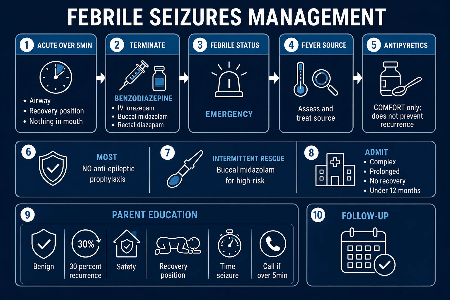
- Place child in recovery position (lateral, head supported), on a soft surface.
- Do NOT restrain, do NOT put anything in the mouth, do NOT attempt to force-feed fluids.
- Time the seizure; call emergency services if it lasts over 5 minutes.
- Antipyretics (paracetamol 15 mg/kg, ibuprofen 10 mg/kg) for the child's comfort and to treat fever — but antipyretics do NOT prevent seizure recurrence; this is a viva trap. [1]
Investigations — minimal for simple FS:
- No routine blood tests, EEG, neuroimaging, or lumbar puncture for a simple febrile seizure with a clearly identifiable fever source and a child who recovers promptly.
- Bloods only if prolonged seizure (glucose, FBC, U&E, calcium, magnesium, CRP), if dehydration is suspected, or if the child is under 6 months.
- Lumbar puncture should be considered in: any child under 12 months with fever and seizure (signs of meningitis may be subtle); children on antibiotics (may mask meningitis); those with persistent altered consciousness, meningeal signs, or signs of raised intracranial pressure (after CT if focal neurology). [1]
Intermittent prophylaxis (rescue medication):
- Buccal midazolam 0.5 mg/kg (max 10 mg) OR rectal diazepam 0.5 mg/kg for any seizure lasting over 5 minutes at home — parents are trained and provided with the drug.
- Intermittent oral clobazam or diazepam during febrile episodes is no longer recommended routinely; risk-benefit does not favour it for simple FS. [1]
Continuous AED prophylaxis (daily phenobarbitone, valproate) is NOT recommended for simple or complex febrile seizures because the risk of side effects outweighs the modest reduction in recurrence, and it does NOT prevent later epilepsy. [1]
Parental counselling (essential and frequently examined):
- Reassure: febrile seizures are common, usually benign, and do not cause brain damage.
- Explain the difference between febrile seizures and epilepsy.
- Discuss recurrence risk (30 to 40 percent) and epilepsy risk (2 to 3 percent for simple).
- Demonstrate the recovery position and teach use of buccal midazolam if provided.
- Fever control measures: antipyretics for comfort, light clothing, fluids — but stress these will NOT reliably prevent recurrence. [1]
Neonatal Seizures
Neonatal seizures differ from older children — the clinical semiology is often subtle, focal, and fragmented because the immature brain cannot sustain generalised synchronous discharges. They are the commonest neurological emergency in the neonatal period, occurring in 1 to 3 per 1000 live births, and frequently signal significant underlying brain injury. [1]
Aetiologies (in approximate order of frequency):
- Hypoxic-ischaemic encephalopathy (HIE) — the commonest cause, typically presenting within the first 24 to 48 hours of life.
- Metabolic — hypoglycaemia, hypocalcaemia, hypomagnesaemia, hyponatraemia, hypernatraemia; inborn errors of metabolism (amino acidopathies, organic acidaemias, urea cycle defects, mitochondrial); pyridoxine-dependent epilepsy (seizures refractory to all AEDs that cease within hours of IV pyridoxine 100 mg).
- Infection — bacterial meningitis (group B streptococcus, E coli), viral encephalitis (HSV, enterovirus), TORCH.
- Structural — perinatal arterial ischaemic stroke, intracranial haemorrhage (intraventricular in preterm), malformations of cortical development.
- Genetic/structural epileptic encephalopathies — benign familial neonatal seizures (KCNQ2/3), early infantile epileptic encephalopathy (Ohtahara, KCNQ2, STXBP1, ARX), early myoclonic encephalopathy.
- Drug withdrawal — maternal opioid, SSRI, or benzodiazepine use. [1]
Clinical classification (Volpe):
- Subtle — the commonest; cycling or rowing movements of limbs, mouthing, tongue thrusting, swimming movements of arms, ocular phenomena (eye deviation, blinking), apnoea. Easily missed and often confused with jitteriness.
- Clonic — rhythmic jerking, usually focal or multifocal, reproducible by touch (unlike jitteriness where stimulus stops it).
- Tonic — sustained posturing of limbs, trunk, or face; may mimic decerebration.
- Myoclonic — brief isolated jerks; may be focal, multifocal, or generalised; often carries a poor prognosis when associated with metabolic disease. [1]
Investigation:
- EEG (ideally continuous video-EEG) — to confirm epileptic origin (many "subtle" events are non-epileptic); interictal abnormalities help localise.
- Bedside glucose, electrolytes (Na, K, Ca, Mg), blood gas, ammonia, lactate, TORCH, septic screen, CSF (cells, protein, glucose, culture, HSV PCR, amino acids in CSF).
- MRI brain — for HIE, stroke, malformations.
- Trial of pyridoxine 50 to 100 mg IV for refractory seizures.
- Genetic/metabolic testing if refractory or family history. [1]
Management:
- Treat the underlying cause first — correct glucose (10 percent dextrose 2 mL/kg), calcium (10 percent calcium gluconate 1 to 2 mL/kg slowly with cardiac monitoring), magnesium, electrolyte disturbances; antibiotics and aciclovir if infection suspected.
- First-line AED: IV phenobarbitone 20 mg/kg loading (then 3 to 5 mg/kg/day maintenance) — still first-line in most units despite concerns about neuroapoptosis.
- Second-line: IV phenytoin 20 mg/kg loading OR IV fosphenytoin (safer) OR IV levetiracetam 40 to 60 mg/kg (increasingly preferred due to safety profile and IV availability).
- Refractory: IV midazolam infusion 0.1 to 0.4 mg/kg/hour, or IV lidocaine infusion (avoid with phenytoin).
- Therapeutic hypothermia (33.5 degrees C for 72 hours) for moderate-severe HIE reduces seizures and improves outcome. [1]
Prognosis: Highly aetiology-dependent. HIE with severe injury, structural lesions, and refractory neonatal seizures carry a poor prognosis (high rates of cerebral palsy, intellectual disability, and epilepsy). Metabolic seizures rapidly corrected have an excellent prognosis. Overall mortality 10 to 15 percent; about 25 to 35 percent of survivors develop epilepsy or neurodevelopmental impairment. [1]
Detailed Epilepsy Syndrome Profiles
Epilepsy syndromes are defined by a cluster of age of onset, seizure types, EEG features, imaging, and prognosis. Recognising the syndrome dictates the first-line drug and prognostic counselling. The seven syndromes below are the high-yield exam syndromes. [1]
1. Childhood Absence Epilepsy (CAE)
Epidemiology: Accounts for 10 to 17 percent of all childhood epilepsies. Age of onset 4 to 10 years (peak 5 to 7). Female-to-male ratio approximately 2:1. Strong polygenic susceptibility with overlap to other idiopathic generalised epilepsies. [1]
Clinical features:
- Multiple daily absence seizures — often 10 to 100 or more per day.
- Each episode: sudden cessation of activity, blank stare, rhythmic eyelid fluttering (3 Hz), sometimes mild automatisms of the hands or mouth. Duration 5 to 15 seconds (rarely over 30).
- No aura, no postictal confusion, no falls, immediate resumption of activity.
- Frequently unrecognised — the teacher reports the child is "daydreaming" or "inattentive", and school performance declines.
- Hyperventilation is a reliable trigger — use it in clinic: have the child blow repeatedly on a pinwheel or a glove for 3 minutes; an absence will often be provoked, and the 3 Hz spike-wave will appear on EEG. [1]
EEG findings:
- Regular, symmetrical 3 Hz generalised spike-and-wave discharges, bisynchronous, superimposed on a normal background.
- Discharges are activated by hyperventilation, drowsiness, and sometimes photic stimulation.
- Typical discharge lasts 3 to 20 seconds and correlates with the clinical absence. [1]
Treatment:
- Ethosuximide — first-line for PURE absence (no GTC seizures); blocks T-type calcium channels in thalamic relay neurons. Dose 20 to 40 mg/kg/day in two divided doses. Side effects: nausea, abdominal pain, headache, behavioural disturbance, rarely leucopenia and lupus-like syndrome.
- Valproate — if absence coexists with generalised tonic-clonic seizures (broader spectrum); 20 to 40 mg/kg/day. Avoid in females of childbearing potential.
- Lamotrigine — alternative; slow titration over 8 weeks to avoid rash (Stevens-Johnson risk).
- AVOID: carbamazepine, phenytoin, gabapentin, tiagabine, vigabatrin — these may WORSEN absence seizures (classic exam trap). [1]
Prognosis:
- Approximately 70 percent achieve complete remission by mid-adolescence.
- Predictors of remission: normal development, absence of GTC seizures, good response to first AED, no family history of epilepsy.
- About 30 percent evolve into juvenile absence epilepsy or juvenile myoclonic epilepsy — warn families that absences may transform in adolescence. [1]
2. Juvenile Myoclonic Epilepsy (JME)
Epidemiology: Accounts for 5 to 10 percent of all epilepsies; the commonest idiopathic generalised epilepsy of adolescence. Age of onset 12 to 18 years (peak 15). Equal sex ratio. [1]
Clinical features — the classical triad:
- Myoclonic jerks on awakening — brief, bilateral, irregular arm jerks, often in clusters; the patient may drop their morning cup of tea or toothbrush. Consciousness is preserved. The single most characteristic feature.
- Generalised tonic-clonic seizures — usually occur shortly after waking, often triggered by myoclonic clustering, sleep deprivation, or alcohol.
- Absence seizures in about 30 percent. [1]
Triggers (the "4 A's and S"): Alcohol, Antidepressant/psychotropic withdrawal, Arousal from sleep, Abnormally flickering light (photosensitivity), and Sleep deprivation (the dominant trigger). Normal intellect and neurological examination. [1]
EEG findings:
- 4 to 6 Hz irregular generalised polyspike-and-wave discharges on a normal background.
- Photosensitivity (paroxysmal response to photic stimulation) in 30 to 50 percent — a defining feature.
- Activated by sleep deprivation and hyperventilation. [1]
Treatment:
- Valproate — the most effective single agent, controlling all three seizure types in 80 to 90 percent; 500 to 2000 mg/day.
- Females: avoid valproate — teratogenic (neural tube defects, cardiac malformations, fetal valproate syndrome with autistic features and reduced IQ, dose-dependent), and risk of PCOS, weight gain. MHRA Pregnancy Prevention Programme (PPP) required if no alternative.
- Alternatives for females: levetiracetam (effective for myoclonus and GTC, broad spectrum, minimal interactions), lamotrigine (effective for GTC but may worsen myoclonus — combine with levetiracetam if needed), topiramate (effective but cognitive side effects, weight loss, nephrolithiasis, teratogenic), zonisamide.
- Lifestyle modification is non-negotiable: regular sleep (at least 8 hours), avoid sleep deprivation, avoid excess alcohol, avoid photic triggers (screen time in moderation, dark glasses for sunlight flicker). [1]
Prognosis:
- LIFELONG TREATMENT — over 90 percent relapse if AEDs are withdrawn, even after years of seizure freedom. This is a critical counselling point that examiners stress.
- Excellent seizure control on medication (80 to 90 percent seizure-free).
- Normal cognition and life expectancy when controlled. [1]
3. Benign Epilepsy with Centrotemporal Spikes (BECTS, Rolandic Epilepsy)
The commonest focal epilepsy of childhood, accounting for 10 to 15 percent of all childhood epilepsies. The term "benign" reflects the excellent prognosis — seizures remit by adolescence — though a subset has cognitive/behavioural comorbidity. [1]
Clinical features:
- Age of onset 3 to 13 years (peak 7 to 9). Boys slightly more affected.
- Hemifacial sensorimotor seizures — twitching of one side of the face and mouth, guttural speech sounds, hypersalivation, drooling, sometimes extension to the arm; seizures are exclusively or predominantly nocturnal and shortly after falling asleep or before waking.
- Often secondarily generalise during sleep.
- Daytime cognition, development, and neurological exam are normal. [1]
EEG findings:
- Centrotemporal (rolandic) spikes — high-amplitude biphasic sharp waves over the C3/C4 and T3/T4/T5/T6 regions, markedly activated by sleep.
- Background otherwise normal. [1]
Treatment:
- Many children need NO treatment (seizures are infrequent, nocturnal, and remit spontaneously). Decision to treat depends on seizure frequency, parental anxiety, and daytime generalisation.
- When treatment is needed: levetiracetam (first-line), carbamazepine, sulthiame, lamotrigine. Sulthiame has particular efficacy for BECTS.
- Avoid over-treatment with broad-spectrum drugs. [1]
Prognosis: Spontaneous remission by age 14 to 16 in virtually all patients. A minority has subtle language or learning difficulties that resolve as the epilepsy remits. [1]
Atypical BECTS variants: A small subset develops Landau-Kleffner syndrome or status epilepticus of BECTS (SEBECT) with cognitive/behavioural regression — blurring the line between BECTS and the epileptic encephalopathies. [1]
4. Infantile Spasms (West Syndrome)
One of the epileptic encephalopathies of infancy — urgent recognition is essential because prompt treatment improves developmental outcome. The classic triad is: (1) epileptic (salaam) spasms, (2) hypsarrhythmia on EEG, (3) psychomotor regression/regression of development. All three need not be present for the diagnosis (West syndrome = all three; infantile spasms = spasms alone). [1]
Clinical features:
- Age of onset peak 4 to 8 months (range 2 to 24 months).
- Salaam spasms — sudden, brief (1 to 3 seconds) flexion of the head, trunk, and arms (jackknife posture), often in clusters of 20 to 100, especially on awakening or after feeding. Extensor and mixed variants occur.
- Crying or irritability between spasms.
- Loss or regression of developmental milestones, loss of visual tracking (often preceding recognition of the spasms). [1]
EEG findings — hypsarrhythmia:
- Chaotic, high-voltage (over 300 microV), asynchronous slow waves intermixed with multifocal spikes — the most abnormal interictal EEG pattern in epilepsy.
- Modified/hypsarrhythmia variants occur, especially during sleep. [1]
Aetiology: Two-thirds are symptomatic — perinatal HIE, tuberous sclerosis complex (a major cause — vigabatrin first-line here), malformations of cortical development, congenital brain malformations, metabolic disease, prior CNS infection, chromosomal (Down, Aicardi). One-third are cryptogenic/idiopathic with better prognosis. [1]
Treatment (time-critical):
- ACTH (adrenocorticotropic hormone) — 150 units/m2/day IM for 2 weeks, then taper. First-line for cryptogenic/idiopathic. Side effects: hypertension, irritability, immunosuppression, GI bleed, electrolyte disturbance.
- High-dose oral prednisolone — 40 to 60 mg/day; alternative where ACTH unavailable.
- Vigabatrin — first-line if tuberous sclerosis complex is the underlying cause; 100 to 150 mg/kg/day. Mandatory visual field monitoring (irreversible concentric visual field defect in 30 to 50 percent).
- Ketogenic diet, hormonal therapy, and pyridoxine trial if refractory. [1]
Prognosis: Poor overall. Only about 25 to 50 percent achieve good developmental outcome. Predictors of poor outcome: symptomatic cause, delayed treatment, persistent hypsarrhythmia, evolution to Lennox-Gastaut. Up to 50 to 70 percent develop other forms of epilepsy (especially Lennox-Gastaut), and 70 to 90 percent have intellectual disability. [1]
5. Lennox-Gastaut Syndrome (LGS)
A severe epileptic encephalopathy of early childhood characterised by the triad: (1) multiple seizure types, (2) slow (1.5 to 2.5 Hz) spike-and-wave on EEG, (3) cognitive impairment with behavioural disturbance. [1]
Clinical features:
- Age of onset 1 to 8 years (peak 3 to 5). Often evolves from West syndrome (about 30 percent of LGS cases had infantile spasms).
- Multiple daily seizure types — the cardinal feature. Mix of: tonic seizures (especially nocturnal — essential to detect with overnight EEG), atonic drop attacks (causing falls and injury), atypical absence, myoclonic, and generalised tonic-clonic. Non-convulsive status epilepticus is common.
- Cognitive impairment — intellectual disability in nearly all; behavioural problems (hyperactivity, aggression, autistic features).
- Aetiology similar to West syndrome: structural (cortical malformation, hypoxic-ischaemic injury, tuberous sclerosis), metabolic, genetic; about one-third cryptogenic. [1]
EEG findings:
- Slow (1.5 to 2.5 Hz) generalised spike-and-wave on an abnormal, slow background.
- Paroxysmal fast activity (10 to 20 Hz) during sleep — the EEG hallmark of tonic seizures. [1]
Treatment — notoriously refractory:
- Valproate — broad-spectrum first-line.
- Lamotrigine — useful adjunct (caution: may worsen myoclonus).
- Topiramate, levetiracetam, zonisamide, clobazam — adjuncts.
- Rufinamide — specifically approved for LGS; effective for drop attacks.
- Felbamate — effective but reserved for refractory cases due to risk of aplastic anaemia and hepatic failure (blood monitoring).
- Cannabidiol (Epidiolex) — FDA-approved adjunct for LGS-associated seizures.
- Ketogenic diet — often very effective.
- Corpus callosotomy or vagus nerve stimulation (VNS) — surgical options for disabling drop attacks.
- AVOID: carbamazepine, oxcarbazepine, phenytoin, gabapentin — may worsen absence/myoclonus. [1]
Prognosis: Poor. Seizures persist into adulthood in over 90 percent; intellectual disability is the rule; shortened life expectancy due to seizure-related injuries, SUDEP, and aspiration. The goal of treatment is seizure reduction, safety (helmets for drop attacks), and quality of life rather than cure. [1]
6. Dravet Syndrome (Severe Myoclonic Epilepsy of Infancy, SMEI)
A genetic epileptic encephalopathy presenting in the first year of life, classically with prolonged fever-triggered seizures, evolving into a refractory multi-type epilepsy with developmental regression. [1]
Genetics: SCN1A mutation (loss of function of the sodium channel alpha-1 subunit) in 70 to 80 percent of cases; mostly de novo autosomal dominant. (Note: gain-of-function SCN2A causes a different, sodium-channel-blocker-responsive epilepsy.) [1]
Clinical evolution — the four stages:
- Febrile stage (0 to 12 months): Prolonged (over 30 minutes), fever-triggered, hemiclonic seizures (often alternating sides); sometimes febrile status epilepticus. Development initially normal.
- Worsening stage (12 to 24 months): Other seizure types appear — myoclonic, atypical absence, focal, atonic; seizures become afebrile and frequent. Developmental regression begins.
- Late childhood: Drug-resistant epilepsy, ataxia, pyramidal signs, moderate-to-severe intellectual disability, behavioural features of autism, hyperactivity, gait deterioration (crouch gait).
- Adolescence/adulthood: Seizure frequency may decline somewhat; cognitive and motor disability persists; risk of SUDEP is high. [1]
Provoking factors: Fever, hot baths, immunisation (the seizures, not the vaccine, are the issue — do NOT withhold vaccines; advise antipyretic cover), photosensitivity, pattern sensitivity. [1]
Treatment:
- Stiripentol combined with valproate + clobazam — first-line specific therapy; stiripentol inhibits clobazam metabolism and enhances GABAergic transmission.
- Fenfluramine — FDA-approved 2020; substantial reduction in convulsive seizure frequency.
- Cannabidiol (Epidiolex) — FDA-approved 2018 for Dravet.
- Ketogenic diet — adjunct.
- AVOID SODIUM CHANNEL BLOCKERS — carbamazepine, oxcarbazepine, lamotrigine, phenytoin — these WORSEN seizures in SCN1A loss-of-function. This is a high-yield exam point.
- Emergency rescue plan: buccal midazolam at home for prolonged seizures; emergency bracelet/ID. [1]
Prognosis: Lifelong drug-resistant epilepsy; intellectual disability in nearly all; reduced life expectancy; risk of SUDEP approximately 1 to 2 percent per year. Early diagnosis and avoidance of sodium channel blockers improve outcome. [1]
7. Landau-Kleffner Syndrome (Acquired Epileptic Aphasia)
A rare epileptic encephalopathy of later childhood in which a previously normal child loses language comprehension (verbal auditory agnosia) against a background of EEG epileptiform activity — often mistaken for deafness or autism. [1]
Clinical features:
- Age of onset 3 to 8 years (peak 5 to 7). Boys more affected (2:1).
- Acquired auditory agnosia / verbal deafness — the child stops understanding spoken language, then loses expressive speech (mutism may follow). Non-verbal cognition may initially be preserved.
- Behavioural disturbance — hyperactivity, irritability, autistic features.
- Overt clinical seizures in about 70 percent (usually infrequent: GTC, focal, absence, atonic); seizures are usually not the presenting complaint.
- Fluctuating course — language may improve and relapse. [1]
EEG findings:
- Bilateral, often posterior temporal, spikes and spike-wave discharges that are markedly activated by sleep.
- Electrical status epilepticus in slow wave sleep (ESES / CSWS) — spike-wave occupying over 85 percent of slow-wave sleep. The defining EEG feature. [1]
Treatment:
- Corticosteroids (prednisolone, methylprednisolone pulses) — high-dose, prolonged courses; often most effective for both EEG and language.
- IV immunoglobulin — alternative immunomodulation.
- AEDs: valproate, levetiracetam, benzodiazepines, sulthiame, ethosuximide. Clobazam and high-dose diazepam can transiently suppress ESES. AVOID carbamazepine and phenytoin (may worsen).
- Multiple subpial transection — surgical option for refractory cases; may improve language by disrupting horizontal epileptic spread.
- Speech and language therapy is essential. [1]
Prognosis: Variable. Language recovery is better with earlier onset and shorter duration. Many children retain residual language and learning difficulties into adulthood. Seizures typically remit by adolescence. [1]
Other Syndromes to Recognise (Briefly)
- Juvenile absence epilepsy (JAE) — onset 10 to 17 years; absences less frequent than CAE but with frequent GTC; treat with valproate or lamotrigine; often lifelong.
- Epilepsy with myoclonic-atonic seizures (Doose syndrome) — onset 1 to 5 years; drop attacks, myoclonus; previously normal child; often responds to ketogenic diet; distinct from LGS by myoclonic-astatic semiology and better cognition.
- Panayiotopoulos syndrome — onset 1 to 14 years; autonomic seizures (nausea, vomiting, pallor, deviation of eyes) lasting hours; occipital spikes on EEG; benign, often untreated.
- Childhood epilepsy with occipital paroxysms (Gastaut type) — late childhood; brief visual seizures with occipital spikes; treated with carbamazepine.
- Benign familial neonatal seizures (KCNQ2/3) — day 2 to 7 of life; multifocal clonic seizures; remit within weeks; excellent prognosis.
- Ohtahara syndrome (early infantile epileptic encephalopathy) — onset under 3 months; tonic spasms; burst-suppression EEG; very poor prognosis; often KCNQ2 or STXBP1. [1]
Status Epilepticus — Full Time-Based Protocol
Definition (ILAE 2015): Status epilepticus is a condition resulting from failure of the mechanisms responsible for seizure termination. Operationally, a continuous seizure lasting over 5 minutes, or two or more seizures without full recovery of consciousness between them. Beyond 30 minutes, the risk of permanent neuronal injury, systemic complications, and refractory status rises sharply. [1]
Refractory SE = SE persisting after first-line (benzodiazepine) AND second-line (AED) therapy, that is, ongoing at 30 minutes. Super-refractory SE = SE continuing or recurring over 24 hours despite anaesthetic therapy. [1]
Why time matters: GABA receptors internalise and NMDA receptors are trafficked to the membrane as SE continues — benzodiazepines become progressively less effective, and the seizure becomes self-sustaining. Treat EARLY and AGGRESSIVELY. [1]
Exam application bank (NEET-PG / INICET)
One-line answer
Seizures affect 5% of children (at least one seizure); epilepsy affects 1%. Febrile seizures are most common (2-5% of children aged 6 months to 5 years). ILAE classification 2017: focal, generalised, unknown onset. Key syndromes: childhood absence (3Hz spike-wave, ethosuximide), juvenile myoclonic (valproate), infantile spasms (ACTH/vigabatrin). Status epilepticus: over 5 min — lorazepam → levetiracetam → phenytoin.
Worked stems (answer without another resource)
Stem 1 — Classic presentation. Map symptoms to mechanism; name the first investigation and first treatment step with dose/route if drug therapy is standard. [1]
Stem 2 — Unstable / complicated. List red flags that force immediate resuscitation, theatre, ICU, antidote, or reperfusion — and what you do in the first 15 minutes. [1]
Stem 3 — Atypical group. Elderly, pregnancy, child, or immunocompromised: how presentation and thresholds change. [1]
Stem 4 — Differential trap. Name the three closest mimics and one discriminator for each. [1]
Stem 5 — Disposition. Who goes home with safety-netting, who is admitted, who needs HDU/ICU/theatre, and what follow-up is mandatory. [1]
Rapid viva checklist
- Definition + classification
- Pathophysiology chain
- Bedside signs / criteria
- Score with exact components (if any)
- Emergency bundle
- Definitive therapy with doses
- Complications of disease and of treatment
- Special populations
- Guideline/trial name if classic
- Three exam traps
Coverage self-check
If you cannot answer any stem above from this page alone, re-read the matching section — the page is intended to be self-sufficient for final-prof and NEET-PG/INICET questions on Seizures in Children.
[1]Stabilisation and Diagnostic Phase (Concurrent with Treatment)
- Airway, breathing, circulation — recovery position, high-flow oxygen 15 L/min via non-rebreather mask, IV access (intraosseous if under 6 years and difficult).
- Bedside glucose — if low, give 5 mL/kg of 10 percent dextrose (avoid 25 percent or 50 percent — risk of hyperosmolality, vein injury).
- Point-of-care blood gas, electrolytes, calcium, magnesium, FBC, CRP, toxicology, AED levels (if known epileptic — check compliance and levels).
- Treat fever if present; screen for sepsis and meningitis (lumbar puncture after stabilisation/CT if indicated).
- Identify the cause — first seizure (workup for structural/metabolic/infectious), known epileptic (non-compliance, intercurrent illness, drug interaction, breakthrough), or acute symptomatic (trauma, metabolic, drug withdrawal, CNS infection). [1]
Second-Line Drug Notes
- Levetiracetam — current preferred second-line; rapid IV, no respiratory/cardiac depression, minimal interactions, broad spectrum. CoRATEt and EcLIPSe trials showed non-inferiority to fosphenytoin/phenytoin with better tolerability.
- Fosphenytoin — preferred over phenytoin (water-soluble, can be given faster, IM option, less tissue necrosis with extravasation); dose expressed as phenytoin equivalents (PE); cardiac monitoring for hypotension, arrhythmias.
- Phenytoin — slow infusion (under 50 mg/min in adults, slower in children); risk of purple glove syndrome, arrhythmia, hypotension; never IM.
- Valproate — effective broad spectrum but avoid in females of childbearing age, liver disease, suspected metabolic disorder (mitochondrial — risk of liver failure). [1]
Refractory and Super-Refractory SE
- Rapid sequence intubation with propofol (2 to 3 mg/kg) or thiopentone (4 to 7 mg/kg); paralysis with rocuronium or vecuronium if needed (note: paralysis masks motor seizures — continuous EEG is mandatory).
- Continuous IV anaesthetic infusion: propofol, midazolam, thiopentone, or ketamine (NMDA antagonist — useful in late SE due to receptor trafficking). Titrate to burst suppression on continuous EEG.
- Continuous EEG monitoring is essential — 30 to 50 percent of convulsive SE patients evolve to non-convulsive status when paralysed.
- Identify and treat the underlying cause: autoimmune encephalitis (NMDA, LGI1 — give steroids/IVIG/plasma exchange early), CNS infection (empirical antibiotics and aciclovir), structural lesion (urgent neurosurgery), metabolic (correct glucose, sodium, calcium).
- Super-refractory: extend anaesthesia, consider ketogenic diet, hypothermia, immunotherapy, surgery (resection/VNS). [1]
Complications of Status Epilepticus
- Respiratory: hypoxia, aspiration pneumonitis, respiratory depression (especially after multiple benzodiazepines).
- Cardiovascular: tachyarrhythmia, hypertension then hypotension, cardiac arrest.
- Metabolic: hyperkalaemia, acidosis, hypoglycaemia, rhabdomyolysis (check CK), acute kidney injury.
- Neurological: hippocampal sclerosis, cognitive impairment, recurrent seizures, SUDEP, permanent neurological deficit.
- Systemic: DIC, multi-organ failure, sepsis. [1]
Post-SE Management
- Continue maintenance AEDs; review and optimise the epilepsy treatment plan.
- Investigate cause (MRI brain, genetic testing, metabolic workup, autoimmune antibodies).
- Address non-compliance; arrange follow-up.
- Assess for non-convulsive status (continuous EEG if not back to baseline within 30 to 60 minutes).
- Safety netting: rescue medication (buccal midazolam) at home; epilepsy nurse referral; SUDEP counselling. [1]
Antiepileptic Drug Pharmacology
AED Selection by Syndrome — The Practical Table
| Syndrome | First-line | Alternatives | Avoid |
|---|---|---|---|
| Childhood absence epilepsy | Ethosuximide | Valproate (if GTC), lamotrigine | Carbamazepine, phenytoin, gabapentin (worsen absence) |
| Juvenile myoclonic epilepsy | Valproate (males) | Levetiracetam, lamotrigine, topiramate (females) | Carbamazepine (worsens myoclonus) |
| Focal epilepsy (any cause) | Levetiracetam or lamotrigine | Carbamazepine, oxcarbazepine, zonisamide, topiramate | — |
| BECTS (Rolandic) | Often none; levetiracetam if needed | Carbamazepine, sulthiame, lamotrigine | — |
| Infantile spasms | ACTH or vigabatrin (TSC) | High-dose prednisolone, ketogenic diet | — |
| Lennox-Gastaut | Valproate | Lamotrigine, topiramate, rufinamide, clobazam, cannabidiol, ketogenic diet | Carbamazepine (worsens absence/myoclonus) |
| Dravet syndrome | Valproate + clobazam + stiripentol | Fenfluramine, cannabidiol, ketogenic diet | Carbamazepine, lamotrigine, phenytoin (sodium channel blockers worsen) |
| Neonatal seizures | Phenobarbitone | Phenytoin/fosphenytoin, levetiracetam, midazolam | — |
Mechanisms of Action — High-Yield
SODIUM BLOCKERS
- Sodium channel blockers (use-dependent blockade): phenytoin, carbamazepine, oxcarbazepine, lamotrigine, lacosamide (slow inactivation), rufinamide. Effective for focal and GTC; can worsen generalised absence/myoclonus.
- Calcium channel blockers: ethosuximide (T-type, thalamic — specific for absence), gabapentin/pregabalin (alpha-2-delta subunit).
- GABAergic enhancers: benzodiazepines (GABA-A), barbiturates (phenobarbitone), vigabatrin (irreversible GABA-transaminase inhibitor), valproate (increases GABA synthesis).
- SV2A modulator: levetiracetam, brivaracetam — binds synaptic vesicle protein SV2A; broad spectrum, unique mechanism.
- AMPA antagonist: perampanel.
- Carbonic anhydrase inhibitor: topiramate, zonisamide (also sodium channel and GABA effects).
- Multiple mechanisms: valproate (sodium channel, GABA, T-type calcium), topiramate. [1]
Key AED Side Effects and Monitoring
| Drug | Key side effects | Monitoring |
|---|---|---|
| Valproate | Weight gain, hair loss, tremor, thrombocytopenia, hepatotoxicity, pancreatitis, PCOS, teratogenicity | LFTs, FBC, pregnancy prevention in females |
| Carbamazepine | Ataxia, diplopia, hyponatraemia (SIADH), Stevens-Johnson syndrome (HLA-B*1502 in Han Chinese/Thai), leukopenia, induces CYP3A4 (interactions with OCP, warfarin) | U&E (sodium), FBC; HLA-B*1502 in at-risk ethnicities |
| Phenytoin | Gingival hyperplasia, hirsutism, acne, coarse facies, ataxia, nystagmus, osteomalacia, megaloblastic anaemia (folate), Purple glove syndrome | Levels (narrow therapeutic index), FBC, LFTs |
| Lamotrigine | Rash (SJS — slow titration essential), insomnia, ataxia; interaction with valproate (valproate inhibits its metabolism — halve the dose) | Rash monitoring; slow titration |
| Levetiracetam | Somnolence, irritability, behavioural disturbance (depression, psychosis — warn families), rare | Clinical; no routine levels |
| Topiramate | Cognitive "dopamax" (word-finding difficulty), weight loss, nephrolithiasis, metabolic acidosis, teratogenic (oral clefts), acute angle-closure glaucoma | Bicarbonate, weight |
| Ethosuximide | GI upset, headache, behavioural changes, rare lupus-like, leucopenia | FBC |
| Vigabatrin | Irreversible concentric visual field defect (30 to 50 percent), sedation | Perimetry (every 6 months) |
| Clobazam | Sedation, tolerance, dependence, drooling (in children) | — |
| Phenobarbitone | Sedation, behavioural disturbance, cognitive impairment, tolerance, osteomalacia | LFTs, bone health |
Withdrawing AEDs
After a seizure-free period of 2 years (syndrome-dependent), withdrawal may be considered. Predictors of relapse: adolescent/adult onset, JME (relapse virtually certain — NEVER withdraw), structural cause, EEG still abnormal, intellectual disability, family history. Withdraw slowly over at least 2 to 3 months (longer for benzodiazepines and barbiturates — up to 6 months) to avoid withdrawal seizures. Driver licensing rules apply (see below). [1]
EEG Interpretation — Basics
The EEG records electrical activity from scalp electrodes placed according to the 10 to 20 international system. Indications in children: classification of seizures and epilepsy syndromes, confirmation of absence seizures (with hyperventilation), evaluation of epileptic encephalopathies, evaluation of first seizure/recurrence risk, and suspected non-epileptic events. [1]
Normal background by age:
- Newborn: discontinuous trace with sleep-wake cycling; delta brush in preterm.
- Infant: 3 to 5 Hz dominant rhythm (slow); matures to adult alpha (8 to 13 Hz) over occipital leads by age 8.
- Activation procedures: hyperventilation (3 minutes), photic stimulation, sleep deprivation, sleep recording (increases yield in many syndromes). [1]
Epileptiform discharges to recognise:
- 3 Hz generalised spike-and-wave — typical absence (CAE).
- 1.5 to 2.5 Hz slow generalised spike-and-wave — Lennox-Gastaut.
- 4 to 6 Hz irregular polyspike-and-wave — JME.
- Centrotemporal spikes activated by sleep — BECTS (Rolandic).
- Hypsarrhythmia — chaotic high-voltage multifocal spikes — West syndrome.
- Burst suppression — Ohtahara, severe HIE, anaesthesia.
- ESES / CSWS — spike-wave occupying over 85 percent of NREM sleep — Landau-Kleffner, CSWS.
- Periodic lateralised epileptiform discharges (PLEDs) — HSV encephalitis, large stroke.
- Triphasic waves — metabolic encephalopathy (hepatic, renal), not necessarily epileptic. [1]
EEG caveats:
- A normal interictal EEG does NOT exclude epilepsy (false-negative 20 to 50 percent on a single recording; repeat with activation, sleep, prolonged recording to increase yield).
- An abnormal EEG alone does NOT diagnose epilepsy — epileptiform discharges in 2 to 5 percent of healthy children.
- Video-EEG monitoring is the gold standard for diagnosing non-epileptic attack disorder and for pre-surgical evaluation. [1]
Differential Diagnosis — Seizure Mimics
Distinguishing epileptic seizures from non-epileptic events is a high-yield viva topic. Many children referred for "seizures" have one of the following: [1]
| Mimic | Key distinguishing features |
|---|---|
| Syncope (vasovagal) | Prodrome (lightheaded, nausea, sweating, dimming vision), brief loss of tone, pallor, rapid recovery within seconds to a minute, no postictal confusion; triggered by standing, pain, sight of blood; myoclonic jerks can occur ("convulsive syncope") — NOT epilepsy |
| Breath-holding spells | Age 6 months to 5 years; precipitated by pain/anger/fright; either cyanotic (cry then holds breath in expiration, turns blue, brief loss of consciousness) or pallid (vagally mediated, brief asystole); family history of febrile seizures in pallid type; resolves by 5 years; iron deficiency check |
| Reflex anoxic seizures | Vagally mediated brief anoxic convulsion from syncopal cause; the convulsion is secondary to cerebral hypoperfusion |
| Night terrors | NREM parasomnia; child sits up, screams, unresponsive, amnestic; occurs in first third of night; no postictal state; resolves by adolescence |
| Parasomnias (somnambulism, confusional arousals) | Stereotyped but lack epileptic semiology; video-EEG if diagnostic doubt |
| Tics / Tourette | Suppressible, stereotyped motor/vocal; not during sleep; wax and wane |
| Sandifer syndrome | Dyskinetic movements of gastro-oesophageal reflux in infants — back arching, torticollis, dystonic posturing after feeds |
| Shuddering attacks | Brief shivering spells in infants; benign, self-limited |
| Benign paroxysmal vertigo | Sudden brief episodes of vertigo/falling in toddlers; normal EEG; resolves |
| Masturbation / self-stimulatory behaviour | Adductive thigh posturing, flushing, distant look; stops on interruption |
| Non-epileptic attack disorder (pseudoseizures) | Variable semiology, not stereotyped, preserved consciousness, no postictal recovery, gradual onset/offset, eye closure during event, pelvic thrusting; often comorbid with epilepsy; diagnosis by video-EEG; psychiatric input |
| Hypoglycaemic spells | Sweating, pallor, confusion; check glucose; treat cause |
| Migraine variants (confusional, basilic) | Aura, headache, family history of migraine |
SPELLS
Investigations in a Child with Seizures
After a first afebrile seizure (NICE CG159):
- EEG — within 4 weeks; use activation procedures; NOT needed for every child but essential for syndrome classification and to exclude absence/photosensitivity.
- MRI brain — if focal seizure, focal EEG, focal neurological deficit, developmental delay, onset under 2 years, or seizures not responding to first-line AED. NOT routine after a clear-cut idiopathic generalised epilepsy with normal EEG.
- Bloods — FBC, U&E, glucose, calcium, magnesium, LFTs — to exclude metabolic cause and as baseline before AED.
- ECG — always — to exclude cardiac syncope masquerading as seizure (long QT, cardiomyopathy); a QTc over 440 ms (boys) or 460 ms (girls) warrants cardiology.
- Genetic testing — if syndromic (Dravet — SCN1A; early infantile epileptic encephalopathy — gene panel); chromosomal microarray as first-tier test for intellectual disability plus epilepsy. [1]
Neuroimaging choice:
- CT in emergency if acute trauma, suspected haemorrhage, or unstable child needing rapid imaging.
- MRI for elective structural evaluation — sequences include T1, T2, FLAIR, coronal hippocampal (for mesial temporal sclerosis), susceptibility (for calcification/haemosiderin in tuberous sclerosis, Sturge-Weber).
- Specific syndromic imaging: tuberous sclerosis (cortical tubers, subependymal nodules, giant cell astrocytoma); Sturge-Weber (leptomeningeal angioma with cortical calcification "tram-track" sign, hemiatrophy); focal cortical dysplasia (thickened cortex, blurring grey-white junction, transmantle sign). [1]
Driving, Contraception, Pregnancy, and Lifestyle Counselling
Driving Regulations (UK DVLA — adapt locally)
- First unprovoked seizure / new diagnosis of epilepsy: must NOT drive and must notify the DVLA. Group 1 (car/motorcycle): seizure-free for 12 months (or 6 months if specialist supports an "isolated seizure" with low recurrence risk and no structural cause). Group 2 (HGV/bus): 5 years seizure-free off all medication.
- Withdrawal of AEDs: must stop driving during withdrawal and for 6 months after the last dose.
- Nocturnal seizures only: 12 months seizure-free from awake seizures (the law has tightened; previously nocturnal-only had a separate category).
- These rules are jurisdiction-specific; USMLE expects awareness of "no driving for at least 6 to 12 months after seizures". [1]
Contraception and AED Interactions
- Enzyme-inducing AEDs (carbamazepine, phenytoin, phenobarbitone, oxcarbazepine, topiramate over 200 mg, rufinamide) reduce effectiveness of combined oral contraceptive pill, progesterone-only pill, implant, and patch — use a higher-dose OCP (50 microgram ethinylestradiol) or a non-hormonal method (copper IUD, condoms); contraceptive patch and implant are unreliable. Levonorgestrel emergency contraception needs a higher dose (3 mg instead of 1.5 mg) and may still fail.
- Non-enzyme-inducing AEDs (valproate, lamotrigine, levetiracetam, ethosuximide, gabapentin, pregabalin, zonisamide) do not reduce OCP efficacy.
- Lamotrigine levels are REDUCED by OCPs (oestrogen induces glucuronidation) — break-through seizures may occur; increase lamotrigine dose when starting OCP.
- Intrauterine devices (copper or hormonal LNG-IUS) are unaffected by AEDs — preferred for women on enzyme inducers. [1]
Pregnancy and Epilepsy
- Pre-conception counselling is essential — optimise seizure control, aim for monotherapy at the lowest effective dose, switch from teratogenic drugs, start 5 mg/day folic acid at least 3 months before conception (reduces neural tube defect risk).
- Valproate — major teratogen; risk of major malformation 6 to 9 percent (vs 2 to 3 percent baseline), dose-dependent; fetal valproate syndrome (neural tube defects, cardiac defects, cleft lip/palate), and impaired cognitive development and autism (IQ reduced by 6 to 9 points). MHRA/EMA: contraindicated in pregnancy and in women of childbearing potential unless a Pregnancy Prevention Programme is in place with highly effective contraception and annual specialist review.
- Lamotrigine — generally regarded as safest in pregnancy; levels fall significantly (increased clearance) — measure levels and increase dose in second/third trimester, then reduce rapidly postpartum to avoid toxicity.
- Levetiracetam — emerging safety data; relatively low teratogenicity.
- Topiramate — oral clefts; avoid in pregnancy.
- Seizure control: most women with epilepsy have uncomplicated pregnancies; about 25 percent have more seizures (sleep deprivation, non-compliance, altered pharmacokinetics). Seizures themselves carry risk to mother (trauma, SUDEP) and fetus (hypoxia).
- Delivery: normal vaginal delivery encouraged; continue AEDs; extra vitamin K to the neonate if mother on enzyme inducers (risk of haemorrhagic disease of the newborn).
- Breastfeeding: generally encouraged; most AEDs cross into breast milk in small amounts. Valproate, lamotrigine, levetiracetam considered compatible. Phenobarbitone and primidone cause neonatal sedation. [1]
SUDEP (Sudden Unexpected Death in Epilepsy)
- Definition: sudden, unexpected, non-traumatic, non-drowning death in a person with epilepsy, with no structural or toxicological cause.
- Incidence: 1 per 1000 person-years in adults with epilepsy (children lower, 0.1 to 0.2 per 1000); higher in refractory epilepsy.
- Risk factors: generalised tonic-clonic seizures, nocturnal seizures, frequent seizures, structural cause, intellectual disability, non-adherence to AEDs.
- Counselling (NICE: discuss SUDEP with all adults and adolescents with epilepsy, and parents of children at high risk): the SINGLE most effective risk reduction is seizure freedom, especially from GTC; supervised nocturnal seizure detection devices may help.
- Prevention: optimal seizure control, AED adherence, avoiding sudden withdrawal, treating nocturnal seizures, supervision overnight. [1]
Lifestyle Counselling
- Sleep: regular sleep, avoid sleep deprivation (the commonest preventable seizure trigger).
- Alcohol: moderate only; binge drinking and withdrawal both provoke seizures.
- Drugs: recreational drugs lower threshold.
- Screen time / photic stimulation: photosensitive patients (mostly JME) should avoid strobe lighting, certain video games; distance from screen, dim lighting, cover one eye if exposure unavoidable.
- Activities with risk: swimming (supervised, never alone), heights (climbing, diving prohibited), bathing (showers preferred — drowning risk), cycling (helmet), cooking (use back rings; avoid carrying boiling liquids).
- Psychological: address stigma, school performance, anxiety, depression; learning difficulties common in epilepsy (co-morbid in 30 to 50 percent). [1]
Complications and Comorbidities
Seizure-related complications:
- Trauma (falls, fractures, head injury — especially in atonic drop attacks of LGS).
- Aspiration and aspiration pneumonitis.
- Hypoxia and neurological injury (especially with prolonged SE).
- Dental trauma, tongue-biting, shoulder dislocation (posterior, during convulsion).
- Mesial temporal sclerosis — acquired hippocampal neuronal loss following prolonged febrile seizures/SE; the most common substrate of temporal lobe epilepsy in later life; cause-or-consequence debate (the "two-hit" hypothesis). [1]
Long-term comorbidities of childhood epilepsy:
- Learning disability — 30 to 50 percent; highest in epileptic encephalopathies.
- Attention deficit hyperactivity disorder (ADHD) — 20 to 40 percent (caution: stimulants may lower seizure threshold — treat ADHD; levetiracetam may worsen behaviour).
- Depression and anxiety — 20 to 30 percent.
- Autism spectrum disorder — common in Dravet, tuberous sclerosis, infantile spasms.
- Migraine — common comorbidity.
- Bone health — enzyme-inducing AEDs cause osteomalacia, osteoporosis, fractures; consider vitamin D and calcium supplementation. [1]
Follow-up Protocols
For children with epilepsy:
- Specialist epilepsy nurse and paediatrician/neurologist review at diagnosis and at intervals depending on control — typically every 6 to 12 months when stable.
- At each visit: seizure diary review, side effects, adherence, school performance, behaviour, mood, growth, development, SUDEP counselling, rescue medication training.
- Routine AED levels NOT needed except phenytoin (narrow index), suspected non-adherence, toxicity, or drug interactions.
- Annual LFTs/FBC for valproate, carbamazepine; biannual for vigabatrin (perimetry).
- Transition to adult epilepsy services at age 16 to 18 with a structured handover. [1]
For febrile seizures: No routine follow-up needed after a simple febrile seizure. Provide written safety-netting advice, rescue medication training if recurrent, and advise parents to seek urgent review for seizure over 5 minutes, recurrent seizures, altered consciousness, or signs of meningitis. [1]
Exam Tips and High-Yield Points
- Ethosuximide = absence; valproate = JME and broad spectrum; levetiracetam = focal and female JME; ACTH/vigabatrin = infantile spasms (vigabatrin for TSC); stiripentol/fenfluramine/cannabidiol = Dravet; rufinamide/cannabidiol/ketogenic = LGS.
- Carbamazepine worsens absence and myoclonus. Lamotrigine may worsen myoclonus. Sodium channel blockers worsen Dravet (SCN1A).
- ILAE 2017 vocabulary: "focal aware" not "simple partial"; "focal impaired-awareness" not "complex partial"; "focal to bilateral tonic-clonic" not "secondarily generalised".
- 3 Hz spike-wave = CAE; 1.5 to 2.5 Hz slow spike-wave = LGS; 4 to 6 Hz polyspike-wave = JME; hypsarrhythmia = West; centrotemporal spikes = BECTS; ESES = Landau-Kleffner.
- Pyridoxine-dependent epilepsy — think in neonatal refractory seizures; trial 100 mg IV.
- HLA-B*1502 — screen before carbamazepine in Han Chinese, Thai, and other South-East Asian populations (Stevens-Johnson).
- Fetal valproate syndrome — facial dysmorphism, neural tube/cardiac defects, autistic features, reduced IQ; Pregnancy Prevention Programme mandatory.
- Febrile seizure = 6 months to 5 years, fever over 38 degrees C, no CNS infection, no prior afebrile seizures.
- Status epilepticus — benzodiazepine (lorazepam 0.1 mg/kg) at 5 minutes, second AED at 10 to 30 minutes (levetiracetam 40 to 60 mg/kg preferred), anaesthetic infusion at 30 minutes; target control within 60 minutes.
- SUDEP — discuss with adults/adolescents; best prevention is seizure freedom from GTC.
- Always check an ECG in a child with "seizures" — long QT presents as syncope/seizure and is treatable/fatal if missed. [1]
AED CHOICE
ABCDE RESCUE
References
- [1]Fisher RS, Cross JH, French JA, Higurashi N, Hirsch E, Jansen FE, Lagae L, Moshé SL, Peltola J, Roulet Perez E, Scheffer IE, Zuberi SM. Operational classification of seizure types by the International League Against Epilepsy: Position Paper of the ILAE Commission for Classification and Terminology. Epilepsia, 2017.PMID 28276060