Paediatrics · Paediatrics
Urinary Tract Infection in Children
Also known as Urinary tract infection in children · Paediatric UTI · Childhood UTI · Vesicoureteric reflux · VUR · Reflux nephropathy · Posterior urethral valves
Urinary tract infection (UTI) in children is common (8 percent of girls, 2 percent of boys by age 7), and in young infants the presentation is often non-specific (fever without source, poor feeding, vomiting, irritability, jaundice) — so a urine sample must be obtained in any unwell or febrile child under 3 months (and considered under 3 years). Causes: E. coli (75 to 85 percent), Klebsiella, Proteus, Enterococcus, Pseudomonas. Risk factors: female sex, uncircumcised boys, constipation, voiding dysfunction, vesicoureteric reflux (VUR), posterior urethral valves (boys), neurogenic bladder. Diagnosis rests on urine culture (SPA gold standard in infants; catheter 10^4 to 10^5; clean-catch over 10^5 CFU/mL). Treat: lower UTI 3 days oral; upper UTI/pyelonephritis 7 to 10 days oral or IV. Investigate recurrent, atypical, or any UTI under 6 months: renal ultrasound, DMSA scintigraphy (scarring at 4 to 6 months), MCUG (VUR selective). Untreated or recurrent pyelonephritis, especially with high-grade VUR, causes permanent renal scarring - hypertension and CKD.
On this page & tools
Your progress
Saved locally on this device.
Exam tags
Red flags
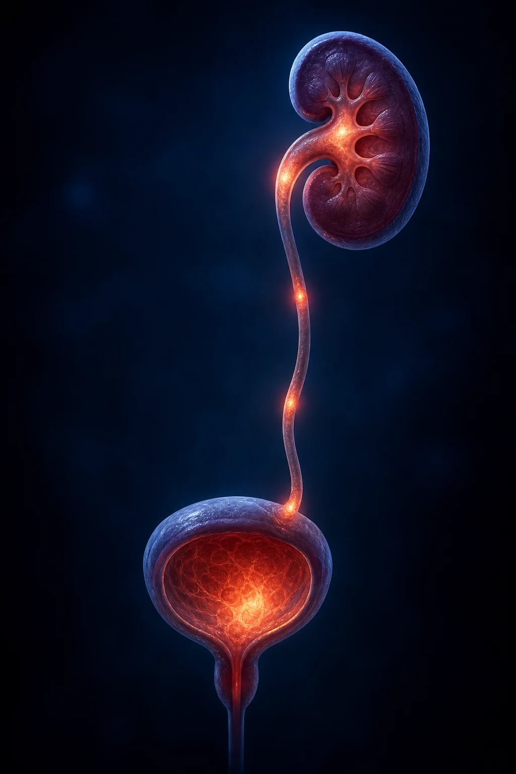
Overview & Definition
A urinary tract infection (UTI) is significant bacterial growth in a properly collected urine sample, with or without symptoms, involving the bladder (cystitis, lower UTI) and/or the kidney (pyelonephritis, upper UTI). It is one of the most common bacterial infections of childhood and a leading reason for paediatric admission and antibiotic prescription in childhood. Its persistently high exam-yield rests on four pivots that recur in every question on this topic: the non-specific presentation of the febrile infant (where missing the diagnosis means losing the narrow window to prevent permanent renal scarring), the diagnostic thresholds that depend entirely on how the urine was collected, the antibiotic ladder with its precise route and duration, and the post-UTI imaging pathway that hunts for vesicoureteric reflux and renal scarring.[1][2]
The clinical reflex this topic exists to teach is unforgettable: a febrile, irritable, vomiting infant has a UTI until proven otherwise — obtain a proper urine sample, do not call it a virus. The reason it matters so much is that renal scarring from recurrent pyelonephritis — especially in the presence of high-grade vesicoureteric reflux — is permanent and cumulative, and is the single most important preventable cause of hypertension and chronic kidney disease attributable to childhood infection. A febrile UTI that is missed, under-treated, or recurrent in infancy becomes adult-onset renal failure two or three decades later. That long shadow is why paediatric UTI is examined relentlessly. [1]
Key definitions the examiner expects you to use precisely: [1]
- Significant bacteriuria — a colony count above the diagnostic threshold for the collection method (see Investigations), of a single organism, in a properly collected sample. Mixed growth or low counts from a contaminated sample are not significant bacteriuria.
- Cystitis (lower UTI) — infection confined to the bladder mucosa; classically dysuria, frequency, urgency, suprapubic pain and haematuria, with little or no fever and a well child.
- Pyelonephritis (upper UTI) — infection of the renal parenchyma; high fever, vomiting, flank pain and systemic illness; carries the risk of permanent cortical scarring.
- Atypical UTI (NICE definition) — a UTI that is seriously ill at presentation, has a poor urine flow, an abdominal or bladder mass, raised creatinine, failure to respond to treatment within 48 hours, septicaemia, or infection with a non-E. coli organism. These features mandate urgent (acute) imaging.
- Recurrent UTI (NICE definition) — two or more episodes of upper UTI, or three or more of lower UTI, or one upper plus one lower UTI. These mandate a full investigation for reflux, scarring and anatomy. [1]
Classification
UTI is classified along three axes: by anatomical site (lower vs upper), by clinical complexity (simple vs atypical vs recurrent), and by the age-dependent mode of presentation (infantile non-specific vs older-child localising). These axes are not academic — together they decide the antibiotic route, the antibiotic duration, and the imaging pathway. A febrile infant with a non-E. coli organism and a palpable bladder is managed very differently from a well 8-year-old girl with a first episode of cystitis. [1]
Lower UTI (cystitis)
bladder only
- Dysuria, frequency, urgency, suprapubic pain, haematuria
- Often afebrile or low-grade fever; child usually well
- Oral antibiotics for 3 days (NICE)
- Low intrinsic risk of renal scarring
- Organism usually E. coli; nitrofurantoin appropriate
Upper UTI (pyelonephritis)
renal parenchyma
- High fever, rigors, vomiting, flank/abdominal pain
- Septic / systemically unwell; risk of permanent scarring
- 7 to 10 days oral or IV antibiotics
- DMSA scarring scan 4 to 6 months later
- Avoid nitrofurantoin — poor renal tissue penetration
Atypical UTI
triggers urgent imaging
- Seriously ill, poor urine flow, abdominal/bladder mass
- Raised creatinine or failure to thrive
- Non-E. coli organism, or no response by 48 hours
- Septicaemia
- Urgent ultrasound during the acute illness
Recurrent UTI
triggers full workup
- Two or more upper UTI, OR three or more lower UTI
- OR one upper plus one lower UTI
- Ultrasound + DMSA + consider MCUG
- Assess for VUR and bladder-bowel dysfunction
- Prophylaxis and prevention programme
Organism-specific patterns worth memorising
The causative organism carries diagnostic information, because certain species point to specific contexts and risk factors: [1]
- E. coli (75 to 85 percent) — the default organism; uropathogenic strains carry P-fimbriae.
- Klebsiella and Proteus — recurrent or hospitalised children; Proteus produces urease, splitting urea to ammonia, which alkalinises urine and precipitates magnesium-ammonium-phosphate (struvite) stones.
- Enterococcus and Pseudomonas — instrumented children, those with anatomical anomalies, or recurrent UTI; Pseudomonas is the signature organism of the child on clean intermittent catheterisation for a neurogenic bladder.
- Staphylococcus saprophyticus — sexually active adolescent girls; a coagulase-negative staph that is novobiocin-resistant.
- Staphylococcus epidermidis — usually a catheter/contaminant; only significant from a properly collected specimen.
- Group B Streptococcus and Candida — neonates and immunocompromised/long-term-catheterised children respectively. [1]
Self-test: a 14-year-old sexually active girl has dysuria and her clean-catch grows a coagulase-negative Staphylococcus. What is the organism?
Staphylococcus saprophyticus — the signature adolescent/sexually-active pathogen. Confirm with a novobiocin resistance test. Treat with a standard lower-UTI regimen (e.g. trimethoprim or nitrofurantoin for 3 days).
Epidemiology & Risk Factors
By age 7, approximately 8 percent of girls and 2 percent of boys have had at least one UTI. After the first year, girls far exceed boys, but there is a striking age/sex inversion in early infancy: in the first three to six months of life boys (and especially uncircumcised boys, who carry a 3 to 7-fold higher risk than circumcised peers) predominate. After infancy the male-to-female ratio inverts because of the female short urethra and ready periurethral colonisation by gut flora. Recurrence is common: roughly 30 percent of children recur within one year of a first UTI, and recurrence risk is highest in children with vesicoureteric reflux and bladder-bowel dysfunction.[1]
Childhood UTI — the numbers that matter
Risk factors divide into the reversible (which you can treat and thereby prevent recurrence) and the structural/congenital (which you investigate and may correct surgically). Examiners reward you for naming the reversible ones, because they are the levers of prevention. [1]
Reversible / modifiable risk factors:
- Constipation and bladder-bowel dysfunction (BBD) — the single most important and most treatable driver of recurrence; a loaded rectum compresses the bladder, raises voiding pressures and impairs emptying.
- Voiding dysfunction — infrequent voiding, incomplete emptying, detrusor overactivity, "holding" postures.
- Dehydration and concentrated urine — reduces the mechanical washout of bacteria.
- Poor perineal hygiene and (in girls) back-to-front wiping.
- Bubble baths and soaps — irritant chemical urethritis (more a cause of dysuria than true UTI). [1]
Structural / congenital risk factors:
- Vesicoureteric reflux (VUR) — present in roughly 30 to 40 percent of children with a febrile UTI.
- Posterior urethral valves — boys; obstructive membrane in the posterior urethra.
- Neurogenic bladder — spina bifida, occult spinal dysraphism, sacral agenesis.
- Structural anomalies — duplex systems, ectopic ureters, ureterocoeles, pelviureteric junction obstruction.
- Instrumentation — long-term catheterisation, repeated instrumentation.
- Immunocompromise — shifts the microbiology toward Pseudomonas, Enterococcus and Candida. [1]
Pathophysiology
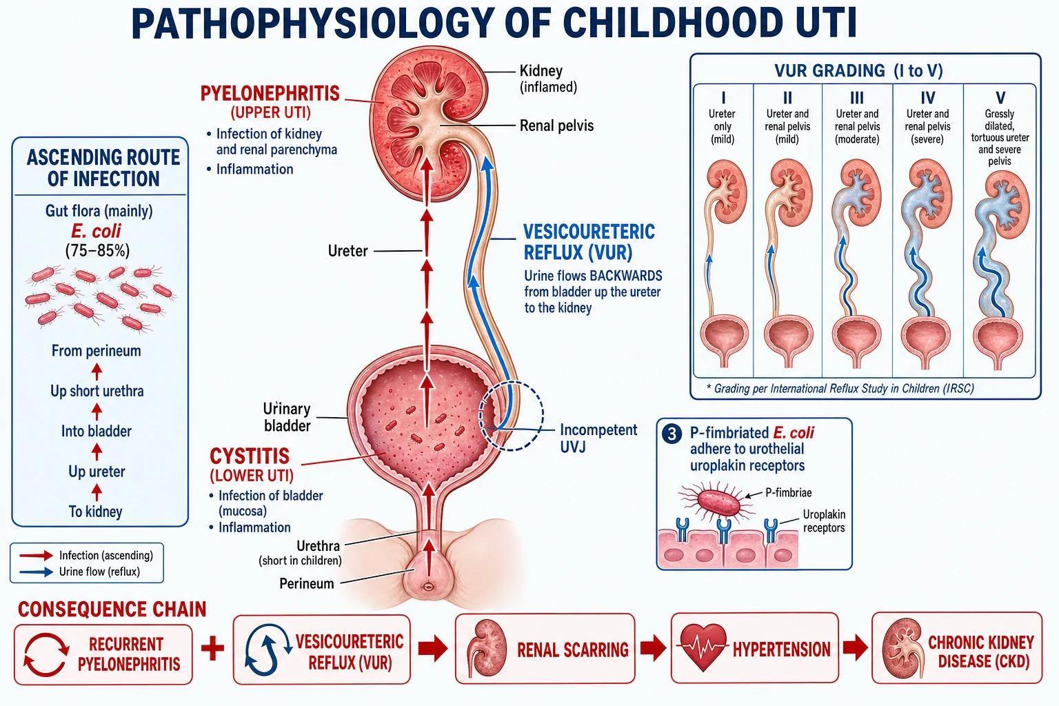
Understanding the mechanism is what makes the management and the investigation pathway logical rather than a list to memorise. The chain is: bacterial entry -> adhesion and multiplication -> host inflammation -> (with VUR) renal parenchymal seeding -> scar formation -> long-term renal damage. [1]
Route of entry: the ascending pathway
The overwhelming route is ascending: periurethral gut flora (originating in the colon) traverse the urethra into the bladder, where they multiply in urine (cystitis), and may ascend the ureters to the kidney (pyelonephritis). Three structural facts of childhood make this route easy: the female urethra is short (about 4 cm in a child), the periurethral mucosa is readily colonised by gut organisms, and the foreskin of the uncircumcised boy is a reservoir for uropathogenic E. coli. Haematogenous spread (bacteria seeding the kidney from the bloodstream) is rare in children and is essentially confined to neonates, where group B streptococcus and E. coli may cause a septicaemic UTI.[1]
Bacterial virulence: P-fimbriated E. coli
Uropathogenic E. coli are not random gut E. coli — they carry specific virulence factors. The most important are P-fimbriae (pyelonephritis-associated pili, encoded by the pap gene cluster), hair-like appendages that terminate in a lectin which binds galactose-alpha(1-4)-galactose-beta residues on uroplakin receptors that carpet the surface of bladder and upper-tract urothelium. By anchoring themselves, P-fimbriated strains resist the mechanical washout that is the bladder's primary defence; strains without fimbriae are simply flushed out. Other virulence factors include haemolysin (tissue-damaging pore-forming toxin), siderophores (which scavenge iron from the iron-poor urinary tract), and capsular K antigen (which resists complement and phagocytosis). The net effect is that a small inoculum of a virulent strain can establish infection where a larger inoculum of an avirulent strain would fail. [1]
Defeat of host defences
The healthy urinary tract clears bacteria mechanically — high urine flow, complete bladder emptying, and the anti-adhesive glycosaminoglycan layer of the urothelium keep it sterile. Anything that impairs these defences sets up infection: urinary stasis (from incomplete emptying, infrequent voiding, or obstruction), high intravesical pressures (from bladder-outlet obstruction or neurogenic bladder), foreign bodies (catheters, stones), and concentrated urine (dehydration). This is why treating constipation and ensuring adequate hydration and regular double-voiding is so effective at preventing recurrence — it restores the mechanical defence the bladder is supposed to have. [1]
Vesicoureteric reflux (VUR): the bridge to kidney damage
Vesicoureteric reflux (VUR) is the pivotal mechanism that links a bladder infection to permanent kidney damage. Normally the ureterovesical junction (UVJ) — the oblique, intramural tunnel through which the ureter enters the bladder — acts as a flap valve: as the bladder fills and pressure rises, the tunnel is compressed shut, preventing retrograde flow. In VUR this tunnel is short and incompetent, so infected bladder urine refluxes back up the ureter to the renal pelvis and, via the renal collecting ducts, into the renal parenchyma (intrarenal reflux). The consequence is that bacteria are delivered directly to the kidney, dramatically increasing the risk of pyelonephritis and scarring. [1]
VUR is most often a congenital malformation of the UVJ (primary VUR); it can also be secondary to high bladder pressures from posterior urethral valves, neurogenic bladder, or voiding dysfunction. It runs in families and shows autosomal dominant inheritance with incomplete penetrance; siblings of an affected child have a roughly 30 percent chance of VUR. [1]
The international reflux grading (I to V) is reproduced exactly because examiners test it verbatim: [1]
International VUR grading (I to V)
Inflammation, scarring and why infants are most vulnerable
Once bacteria and the neutrophil-rich inflammatory response reach the renal parenchyma, the result is acute pyelonephritis — interstitial oedema, neutrophil infiltration, microabscess formation and tubular damage. The renal papillary anatomy matters: in infants and young children the papillae are largely of the "simple" type whose collecting ducts open at right angles and permit intrarenal reflux, whereas adult papillae are predominantly "compound" and slit-like, resisting reflux. This anatomical immaturity, combined with an immature immune system, is why infants scar far more readily than adults for the same infection. [1]
If the infection is treated promptly and completely, the inflammation resolves and the kidney may recover. If it is recurrent, severe, or delayed in treatment, healing occurs by fibrosis — a permanent focal cortical scar that contracts over time, deforming the renal outline and destroying nephrons in that segment. Bilateral scarring reduces total functioning renal mass; over years to decades this drives hypertension (from impaired sodium handling and relative ischaemia), proteinuria, and progressive chronic kidney disease that can culminate in end-stage renal failure. This is the entire reason childhood UTI is taken so seriously: a preventable infection in infancy becomes irreversible renal disease in adulthood.[1][2]
[1]Clinical Presentation
The presentation is age-dependent, and the infantile form is the one examiners test most deliberately because it carries no localising clues — the diagnosis is missed unless the clinician has the reflex to sample urine. [1]
Infants and young children (under 2 to 3 years): the non-specific presentation
Infants cannot localise or report urinary symptoms, and UTI presents as a systemic illness without a focus. The cardinal features are: [1]
- Fever without an obvious source — often the only sign; may be high.
- Irritability and inconsolable crying.
- Poor feeding, lethargy, floppiness.
- Vomiting and/or diarrhoea.
- Failure to thrive if recurrent or chronic.
- Prolonged jaundice in the neonate (conjugated hyperbilirubinaemia from sepsis is a recognised neonatal UTI presentation).
- Frank sepsis — hypothermia, apnoea, grunting, mottling in the very young. [1]
The single reflex the examiner wants: any febrile, irritable, vomiting infant under 3 months (and considered up to 3 years) with no obvious focus must have urine sampled for culture — UTI is present until proven otherwise. [1]
Older children (over 3 years): the localising presentation
Toilet-trained children can report symptoms and UTI declares itself in the urinary tract: [1]
- Dysuria (painful urination), frequency, urgency.
- Suprapubic or abdominal pain.
- Secondary enuresis or new daytime incontinence (a child who was dry now wetting).
- Haematuria (visible or dipstick).
- Cloudy or foul-smelling urine.
- Fever — low-grade or absent in lower UTI. [1]
Upper UTI (pyelonephritis) at any age
Pyelonephritis adds the features of a systemic, septicaemic illness superimposed on urinary symptoms: [1]
- High fever (often over 39 C), with rigors.
- Flank or loin pain, and renal angle tenderness.
- Vomiting and marked systemic upset.
- In the infant: a septic-appearing, febrile child with no localising signs — distinguished from cystitis only by the fever and systemic illness. [1]
Self-test: which features move a UTI from 'lower' to 'upper'?
High fever (often over 39 C), rigors, flank/loin pain, renal angle tenderness, and marked systemic illness (vomiting, sepsis). These change management: the antibiotic must penetrate renal tissue (so not nitrofurantoin), the course is 7 to 10 days, and the route is IV if the child is young or unwell.
Differential Diagnosis
The differential splits into (a) conditions that mimic UTI, and (b) the mimics of the febrile infant that you must not mistake for "just a virus" — in the latter, the reflex is always to sample urine. [1]
Viral fever / URTI
the trap
- Febrile infant with coryza
- BUT a febrile infant can have BOTH a virus and a UTI
- Reflex: sample urine in any febrile infant under 3 months without a clear source
- Missing UTI here causes preventable scarring
Vulvovaginitis / balanitis
external irritation
- Dysuria and external soreness
- Urine culture is sterile (or contaminated)
- Examine the genitalia; treat locally
- Excludes true UTI once culture is negative
Gastroenteritis / appendicitis
abdominal overlap
- Vomiting and abdominal pain overlap
- Appendicitis localises to right iliac fossa with guarding
- Diarrhoea suggests gastroenteritis
- Beware: bag urine contamination is misleading
Diabetes mellitus
polyuria mimic
- Polyuria, polydipsia, secondary enuresis
- Weight loss; check urine and blood glucose
- Can present with DKA
- Concentrated urine or crystalluria also causes dysuria without infection
Further differentials to keep on the list: sexually transmitted infection in adolescents (chlamydia/gonorrhoea urethritis — send NAAT), renal stones and renal colic (Proteus/struvite; flank pain with haematuria), pinworm infestation (perineal irritation and scratching in young children), and chemical urethritis from bubble baths. A useful discriminator: in true UTI the urine culture is positive with a single organism above threshold; in mimics it is negative or mixed. [1]
Clinical & Bedside Assessment
A focused history and examination often reveal the risk factor that explains the UTI and that, once treated, prevents the next one. [1]
History — establish the fever (duration, height, response to antipyretics), urinary symptoms (dysuria, frequency, urgency, haematuria, enuresis), fluid intake, and crucially the bowel habit (constipation is the key reversible driver). Ask specifically about the urinary stream in boys — a weak, dribbling or straining stream suggests posterior urethral valves or bladder-outlet obstruction. Take a history of prior UTI (number and type), any antenatally detected renal tract abnormality (hydronephrosis, duplex system), and family history of VUR or renal disease. [1]
Examination — assess:
- General: illness, hydration, temperature, capillary refill.
- Growth: weight and height centiles (failure to thrive suggests chronic or recurrent disease).
- Abdomen: palpable bladder, abdominal masses, renal angle tenderness (pyelonephritis), distended loops (obstruction).
- Genitalia: phimosis, vulvovaginitis, labial adhesions, meatal stenosis.
- Blood pressure: hypertension in a child implies renal disease (scarring, obstructive uropathy) — always measure it, and it is frequently omitted in the real world, which is exactly why examiners test it.
- Spine and lower-limb neurology: look for the lumbosacral dimple, hair tuft, lipoma, or haemangioma of occult spinal dysraphism, and any lower-limb neurology suggesting a neurogenic bladder.
- Stream observation in a boy (posterior urethral valves). [1]
Distinguish upper from lower UTI at the bedside: high fever, flank pain, vomiting and systemic illness point to pyelonephritis and change the antibiotic route, duration and tissue-penetration requirement. Identify the atypical case (poor stream, mass, FTT, hypertension, non-E. coli, no response at 48 hours) at the bedside, because it changes the imaging pathway. [1]
Investigations
Step 1 — collect the urine correctly
Diagnosis rests on urine culture from a properly collected sample, and the validity of the culture depends almost entirely on how the urine was collected. A contaminated sample leads to overdiagnosis, unnecessary imaging and antibiotics; a missed sample leads to under-treatment and scarring. The hierarchy is: [1]
Urine collection — best to worst
SCCB
gold standard in infants; bladder-wall puncture, near-zero contamination; any growth diagnostic
practical alternative to SPA; sterile catheter into the bladder; low contamination
for toilet-trained children; clean genitalia, midstream into a sterile pot
high contamination; NOT diagnostic; useful only if culture is negative (rules out UTI)
Suprapubic aspiration (SPA) is the gold standard in non-toilet-trained infants. Performed with a full bladder (ideally ultrasound-guided, which raises yield from about 50 percent to over 90 percent): clean the suprapubic skin, insert a fine needle 1 to 2 cm above the symphysis pubis in the midline, aiming slightly towards the pelvis, and aspirate. Because bladder urine is normally sterile, any growth of a single organism is diagnostic. Its near-zero contamination is why it has the highest specificity of any method. [1]
Bladder catheterisation is the practical alternative: a sterile feeding tube or catheter is passed through the urethra into the bladder; the first few millilitres are discarded, and the mid-sample cultured. It has a small contamination rate but is reliable. [1]
Clean-catch midstream is appropriate for toilet-trained children: clean the genitalia, ask the child to begin voiding, and catch the midstream portion in a sterile container. [1]
Bag (or pad) urine is the least reliable: an adhesive bag is applied to the perineum. It is so contaminated by skin and perineal flora that a positive result is not diagnostic and must be confirmed by SPA, catheter or clean-catch. Its only legitimate use is as a rule-out — a truly negative culture excludes UTI, which can spare a well child an invasive sample. [1]
Step 2 — interpret the culture with the correct threshold
The colony count that confirms UTI depends on the collection method, because the threshold compensates for the contamination rate of each method. The NICE / AAP thresholds are: [1]
Culture thresholds (NICE / AAP)
Mixed growth (two or more organisms), or any growth from a bag sample, reflects contamination and is not diagnostic. Pyuria supports infection and is required to interpret borderline (lower) colony counts. The AAP definition for febrile UTI in infants requires both pyuria and a culture above threshold — pyuria without bacteriuria (or vice versa) should prompt a search for an alternative diagnosis.[2]
Step 3 — urine dipstick at the bedside
A bedside dipstick is fast but imperfect, and is a triage tool rather than a definitive test in young children: [1]
- Leukocyte esterase — detects neutrophil esterase, a proxy for pyuria; sensitivity around 80 percent, specificity around 80 to 90 percent. A negative test reduces but does not exclude UTI.
- Nitrites — detect bacterial conversion of dietary nitrate to nitrite; high specificity (over 90 percent) but poor sensitivity in children, because children void frequently and may not store urine in the bladder long enough (4 hours) for the conversion to occur. So nitrite-negative does not exclude UTI in a child.
- Blood and protein are non-specific and do not confirm UTI. [1]
Practical rule: a dipstick is most useful to support a clinical suspicion while awaiting culture, or to triage whether to start empirical antibiotics before culture returns. A child with a high pre-test probability (febrile infant) should have urine sent regardless of the dipstick. [1]
Step 4 — blood tests
Send FBC, CRP, blood culture and renal function (urea, creatinine, electrolytes) when the child is young (under 3 months), systemically unwell, failing to respond to treatment, or recurrent/atypical. In the unwell infant, blood cultures may grow the same organism as the urine. Renal function is the baseline against which any later impairment is compared, and a raised creatinine is an atypical feature mandating imaging. In the neonate, a full septic screen including lumbar puncture is indicated, because UTI in this age group is part of neonatal sepsis. [1]
Step 5 — imaging: the post-UTI pathway
The imaging pathway follows NICE CG54 (UK) and is dictated by the child's age and by whether the UTI was atypical or recurrent. The aim is to find the conditions that predispose to scarring — obstruction, high-grade VUR, dysplasia — and the scars themselves. Three modalities do different jobs: [1]
Renal ultrasound
anatomy and obstruction
- First-line: shows hydronephrosis, duplex systems, ureterocoeles, stones, bladder wall
- Detects obstruction (posterior urethral valves, PUJ)
- Poor at detecting VUR and scarring
- Performed acutely in atypical UTI, or within 6 weeks routinely
DMSA scintigraphy
scarring and function
- Detects permanent renal cortical SCARRING and relative function
- Performed 4 to 6 MONTHS after the UTI
- Delay lets acute inflammation resolve so only fixed scars are counted
- Acute DMSA can show pyelonephritis but is not routine
MCUG (micturating cystourethrogram)
VUR and PUV
- The ONLY test that grades VUR (reflux) and diagnoses posterior urethral valves
- Invasive and involves radiation; use SELECTIVELY
- Indicated: atypical/recurrent UTI, boys under 6 months (exclude PUV), suspected high-grade VUR
- Shows a dilated posterior urethra with abrupt narrowing in PUV
When to image (NICE pathway): [1]
- UTI responding well to antibiotics within 48 hours in a child under 6 months — routine ultrasound only.
- UTI responding well in a child over 6 months — no routine imaging.
- Atypical UTI (any age) — urgent ultrasound during the acute illness; consider MCUG and DMSA.
- Recurrent UTI (any age) — ultrasound plus DMSA at 4 to 6 months; consider MCUG, especially in young children and boys. [1]
Self-test: a 4-year-old girl has a first, typical, E. coli UTI that responds in 24 hours. Does she need imaging?
No. A first, typical, E. coli UTI that responds to antibiotics within 48 hours in a child over 6 months needs no routine imaging. Imaging is reserved for the under-6-months, atypical, and recurrent groups.
Management — Resuscitation
Assess ABCDE. The septic infant needs oxygen, IV access, a 10 to 20 mL/kg crystalloid fluid bolus (repeated if needed), and broad-spectrum IV antibiotics. The cardinal rule: sample the urine before giving antibiotics whenever possible (so the culture is interpretable), but never delay empirical antibiotics in a septic child — treat first and sample immediately. Correct dehydration with oral or IV fluids and give antipyretics (paracetamol 15 mg/kg, ibuprofen 5 to 10 mg/kg) for comfort and to reduce insensible fluid losses. [1]
Admit for IV antibiotics any: infant under 1 to 3 months; child who is septic, dehydrated, vomiting or unable to tolerate oral therapy; or child failing oral therapy (no improvement at 48 hours). The neonate (under 1 month) with suspected UTI is managed as neonatal sepsis: full septic workup including lumbar puncture, and IV ampicillin plus gentamicin (or cefotaxime) — a regimen active against E. coli, group B streptococcus and Listeria.[1]
Management — Definitive & Stepwise
Empirical antibiotics are started after the urine is obtained (treat first and sample immediately if septic), then narrowed once sensitivities return. Always respect local E. coli resistance patterns — amoxicillin monotherapy resistance is common (often over 30 to 40 percent in many regions), so empirical amoxicillin alone is unreliable. The choice of agent, route and duration is determined by whether the infection is lower or upper, the child's age and illness severity, and local guidelines. [1]
Antibiotic therapy by syndrome
Oral trimethoprim, nitrofurantoin, cefalexin, or co-amoxiclav (if susceptible), for 3 days (NICE). Choose by local resistance and allergy. Avoid nitrofurantoin if upper UTI is suspected — it penetrates renal tissue poorly.
Oral co-amoxiclav, cefixime or cefalexin if the child is well, over 3 months, and can tolerate oral therapy. Duration 7 to 10 days (up to 14 in severe or slow-to-resolve cases).
IV ceftriaxone, co-amoxiclav or gentamicin if young (under 3 months), unwell, dehydrated, vomiting, septic, or non-responsive. Switch to oral once afebrile and clinically improving (typically 48 hours).
Manage as neonatal sepsis: IV ampicillin plus gentamicin (or cefotaxime), full workup including lumbar puncture. Avoid ceftriaxone in the neonate (biliary sludging, displacement of bilirubin).
Antibiotic doses (paediatric)
The doses an examiner expects you to be able to state precisely — agent, dose, route, frequency and duration: [1]
Trimethoprim
lower UTI; prophylaxis
- Treatment: 4 mg/kg twice daily for 3 days (lower UTI)
- Prophylaxis: 2 mg/kg once at night
- Avoid in early pregnancy (folate antagonist); avoid long-term if possible
Nitrofurantoin
lower UTI only
- Treatment: 3 mg/kg/day in divided doses (children over 3 months)
- Prophylaxis: 1 mg/kg once at night
- AVOID under 3 months (haemolysis) and in UPPER UTI (poor tissue penetration)
Cefalexin
lower or upper, oral
- Treatment: 12.5 mg/kg twice daily (or 25 mg/kg/day divided)
- Safe in infants; good urinary levels
- Suitable for upper UTI oral step-down
Co-amoxiclav
oral or IV
- Dose by amoxicillin component: 25 to 40 mg/kg/day divided
- Broad cover; use if local E. coli susceptible
- Good oral step-down for pyelonephritis
Cefixime
oral cephalosporin
- 8 mg/kg/day (once or twice daily)
- Good oral option for pyelonephritis step-down
- Third-generation; useful if resistant organisms
Ceftriaxone
IV for severe/young
- 50 mg/kg once daily IV (max 2 g)
- Once-daily IV; excellent tissue levels
- Avoid in neonates (use cefotaxime)
Gentamicin
IV, severe
- Once-daily extended-interval: 6 to 7 mg/kg IV
- Monitor levels and renal function
- Good for severe/pyelonephritis and neonatal sepsis
Prophylaxis
Continuous low-dose antibiotic prophylaxis — a single nocturnal dose at roughly half the treatment dose — is reserved for recurrent UTI and high-grade VUR while investigation and definitive management proceed, and is not routine after a first uncomplicated UTI. Regimens: trimethoprim 2 mg/kg nocte or nitrofurantoin 1 mg/kg nocte (rotate every 3 months in long-term use to limit resistance). The RIVUR trial showed prophylaxis reduces recurrence in children with VUR but does not reduce renal scarring, which is why it is increasingly reserved for high-grade VUR and recurrence rather than applied universally.[1]
Treating the reversible drivers (the real prevention)
The most effective prevention of recurrence addresses the modifiable factors: [1]
- Treat constipation aggressively — laxatives (e.g. macrogol), adequate fibre and fluids, regular toileting. A loaded rectum compressing the bladder is the commonest unrecognised cause of recurrence.
- Treat bladder-bowel dysfunction — timed double-voiding, biofeedback for detrusor-sphincter dyssynergia, urotherapy.
- Ensure adequate hydration and frequent voiding (every 2 to 3 hours) to maintain mechanical washout.
- Counsel on perineal hygiene (front-to-back wiping in girls). [1]
Circumcision (selective, not routine)
Circumcision reduces the risk of UTI in boys by removing the foreskin reservoir for uropathogenic E. coli, but it is recommended only selectively — for boys with recurrent UTI plus high-grade VUR or a structural anomaly — where the benefit (reduced recurrence) clearly outweighs procedural risk. It is not routine after a single UTI. [1]
Surgical management of high-grade VUR
High-grade VUR (grades IV to V) with recurrent pyelonephritis and scarring, or VUR failing conservative management, is managed surgically: [1]
- Endoscopic bulking (the STING procedure) — injection of a bulking agent (e.g. dextranomer-hyaluronic acid copolymer, Deflux) beneath the ureteric orifice to augment the incompetent UVJ tunnel. Minimally invasive, day-case; success around 70 to 80 percent per injection (lower for high grades; may need repeat).
- Open ureteric reimplantation — the definitive operation; the ureter is re-tunnelled through the bladder wall to create a competent flap valve (e.g. Cohen cross-trigonal or Politano-Leadbetter techniques). Success over 95 percent; reserved for failure of conservative/endoscopic management or very high-grade reflux. [1]
Specific Subtypes & Scenarios
Lower UTI (cystitis)
Bladder-only infection in a well, older child. Manage as an outpatient with a 3-day oral course; imaging not required if it is a first, typical, E. coli infection responding within 48 hours. The risk of renal scarring is low. [1]
Upper UTI (pyelonephritis)
Renal parenchymal infection with systemic illness. Treat for 7 to 10 days (oral if well; IV if young or unwell), and arrange a DMSA at 4 to 6 months to detect scarring. Ultrasound during the acute illness if the child is under 6 months or has atypical features. [1]
Recurrent UTI — the workup
Two or more upper UTI, three or more lower UTI, or one of each, mandates a full investigation: ultrasound (anatomy), DMSA at 4 to 6 months (scarring), and consider MCUG (VUR). Address bladder-bowel dysfunction, start prophylaxis if appropriate, and refer to paediatric nephrology/urology if scarring or high-grade VUR is found. The goal is to interrupt the recurrence -> scarring -> CKD cycle. [1]
Atypical UTI — exclude obstruction
Seriously ill, poor stream, palpable bladder or mass, raised creatinine, non-E. coli organism, or no response at 48 hours mandates an urgent ultrasound during the acute illness to exclude a surgical emergency — obstruction (posterior urethral valves, stone, PUJ obstruction), abscess, or a badly dysplastic kidney. A child with obstruction and infection needs decompression (catheter, nephrostomy or drainage) as well as antibiotics. [1]
Vesicoureteric reflux (VUR)
Congenital incompetence of the ureterovesical junction, graded I to V. Low-grade (I to II) usually resolves spontaneously with growth of the UVJ tunnel and is managed conservatively (surveillance, prophylaxis if recurrent, treat BBD). High-grade (IV to V) with recurrent pyelonephritis and scarring needs surgical correction (STING or reimplantation). All children with VUR need BP and urinalysis surveillance for scarring sequelae. [1]
Posterior urethral valves (boys)
A membranous obstruction of the posterior urethra occurring only in boys, and the most common cause of lower-urinary-tract obstruction in male infants. The presentation and management span antenatal to long-term renal care: [1]
- Antenatal: bilateral hydronephrosis, a distended thick-walled bladder, oligohydramnios, and (if severe) the Potter sequence (pulmonary hypoplasia, limb deformities, characteristic facies from intrauterine compression).
- Neonatal: a weak, dribbling urinary stream, a palpable distended bladder, bilateral flank masses (hydronephrosis), respiratory distress from pulmonary hypoplasia, and renal dysplasia with raised creatinine.
- Diagnosis: MCUG (micturating cystourethrogram) is diagnostic — it shows a dilated posterior urethra with an abrupt transition to a narrow distal urethra at the valve.
- Acute management: urinary catheterisation to decompress the bladder, correct fluid and electrolyte disturbances, then endoscopic valve ablation. A vesicostomy may be needed if ablation is not immediately feasible or the baby is too small.
- Long-term: these boys are at high risk of renal dysplasia, CKD and end-stage renal failure, and of bladder dysfunction (the "valve bladder"). They need lifelong nephrology and urology follow-up, monitoring of renal function and BP, and management of any associated VUR. [1]
Self-test: a 2-day-old boy has antenatal bilateral hydronephrosis, a palpable bladder and a poor stream. What is the diagnosis and the diagnostic test?
Posterior urethral valves until proven otherwise. The diagnostic test is MCUG, showing a dilated posterior urethra with abrupt narrowing at the valve. Acute management is catheterisation to decompress, correct electrolytes, then endoscopic valve ablation.
Complications & Pitfalls
Long-term complications
The defining long-term complication is renal scarring — permanent, detected on DMSA 4 to 6 months after infection, and concentrated in young infants, recurrent upper UTI, high-grade VUR, delayed treatment and atypical organisms. Around 5 to 15 percent of children with a febrile UTI scar, and scarring is cumulative with each recurrent pyelonephritic episode. [1]
- Hypertension — consequence of bilateral scarring; all scarred children need lifelong BP surveillance. Often the first manifestation of "burnt-out" reflux nephropathy in adolescence or adulthood.
- Proteinuria — indicates hyperfiltration injury to remaining nephrons; a marker of progressive renal damage.
- Chronic kidney disease / end-stage renal failure — from extensive bilateral scarring or congenital dysplasia (posterior urethral valves, high-grade VUR). Reflux nephropathy accounts for a significant fraction of paediatric and adult ESRF.
- Impaired growth / failure to thrive — from chronic illness and recurrent infection. [1]
Acute complications
- Septicaemia and septic shock — particularly in infants.
- Renal or perinephric abscess — a collection requiring drainage; suggested by persistent fever and a mass despite antibiotics.
- Acute kidney injury — rare, from dehydration and sepsis.
- Electrolyte disturbance — hyponatraemia, hyperkalaemia in sick infants. [1]
The classic, preventable pitfalls
Exam application bank (NEET-PG / INICET)
One-line answer
Urinary tract infection (UTI) in children is common (8 percent of girls, 2 percent of boys by age 7), and in young infants the presentation is often non-specific (fever without source, poor feeding, vomiting, irritability, jaundice) — so a urine sample must be obtained in any unwell or febrile child under 3 months (and considered under 3 years). Causes: E. coli (75 to 85 percent), Klebsiella, Proteus, Enterococcus, Pseudomonas. Risk factors: female sex, uncircumcised boys, constipation, voiding dysfunction, vesicoureteric reflux (VUR), posterior urethral valves (boys), neurogenic bladder. Diagnosis rests on urine culture (SPA gold standard in infants; catheter 10^4 to 10^5; clean-catch over 10^5 CFU/mL). Treat: lower UTI 3 days oral; upper UTI/pyelonephritis 7 to 10 days oral or IV. Investigate recurrent, atypical, or any UTI under 6 months: renal ultrasound, DMSA scintigraphy (scarring
Worked stems (answer without another resource)
Stem 1 — Classic presentation. Map symptoms to mechanism; name the first investigation and first treatment step with dose/route if drug therapy is standard. [1]
Stem 2 — Unstable / complicated. List red flags that force immediate resuscitation, theatre, ICU, antidote, or reperfusion — and what you do in the first 15 minutes. [1]
Stem 3 — Atypical group. Elderly, pregnancy, child, or immunocompromised: how presentation and thresholds change. [1]
Stem 4 — Differential trap. Name the three closest mimics and one discriminator for each. [1]
Stem 5 — Disposition. Who goes home with safety-netting, who is admitted, who needs HDU/ICU/theatre, and what follow-up is mandatory. [1]
Rapid viva checklist
- Definition + classification
- Pathophysiology chain
- Bedside signs / criteria
- Score with exact components (if any)
- Emergency bundle
- Definitive therapy with doses
- Complications of disease and of treatment
- Special populations
- Guideline/trial name if classic
- Three exam traps
Coverage self-check
If you cannot answer any stem above from this page alone, re-read the matching section — the page is intended to be self-sufficient for final-prof and NEET-PG/INICET questions on Urinary Tract Infection in Children.
Further pitfalls: using amoxicillin alone empirically (high E. coli resistance), using nitrofurantoin for pyelonephritis (poor renal-tissue penetration), omitting the blood pressure measurement, and not treating constipation in a child with recurrent UTI (the commonest missed reversible driver). [1]
Prognosis & Disposition
Most childhood UTIs resolve completely with appropriate antibiotics; a single uncomplicated lower UTI has an excellent outcome. The risk of permanent renal scarring is concentrated in young infants, recurrent upper UTI, high-grade VUR, delayed treatment and atypical organisms — around 5 to 15 percent of children with a febrile UTI scar, and scarring is cumulative with each recurrent pyelonephritic episode. Roughly 30 percent of children recur within one year. [1]
Disposition:
- Lower UTI in an older, well child — managed as an outpatient with oral antibiotics.
- Admit infants under 3 months, any septic or dehydrated child, any child failing oral therapy, and any child with atypical features (obstruction suspected).
- Long-term safety-net: annual blood pressure and urinalysis for any child with confirmed scarring; nephrology referral for hypertension, proteinuria or a falling GFR. [1]
Special Populations
- Neonates (under 1 month): non-specific septic presentation; SPA/catheter urine; manage as neonatal sepsis with LP and IV ampicillin plus gentamicin (or cefotaxime); ultrasound mandatory. Avoid ceftriaxone (biliary sludging, bilirubin displacement).
- Infants 1 to 3 months: non-specific febrile illness; SPA/catheter urine; IV ceftriaxone or co-amoxiclav; low threshold for admission and ultrasound.
- Toilet-trained children: clean-catch urine; oral antibiotics if well; outpatient management reasonable.
- Adolescent girls: consider sexually transmitted infection (send chlamydia/gonorrhoea NAAT); Staphylococcus saprophyticus is a recognised pathogen; counsel on hygiene and post-coital voiding.
- Spina bifida / neurogenic bladder: recurrent UTI from stasis and clean intermittent catheterisation; prophylaxis and urology input; Pseudomonas is common; manage with urodynamics and anticholinergics.
- Immunocompromised / instrumented: atypical organisms (Pseudomonas, Enterococcus, Candida) — broaden empirically and culture early; consider fungal UTI in the long-term-catheterised or immunosuppressed child.
- Pregnant adolescents: UTI in pregnancy carries higher risk of pyelonephritis, prematurity and low birth weight; screen and treat all bacteriuria, avoid nitrofurantoin near term, and use a penicillin or cephalosporin. [1]
Evidence, Guidelines & Regional Differences
[1] [1]The RIVUR trial (NEJM 2014) randomised children with VUR to continuous trimethoprim-sulfamethoxazole prophylaxis versus placebo: prophylaxis reduced recurrent UTI but did not reduce renal scarring — the central evidence behind the move away from routine prophylaxis for low-grade VUR. Three active controversies remain: antibiotic duration (3 days lower vs 7 to 14 days febrile, regional variation), routine prophylaxis (increasingly reserved for high-grade VUR and recurrence), and circumcision (selective, not routine, balancing benefit against procedural risk and parental values).
Exam Pearls
- Any febrile unwell child under 3 months — urine sample (SPA gold standard); UTI until proven otherwise.
- Infants present non-specifically (fever without source, vomiting, irritability, prolonged jaundice) — the reflex is to get urine, not to call it a virus.
- E. coli 75 to 85 percent; Proteus links to stones (struvite, urease); Staph saprophyticus to adolescent girls; Pseudomonas to instrumented/recurrent; Enterococcus to structural anomaly.
- Culture thresholds: SPA any growth; catheter 10^4 to 10^5; clean-catch over 10^5 (over 10^4 with pyuria). Bag urine is not diagnostic — confirm positives.
- Lower UTI = 3 days oral (trimethoprim/nitrofurantoin/cefalexin). Pyelonephritis = 7 to 10 days oral/IV (co-amoxiclav/cefixime oral; ceftriaxone/gentamicin IV). Avoid nitrofurantoin for upper UTI (poor tissue penetration) and under 3 months (haemolysis).
- Investigate: ultrasound (under 6 months, atypical, recurrent); DMSA scarring at 4 to 6 months; MCUG selective (atypical/recurrent, boys under 6 months for posterior urethral valves, suspected high-grade VUR).
- VUR graded I to V: low-grade conservative (usually resolves), high-grade surgical (STING endoscopic bulking or ureteric reimplantation).
- Untreated or recurrent pyelonephritis plus VUR leads to renal scarring -> hypertension, proteinuria, CKD.
- Treat constipation/voiding dysfunction — the single most effective reversible prevention of recurrence.
- Posterior urethral valves (boys): bilateral hydronephrosis plus distended bladder, poor stream; MCUG diagnostic; catheterise then ablate valves; renal dysplasia/CKD risk.
- DMSA is delayed 4 to 6 months so acute inflammation resolves and only permanent scars are counted.
- Always measure blood pressure in a child with UTI — hypertension implies renal disease. [1]
References
- [1]Shaikh N, Haralam MA, Shope TR, et al. Contemporary Management of Urinary Tract Infection in Children Pediatrics, 2021.PMID 33479164
- [2]Subcommittee on Urinary Tract Infection, Roberts KB. Urinary Tract Infection in Children Pediatr Clin North Am, 2022.PMID 36880924