Respiratory · General Medicine
Allergic Bronchopulmonary Aspergillosis & Aspergillus Lung Disease
Also known as Allergic bronchopulmonary aspergillosis · ABPA · Aspergilloma · Chronic pulmonary aspergillosis · Invasive pulmonary aspergillosis · Fungal asthma
Aspergillus fumigatus causes a spectrum of lung disease whose form is set entirely by the host: Aspergillus sensitisation (skin/IgE positive, asthma may worsen, no structural change), allergic bronchopulmonary aspergillosis (ABPA) — a Th2 hypersensitivity reaction to Aspergillus colonising the airways of patients with asthma or cystic fibrosis, producing poorly controlled asthma, brownish sputum plugs, blood eosinophilia, a markedly raised total IgE (over 500 to 1000 IU/mL) and central (proximal) bronchiectasis with mucus plugging; aspergilloma — a saprophytic fungal ball in a pre-existing cavity causing haemoptysis (which can be massive); chronic pulmonary aspergillosis — slow cavitating disease in mild immunocompromise; and invasive pulmonary aspergillosis — an angioinvasive pneumonia in the neutropenic or transplant patient, treated first-line with voriconazole. ABPA is diagnosed by the ISHAM criteria (predisposing asthma/CF plus obligatory Aspergillus sensitisation, with a raised total IgE and characteristic imaging and/or eosinophilia), staged I to V (acute, remission, exacerbation, steroid-dependent, fibrotic), and treated with oral corticosteroids plus an antifungal (itraconazole or voriconazole) — the antifungal is steroid-sparing — with omalizumab (anti-IgE) for steroid-dependent or refractory disease.
On this page & tools
Your progress
Saved locally on this device.
Exam tags
Red flags
Overview & Definition
A single environmental mould — Aspergillus fumigatus, a ubiquitous soil saprophyte whose spores (conidia) are inhaled daily by every human — produces five clinically distinct pulmonary syndromes, and which syndrome develops is determined almost entirely by the state of the host's airway and immune system.[1][7] The lung is the portal of entry, but whether the organism is cleared silently, ignites an allergic reaction, colonises a cavity, or invades tissue depends on whether the host has asthma, structural lung disease, mild immunocompromise, or profound neutropenia. This host-determined spectrum is the single most examinable idea in the topic.
Allergic bronchopulmonary aspergillosis (ABPA) is a Th2-mediated hypersensitivity reaction to Aspergillus fumigatus colonising the airways of a person with asthma or cystic fibrosis.[1] The allergic inflammation damages the larger, more central airways (the segmental and subsegmental bronchi), producing the condition's radiological signature — central (proximal) bronchiectasis with mucus plugging — and the bedside clue of brownish sputum plugs (casts of inspissated mucus loaded with eosinophils and fungal hyphae).[7][8] The other syndromes are Aspergillus sensitisation (skin or specific-IgE positivity, asthma may worsen, but no bronchiectasis or tissue damage yet), aspergilloma (saprophytic growth of a fungal ball within a pre-existing cavity), chronic pulmonary aspergillosis (a slow, progressive cavitating form in mild immunocompromise), and invasive pulmonary aspergillosis (an angioinvasive pneumonia in the profoundly immunocompromised).[1][4]
The clinical importance of recognising ABPA early is that, unlike ordinary severe asthma, it responds specifically to corticosteroids plus an antifungal; missing the diagnosis and treating it as refractory asthma for years allows irreversible bronchiectasis and fibrotic end-stage lung disease to develop.[1][8]
ABPA — the headline numbers
Classification
Aspergillus lung disease is classified along two axes: the clinical syndrome (set by the host), and — within ABPA itself — the radiological form and the clinical stage.[1][7]
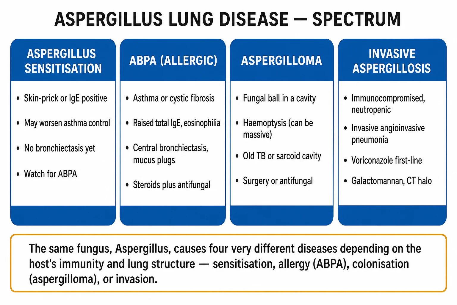
Aspergillus sensitisation
Allergy without damage
- **Positive skin-prick test or serum Aspergillus-specific IgE** in an asthmatic
- Asthma may be more severe, but **no bronchiectasis, no mucus plugging, normal IgE range**
- Overlaps with **severe asthma with fungal sensitisation (SAFS)**
- Antifungal trial may help symptoms; not the same entity as ABPA
ABPA (allergic)
Hypersensitivity + structural change
- **Asthma or cystic fibrosis** + Aspergillus sensitisation + **raised total IgE** + eosinophilia
- **Central (proximal) bronchiectasis, mucus plugging, high-attenuation mucus**
- Diagnosed by the **ISHAM criteria**; staged I–V (Agarwal modification of Patterson)
- **Corticosteroid plus antifungal**; **omalizumab** for steroid-dependent or refractory disease
Aspergilloma
Saprophytic fungal ball in a cavity
- **Fungal ball in a pre-existing cavity** (old TB, sarcoid, bullae, bronchiectasis)
- **Cough, haemoptysis** that may be **massive and life-threatening**
- **Mobile intracavitary mass with air-crescent sign** on imaging; strong serum **Aspergillus IgG/precipitins**
- **Surgery or bronchial-artery embolisation** for bleeding; azoles have poor cavity penetration
Chronic pulmonary aspergillosis (CPA)
Slow cavitating disease
- Subacute–chronic cavitating disease over **months** in **mild immunocompromise** or structural lung disease (TB-destroyed lung, COPD)
- Weight loss, chronic cough, haemoptysis; **progressive upper-lobe cavities, pleural thickening**
- **Long-term oral azole** (itraconazole/voriconazole) for at least 6 months; surgery for localised disease
Invasive pulmonary aspergillosis
Angioinvasive, immunocompromised host
- **Prolonged neutropenia, stem-cell or solid-organ transplant, high-dose steroids, advanced AIDS**, chronic granulomatous disease
- Fever unresponsive to antibiotics, pleuritic pain, haemoptysis, **rapidly progressive infiltrates**
- **CT halo sign** (haemorrhagic nodule) early, **air-crescent sign** with neutrophil recovery
- **Voriconazole first-line** (Herbrecht 2002 NEJM); alternatives liposomal amphotericin B, isavuconazole
Radiological classification of ABPA
ABPA is further divided by whether structural change is present on imaging, because this predicts severity and steroid-dependence:[1][7]
- ABPA-S (seropositive ABPA) — meets the immunologic criteria without bronchiectasis on CT. Often an earlier stage; treatable before structural damage accrues.
- ABPA-B (ABPA with bronchiectasis) — characteristic central bronchiectasis on imaging. A more advanced, structural form.
- ABPA with high-attenuation mucus (HAM) — a distinct, pathognomonic subtype in which impacted mucus is denser than skeletal muscle on unenhanced CT. HAM-ABPA is usually severe, often steroid-dependent, and carries a higher treatment burden.[7]
Clinical staging of ABPA (Agarwal modification of Patterson/Greenberger)
ABPA runs a relapsing–remitting course that is tracked by the total IgE trend, and is classically described in five stages.[1][7]
The five stages of ABPA — ARESF
ARESF
Active disease: symptoms, eosinophilia, raised IgE, infiltrates/bronchiectasis; responds briskly to steroids with falling IgE
Sustained (over 6 months) clinical and immunologic improvement off treatment; IgE falls by over 25–50%
Relapse: IgE rises by over 2 times the remission baseline, with new symptoms/infiltrates; treat as acute
Relapses whenever steroids are tapered below a threshold; needs long-term steroid-sparing strategy (antifungal ± omalizumab)
Irreversible fibrotic bronchiectasis, type-2 respiratory failure and cor pulmonale; largely irreversible
Epidemiology & Risk Factors
Aspergillus fumigatus is ubiquitous — its spores are present in soil, decaying vegetation, compost, damp buildings and air-conditioning systems — so exposure is universal and the deciding factor is the host, not the organism.[1]
ABPA complicating asthma. Roughly 1 to 2 percent of all patients with asthma have ABPA, but the prevalence rises steeply with asthma severity — to around 7 to 15 percent or more in patients attending specialist asthma clinics, and to over 20 percent in intensive-care admissions for status asthmaticus.[1][9] A systematic review of Indian studies found a pooled ABPA prevalence of about 2.5 percent among all asthmatics and substantially higher among difficult-to-treat asthmatics, making India one of the highest-burden settings given the size of its asthma population.[9]
ABPA complicating cystic fibrosis. ABPA occurs in approximately 2 to 15 percent of CF patients (commonly quoted around 8 percent), with a peak in adolescence and young adulthood; because it accelerates lung destruction, annual screening (total IgE and Aspergillus-specific IgE/IgG) is built into CF care programmes.[6]
Host risk factors cluster into two groups. The obligate airway predisposition is asthma or cystic fibrosis — without one of these, ABPA is vanishingly rare. The immunogenetic background that tilts an atopic asthmatic toward ABPA includes atopy, the HLA-DR2 and HLA-DR5 haplotypes (which present Aspergillus antigens to T cells), interleukin-10 promoter polymorphisms, and even CFTR heterozygosity in non-CF ABPA. Environmental exposure — farming, composting, damp/mouldy housing, leaf mould — raises the inhaled antigen load.[1][10]
Aspergilloma is epidemiologically distinct: it is a saprophytic coloniser of a pre-existing pulmonary cavity — most often the residue of old pulmonary tuberculosis, but also sarcoidosis, bronchiectasis, bullous emphysema, a lung abscess, or a cavitating carcinoma.[4]
Invasive aspergillosis is the disease of the immunocompromised: the dominant risk is prolonged profound neutropenia (under 500 neutrophils/microL for over 10 days), classically during induction chemotherapy for acute leukaemia; allogeneic haematopoietic stem-cell transplantation, particularly with graft-versus-host disease and high-dose steroids; solid-organ transplantation (especially lung); prolonged high-dose corticosteroids (e.g. COPD exacerbations treated heavily); advanced AIDS; and the inherited defects of phagocyte killing — chronic granulomatous disease and hyper-IgE (Job) syndrome.[3][4]
Pathophysiology
The pathogenesis of each Aspergillus syndrome is best understood as a dialogue between a virulent environmental mould and a particular host deficit.[1][10]
Why Aspergillus colonises the asthmatic/CF airway
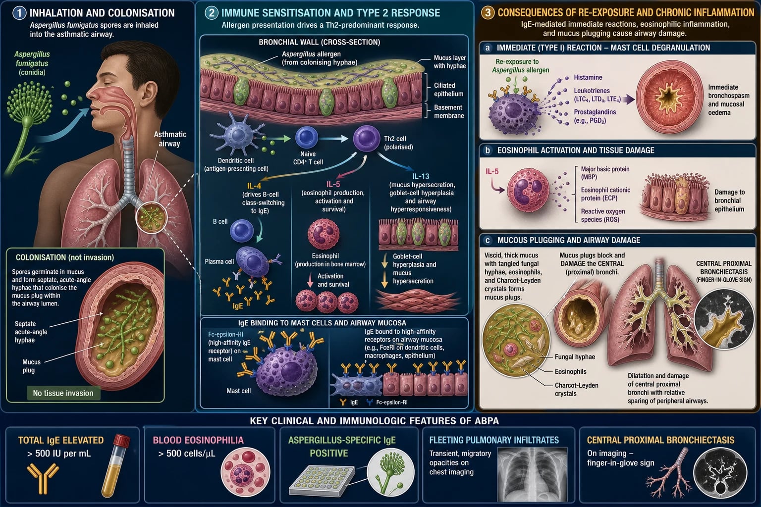
Aspergillus fumigatus is exquisitely adapted to the human airway. Its small conidia (2 to 3 micrometres) penetrate to the terminal bronchioles and alveoli; it is thermotolerant (growth at 37 degrees Celsius and above); it secretes proteases (alkaline protease, elastase) and gliotoxin that strip epithelium and dampen the macrophage oxidative burst; and it scavenges iron through siderophores.[10] In a normal host these conidia are cleared by alveolar macrophages (innate) and neutrophils (adaptive). In the asthmatic — and especially the CF — airway, viscous mucus, impaired mucociliary clearance, and an inflamed epithelium allow the organism to persist and germinate, releasing a continuous stream of antigen into a Th2-primed mucosa.[1][10]
The Th2 hypersensitivity cascade
In the genetically susceptible host (HLA-DR2/DR5, atopic background), airway dendritic cells present Aspergillus antigens to naive CD4 T cells and skew them toward a Th2 phenotype. The Th2 cytokines do the damage: [1]
- IL-4 and IL-13 drive B-cell IgE class-switching, producing the massively elevated total serum IgE and Aspergillus-specific IgE that define ABPA; IgE-coated mast cells degranulate on antigen contact, releasing histamine, leukotrienes and cytokines.
- IL-5 drives eosinophil production, maturation and survival in the bone marrow, producing the blood and sputum eosinophilia; eosinophils then infiltrate the airway wall and release major basic protein, eosinophil cationic protein and reactive oxygen species, which strip the bronchial epithelium and damage the airway wall.
- The result, histologically, is eosinophilic bronchitis, mucoid impaction (inspissated mucus packed with eosinophils and fungal hyphae), and bronchocentric granulomatosis — a necrotising granulomatous inflammation centred on the bronchiole.[1][10]
Why the bronchiectasis is central and upper-lobe
The allergic inflammation targets the larger, more central airways — the segmental and subsegmental bronchi — which is why ABPA produces central (proximal, perihilar) bronchiectasis with an upper-lobe predominance, in contrast to the peripheral distribution of post-infective bronchiectasis or the traction bronchiectasis of pulmonary fibrosis.[7][8] This central, upper-lobe, varicose-or-cystic pattern on HRCT is one of the most characteristic and most examinable radiological findings in respiratory medicine. The mucus that fills these ectatic bronchi casts the bronchus, producing the 'gloved-finger' and 'tram-track' shadows on imaging and the brownish sputum plugs the patient coughs up.
High-attenuation mucus (HAM) — nearly pathognomonic
In a subset of patients the impacted mucus is denser than skeletal muscle on unenhanced (non-contrast) CT — a finding called high-attenuation mucus. Its density comes from heavy loading with calcium oxalate crystals (a metabolic product of Aspergillus) and fungal hyphae.[7] HAM is nearly pathognomonic for ABPA, identifies a severe and often steroid-dependent phenotype, and correlates with higher total IgE and Aspergillus-specific IgG.[7]
Aspergilloma — saprophytic colonisation
A pre-existing cavity (old TB, sarcoid, bulla) provides a warm, oxygen-rich, immune-privileged space in which inhaled A. fumigatus grows saprophytically as a ball of branching septate hyphae, fibrin and debris.[4] The ball is mobile within the cavity (changing position with posture) and is separated from the cavity wall by a crescent of air (the air-crescent sign). The surrounding cavity wall is highly vascular, with hypertrophied bronchial arteries; erosion of these vessels is the mechanism of the haemoptysis — occasionally massive — that dominates the clinical picture.[4]
Invasive aspergillosis — angioinvasion
In the neutropenic or steroid-immunosuppressed host, the organism breaches the epithelium and invades the pulmonary vasculature. Hyphae grow across and within pulmonary arteries, producing septate acute-angle branching that occludes the vessel; the downstream lung undergoes ischaemic and haemorrhagic infarction.[3] Early in the course this produces the CT halo sign — a nodule of infarcted lung surrounded by a halo of ground-glass haemorrhage; as the neutrophil count recovers and the infarcted core sequesters and cavitates, the air-crescent sign appears.[4] The same angioinvasive mechanism allows haematogenous dissemination — classically to the brain, skin, liver and eye.[3][4]
Clinical Presentation
The presentation of Aspergillus lung disease is dictated by which syndrome the host has developed. ABPA itself rarely presents as an acute emergency; its importance is as a cause of poorly controlled asthma, and its acute presentations are usually an asthma exacerbation or an ABPA exacerbation triggered by rising antigen load.[1][8]
ABPA in the asthmatic
The classical patient is a young-to-middle-aged atopic asthmatic whose asthma has become difficult to control — escalating reliever use, recurrent exacerbations, worsening peak-flow variability — over weeks to months, with one or more of the following:[1][7]
Respiratory symptoms
Beyond ordinary asthma
- **Worsening asthma control** despite adherence and inhaled corticosteroids — the most common and earliest clue
- **Productive cough with brownish-black or rust-coloured sputum plugs** — casts of inspissated mucus, eosinophils, Charcot-Leyden crystals and fungal hyphae
- **Recurrent 'pneumonias' in different lobes** — the fleeting, migratory pulmonary infiltrates of mucoid impaction and atelectasis
- **Pleuritic chest pain, fever and malaise** during exacerbations, with wheeze and dyspnoea
Signs
Often deceptively ordinary
- **Diffuse polyphonic wheeze** and signs of airflow obstruction; locally reduced breath sounds where a lobe is collapsed by a mucus plug
- **Fleeting crackles** over an area of infiltrate; occasionally **clubbing** in long-standing, advanced (fibrotic) disease
- Stigmata of **atopy** (eczema, allergic rhinitis) and, where relevant, of **cystic fibrosis** (failure to thrive, recurrent infection, bronchiectasis)
Atypical / mimics
The corners examiners probe
- **Collapse-consolidation of a lobe by a single large mucus plug**, mimicking a tumour or foreign body (mucoid impaction, 'gloved-finger' sign)
- **Years of misdiagnosis as refractory asthma** before the raised IgE and central bronchiectasis are found
- **Pneumonia that recurs in the same or different upper lobes**, treated repeatedly with antibiotics without resolution
- **ABPA presenting with type-1 respiratory failure** during a severe exacerbation
The brownish sputum plug is the single highest-yield bedside clue. It is a cast of the bronchus — a firm, cylindrical or branching plug of inspissated mucus teeming with eosinophils, Charcot-Leyden crystals (bipyramidal eosinophil breakdown products) and branching septate fungal hyphae.[7] Other causes of brown/rust-coloured sputum (old haemoptysis, 'rusty' pneumococcal sputum) should be considered, but in an asthmatic a brown plug plus eosinophilia is ABPA until excluded.
ABPA exacerbation
An ABPA exacerbation resembles a severe asthma exacerbation but carries systemic features and an immunologic signature: worsening dyspnoea and wheeze, new or worsening brownish sputum plugs, low-grade fever, malaise, a rising peripheral eosinophil count, new fleeting pulmonary infiltrates, and a rising total IgE (typically doubling the patient's remission baseline).[1][7]
Aspergilloma
The patient is usually known to have an old tuberculous cavity, sarcoidosis or bullous lung disease, and presents with chronic cough and haemoptysis that can range from blood-streaked sputum to massive, life-threatening bleeding (the dominant threat). Systemic features are modest; weight loss and malaise may indicate evolution to chronic pulmonary aspergillosis.[4]
Invasive aspergillosis
In the neutropenic, transplant or heavily steroid-treated patient, the presentation is fever unresponsive to broad-spectrum antibiotics after 4 to 7 days, with pleuritic chest pain, non-productive or slightly blood-streaked cough, and dyspnoea, and rapidly progressive nodular or wedge-shaped pulmonary infiltrates that often cavitate with neutrophil recovery.[3][4] Haemoptysis is common and indicates angioinvasion. A high index of suspicion is mandatory, because delayed therapy carries a high mortality.
Chronic pulmonary aspergillosis
A subacute illness of months: chronic productive cough, weight loss, fatigue and haemoptysis in a patient with mild immunocompromise (mild steroid use, diabetes, alcoholism, COPD) or structural lung disease (TB-destroyed lung); imaging shows progressive upper-lobe cavitation, pleural thickening and adjacent infiltrates.[4]
Differential Diagnosis
The clinical question — "Is this ABPA, or something else that looks like it?" — usually arises in one of three settings: a difficult-to-treat asthmatic with eosinophilia, a patient with upper-lobe/central bronchiectasis, or a patient with a cavity containing a mass. The high-yield differentials in each:[1][7]
Severe eosinophilic asthma (non-fungal)
The closest mimic
- **Difficult asthma with eosinophilia but negative Aspergillus skin-prick and specific IgE**; total IgE is normal or only mildly raised, **no central bronchiectasis**
- Responds to inhaled corticosteroid/LABA optimisation and anti-IL-5 biologics; no role for azoles
- **Always check sensitisation before labelling non-fungal** — sensitisation is the ISHAM obligatory criterion
Eosinophilic granulomatosis with polyangiitis (EGPA, Churg-Strauss)
Systemic vasculitis
- **Adult-onset asthma, eosinophilia, sinusitis, mononeuritis multiplex** and other organ involvement (cardiac, renal)
- **p-ANCA / anti-MPO positive** in 40 percent; **extrapulmonary features distinguish it from ABPA**
- Treated with systemic steroids and immunosuppression (cyclophosphamide, mepolizumab)
Hypereosinophilic syndrome / Tropical pulmonary eosinophilia
Other eosinophilic lung diseases
- **Strongyloides** (migrating larvae, eosinophilia, GI and skin features, serology positive)
- **Tropical pulmonary eosinophilia** (Wuchereria bancrofti/Brugia; high IgE, reticulonodular infiltrates, responds to diethylcarbamazine)
- **Drug-induced and idiopathic eosinophilic pneumonia**; no Aspergillus sensitisation
Cystic fibrosis and other bronchiectasis
Central/upper-lobe bronchiectasis
- **CF bronchiectasis** is also central/upper-lobe; **ABPA complicates CF** so they co-exist — screen for both
- **Post-tuberculous bronchiectasis** — upper-lobe, focal, history of TB; traction from apical fibrosis
- **Primary ciliary dyskinesia, hypogammaglobulinaemia** — distinguish by their own phenotypes
Aspergilloma — cavity with a mass
Cavitating mimics
- **Cavitating squamous-cell carcinoma** — eccentric thick wall, **intracavitary mass, no air-crescent change with posture**, no surrounding consolidation, biopsy diagnostic
- **Hydatid cyst**, **lung abscess with sequestered debris**, **Rasmussen aneurysm** (TB cavity with a pseudoaneurysm), **organising haematoma**
- **Aspergilloma** is suggested by a **mobile intracavitary mass** with an air crescent that **changes with posture** and **strongly positive serum Aspergillus IgG/precipitins**
CT halo sign — other causes
Not always invasive aspergillosis
- **Haemorrhagic metastases** (renal cell, choriocarcinoma, melanoma), **Kaposi sarcoma**, **granulomatosis with polyangiitis (GPA/Wegener)**, **tuberculosis**, **haemorrhagic viral pneumonia**
- In the **neutropenic/haematology host**, a halo is treated as **invasive aspergillosis until proven otherwise** (start voriconazole, send galactomannan)
A recurring theme: ABPA is frequently missed for years because its presentation overlaps with ordinary severe asthma. Any asthmatic with a blood eosinophil count over 500/microL, a total IgE over 500 IU/mL, recurrent pulmonary infiltrates or brown sputum plugs should be screened for Aspergillus sensitisation.[1][9]
Clinical & Bedside Assessment
The focused assessment has three jobs: characterise the asthma control and any respiratory compromise, look for the bedside clues of ABPA (the sputum cup, the chest), and screen for Aspergillus sensitisation.[1]
Asthma control and compromise. Quantify symptom control (symptom frequency, night waking, reliever use, activity limitation), exacerbation frequency and severity, and current controller therapy and adherence. Examine for wheeze, prolonged expirium, hyperinflation, use of accessory muscles; in an exacerbation, assess severity against the standard asthma-danger features (silent chest, exhaustion, poor effort, cyanosis, bradycardia, normalising pCO2). Look for signs of a collapsed lobe from mucoid impaction (dullness, reduced breath sounds).[7]
The sputum cup is part of the examination. The brownish-black or rust-coloured plugs of ABPA are firm, branching casts of inspissated mucus; a wet mount shows branching septate acute-angle hyphae and Charcot-Leyden crystals.[7] (Distinguish from 'rusty' pneumococcal sputum, frank haemoptysis, and the currant-jelly sputum of Klebsiella.)
General examination should seek atopy (eczema, allergic conjunctivitis, nasal polyps), clubbing and crackles in long-standing/advanced disease, and — where relevant — the stigmata of cystic fibrosis (failure to thrive, nasal polyps, bronchiectasis, malabsorption) and of the immunocompromised host (indwelling central lines, post-transplant signs, oral candidiasis).[1]
Screening the difficult asthmatic is a laboratory extension of the bedside assessment: a full blood count (eosinophil count), a total serum IgE, and an Aspergillus skin-prick test or serum Aspergillus-specific IgE. A positive screen prompts HRCT.[1][9]
Investigations
Investigations confirm the diagnosis against the ISHAM criteria, define the radiological form (ABPA-S, ABPA-B, HAM), exclude mimics, and — for the invasive and aspergilloma syndromes — provide microbiologic and radiologic confirmation.[1][7]
The ISHAM criteria (reproduced verbatim)
The International Society for Human and Animal Mycology (ISHAM) 2013 working group (Agarwal, Chakrabarti, Denning et al.) criteria are the global standard.[1]
Total IgE — the single most important test
The total serum IgE is the cornerstone of diagnosis, staging and follow-up. In active untreated ABPA it is markedly elevated (typically over 1000 IU/mL, often several thousand); ISHAM accepts above 500 IU/mL in the right context. Corticosteroids lower IgE, so the level must always be interpreted with the patient's steroid status — a "normal" IgE on steroids does not exclude ABPA; re-test off steroids or interpret cautiously. The trend is more useful than a single value: a fall of at least 20 to 25 percent indicates treatment response, and a rise of more than two times the remission baseline indicates relapse/exacerbation.[1][7]
Aspergillus-specific tests
- Skin-prick test (type I immediate hypersensitivity to Aspergillus fumigatus extract) — a sensitive screen; a negative skin-prick test excludes ABPA in practical terms (sensitivity is very high).[1]
- Serum Aspergillus-specific IgE (ImmunoCAP/RAST) — equivalent or superior to skin-prick; used when skin testing is impractical or suppressed by antihistamines.[7]
- Serum Aspergillus-specific IgG and precipitins — a marker of sensitisation/immune complex formation; supports the diagnosis and is strongly positive in aspergilloma and chronic pulmonary aspergillosis.[4]
Imaging
Chest X-ray may be normal early, but classically shows one or more of: fleeting or migratory pulmonary infiltrates (appear, shift, resolve), 'tram-track' and 'gloved-finger' (branching tubular) opacities of mucoid impaction in central bronchi, 'ring' shadows of ectatic bronchi seen end-on, band shadows, and atelectasis from mucus plugging.[7]
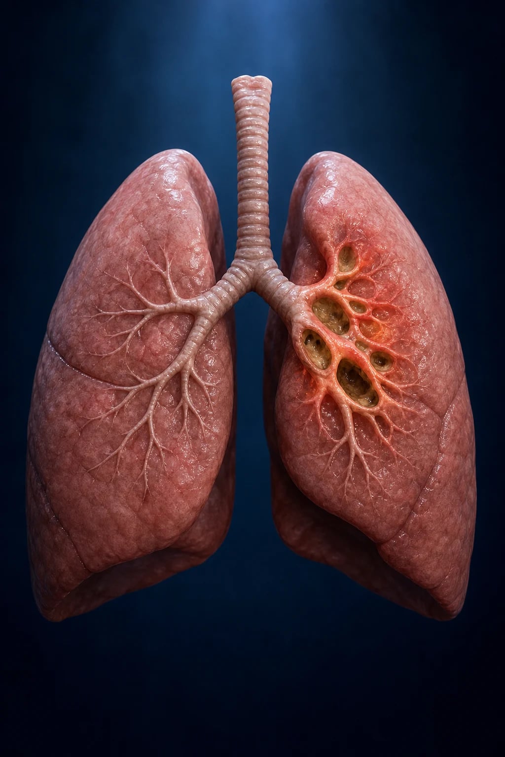
High-resolution CT (HRCT) chest is the definitive imaging. Findings include central (proximal, perihilar, upper-lobe predominant) bronchiectasis — varicose or cystic in advanced disease; mucus plugging in ectatic bronchi; atelectasis/collapse; and — in the pathognomonic subtype — high-attenuation mucus (HAM), mucus denser than paraspinal skeletal muscle on unenhanced CT.[7] In aspergilloma, CT shows a mobile intracavitary mass with an air-crescent sign in a pre-existing cavity. In invasive aspergillosis, the CT halo sign (nodular consolidation surrounded by a ground-glass halo of haemorrhage) is the early hallmark, evolving to the air-crescent sign as the infarct cavitates.[3][4]
Spirometry and lung function
Spirometry confirms the obstructive pattern of asthma and tracks response; a restrictive component or fixed obstruction suggests advanced/fibrotic disease. Serial spirometry is part of follow-up.[7]
Investigations for the other syndromes
Invasive aspergillosis in the at-risk host: serum and BAL galactomannan (Platelia ELISA; a polysaccharide cell-wall component released by Aspergillus — a single serum index over 0.5 or BAL over 1.0 is positive), 1,3-beta-D-glucan (non-specific marker of invasive fungal infection), high-resolution CT (halo and air-crescent signs), sputum/BAL culture (Aspergillus grows on Sabouraud agar), PCR, and where feasible histology of biopsy (septate acute-angle branching hyphae invading tissue).[3][4]
Aspergilloma: chest radiograph (mobile intracavitary mass with air-crescent), CT confirming the fungal ball, serum Aspergillus precipitins/IgG (typically strongly positive), and sputum culture.[4]
Sputum cytology
A wet mount or cytology of sputum may show branching septate acute-angle hyphae, Charcot-Leyden crystals (bipyramidal eosinophil breakdown products) and eosinophils — supportive but not diagnostic.[7]
Management — Resuscitation
ABPA itself is rarely a resuscitation emergency, but three acute presentations demand immediate action: a severe asthma exacerbation with respiratory failure complicating ABPA, a massive haemoptysis from an aspergilloma, and rapidly progressive invasive aspergillosis in the immunocompromised.[4][7]
Severe asthma exacerbation
ABCDE first. Supplemental oxygen to maintain saturations 94 to 98 percent; nebulised salbutamol 5 mg driven by oxygen with ipratropium bromide 0.5 mg, repeated or continuous; systemic corticosteroid — prednisolone 40 to 50 mg orally (or hydrocortisone 100 mg IV if unable to tolerate oral or very severe); IV magnesium sulphate 1.2 to 2 g over 20 minutes for life-threatening features. Escalate to non-invasive ventilation, invasive mechanical ventilation and ICU for life-threatening asthma (silent chest, exhaustion, cyanosis, bradycardia, poor respiratory effort, rising or normalising pCO2).[7]
Massive haemoptysis from an aspergilloma
This is a life-threatening emergency. Protect the uninvolved lung by placing the patient in the lateral decubitus position with the bleeding side down; secure the airway with a large-bore single-lumen endotracheal tube or a double-lumen tube; arrange urgent bronchial-artery embolisation (the first-line intervention); involve thoracic surgery early (cavernostomy or lobectomy for uncontrolled bleeding).[4]
Invasive aspergillosis in the neutropenic patient
Start empiric antifungal therapy without waiting for confirmation — voriconazole (the first-line agent) or liposomal amphotericin B; send serum and BAL galactomannan; obtain an urgent high-resolution CT (halo sign); reverse the immunocompromise (G-CSF, reduce immunosuppression where possible); and treat respiratory failure supportively. Delay is the single largest contributor to mortality.[3][4]
ABPA exacerbation
A rising IgE, new infiltrates, worsening wheeze and brown plugs in a known ABPA patient indicate an exacerbation. The immediate pharmacologic step is a course of systemic corticosteroid (prednisolone 0.5 to 0.75 mg/kg/day), with addition or re-introduction of an antifungal if not already on one.[1][7]
Management — Definitive & Stepwise
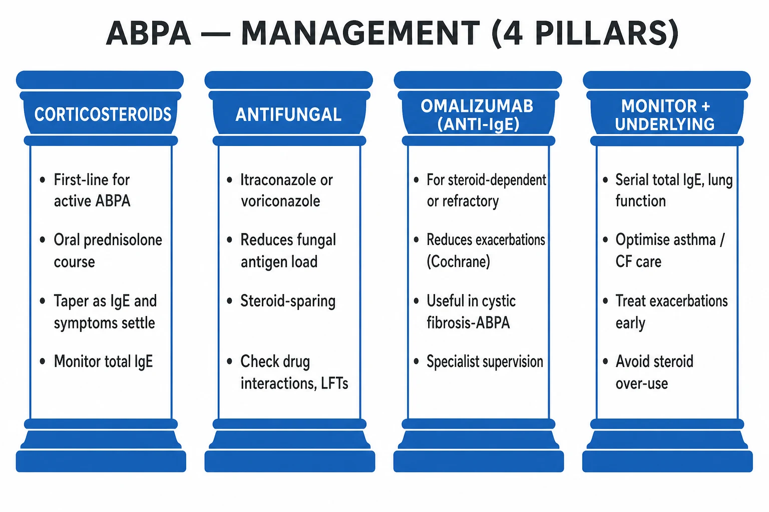
Definitive ABPA management rests on four pillars: systemic corticosteroids to suppress the allergic inflammation, an antifungal to reduce the antigen load (the two together are steroid-sparing), anti-IgE (and emerging anti-IL-5) biologics for steroid-dependent or refractory disease, and serial monitoring of total IgE and lung function with optimisation of the underlying asthma or CF.[1][2][7]
Systemic corticosteroids — drug, dose, route, duration, rationale
Prednisolone — first line
The fire-extinguisher
- **Prednisolone 0.5 to 0.75 mg/kg/day orally** (typically 20 to 40 mg) for **4 to 6 weeks**, then **taper by 5 to 10 mg every 2 weeks** over a further 3 to 6 months guided by total IgE and symptoms
- Rationale: **suppress the Th2-driven allergic inflammation** and the eosinophil-mediated airway-wall damage
- Monitor **blood glucose, bone density, weight, blood pressure**; consider **calcium/vitamin D and gastric protection** for prolonged courses
IV corticosteroid — for severe exacerbation
When oral is not enough
- **Hydrocortisone 100 mg IV every 6 hours** (or **methylprednisolone 40 to 60 mg IV daily**) for a severe ABPA/asthma exacerbation, switching to oral prednisolone as the patient improves
- Bridges to the oral taper regimen above once stable
Inhaled corticosteroids
Not enough for active ABPA
- **Essential for the underlying asthma** but **insufficient as sole therapy** for active ABPA — the antigen load and systemic Th2 response require systemic steroids
- Maintain high-dose ICS/LABA throughout and after the systemic course
Antifungals — steroid-sparing antigen control
The rationale is mechanistic: corticosteroids suppress the inflammation; azoles cut the antigen load that drives it. Used together, azoles allow a lower cumulative steroid dose (steroid-sparing) and reduce relapse.[2][5][10]
Itraconazole — the standard azole
First-line oral antifungal
- **Itraconazole 200 mg orally twice daily for 16 weeks, then 200 mg once daily for 16 weeks**
- Rationale: **reduce Aspergillus antigen load** in the airway; **steroid-sparing** (Stevens 2000 NEJM showed fewer treatment failures and lower cumulative steroid dose)
- **Monitor levels (trough) and LFTs**; absorption is pH-dependent (avoid H2-blockers/PPIs; take with acidic beverage/capsule over solution)
- **Major CYP3A4 interactions**: statins, calcineurin inhibitors, warfarin, vincristine, midazolam, digoxin; **negative inotrope (heart failure caution)**
Voriconazole — refractory / preferred
Better bioavailability
- **Voriconazole** (e.g. 200 mg orally twice daily, weight-based loading) for **itraconazole intolerance, failure or malabsorption**, or refractory disease
- Superior tissue penetration; also **first-line for invasive aspergillosis** (Herbrecht 2002 NEJM, superior response and survival versus amphotericin B)
- **Monitor levels, LFTs, and for visual disturbance** (photopsia), photosensitivity, **QT prolongation and skin cancer**; broad CYP interactions
Posaconazole / isavuconazole
Alternatives
- **Posaconazole** and **isavuconazole** for intolerance or resistance; isavuconazole has a **favourable QT profile** (shortens QT)
- Used mainly for invasive/chronic disease and as alternatives in ABPA salvage
Omalizumab (anti-IgE) and the biologics
Omalizumab, a recombinant humanised monoclonal antibody that binds free IgE and downregulates the high-affinity IgE receptor on effector cells, has become the key steroid-sparing agent for steroid-dependent or refractory ABPA — particularly in cystic fibrosis, where systemic steroids carry special morbidity (growth, glucose, infection).[6] A Cochrane review (Jat 2021) found anti-IgE therapy improves clinical and immunologic outcomes in ABPA complicating CF, supporting its use when standard therapy fails or steroid-toxicity is unacceptable.[6] Dosing is subcutaneous every 2 to 4 weeks, dosed by body weight and baseline total IgE.[6][7]
The anti-IL-5 biologics — mepolizumab (anti-IL-5) and benralizumab (anti-IL-5 receptor alpha, depleting eosinophils) — target the eosinophil arm of the cascade and show promise in small series and case reports of steroid-dependent ABPA, but the evidence base is much smaller than for omalizumab and they remain off-label.[7]
Treatment response and follow-up
Total IgE is the key biomarker. Check it every 6 to 8 weeks during treatment — it should fall by at least 20 to 25 percent; symptom control, spirometry and (when indicated) repeat imaging should improve in parallel. After remission, continue serial IgE every 3 to 6 months indefinitely, because a rise of more than two times the remission baseline signals relapse and triggers a treatment course.[1][7]
Management of aspergilloma
- Asymptomatic aspergilloma — observe; many remain stable.[4]
- Symptomatic / haemoptysis — bronchial-artery embolisation is the first-line intervention for bleeding; surgical resection (cavernostomy or lobectomy) is definitive but carries significant morbidity in poor-reserve patients and is reserved for recurrent or massive haemoptysis, or localised disease in a fit patient.[4]
- Systemic azoles have poor penetration into the avascular cavity and limited role; intracavitary instillation of amphotericin B or itraconazole has been used in selected cases.[4]
Management of invasive aspergillosis
Voriconazole is first-line on the strength of the Herbrecht 2002 NEJM randomised trial, which showed superior response and survival versus amphotericin B for primary therapy of invasive aspergillosis.[3] Alternatives and salvage include liposomal amphotericin B, isavuconazole, posaconazole, and the echinocandins (caspofungin, micafungin) as salvage or combination. Therapy continues for 6 to 12 weeks and until clinical, radiologic and microbiologic resolution plus immunorecovery. Primary antifungal prophylaxis (posaconazole during prolonged neutropenia or GVHD) is standard in the highest-risk haematology patients.[3][4]
Management of chronic pulmonary aspergillosis (CPA)
Long-term oral itraconazole or voriconazole for at least 6 months (often years), with monitoring of Aspergillus IgG, CRP and imaging; surgical resection for localised, surgically-fit disease.[4]
Escalation triggers in ABPA
Escalate (add or re-introduce an antifungal; add a biologic; reconsider the diagnosis) when: the total IgE fails to fall by 6 to 8 weeks or rises; new infiltrates appear; airflow obstruction worsens; or exacerbations recur despite tapering steroids (steroid-dependent ABPA, Stage IV).[1][7]
Specific Subtypes & Scenarios
Each clinical form of Aspergillus lung disease reframes the priority — suppress allergy, control bleeding, or stop invasion. [1]
- ABPA-S (seropositive, no bronchiectasis) — an earlier form; meets immunologic criteria but HRCT shows no bronchiectasis. Treat to prevent progression to structural disease.[1]
- ABPA-B (with bronchiectasis) — the classic radiological form; central bronchiectasis on HRCT; higher relapse risk and closer monitoring.[7]
- ABPA with high-attenuation mucus (HAM) — pathognomonic, severe, often steroid-dependent; needs aggressive combined therapy and often a biologic.[7]
- ABPA in cystic fibrosis — diagnostic caveat: the IgE cut-off may be applied more flexibly in CF; treat actively but steroid-sparingly (steroids harm growth, glucose control and infection control in CF); omalizumab is particularly useful.[6]
- Aspergilloma — pre-existing cavity, haemoptysis (may be massive), embolisation or surgery; azoles have poor cavity penetration.[4]
- Invasive aspergillosis — neutropenic/transplant host, voriconazole, galactomannan, CT halo sign.[3][4]
- Chronic pulmonary aspergillosis — subacute cavitating disease in mild immunocompromise; long-term azole.[4]
- Allergic Aspergillus sinusitis — frequently co-exists with ABPA (the 'united airway'); manage with topical and systemic steroids ± surgery.[7]
- Severe asthma with fungal sensitisation (SAFS) — severe asthma with fungal sensitisation, total IgE below 1000 IU/mL and no bronchiectasis; an antifungal trial may help, but it is not ABPA and does not meet ISHAM criteria.[1][9]
Complications & Pitfalls
Long-term complications of ABPA flow directly from the central-airway damage and the cumulative steroid burden: progressive irreversible central bronchiectasis, recurrent secondary bacterial infection (often with Pseudomonas or Haemophilus), the fibrotic end-stage with type-2 respiratory failure and cor pulmonale, and the chronic toxicity of oral corticosteroids — osteoporosis and vertebral fractures, diabetes mellitus, hypertension, cataract, skin atrophy, and hypothalamic-pituitary-adrenal suppression.[1][7] Aspergilloma threatens massive life-threatening haemoptysis from bronchial-artery erosion and may progress to chronic pulmonary aspergillosis.[4] Invasive aspergillosis threatens dissemination (especially to the brain), massive haemoptysis, airway obstruction and a mortality of 30 to 60 percent despite treatment.[3][4]
Treating ABPA as ordinary severe asthma
Missed diagnosis
- Years of escalating asthma therapy without the **corticosteroid-plus-antifungal** combination allows **irreversible bronchiectasis** to accrue
- Always screen the difficult asthmatic: **eosinophils, total IgE, Aspergillus skin-prick or specific IgE**
Corticosteroid alone, no antifungal
Higher cumulative steroid
- Adding an **azole reduces the antigen load** (Stevens 2000 NEJM), is **steroid-sparing**, and reduces relapse — corticosteroid monotherapy drives steroid toxicity
Forgetting azole interactions
CYP3A4 danger
- **Itraconazole and voriconazole are potent CYP3A4 inhibitors/substrates** — check **statins, calcineurin inhibitors, warfarin, vincristine, midazolam, digoxin**; **itraconazole is a negative inotrope** (heart-failure caution); **voriconazole** — LFTs, visual disturbance, photosensitivity, **skin cancer**; **monitor drug levels and LFTs**
Over-diagnosing ABPA on IgE alone
False positive
- A raised IgE is common in atopy; ABPA requires **Aspergillus sensitisation** (skin-prick or specific IgE) AND characteristic imaging/eosinophilia — the **obligatory ISHAM criterion**
Missing galactomannan in the neutropenic patient
Treatable, fatal if missed
- In the at-risk host, **fever unresponsive to antibiotics** mandates **serum/BAL galactomannan and CT** — start **voriconazole** empirically while awaiting results
Steroid over-treatment in CF-ABPA
Special-population harm
- In CF, steroids impair **growth, glucose control and infection control** — use **omalizumab** and azoles early as steroid-sparing
Prognosis & Disposition
ABPA, treated early with corticosteroid plus an antifungal, responds in the majority — symptoms settle, the IgE falls, and imaging improves. Relapse is common, however, and is detected by a rising total IgE (more than two times the remission baseline), driving the need for lifelong surveillance.[1][7] The worst outcomes are in fibrotic end-stage (Stage V) disease, in steroid-dependent ABPA, and in ABPA with high-attenuation mucus.[7] Aspergilloma has a variable course dominated by the risk of massive haemoptysis; surgical resection is curative but carries morbidity in the poor-reserve patient.[4] Invasive aspergillosis carries a mortality of 30 to 60 percent even with treatment, highest with delayed therapy, disseminated disease, persistent neutropenia and failure of immunorecovery.[3][4]
Disposition for ABPA is specialist respiratory outpatient care with serial total IgE (every 6 to 8 weeks during treatment, then every 3 to 6 months long-term), lung function, and periodic imaging as clinically indicated; admission is reserved for severe exacerbations, suspected invasive transformation, or complications such as massive haemoptysis. The underlying asthma or CF must be optimised in parallel.[1][7]
Special Populations
- Cystic fibrosis — high ABPA prevalence (around 8 percent); annual screening (total IgE and Aspergillus-specific IgE/IgG) is built into CF care; the IgE threshold may be applied more flexibly; treat actively but steroid-sparingly; omalizumab is particularly useful.[6]
- Children — same diagnostic approach and first-line therapy, but minimise systemic steroid exposure (growth, adrenal suppression); use azoles and biologics early as steroid-sparing.[7]
- Pregnancy — prednisolone (the corticosteroid of choice in pregnancy) is the first-line controller; avoid itraconazole in the first trimester where possible and use it cautiously thereafter; omalizumab has limited pregnancy data but is used in refractory disease; manage in a multidisciplinary setting.[7]
- The elderly — heightened corticosteroid toxicity (osteoporosis, diabetes, hypertension) and polypharmacy increase the value of steroid-sparing azoles/biologics; check azole drug interactions carefully.[7]
- Immunocompromised / transplant — the dominant consideration shifts from ABPA to invasive aspergillosis: posaconazole prophylaxis in prolonged neutropenia/GVHD, galactomannan surveillance, voriconazole first-line treatment.[3][4]
- Aspergilloma in old TB — assess fitness for surgery; weigh embolisation against resection.[4]
- Diabetic patient with ABPA — heightened steroid-induced hyperglycaemia and opportunistic-infection risk; tight glucose control and steroid-sparing.[7]
Evidence, Guidelines & Regional Differences
The ISHAM 2013 working group criteria (Agarwal, Chakrabarti, Denning et al.) unified and relaxed the older Rosenberg-Patterson criteria — making the IgE threshold above 500 IU/mL acceptable when all other criteria are met — and are now the global diagnostic standard.[1] The pivotal Stevens 2000 NEJM randomised trial of itraconazole in ABPA showed that adding itraconazole for 16 weeks reduced treatment failure and lowered the cumulative steroid dose, anchoring the steroid-sparing antifungal strategy.[2] The pivotal Herbrecht 2002 NEJM randomised trial established voriconazole over amphotericin B as first-line for invasive aspergillosis (better response and survival at 12 weeks).[3] The Cochrane review by Wark (2004) confirmed that azoles produce immunologic and radiologic improvement in ABPA associated with asthma,[5] and the Cochrane review by Jat (2021) supported anti-IgE therapy for ABPA complicating cystic fibrosis.[6]
The IDSA 2008 aspergillosis guidelines (Walsh et al; updated 2016) codify voriconazole first-line for invasive disease and azoles for chronic forms.[4] The ERS/ECMM/ESCMID 2017 joint guideline harmonises European practice. The Cystic Fibrosis Foundation consensus drives the annual screening programme for ABPA in CF.[6]
Regional deltas: [1]
| Region / guideline | Approach to ABPA and fungal asthma |
|---|---|
| India (Agarwal / FASE consensus) | High ABPA prevalence; Agarwal staging (I–V) widely used; lower threshold to treat; Lung India / Indian J Med Res reviews are leading references.[8][9] |
| UK (BTS severe asthma pathway) | Screen difficult asthma for fungal sensitisation; antifungal stewardship to limit resistance; omalizumab commissioned for severe allergic asthma. |
| US (IDSA 2008/2016) | Voriconazole first-line for invasive disease; azoles for chronic; biologics off-label.[4] |
| CF Foundation consensus (global CF) | Annual screening for ABPA in CF (total IgE and Aspergillus-specific IgE/IgG).[6] |
Where the evidence is weak — there is a paucity of large randomised trials in ABPA itself (the field is dominated by observational cohorts, mostly from Agarwal's group); the anti-IL-5 biologics (mepolizumab, benralizumab) and even omalizumab in non-CF ABPA remain off-label; the optimal duration of antifungal therapy and the role of antifungals in SAFS are debated.[5][7]
Exam Pearls
The Aspergillus lung spectrum — SAM-CI
SAM-CI
Skin-prick or specific IgE positive; asthma may worsen; NO bronchiectasis; watch for ABPA
Asthma/CF + sensitisation + raised IgE (>500) + eosinophilia + central bronchiectasis; ISHAM criteria; steroid + antifungal
Fungal ball in a cavity; air-crescent sign; HAEMOPTYSIS (may be massive); embolisation or surgery
Slow cavitating disease in mild immunocompromise; long-term azole
Neutropenic/transplant host; angioinvasive; CT halo sign; VORICONAZOLE first-line
ISHAM criteria — POSITIVE
POSITIVE
Obligatory — without one of these, ABPA is not the diagnosis
Positive Aspergillus skin-prick OR raised Aspergillus-specific IgE
Over 1000 IU/mL classically, or above 500 IU/mL if all other criteria met
Central bronchiectasis, mucus plugging, high-attenuation mucus, fleeting infiltrates
Raised Aspergillus-specific IgG/precipitins, eosinophils over 500/microL
Falls 20–25% on treatment; doubles = relapse
Antifungals reduce antigen load — steroid-sparing
Blood eosinophilia is a core feature
Why is the bronchiectasis of ABPA characteristically central (proximal)?
The allergic inflammation targets the larger, central segmental and subsegmental bronchi — where mucoid impaction, bronchocentric granulomatosis and eosinophil-mediated wall injury concentrate. The result is central (perihilar), upper-lobe predominant, varicose-or-cystic bronchiectasis — the radiological signature that distinguishes ABPA from the peripheral bronchiectasis of post-infective disease or the traction bronchiectasis of fibrosis.
Why is voriconazole first-line for invasive aspergillosis?
The Herbrecht 2002 NEJM randomised trial showed voriconazole produced a better response (53% vs 32%) and higher survival (71% vs 58%) at 12 weeks than amphotericin B for primary therapy of invasive aspergillosis, with less nephrotoxicity. This established voriconazole as the first-line agent (Walsh/IDSA guidelines).[3][4]
- The one-liner: ABPA is a Th2 hypersensitivity to Aspergillus fumigatus in asthma or CF — raised total IgE, Aspergillus sensitisation, eosinophilia, central bronchiectasis, brownish sputum plugs; ISHAM criteria; treated with oral corticosteroid plus an antifungal (itraconazole or voriconazole), with omalizumab for steroid-dependent or refractory disease.[1]
- High-attenuation mucus (HAM) on unenhanced CT (mucus denser than paraspinal muscle) is nearly pathognomonic.[7]
- Frequently misremembered facts, correctly stated: itraconazole is steroid-sparing, not a replacement for corticosteroid in active ABPA; galactomannan is a marker of invasive disease, not ABPA; omalizumab is steroid-sparing for refractory/CF disease, not first-line; a negative Aspergillus skin-prick test excludes ABPA.[1][7]
- Aspergilloma → haemoptysis; invasive aspergillosis → voriconazole in the neutropenic.[3][4]
- Five stages (ARESF): Acute, Remission, Exacerbation, Steroid-dependent, Fibrotic.[1]
- Same fungus, different host.[1]
Exam application bank (NEET-PG / INICET)
One-line answer
Aspergillus fumigatus causes a spectrum of lung disease whose form is set entirely by the host: Aspergillus sensitisation (skin/IgE positive, asthma may worsen, no structural change), allergic bronchopulmonary aspergillosis (ABPA) — a Th2 hypersensitivity reaction to Aspergillus colonising the airways of patients with asthma or cystic fibrosis, producing poorly controlled asthma, brownish sputum plugs, blood eosinophilia, a markedly raised total IgE (over 500 to 1000 IU/mL) and central (proximal) bronchiectasis with mucus plugging; aspergilloma — a saprophytic fungal ball in a pre-existing cavity causing haemoptysis (which can be massive); chronic pulmonary aspergillosis — slow cavitating disease in mild immunocompromise; and invasive pulmonary aspergillosis — an angioinvasive pneumonia in the neutropenic or transplant patient, treated first-line with voriconazole. ABPA is diagnosed by t [1]
Worked stems (answer without another resource)
Stem 1 — Classic presentation. Map symptoms to mechanism; name the first investigation and first treatment step with dose/route if drug therapy is standard. [1]
Stem 2 — Unstable / complicated. List red flags that force immediate resuscitation, theatre, ICU, antidote, or reperfusion — and what you do in the first 15 minutes. [1]
Stem 3 — Atypical group. Elderly, pregnancy, child, or immunocompromised: how presentation and thresholds change. [1]
Stem 4 — Differential trap. Name the three closest mimics and one discriminator for each. [1]
Stem 5 — Disposition. Who goes home with safety-netting, who is admitted, who needs HDU/ICU/theatre, and what follow-up is mandatory. [1]
Rapid viva checklist
- Definition + classification
- Pathophysiology chain
- Bedside signs / criteria
- Score with exact components (if any)
- Emergency bundle
- Definitive therapy with doses
- Complications of disease and of treatment
- Special populations
- Guideline/trial name if classic
- Three exam traps
Coverage self-check
If you cannot answer any stem above from this page alone, re-read the matching section — the page is intended to be self-sufficient for final-prof and NEET-PG/INICET questions on Allergic Bronchopulmonary Aspergillosis & Aspergillus Lung Disease.
References
- [1]Agarwal R, Chakrabarti A, Shah A, Gupta D, Meis JF, Guleria R, Moss R, Denning DW. Allergic bronchopulmonary aspergillosis: review of literature and proposal of new diagnostic and classification criteria Clin Exp Allergy, 2013.PMID 23889240
- [2]Stevens DA, Schwartz HJ, Lee JY, Moskovitz BL, Jerome DC, Catanzaro A, Bamberger DM, et al. A randomized trial of itraconazole in allergic bronchopulmonary aspergillosis N Engl J Med, 2000.PMID 10717010
- [3]Herbrecht R, Denning DW, Patterson TF, Bennett JE, Greene RE, Oestmann JW, et al. Voriconazole versus amphotericin B for primary therapy of invasive aspergillosis N Engl J Med, 2002.PMID 12167683
- [4]Walsh TJ, Anaissie EJ, Denning DW, Herbrecht R, Kontoyiannis DP, Marr KA, et al. Treatment of aspergillosis: clinical practice guidelines of the Infectious Diseases Society of America Clin Infect Dis, 2008.PMID 18177225
- [5]Wark PA, Gibson PG, Wilson AJ. Azoles for allergic bronchopulmonary aspergillosis associated with asthma Cochrane Database Syst Rev, 2004.PMID 15266440
- [6]Jat KR, Walia DK, Khairwa A. Anti-IgE therapy for allergic bronchopulmonary aspergillosis in people with cystic fibrosis Cochrane Database Syst Rev, 2021.PMID 34550603
- [7]Agarwal R, Muthu V, Sehgal IS, Dhooria S, Prasad KT, Aggarwal AN. Allergic Bronchopulmonary Aspergillosis Clin Chest Med, 2022.PMID 35236565
- [8]Agarwal R, Sehgal IS, Dhooria S, Aggarwal AN. Allergic bronchopulmonary aspergillosis Indian J Med Res, 2020.PMID 32719226
- [9]Agarwal R, Muthu V, Sehgal IS. Prevalence of Aspergillus sensitization and Allergic Bronchopulmonary Aspergillosis in bronchial asthma: A systematic review of Indian studies Lung India, 2023.PMID 37961961
- [10]Wark P. Pathogenesis of allergic bronchopulmonary aspergillosis and an evidence-based review of azoles in treatment Respir Med, 2004.PMID 15481266