Respiratory · General Medicine
Acute Bronchitis
Also known as Acute bronchitis · Chest cold · Acute tracheobronchitis
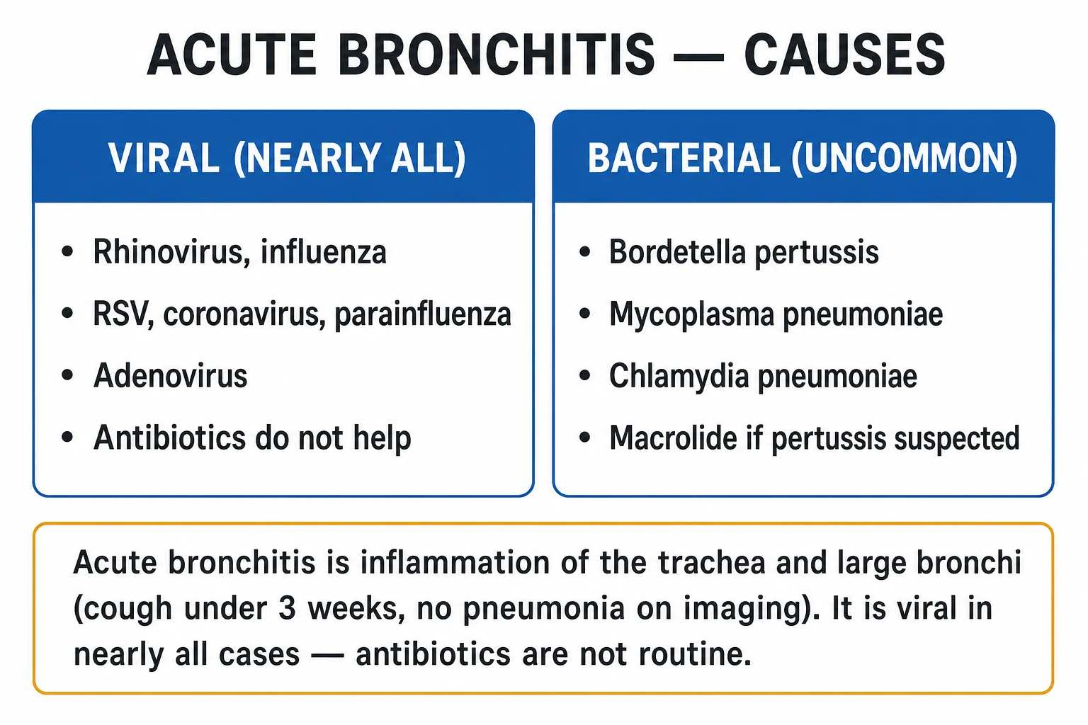
Acute bronchitis is acute inflammation of the trachea and large bronchi producing a self-limiting cough of up to three weeks, nearly always viral (rhinovirus, influenza, RSV, coronavirus, parainfluenza, adenovirus) and without the radiographic consolidation of pneumonia. The cough often lasts one to three weeks (sometimes up to eight from post-viral airway hyper-reactivity). The pivotal clinical tasks are to exclude pneumonia (normal vital signs, no focal chest signs, normal oxygenation) and to avoid unnecessary antibiotics — they offer minimal benefit and real harm, because the cause is viral. Management is symptomatic (rest, fluids, analgesia, honey or an antitussive). Consider pertussis (cough beyond three weeks or known exposure) — treat with a macrolide and notify public health.
On this page & tools
Your progress
Saved locally on this device.
Exam tags
Red flags
Overview & Definition
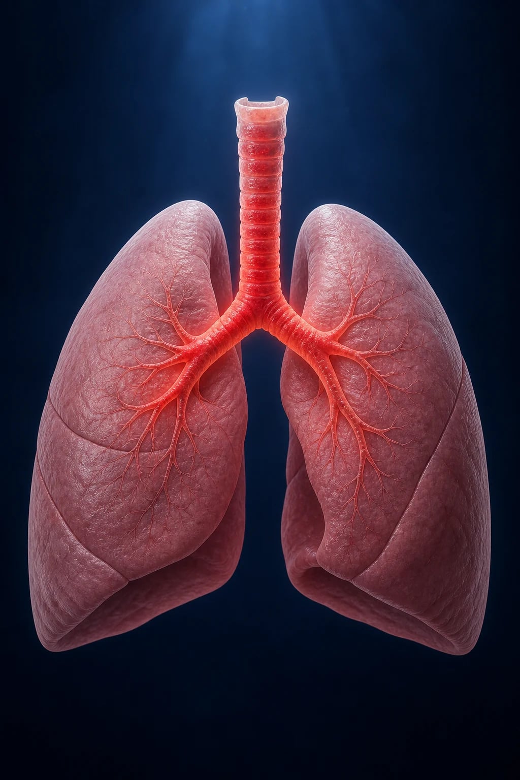
Acute bronchitis is acute inflammation of the trachea and the large bronchi — the conducting airways above the respiratory bronchioles — producing a self-limiting cough lasting up to three weeks. The lung parenchyma is spared; this is the single anatomical fact that distinguishes acute bronchitis from pneumonia, and it is the pivot on which the entire clinical decision turns.[2]
Acute bronchitis overlaps clinically with the common cold and other upper-respiratory infections, and it is one of the commonest reasons adults consult a clinician and receive an antibiotic. The pivotal judgement is twofold: separate it from pneumonia (which needs antibiotics, oxygen and sometimes admission) and withhold antibiotics from the far larger viral majority — antibiotics offer only a small, clinically insignificant reduction in cough duration at the cost of real adverse effects and resistance.[1][2]
Classification
Acute bronchitis is best classified by aetiology (viral vs bacterial) and by context (the host in whom it occurs), because these axes drive management: [1]
By aetiology: [1]
- Viral (the overwhelming majority, over 90% of cases): rhinovirus (commonest), influenza A and B, respiratory syncytial virus (RSV), parainfluenza, coronavirus (including SARS-CoV-2), adenovirus, human metapneumovirus, and human bocavirus. Influenza and RSV are the more likely to produce severe or lower-airway disease, especially in the elderly and those with cardiopulmonary comorbidity.
- Bacterial (uncommon as a primary cause): Bordetella pertussis (whooping cough — the single most important treatable cause of prolonged acute cough), Mycoplasma pneumoniae, and Chlamydophila pneumoniae (atypical organisms that produce a syndrome overlapping with acute bronchitis / atypical pneumonia). Bordetella parapertussis causes a milder pertussis-like illness.
- Secondary bacterial infection of virally damaged airways (Streptococcus pneumoniae, Haemophilus influenzae, Moraxella catarrhalis) may complicate the course, particularly in COPD — but in the uncomplicated adult this does not justify routine antibiotics. [1]
By clinical context (drives threshold and therapy): [1]
- Uncomplicated acute bronchitis in an otherwise-well adult — symptomatic management, no antibiotics, no investigations.
- Acute bronchitis in COPD or asthma — a common exacerbation trigger; treat the underlying disease (bronchodilators ± steroids; antibiotics in COPD by Anthonisen criteria — see Specific Subtypes).
- Pertussis syndrome — paroxysmal cough over three weeks, whoop, post-tussive vomiting; macrolide and public-health notification.
- Acute bronchitis with significant comorbidity (heart failure, immunocompromise, chronic lung disease, the very young or old) — lower threshold to investigate and admit. [1]
Acute bronchitis
- Inflammation of **trachea + large bronchi**; parenchyma normal
- Cough **under 3 weeks**, often following URTI symptoms
- **Viral** in over 90% (rhinovirus, influenza, RSV)
- Normal vitals, **no consolidation**, normal CXR
- **No routine antibiotics** — symptomatic management
Pneumonia
- Infection of the **alveoli / lung parenchyma** — consolidation
- Fever, **productive cough**, dyspnoea, **pleuritic pain**
- **Strep pneumoniae**, atypicals, viruses
- Tachypnoea, **focal signs**, consolidation on CXR
- **Antibiotics within 4 hours**; severity by CURB-65
Chronic bronchitis (COPD)
- Chronic productive cough on **most days for 3 months in 2 successive years**
- **Irreversible** airflow obstruction (COPD)
- Smoking-related; exacerbations often infective
- Exacerbation: Anthonisen criteria guide antibiotics
- Bronchodilators, steroids, pulmonary rehabilitation
Epidemiology & Risk Factors
Acute bronchitis is one of the commonest acute respiratory illnesses in adults, with an incidence of roughly 40–50 cases per 1000 adults per year, peaking in autumn and winter. It affects all ages but is most frequent in young adults (under 25), the elderly, and those with underlying lung disease. It is a leading reason for ambulatory antibiotic prescribing, and inappropriate antibiotic use for acute bronchitis remains a major stewardship target worldwide. [1]
From a health-economic perspective, acute bronchitis generates substantial direct costs (consultations, investigations, prescriptions) and indirect costs (time off work, reduced productivity). A large proportion of these costs are driven by unnecessary antibiotics and chest X-rays. Even a modest reduction in inappropriate antibiotic prescribing for acute bronchitis yields significant population-level savings and slows the rise of antimicrobial resistance. Quality-improvement initiatives that combine clinician education, patient information leaflets, delayed prescribing and point-of-care CRP testing have demonstrated meaningful reductions in antibiotic use for acute respiratory infections in primary care. [1]
Host and environmental risk factors: [1]
- Smoking (active and passive) — impairs mucociliary clearance, increases susceptibility and severity.
- Chronic lung disease — COPD, asthma, bronchiectasis.
- Immunocompromise — HIV, immunosuppressants, malignancy.
- Air pollution and indoor biomass-fuel smoke (a major driver in the developing world).
- Crowded living conditions — facilitate viral spread and pertussis transmission.
- Waning pertussis immunity — vaccination or natural infection does not confer lifelong immunity; adults are the main reservoir for infant pertussis, and a notable fraction of adults with cough lasting over three weeks have pertussis.
- Extremes of age and cardiac comorbidity — higher risk of complication and lower threshold to investigate. [1]
Seasonality: viral acute bronchitis mirrors the circulation of its causative viruses — rhinovirus year-round with autumn/spring peaks; influenza and RSV in winter; enteroviruses in summer. [1]
Acute bronchitis — key numbers
Pathophysiology
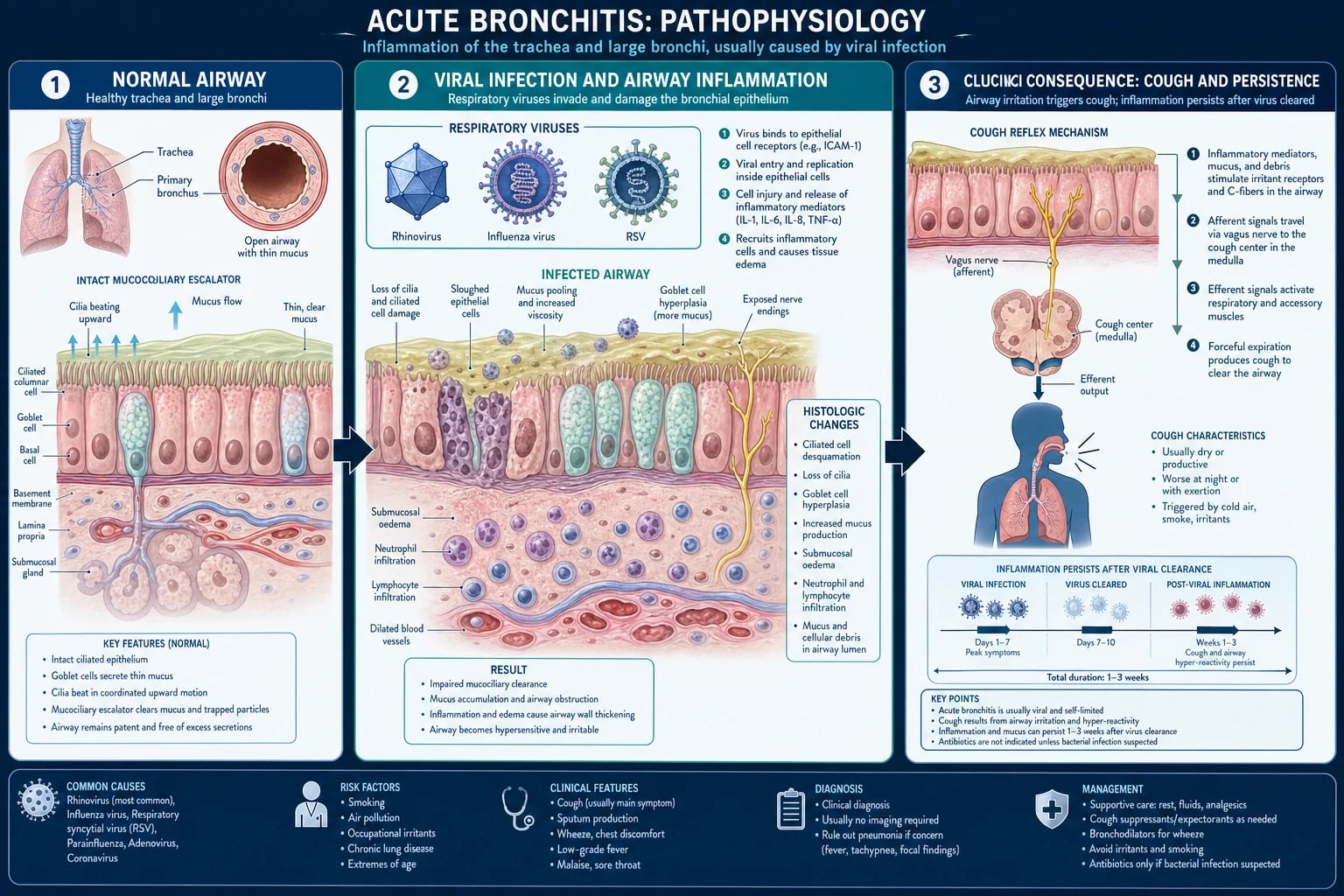
The conducting airways are normally defended by the mucociliary escalator (a coordinated beat of cilia propelling mucus and trapped particles upward, atop a thin sol layer bathing the cilia), by goblet-cell and submucosal-gland mucus, and by innate immune cells (alveolar and airway macrophages, secretory IgA, antimicrobial peptides such as defensins). Acute bronchitis occurs when a respiratory virus breaches these defences.[2]
The mechanism, step by step: [1]
- Inoculation and attachment. Respiratory viruses reach the bronchial epithelium after a primary upper-respiratory infection (the common-cold prodrome) and attach to epithelial receptors — e.g. rhinovirus binds ICAM-1, influenza binds sialic-acid residues, RSV the G protein to host cells. Viral replication in ciliated epithelial cells follows.
- Epithelial injury and loss of cilia. Infected cells undergo apoptosis and desquamation, stripping the mucociliary escalator. Ciliary beat frequency falls and cilia are lost, so mucus and debris pool in the airway lumen. This is the structural lesion of acute bronchitis.
- Inflammatory infiltrate. Damaged epithelium releases cytokines and chemokines — notably interleukin-6 (IL-6), interleukin-8 (IL-8, a potent neutrophil chemoattractant), TNF-alpha, and RANTES — recruiting neutrophils and lymphocytes into the submucosa and airway lumen. Bronchial-wall vasodilatation, hyperaemia and oedema follow, together with goblet-cell hyperplasia and increased mucus secretion.
- Bronchial hyper-reactivity. Epithelial denudation exposes irritant (rapidly-adapting stretch) receptors and C-fibre endings in the submucosa, lowering their threshold to tussive stimuli; mediators such as bradykinin and tachykinins sensitise these endings. The result is airway hyper-reactivity and the characteristic persistent, often dry-then-productive cough.
- Systemic features. The same cytokines (IL-1, IL-6, TNF-alpha, interferons) produce the fever, malaise, myalgia and headache via the hypothalamic prostaglandin-E2 set-point and systemic effects.
- Resolution and the lingering cough. As the virus is cleared by innate and adaptive immunity, the epithelium regenerates over one to three weeks, but mucociliary repair and resolution of airway hyper-reactivity lag, so the cough can persist for weeks — and in a minority (post-viral airway hyper-reactivity / post-bronchitic cough) for up to eight weeks, even after the patient feels otherwise well. [1]
Why antibiotics are ineffective: because the causative organism is viral and there is no bacterial target in uncomplicated disease. Antibiotics do not shorten the viral course, do not prevent bacterial superinfection in the well adult, and carry real harms — gastrointestinal adverse effects, rash, Clostridioides difficile infection, selection of resistance, cost, and anaphylaxis. This is the evidence base of the Cochrane review.[1]
How pertussis differs: Bordetella pertussis adheres to ciliated respiratory epithelium via filamentous haemagglutinin, pertactin and fimbriae, evades clearance, and secretes pertussis toxin (PT), adenylate cyclase toxin, tracheal cytotoxin and dermonecrotic toxin. These toxins paralyse and destroy cilia and drive a marked lymphocytosis (PT-driven), producing the paroxysmal cough (from sensory-neuron sensitisation by PT and bradykinin), the inspiratory whoop (forced inspiration against a narrowed glottis after a paroxysm), and post-tussive vomiting. The cough lasts weeks because ciliary recovery is slow — hence the older name "the hundred-day cough". [1]
Clinical Presentation
Typical uncomplicated acute bronchitis in an otherwise-well adult: [1]
- Acute cough — the defining symptom, under three weeks in duration; often dry at first, then productive of clear, yellow or green sputum. Sputum colour reflects inflammatory cells (neutrophils), not bacterial infection — green/yellow sputum does not by itself justify an antibiotic.
- Prodromal upper-respiratory symptoms — rhinorrhoea, sore throat, sneezing, nasal congestion, low-grade fever, malaise, myalgia, headache (the viral prodrome).
- Wheeze and chest tightness or retrosternal discomfort — from airway hyper-reactivity and coughing; a mild, transient wheeze is common.
- Dyspnoea is typically mild; significant dyspnoea, especially at rest, should prompt reconsideration of pneumonia, asthma/COPD exacerbation, or another diagnosis. [1]
Vital signs are typically normal or near-normal — this is the bedside marker that distinguishes bronchitis from pneumonia. Fever is usually low-grade (under 38°C); a high fever (over 38.5°C), tachypnoea, hypoxia, or haemodynamic upset should redirect the diagnosis toward pneumonia, influenza, COVID-19, or sepsis. [1]
Examination findings: the chest is usually clear, though coarse crackles and wheeze may be heard that clear or change with coughing. Critically, signs of consolidation are absent — no dullness to percussion, no bronchial breath sounds, no increased vocal resonance/tactile fremitus. Upper-airway examination may reveal pharyngeal injection or viral coryza. [1]
Course: most symptoms resolve within one to three weeks; the cough is often the last symptom to clear. A minority have a post-bronchitic cough lasting up to eight weeks from persistent airway hyper-reactivity, which then warrants the chronic-cough pathway if it persists. [1]
Atypical presentations to recognise: [1]
- Elderly: blunted febrile response, may present with confusion, falls, functional decline, anorexia; lower threshold to investigate and admit.
- COPD/asthma: the viral trigger produces an exacerbation with increased dyspnoea, wheeze, sputum volume or purulence — manage the underlying disease (see Specific Subtypes).
- Pregnancy: usual viral course; symptomatic treatment options narrowed by safety; pertussis in pregnancy is relevant to neonatal protection (maternal vaccination).
- Immunocompromised: higher risk of severe viral disease, secondary infection, and atypical organisms — lower threshold to investigate.
- Pertussis syndrome: paroxysmal cough lasting over three weeks, inspiratory whoop, post-tussive vomiting, and paroxysms at night — classically in an adolescent or adult whose childhood immunity has waned. [1]
Differential Diagnosis
An acute cough has a wide differential. The clinical task is to exclude the serious/treatable causes (pneumonia, PE, heart failure, pertussis, foreign body) and to recognise when the cough is not a simple viral bronchitis.[2]
The most important distinctions: [1]
- Community-acquired pneumonia — fever, tachypnoea, focal chest signs (consolidation), pleuritic pain, hypoxia; CXR shows consolidation. The single bedside discriminator is the vital signs plus chest examination — pneumonia declares itself; bronchitis does not. Use CRB-65/CURB-65 if pneumonia is present to grade severity.
- Asthma exacerbation / post-viral bronchial hyper-reactivity — wheeze, nocturnal cough, atopy, prior history; peak flow / spirometry with reversibility clinches it; treat with inhaled bronchodilators and steroids.
- COPD acute exacerbation — known COPD, smoking history, increased dyspnoea/sputum; treat with bronchodilators, steroids, and antibiotics by Anthonisen criteria.
- Upper-airway cough syndrome (post-nasal drip) — chronic allergic or infective rhinitis/sinusitis; cough triggered by drainage; treat the cause.
- Gastro-oesophageal reflux disease (GORD/GERD) — chronic cough, often without heartburn; trial of PPI and lifestyle measures.
- Pertussis — paroxysmal cough over 3 weeks, whoop, post-tussive vomiting; nasopharyngeal swab for PCR; macrolide; notify public health.
- ACE-inhibitor cough — dry cough within weeks of starting an ACE inhibitor (e.g. ramipril, lisinopril, enalapril); stop the drug.
- Acute viral tracheitis — prominent retrosternal pain and dry cough; usually a severe viral upper-airway syndrome.
- Pulmonary embolism — pleuritic pain, dyspnoea out of proportion, risk factors (immobility, malignancy); not usually febrile; Wells score + D-dimer/CTPA.
- Heart failure — orthopnoea, PND, bibasal crackles, oedema, raised JVP; BNP/NT-proBNP and echo.
- Lung cancer / TB / atypical infection — in persistent cough over 3 weeks with red flags (haemoptysis, weight loss, night sweats, smoking history, older age): CXR ± sputum for AFB / CT chest. [1]
The chronic-cough pathway (cough over 8 weeks): the commonest causes are asthma, reflux, post-nasal drip (the "triad") plus ACE inhibitors and non-asthmatic eosinophilic bronchitis; investigate with CXR, spirometry and a trial of therapy, escalating to high-resolution CT and bronchoscopy if red flags. [1]
DDx of acute cough — think CHOPS
CHOPS
known COPD, smoking, increased dyspnoea — Anthonisen criteria for antibiotics
orthopnoea, PND, crackles, raised JVP — BNP/echo
pneumonia (consolidation, tachypnoea), pertussis (whoop, over 3 wk)
pleuritic pain, dyspnoea, risk factors — Wells + D-dimer
post-nasal drip, GORD, drug-induced cough — triad of chronic cough
Clinical & Bedside Assessment
Acute bronchitis is a clinical diagnosis made at the bedside. The examination is directed at excluding pneumonia and other serious causes, not at "confirming" bronchitis. [1]
Vital signs (the most important data): [1]
- Respiratory rate — the single most sensitive marker of a lower-respiratory infection; a raised RR redirects to pneumonia/sepsis. Normal adult RR is 12–20.
- Oxygen saturation — normal (typically 95% or above in a non-COPD adult). Hypoxia mandates investigation.
- Temperature — usually low-grade; high fever (over 38.5°C) suggests influenza, pneumonia, or another infection.
- Heart rate and blood pressure — tachycardia or hypotension suggests sepsis, dehydration, PE, or significant illness.
- Conscious level — confusion in the elderly is a red flag (and a CURB-65 component for pneumonia). [1]
Focused respiratory examination: [1]
- Inspection — work of breathing, use of accessory muscles, cyanosis, chest wall movement.
- Palpation — chest expansion (reduced focally in consolidation/effusion); tactile fremitus.
- Percussion — resonant in bronchitis; dullness indicates consolidation, effusion, or collapse.
- Auscultation — wheeze and coarse crackles that clear or shift with cough are compatible with bronchitis; bronchial breath sounds, fine crackles, and increased vocal resonance indicate consolidation (pneumonia). A silent hemithorax with dullness suggests effusion or collapse.
- Upper-airway, ENT, and cardiovascular examination complete the assessment — pharyngitis, sinus tenderness, otitis, and signs of heart failure all redirect the diagnosis. [1]
Assess for sepsis (qSOFA: RR 22 or more, altered mentation, systolic BP 100 or less) and dehydration — neither should be present in uncomplicated acute bronchitis. [1]
Investigations
Acute bronchitis is a clinical diagnosis and, in the typical uncomplicated case, no investigations are required. The role of testing is to exclude alternative diagnoses when clinical features are not typical.[2]
Usually NOT required in uncomplicated acute bronchitis: [1]
- Chest X-ray — the parenchyma is normal; routine CXR does not change management and exposes the patient to radiation and cost.
- Full blood count, CRP — non-specific; a marked lymphocytosis may hint at pertussis.
- Sputum culture / Gram stain — viral sputum is not cultured; sputum colour does not predict bacterial infection.
- Viral PCR / multiplex respiratory panel — rarely changes management in the community; useful in outbreak settings, severe disease, immunocompromise, or in pregnancy/neonatal contacts. [1]
Indications for a chest X-ray (red flags / diagnostic uncertainty): [1]
- Abnormal vital signs — tachypnoea, hypoxia, high fever, tachycardia, hypotension.
- Focal chest signs on auscultation/percussion.
- Diagnostic uncertainty — clinician cannot confidently exclude pneumonia.
- Haemoptysis, pleuritic pain, or significant dyspnoea.
- Comorbidity — COPD, heart failure, immunocompromise, malignancy.
- Failure to improve within an expected timeframe, or deterioration after initial assessment.
- Older age, especially smokers over 50, to exclude underlying malignancy if symptoms persist. [1]
When to investigate for pertussis: [1]
- Cough over 3 weeks, paroxysmal, with whoop or post-tussive vomiting, or known exposure.
- Nasopharyngeal swab PCR is the test of choice in the first 3–4 weeks of illness (most sensitive in the catarrhal/early paroxysmal stage); culture is specific but less sensitive and only positive early; serology (anti-pertussis toxin IgG) is useful later (after 2 weeks, single high titre or rising titre).
- A marked absolute lymphocytosis (often over 15–20 × 10⁹/L in children) supports the diagnosis. [1]
When to do spirometry / peak flow: [1]
- Suspected asthma or COPD — demonstrate airflow obstruction and reversibility; peak-flow monitoring tracks an exacerbation. [1]
Point-of-care tests and stewardship: [1]
- CRP and procalcitonin — low levels support withholding antibiotics; procalcitonin-guided algorithms reduce antibiotic use in respiratory tract infections without worsening outcomes, and are endorsed by some stewardship programmes — though not routinely required in simple acute bronchitis.
- Rapid influenza and SARS-CoV-2 testing — when these viruses are prevalent and treatment/isolation decisions hinge on the result (pregnancy, immunocompromise, frail elderly, outbreak). [1]
Management — Resuscitation
Uncomplicated acute bronchitis does not require resuscitation — the patient is systemically well. Resuscitation is only relevant when the diagnosis is not simple bronchitis, i.e. when a red flag has surfaced: [1]
- Airway/Breathing — give oxygen to target SpO2 94–98% in most adults (or 88–92% in COPD / chronic CO2 retainers) for any patient who is hypoxic. Hypoxia mandates CXR and re-diagnosis.
- Circulation — IV access and balanced crystalloid if hypotensive or dehydrated; manage suspected sepsis with the Surviving Sepsis hour-1 bundle (oxygen, blood cultures, lactate, broad-spectrum antibiotics within 1 hour, 30 mL/kg crystalloid, noradrenaline for fluid-refractory shock). Sepsis means the diagnosis is pneumonia or another infection, not simple bronchitis.
- Disability — confusion (especially in the elderly) is a red flag; reassess for hypoxia, sepsis, metabolic cause.
- Escalate to hospital if any of: tachypnoea, hypoxia, confusion, hypotension, inability to maintain oral intake, significant comorbidity, or social factors preventing safe management at home. [1]
Management — Definitive & Stepwise
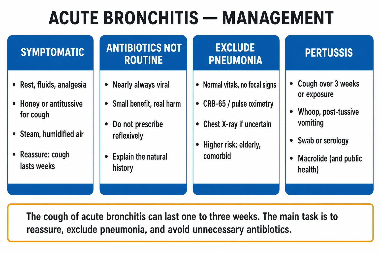
The definitive management of uncomplicated acute bronchitis is symptomatic, with explicit reassurance and a no-antibiotic / delayed-antibiotic strategy.[1][2]
Step 1 — Reassure and set expectations (the most therapeutic intervention). [1]
- Explain that the illness is viral and self-limiting; the cough will usually last one to three weeks and may run to eight weeks; antibiotics will not help and may cause harm.
- A no-antibiotic strategy, or a delayed (post-dated / "come back if not better in 10–14 days") prescription, reduces antibiotic use without worsening outcomes and is recommended by NICE (UK) and the ACP/ASIM–CDC/APP stewardship principles. [1]
Step 2 — Symptomatic relief. [1]
- Rest and adequate oral fluids.
- Antipyretic / analgesia: paracetamol 1 g orally every 6 hours (maximum 4 g per 24 hours in an adult) for fever, myalgia, headache, sore throat; or ibuprofen 400 mg orally every 8 hours with food (avoid in pregnancy third trimester, peptic ulcer, significant renal disease).
- Cough: honey (1–2 teaspoons, or ~2.5–5 g) — modestly reduces cough frequency and severity in adults and children over 1 year (do not give honey to infants under 1 year — risk of infant botulism). An antitussive such as dextromethorphan 10–20 mg orally every 4 hours (maximum 120 mg/24 h) may help a troublesome dry cough; codeine (15–30 mg up to four times daily) is an alternative but with sedation/constipation/dependence risks and is contraindicated in children under 12 and to be avoided in breastfeeding and in rapid CYP2D6 metabolisers. Avoid antitussives when the cough is productive of significant sputum.
- Steam inhalation / humidified air — unproven but harmless and subjectively soothing for some; warm fluids and throat lozenges ease the associated sore throat.
- Smoking cessation advice — accelerates mucociliary recovery and reduces recurrence. [1]
Step 3 — When antibiotics ARE indicated (the exceptions). [1]
- Suspected or confirmed pertussis — give a macrolide promptly to reduce transmission and shorten the course if given early (in the catarrhal/early paroxysmal stage):
- Azithromycin 500 mg orally on day 1, then 250 mg daily on days 2–5 (5-day course). Preferred in pregnancy and in neonates/children.
- Clarithromycin 500 mg orally twice daily for 7 days (or 250 mg BD for 7 days in lower-weight/children by age) — alternative in non-pregnant adults.
- Erythromycin 500 mg four times daily for 7 days — traditional but more gastrointestinal adverse effects; avoid in late pregnancy (hepatotoxicity risk) and in long-QT.
- For macrolide-allergic or intolerant patients: co-trimoxazole for 14 days is an alternative (limited evidence).
- Notify public health and arrange chemoprophylaxis of household and high-risk contacts, and vaccination status review.
- COPD exacerbation with Anthonisen criteria (see Specific Subtypes) — antibiotics are indicated when two of the three Anthonisen cardinal symptoms are present: increased sputum purulence, increased sputum volume, increased dyspnoea; choose an agent against H. influenzae, S. pneumoniae, M. catarrhalis (e.g. amoxicillin–clavulanate, doxycycline, or a macrolide per local guidance) for 5–7 days.
- Clear secondary bacterial infection (e.g. pneumonia, sinusitis, otitis media) — treat the complication, not the bronchitis. [1]
Step 4 — Bronchodilators and steroids. [1]
- Inhaled β2-agonists (salbutamol 100–200 µg, 1–2 puffs up to four times daily) — for patients with demonstrable wheeze or airway hyper-reactivity, especially those with underlying asthma/COPD. Not routinely indicated in non-wheezy adults (limited evidence; adverse effects of tremor/tachycardia).
- Oral or inhaled corticosteroids — not routinely indicated in uncomplicated acute bronchitis; a short course may be considered in severe post-viral hyper-reactivity or in patients with asthma/COPD exacerbation, but evidence of benefit in non-asthmatic adults is weak. [1]
Step 5 — Safety-net and follow-up. [1]
- Advise the patient to re-present urgently if they develop breathlessness, chest pain, haemoptysis, high fever, confusion, or failure to improve by 3 weeks.
- Review at 3 weeks for persistent cough — enter the chronic-cough pathway and consider pertussis testing, CXR in smokers over 50 or those with red flags, spirometry, and a trial of reflux/asthma therapy.[2]
Antibiotic stewardship and delayed prescribing
The single most important management decision in acute bronchitis is whether to give an antibiotic at all. In uncomplicated cases the answer is no. However, many patients expect antibiotics, and a confrontational "no" can damage the therapeutic relationship and lead to re-consultation. The evidence-based compromise is delayed prescribing: a post-dated prescription, or a prescription left at reception to be collected only if the patient is not better after 10–14 days. Randomised trials show that delayed prescribing reduces antibiotic consumption compared with immediate prescribing, while patient satisfaction and clinical outcomes remain similar to a no-antibiotic strategy. The patient should be told that green or yellow sputum reflects inflammatory cells, not bacterial infection, and that antibiotics are more likely to cause diarrhoea or rash than to shorten the cough. When antibiotics are genuinely indicated — pertussis, pneumonia, or COPD exacerbation by Anthonisen criteria — they should be given promptly, but the clinician should make it clear that this is an exception, not the rule.[1][2]
Specific Subtypes & Scenarios
Pertussis (whooping cough). Bordetella pertussis produces a catarrhal stage (1–2 weeks, indistinguishable from a common cold), a paroxysmal stage (2–6 weeks or longer — paroxysms of coughing, inspiratory whoop, post-tussive vomiting, exhaustion), and a convalescent stage (weeks to months of gradual resolution). In adolescents and adults, the whoop is often absent and the presentation is simply a prolonged paroxysmal cough. Diagnosis is by nasopharyngeal swab PCR (early) or serology (later); a lymphocytosis supports it. Treat with a macrolide (azithromycin preferred, including in pregnancy), notify public health, and give chemoprophylaxis to household and high-risk contacts (neonates, pregnant women, the immunocompromised). Maternal pertussis vaccination (Tdap between 16 and 32 weeks of every pregnancy, in the UK from 20 weeks) and childhood immunisation are the cornerstone of prevention — protecting the vulnerable neonate who bears the brunt of severe disease. [1]
Acute bronchitis in COPD. A viral infection is a common trigger of an acute exacerbation of COPD (AECOPD). The exacerbation is managed with short-acting bronchodilators (salbutamol ± ipratropium via nebuliser/spacer), systemic corticosteroids (prednisolone 30–40 mg orally for 5–7 days, or IV hydrocortisone if severe), and antibiotics guided by the Anthonisen criteria. The Anthonisen criteria (cardinal symptoms): increased sputum volume, increased sputum purulence, increased dyspnoea. Antibiotics are indicated when at least two of three are present (especially if all three, or purulence plus one other). Choose an agent covering H. influenzae, S. pneumoniae, M. catarrhalis: amoxicillin–clavulanate 500/125 mg TDS, doxycycline 100 mg BD, or a macrolide, for 5–7 days; broaden cover in severe exacerbation or bronchiectasis. Consider non-invasive ventilation for exacerbations with respiratory acidosis (pH under 7.35, PaCO2 over 6 kPa/45 mmHg). [1]
Acute bronchitis in asthma. Viral infections are the commonest trigger of an acute asthma attack. Manage with inhaled short-acting β2-agonist (salbutamol via spacer or nebuliser), ipratropium if severe, systemic corticosteroids (prednisolone 40–50 mg for 5 days in adults), and oxygen to target SpO2 94–98%; consider magnesium sulphate (2 g IV over 20 min) in life-threatening asthma. Antibiotics are not indicated unless there is clear bacterial infection. [1]
Mycoplasma pneumoniae and Chlamydophila pneumoniae. These atypical organisms can produce a syndrome of acute bronchitis / atypical pneumonia (dry cough, fever, headache, malaise, extrapulmonary features — with Mycoplasma, erythema multiforme, haemolytic anaemia from cold agglutinins, neurological and joint features). When suspected (especially in outbreaks among young adults), treat with a macrolide (azithromycin 500 mg day 1 then 250 mg days 2–5) or doxycycline 100 mg BD for 7–14 days. Mycoplasma lacks a cell wall, so beta-lactams are ineffective — an exam favourite. [1]
Acute bronchitis in the immunocompromised. Viral lower-respiratory infections can be severe (e.g. RSV, influenza, CMV, parainfluenza in transplant recipients). Lower threshold to test (multiplex respiratory PCR), image (CXR ± CT), and consider antiviral therapy (e.g. oseltamivir for influenza, ribavirin or palivizumab in selected RSV cases under specialist guidance). [1]
Complications & Pitfalls
Complications: [1]
- Post-bronchitic cough — persistent airway hyper-reactivity for up to eight weeks; manage with reassurance and, if troublesome, inhaled bronchodilator or a short trial of inhaled steroid.
- Secondary bacterial infection — sinusitis, otitis media, and occasionally pneumonia (watch for deterioration, new fever, new focal signs).
- Bronchiolitis in infants and young children — RSV and other viruses can cause lower-airway disease in the under-twos (distinct topic).
- Pertussis complications — pneumonia, rib fractures (from coughing), syncope, subconjunctival haemorrhage, inguinal/epigastric herniation, seizures, encephalopathy, and (rarely) death in infants; pneumonia is the commonest fatal complication in adults.
- Exacerbation of underlying disease — COPD, asthma, heart failure. [1]
Classic pitfalls: [1]
- Prescribing antibiotics unnecessarily — the commonest error; contributes to resistance, adverse effects, C. difficile, and patient expectation. Use a delayed prescription and patient education instead.
- Missing pneumonia — reassuring a patient who actually has consolidation; always check vital signs and examine the chest, and have a low threshold for CXR in the elderly, comorbid, or deteriorating patient.
- Missing pertussis — attributing a prolonged paroxysmal cough to "just a virus"; test and treat if cough exceeds three weeks or there is known exposure.
- Over-investigating mild disease — routine CXR, bloods and sputum cultures in a well adult with classic acute bronchitis waste resources and cause false-positive cascades.
- Withholding investigation when red flags are present — haemoptysis, weight loss, night sweats in a smoker demand CXR and further work-up for malignancy and TB.
- Attributing serious disease to a viral illness — PE, heart failure, and early sepsis can masquerade as "a bad cold with a cough." [1]
Prognosis & Disposition
Acute bronchitis is self-limiting. The cough resolves within one to three weeks in most adults; a minority have a post-bronchitic cough lasting up to eight weeks. Prognosis is excellent in the otherwise-well adult; comorbidity (COPD, immunocompromise, heart failure, extremes of age) raises the risk of complication and the threshold to admit. [1]
Disposition: [1]
- Community management is appropriate for the vast majority — symptomatic treatment, safety-net advice, and a delayed antibiotic prescription if desired.
- Admit when red flags are present (tachypnoea, hypoxia, confusion, hypotension, significant comorbidity, immunocompromise, social factors precluding safe outpatient care), or when the diagnosis is uncertain and a serious alternative cannot be excluded.
- Follow-up CXR at 6–8 weeks is not routinely required for uncomplicated acute bronchitis, but is indicated for persistent symptoms, smokers over 50, or any non-resolving opacity to exclude underlying malignancy. [1]
Special Populations
- Pregnancy. Symptomatic management is the mainstay; paracetamol is the preferred antipyretic/analgesic throughout pregnancy; NSAIDs are avoided from 20 weeks and contraindicated in the third trimester (premature closure of the ductus arteriosus, oligohydramnios). Honey is safe in the pregnant adult; antitussives should be used judiciously. For pertussis, azithromycin is the macrolide of choice in pregnancy. Crucially, maternal pertussis vaccination (Tdap) in every pregnancy protects the neonate via transplacental antibody transfer.
- Children. Acute bronchitis is usually viral and self-limiting; honey is contraindicated under 1 year (infant botulism) and codeine is contraindicated under 12 years and in breastfeeding (FDA/EMA). Use paracetamol or ibuprofen (weight-based) for symptom relief. Be alert to bronchiolitis in infants, foreign-body aspiration (sudden onset, monophonic wheeze), and pertussis (apnoea in infants is a danger sign). Always consider the non-accidental/serious-differential in a child with atypical features.
- Elderly. Blunted febrile and inflammatory responses; may present with confusion, falls, functional decline, anorexia rather than classic cough. Lower threshold to investigate (CXR) and admit; watch for aspiration, delirium, and underlying pneumonia. Review medications (avoid over-sedating antitussives that risk delirium and secretion retention).
- Immunocompromised. Higher risk of severe viral disease (influenza, RSV, CMV), secondary infection, and atypical organisms; lower threshold to test (multiplex PCR), image, and involve the specialist team; consider early antivirals (e.g. oseltamivir empirically for influenza in high-risk patients within 48 hours of symptom onset).
- COPD / asthma. Treat the exacerbation (bronchodilators, steroids; antibiotics in COPD by Anthonisen criteria). A viral trigger does not change the underlying-disease management.
- Anticoagulated / cardiac patients. A vigorous cough can rarely cause non-pulmonary bleeding (e.g. epistaxis, subconjunctival haemorrhage); check INR if on warfarin. Exclude heart failure as the cause of "acute cough." [1]
Evidence, Guidelines & Regional Differences
Key evidence: [1]
- Cochrane review (Smith 2017) — antibiotics, compared with placebo, shortened the duration of cough by less than a day with no effect on illness limitation, while adverse effects (nausea, vomiting, diarrhoea, rash) increased significantly. Conclusion: antibiotics provide small, clinically insignificant benefits and are not justified in uncomplicated acute bronchitis.[1]
- ACP/ASIM principles of appropriate antibiotic use (Snow/Gonzales 2001) — clinicians should not prescribe antibiotics for uncomplicated acute bronchitis; the focus is on patient communication about expected duration and the harms of antibiotics.[2]
Guidelines and stewardship: [1]
- NICE / CKS (UK): acute bronchitis is usually self-limiting; do not routinely offer an antibiotic; offer a delayed prescription (post-dated or "use if not better in 10–14 days") with advice; no routine CXR unless red flags. Consider an immediate antibiotic only when there is pertussis (macrolide), or the patient is systemically very unwell or at high risk of complication (significant heart, lung, renal, liver or neuromuscular disease, immunocompromise, cystic fibrosis, young children born prematurely).
- CDC / ACP–ASIM–APP (US) stewardship: "Do not prescribe antibiotics for viral upper respiratory infections or acute bronchitis" is a flagship Choosing Wisely / stewardship recommendation.
- Indian (NMC/ICMR/NCDC) context: antibiotic over-prescription for acute respiratory infections is common, driving community antimicrobial resistance; national AMR-surveillance and stewardship programmes emphasise symptomatic management, delayed prescribing, and patient education for acute bronchitis, reserving antibiotics for clear bacterial infection (pneumonia, pertussis, exacerbations of COPD). Seasonal influenza and COVID-19 testing/isolation are relevant during outbreaks. [1]
Controversies and emerging tools: [1]
- Procalcitonin-guided antibiotics — algorithms using a low procalcitonin to withhold/stop antibiotics reduce exposure without worsening outcomes in respiratory tract infection; not routinely needed in simple acute bronchitis but useful in the undifferentiated acute-cough presentation.
- β2-agonists — limited, weak evidence of benefit in non-asthmatic adults; reserved for those with wheeze.
- Honey — modest, consistent evidence of benefit in acute cough in adults and children over 1 year; cheap, safe, and a reasonable first-line antitussive.
- Pertussis in adults — increasingly recognised as a cause of prolonged cough; adult Tdap boosters and maternal vaccination are the public-health levers. [1]
Exam Pearls
- Acute bronchitis = acute inflammation of the trachea and large bronchi; cough under 3 weeks; nearly always viral; no consolidation on CXR.
- Viruses: rhinovirus (commonest), influenza A/B, RSV, parainfluenza, coronavirus (incl. SARS-CoV-2), adenovirus, human metapneumovirus.
- Excluded at the bedside by normal vitals and no focal chest signs — CXR only if uncertain or red flags.
- Antibiotics NOT routine — small, clinically insignificant benefit, real harm (Cochrane). Pertussis is the exception — give a macrolide.
- Pertussis: paroxysmal cough over 3 weeks, whoop, post-tussive vomiting, lymphocytosis; nasopharyngeal PCR; azithromycin 500 mg then 250 mg × 4 days; notify public health; chemoprophylaxis of contacts; maternal Tdap prevents neonatal disease.
- Symptomatic: paracetamol 1 g QDS / ibuprofen 400 mg TDS; honey (over 1 yr) or dextromethorphan / codeine (over 12 yr) for cough; rest, fluids, smoking-cessation advice.
- In COPD, antibiotics by Anthonisen criteria — increased sputum purulence + volume + dyspnoea (two of three).
- Honey is contraindicated under 1 year (infant botulism); codeine is contraindicated under 12 years.
- Mycoplasma lacks a cell wall → beta-lactams are ineffective; use a macrolide or doxycycline.
- Post-bronchitic cough can last up to 8 weeks from airway hyper-reactivity; the chronic-cough (over 8 weeks) triad is asthma, reflux, post-nasal drip (plus ACE inhibitors). [1]
Exam application bank (NEET-PG / INICET)
One-line answer
Acute bronchitis is acute inflammation of the trachea and large bronchi producing a self-limiting cough of up to three weeks, nearly always viral (rhinovirus, influenza, RSV, coronavirus, parainfluenza, adenovirus) and without the radiographic consolidation of pneumonia. The cough often lasts one to three weeks (sometimes up to eight from post-viral airway hyper-reactivity). The pivotal clinical tasks are to exclude pneumonia (normal vital signs, no focal chest signs, normal oxygenation) and to avoid unnecessary antibiotics — they offer minimal benefit and real harm, because the cause is viral. Management is symptomatic (rest, fluids, analgesia, honey or an antitussive). Consider pertussis (cough beyond three weeks or known exposure) — treat with a macrolide and notify public health.
Worked stems (answer without another resource)
Stem 1 — Classic presentation. Map symptoms to mechanism; name the first investigation and first treatment step with dose/route if drug therapy is standard. [1]
Stem 2 — Unstable / complicated. List red flags that force immediate resuscitation, theatre, ICU, antidote, or reperfusion — and what you do in the first 15 minutes. [1]
Stem 3 — Atypical group. Elderly, pregnancy, child, or immunocompromised: how presentation and thresholds change. [1]
Stem 4 — Differential trap. Name the three closest mimics and one discriminator for each. [1]
Stem 5 — Disposition. Who goes home with safety-netting, who is admitted, who needs HDU/ICU/theatre, and what follow-up is mandatory. [1]
Rapid viva checklist
- Definition + classification
- Pathophysiology chain
- Bedside signs / criteria
- Score with exact components (if any)
- Emergency bundle
- Definitive therapy with doses
- Complications of disease and of treatment
- Special populations
- Guideline/trial name if classic
- Three exam traps
Coverage self-check
If you cannot answer any stem above from this page alone, re-read the matching section — the page is intended to be self-sufficient for final-prof and NEET-PG/INICET questions on Acute Bronchitis.
References
- [1]Smith SM, Fahey T, Smucny J, Becker LA, Little P. Antibiotics for acute bronchitis Cochrane Database Syst Rev, 2017.PMID 28626858
- [2]Snow V, Mottur-Pilson C, Gonzales R, et al. (American College of Physicians–American Society of Internal Medicine). Principles of appropriate antibiotic use for treatment of acute bronchitis in adults Ann Intern Med, 2001.PMID 11255531