Respiratory · General Medicine
Aspiration Pneumonia & Pneumonitis
Also known as Aspiration pneumonia · Aspiration pneumonitis · Mendelson syndrome · Aspiration syndromes · Inhalation pneumonia
Aspiration lung injury is not one disease but a spectrum. Aspiration pneumonitis (Mendelson syndrome) is an acute chemical burn of the lung from sterile, acidic gastric contents, typically after a witnessed large aspiration under anaesthesia or in impaired consciousness — usually no antibiotics initially, resolving over days if uncomplicated. Aspiration pneumonia is bacterial infection from aspiration of oropharyngeal secretions rich in anaerobes and mixed mouth flora, in a host with impaired swallowing or consciousness (stroke, dementia, seizures, alcohol, neuromuscular disease, nasogastric tubes, poor dentition), producing infiltrates in the dependent segments (posterior upper lobes, superior lower lobes). Aspiration pneumonia needs antibiotics covering anaerobes (amoxicillin-clavulanate or clindamycin). Prevention is the real treatment — swallow assessment, meticulous oral hygiene, head-of-bed elevation, modified diet, and thoughtful feeding-tube decisions — which reduce recurrence far more than any antibiotic.
On this page & tools
Your progress
Saved locally on this device.
Exam tags
Red flags
Overview & Definition
"Aspiration" is the entry of gastric or oropharyngeal material into the larynx and lower airway, below the vocal cords. Microaspiration occurs silently in up to half of healthy adults during sleep and is cleared by cough and mucociliary defence; it becomes disease when the inoculum is large, the material toxic, or the host unable to clear or contain it.[3]
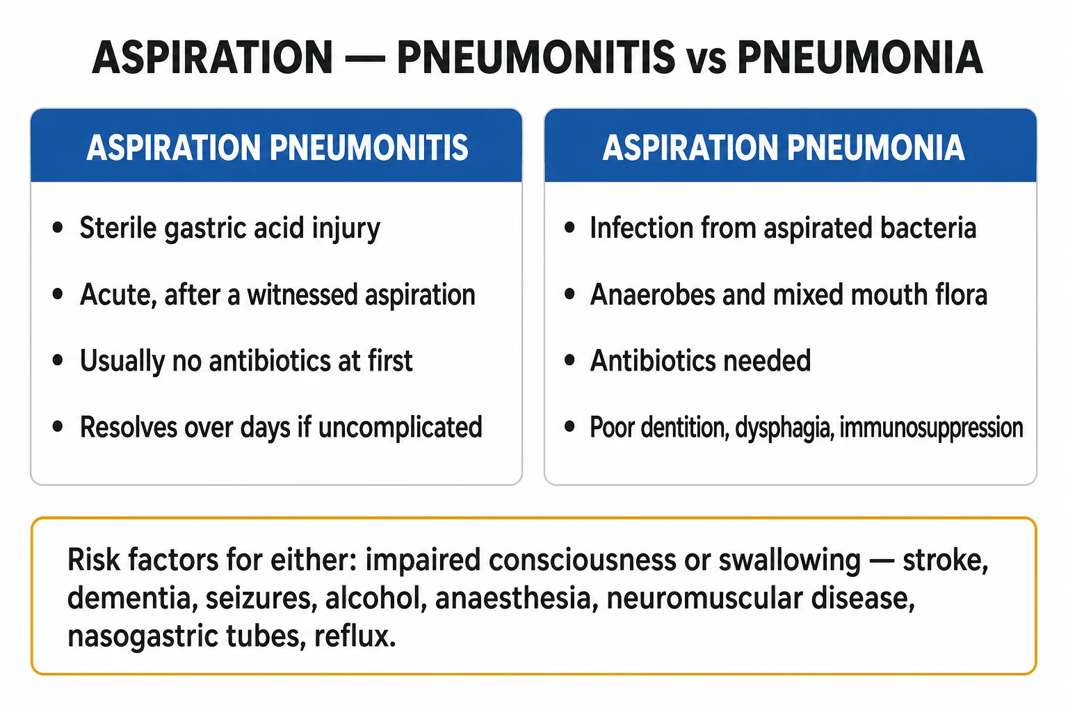
The crucial clinical distinction — one examiners probe relentlessly — is between the two ends of the aspiration spectrum:[1][3]
- Aspiration pneumonitis (Mendelson syndrome) is an acute chemical injury of the lung produced by the inhalation of sterile, acidic gastric contents. It classically follows a witnessed large aspiration in a patient with depressed consciousness (general anaesthesia, obstetric anaesthesia, seizures, alcohol or drug overdose, head injury). Because the injury is chemical, not infective, the standard teaching is supportive care without routine initial antibiotics, with antibiotics reserved for the minority who develop secondary bacterial infection (fever or purulent sputum persisting or worsening beyond 48 hours).[3]
- Aspiration pneumonia is a bacterial infection of the lung parenchyma that follows aspiration of oropharyngeal secretions colonised by bacteria — predominantly anaerobes and mixed mouth flora. It develops more insidiously in a host with impaired swallowing or consciousness (stroke, dementia, neuromuscular disease, prolonged intubation, poor dentition, nasogastric tubes), producing consolidation in the dependent lung segments and typically foul-smelling sputum. It requires antibiotics that cover anaerobes.[5]
The distinction is not always clean — the two overlap, and a patient with chemical pneumonitis may secondarily infect — but the question "is this primarily chemical or primarily infective?" drives the single most tested decision: whether to give antibiotics on day one.[3]
Classification
Aspiration lung injury is best classified along two axes: the nature of the aspirated material (which determines the mechanism), and the setting of acquisition (which shapes empiric antibiotic breadth).[1][5]
Aspiration pneumonitis
Mendelson syndrome — chemical injury
- **Sterile, acidic gastric contents** (classically pH under 2.5) inhaled into the lung
- Mechanism is **direct chemical burn** of the alveolar-capillary membrane — epithelial necrosis, surfactant inactivation, capillary leak
- **Acute onset within hours** of a witnessed aspiration (anaesthesia, seizure, vomiting in impaired consciousness)
- **No initial antibiotics** — supportive care; antibiotics only if secondary infection develops at or beyond 48 hours
Aspiration pneumonia
Infection from oropharyngeal flora
- **Bacterial infection** from aspirated oropharyngeal secretions — **anaerobes and mixed mouth flora**
- Mechanism is **alveolar macrophage cytokine release (IL-1, TNF-alpha) and neutrophil influx → consolidation**
- **Insidious onset over days** in impaired swallowing/consciousness (stroke, dementia, poor dentition, NG tubes)
- **Antibiotics required**, covering anaerobes (amoxicillin-clavulanate or clindamycin)
Aspiration lung abscess
Late complication of inadequately treated anaerobic infection
- Develops **1 to 2 weeks** after a clinically evident aspiration event
- **Anaerobes** (Bacteroides, Fusobacterium, Peptostreptococcus) predominate; cavitation with **air-fluid level**
- **Prolonged antibiotics (4 to 6 weeks)**; drainage only if large, fail medical therapy, or rupture to pleura
By the setting of acquisition (which sets the empiric antibiotic breadth): aspiration pneumonia acquired in the community behaves microbiologically like CAP with added anaerobe cover, while hospital or nursing-home acquisition demands broader cover including Gram-negatives and MRSA. The old healthcare-associated pneumonia (HCAP) category — which once triggered broad-spectrum cover purely because a patient came from a nursing home — has been retired by the ATS/IDSA 2019 guideline, because the risk of resistant organisms is now stratified by individual patient risk factors (prior IV antibiotics, recent hospitalisation, chronic dialysis, wound care) rather than by location alone.[2]
Epidemiology & Risk Factors
Aspiration pneumonia accounts for roughly 5 to 15 percent of community-acquired pneumonia cases, and a far higher proportion of pneumonia in care homes, hospitals, and the post-stroke population. It is a leading infectious cause of death in the elderly and the neurologically impaired.[1][5]
Aspiration pneumonia — headline numbers
The unifying mechanism of all aspiration is failure of one or more of the three protective layers: (1) glottic closure and cough reflex (conscious level), (2) swallowing coordination, and (3) mucociliary and alveolar-macrophage clearance. Risk factors cluster by which layer fails.[1]
[1]Risk factors and what they favour: [1]
| Risk factor / host | What it predisposes to |
|---|---|
| General anaesthesia, obstetric anaesthesia | Pneumonitis (Mendelson) — large-volume acid aspiration |
| Seizures, alcohol intoxication, drug overdose | Pneumonitis or pneumonia — impaired consciousness |
| Acute stroke (especially brainstem, large cortical) | Pneumonia — dysphagia; the commonest cause of post-stroke infection |
| Dementia / advanced age | Pneumonia — slow swallow, poor oral hygiene, reduced clearance |
| Neuromuscular disease (Parkinson, MND, MG, bulbar) | Pneumonia — weak swallow and cough |
| Nasogastric / PEG tube | Pneumonia — the tube splints the gastro-oesophageal sphincter and pools oropharyngeal secretions; it does NOT abolish aspiration |
| Poor dentition, xerostomia, gum disease | Pneumonia — heavier anaerobe colonisation of secretions |
| Oesophageal disease (stricture, achalasia, severe reflux, Zenker diverticulum) | Pneumonia — retained food and reflux reach the airway |
| Prolonged intubation / ICU | Ventilator-associated aspiration pneumonia |
| Proton-pump inhibitor / H2-blocker long-term use | Raises the pH of gastric contents, permitting bacterial overgrowth that converts sterile pneumonitis into infected pneumonia |
Pathophysiology
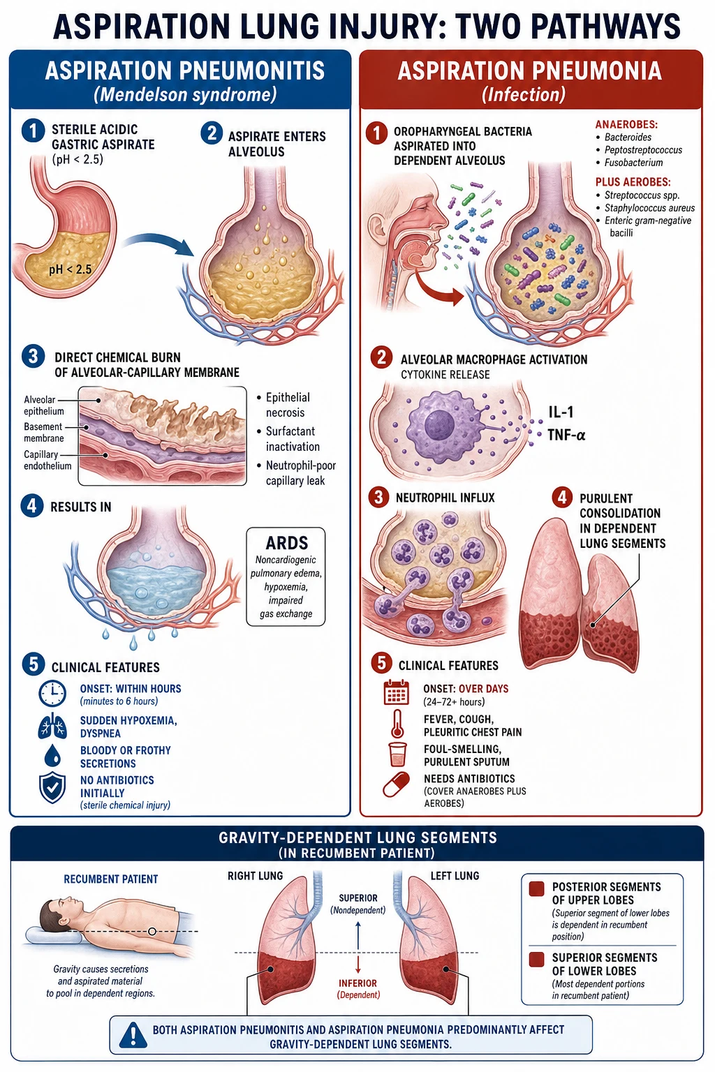
The lung is normally sterile below the vocal cords, defended by the cough reflex, glottic closure, mucociliary escalator, and alveolar macrophages. Aspiration causes injury when one or more of these defences is bypassed or overwhelmed by a large or toxic inoculum.[1]
Aspiration pneumonitis — the chemical cascade
When acidic gastric contents (the classical threshold is a pH under 2.5) reach the alveoli, the injury is immediate and dose-dependent: the acid denatures and lyses type I and II pneumocytes and the capillary endothelium within seconds. The death of type II pneumocytes cripples surfactant production, so alveoli collapse; the disrupted endothelium allows a protein-rich exudate to flood the alveolar space. The process is neutrophil-poor initially (it is a chemical burn, not infection), but within 24 to 48 hours neutrophils are recruited into the damaged tissue, and the inflammatory exudate becomes a fertile substrate for secondary bacterial infection — which is why pneumonitis may "tip over" into pneumonia if watched passively.[3]
The cardinal gas-exchange consequence is shunt and ventilation-perfusion mismatch from alveolar flooding and collapse, producing hypoxaemia that can progress to acute respiratory distress syndrome (ARDS) in severe cases. The injury is largely confined to the zones the acid reaches, which — by gravity — are the dependent segments.[3]
Aspiration pneumonia — the infective cascade
When oropharyngeal secretions carrying a heavy bacterial load (notably anaerobes that colonise the gingival crevices and plaque of patients with poor dentition) reach the alveoli, the inoculum is initially contained by alveolar macrophages. When the load exceeds macrophage capacity, macrophages release IL-1 and TNF-alpha, recruiting neutrophils that flood the alveolus and generate the purulent exudate we call consolidation. The same cytokines produce the fever, tachycardia and systemic upset; the bacterial breakdown products give the sputum its characteristic foul smell.[5]
The microbiology reflects the oropharynx: in community-acquired aspiration the flora is mixed anaerobic (Bacteroides, Prevotella, Fusobacterium, Peptostreptococcus, Actinomyces) plus aerobes such as Streptococcus pneumoniae, Haemophilus influenzae and Staphylococcus aureus; in hospital or nursing-home acquisition Gram-negative bacilli (including Klebsiella, Pseudomonas) and MRSA become important.[5] Anaerobes are notoriously missed on routine expectorated-sputum culture because they die in transit and in the oxygenated sputum cup — so a "sterile" culture in a typical clinical picture does not exclude an anaerobic infection; the foul smell and the dependent-segment distribution are the real diagnostic clues.
Why the dependent segments? — gravity decides the X-ray
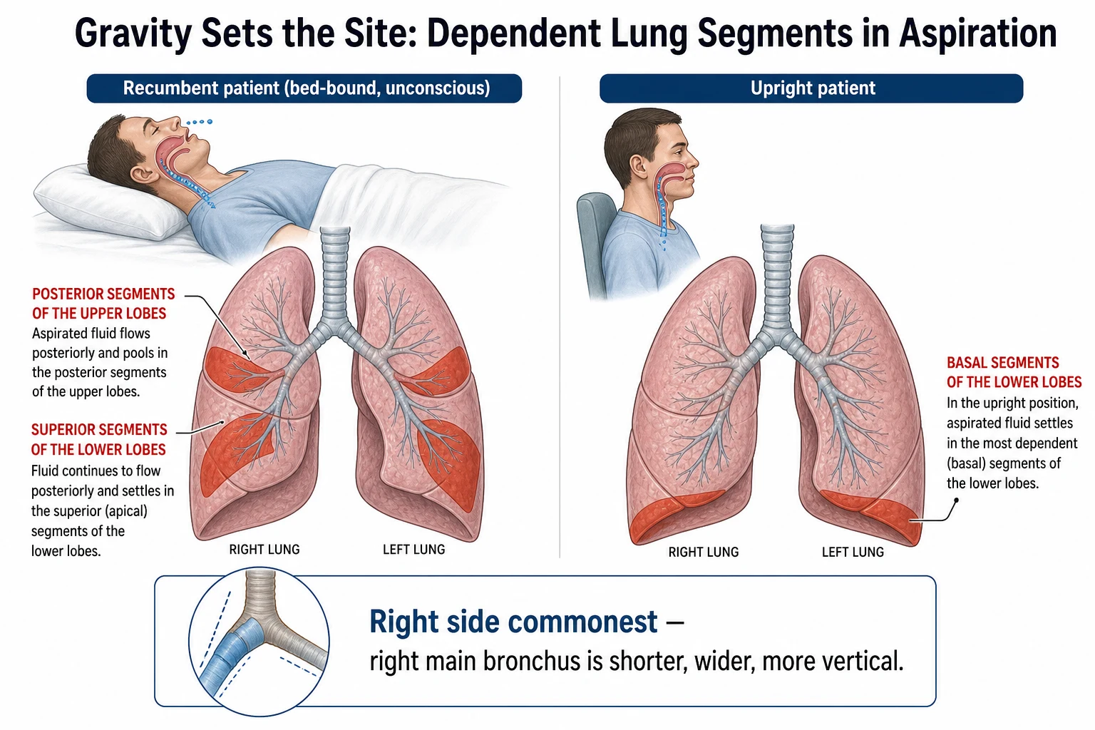
Aspirated material is liquid or semi-liquid, so it flows to the lowest point the patient's posture offers. In a recumbent (bed-bound, unconscious, post-ictal) patient the dependent bronchopulmonary segments are the posterior segments of the upper lobes and the superior segments of the lower lobes — the classic X-ray location. In a patient who aspirates upright, the material settles in the basal segments of the lower lobes (the right side more often, because the right main bronchus is shorter, wider and more vertical). This gravity-driven distribution, combined with a swallowing or consciousness risk factor, is what clinically flags an opacity as aspirated rather than just "pneumonic".[1][5]
Clinical Presentation
The tempo and the setting separate the two syndromes more reliably than any single sign.[3][5]
Aspiration pneumonitis is acute: within minutes to hours of a witnessed aspiration (during induction of anaesthesia, after a seizure, after forceful vomiting in an intoxicated patient) the patient develops sudden dyspnoea, wheeze, hypoxaemia, cough, and a low-grade fever. Cyanosis, tachypnoea and diffuse crackles may be present; signs of consolidation are less prominent than the hypoxia. A chest X-ray taken within a few hours shows patchy or bilateral dependent infiltrates (sometimes bilateral, because the inoculum distributes widely). The patient is typically previously well, and the aspiration event is observed.[3]
Aspiration pneumonia is insidious: over days, a patient with an obvious swallow or consciousness risk develops fever, cough with purulent and often foul-smelling sputum, pleuritic chest pain, dyspnoea, and malaise. There may be haemoptysis and, in advanced or cavitated disease, a fetid breath. Examination reveals signs of consolidation in a dependent segment (reduced expansion, dullness, bronchial breath sounds, crackles, increased vocal resonance). The patient is typically elderly, neurologically impaired, or institutionalised, and the aspiration is not witnessed — the history must be sought.[5]
Atypical presentation — the corners examiners live in
Elderly / dementia
Blunted inflammatory response
- **Confusion, falls, new incontinence, functional decline, anorexia** may be the only signs
- Fever and cough may be **absent**; a normal temperature does not exclude pneumonia
- Lower threshold to investigate and admit; tachypnoea is the most sensitive vital sign
Post-stroke
Dysphagia-driven, often silent
- **Aspiration is frequently silent** — no cough, because the cough reflex is impaired along with the swallow
- Pneumonia develops in the **first week**; bedside swallow screen must precede ANY oral intake
- **Wet voice, cough on fluids, pocketing of food, dysarthria** signal an unsafe swallow
Immunocompromised
Broader differential
- May be afebrile with neutropenia; consider **Gram-negatives, Pseudomonas, MRSA**
- Co-existing opportunists (Pneumocystis, fungal) widen the differential
Signs of an unsafe swallow at the bedside — the gateway finding that should trigger a formal swallow assessment before anything is given by mouth — are a wet-sounding voice or cough after a sip of water, pocketing of food in the cheek, dysarthria, facial weakness, and a poor or absent cough. [1]
Differential Diagnosis
A dependent-segment opacity in a frail patient is not always aspiration pneumonia, and an acute bilateral infiltrate after vomiting is not always pneumonitis. The high-yield differentials:[2][5]
Community-acquired pneumonia (non-aspiration)
Compare organism profile and distribution
- *S. pneumoniae* commonest; **no specific dysphagia signal**, distribution often **lobar, not dependent**
- Sputum not foul-smelling; anaerobe cover not routinely needed unless aspiration risk present
Cardiogenic pulmonary oedema
Mimic of acute pneumonitis
- **Bilateral perihilar (bat-wing) infiltrates**, gallop, raised JVP, response to **diuretics**
- No fever/purulent sputum; **BNP elevated**, echo shows reduced ejection fraction
ARDS / other causes
Mimic of severe pneumonitis
- Bilateral opacities, **PaO2/FiO2 under 300**, not fully cardiac; trigger may be sepsis, trauma, pancreatitis
- Aspiration itself is a cause of ARDS — so the two are not mutually exclusive
Lung abscess / cavitating lesion
Mimic of complicated aspiration
- **Air-fluid level** in a cavity; TB (upper lobe, night sweats), squamous-cell carcinoma (thick, irregular wall)
- Aspiration abscess sits in **dependent segments** with anaerobic polymicrobial flora
Atelectasis
Mimic in immobilised post-stroke patients
- **Volume loss**, no fever or purulent sputum, resolves with physiotherapy and mobilisation
- Distinguish from consolidation by the **shifted fissure/mediastinum** toward the opacity
Parapneumonic effusion / empyema
Complication, not a pure mimic
- Dullness with **absent breath sounds**; **Light's criteria** exudate; drain if purulent or pH under 7.2
Always consider tuberculosis (upper-lobe cavity, night sweats, risk factors) and underlying malignancy (a post-obstructive opacity behind a tumour) when an "aspiration pneumonia" is recurrent or slow to resolve.[2]
Clinical & Bedside Assessment
The focused examination has two jobs: characterise the respiratory lesion, and assess the swallow and consciousness that caused it. [1]
Respiratory examination in dependent-segment consolidation shows the classic signs of consolidation: reduced expansion, dullness to percussion, bronchial breath sounds, crackles, increased vocal resonance and a possible pleural rub. A silent hemithorax with dullness suggests an effusion or collapse. Vital signs drive severity — the respiratory rate is the single most sensitive marker of a lower-respiratory infection, and tachypnoea should never be dismissed.[2]
Swallow assessment at the bedside is the gateway to every aspiration question. The simplest validated tool is the 3-ounce (90 mL) water swallow test or a structured screen such as the Gugging Swallowing Screen (GUSS): give the patient sips of water and watch for cough or a wet voice within one minute — a positive screen means an unsafe swallow, and the patient must remain nil by mouth until a formal assessment is done. In acute stroke, a swallow screen must be performed within four hours of admission and before any oral intake, fluids, or medication.[5]
[1]A "wet voice" — a gurgling, liquid quality to the voice after swallowing — and a "wet cough" after a sip are the bedside hallmarks of material pooling in the larynx or pharynx and signal an unsafe swallow. [1]
Investigations
Investigations confirm the opacity, characterise severity, identify the organism where possible, and — most distinctively — evaluate the swallowing mechanism.[2][5]
First-line imaging: [1]
- Chest X-ray — mandatory to confirm consolidation in a dependent segment (posterior upper lobe, superior lower lobe). It may also show cavitation with an air-fluid level (lung abscess) or an effusion.
- CT chest — when the CXR is equivocal, to define cavitation, empyema, necrotising pneumonia, or an underlying mass (suspect malignancy in a non-resolving or recurrent opacity). [1]
Laboratory: [1]
- Full blood count — leucocytosis; CRP and procalcitonin (the latter may help separate bacterial pneumonia from sterile pneumonitis).
- Urea and electrolytes, liver function, blood glucose — severity and comorbidity.
- Arterial blood gas — to quantify hypoxaemia, hypercapnia, and acid-base status in severe disease.
- Blood cultures (before antibiotics) and sputum Gram stain and culture — note that anaerobes are usually missed on expectorated sputum; this does not exclude them.
- Pleural fluid (if an effusion is present) — apply Light's criteria (pleural/serum protein ratio over 0.5, LDH ratio over 0.6, or fluid LDH over two-thirds the upper limit of normal for an exudate); drain if complicated (purulent, pH under 7.2, glucose under 40 mg/dL, LDH over 1000, or positive culture/Gram stain). [1]
Severity scores — apply the same scores used for CAP. CURB-65 (1 point each: Confusion, Urea over 7 mmol/L, Respiratory rate 30 or more, Blood pressure systolic under 90 or diastolic under 60, age 65 or more; 0 to 1 home, 2 hospital, 3 to 5 consider ICU) and the Pneumonia Severity Index (PSI/PORT) stratify severity and site of care. Their limitation in aspiration pneumonia is that they do not capture the swallowing deficit or recurrence risk, which are the real drivers of outcome here — so a low CURB-65 must not be read as "no swallow problem".[2]
Instrumental swallow assessment — the definitive evaluation when the bedside screen fails or is equivocal: [1]
- Videofluoroscopic swallow study (VFSS / modified barium swallow) — the gold standard; visualises the oral, pharyngeal and oesophageal phases radiographically and identifies the consistency at which aspiration occurs.
- Fiberoptic endoscopic evaluation of swallowing (FEES) — bedside endoscopic assessment of swallow and secretion pooling; useful where transport to fluoroscopy is impractical (ICU, stroke unit). [1]
Evaluating the cause — every case deserves a hunt for the predisposing condition: CT brain (stroke, mass), EEG (seizures), oesophageal studies (stricture, achalasia, reflux, Zenker diverticulum), review of dentition, and a medication review for sedatives that depress consciousness and swallow. [1]
Management — Resuscitation
ABCDE first. The immediate priority in a witnessed large aspiration is the airway and oxygen, not antibiotics.[3]
- Airway — place the patient in the recovery position, suction the oropharynx under direct vision, and intubate early if the airway is unprotected or the patient cannot maintain oxygenation. In a semi-conscious patient who is actively vomiting, protect the airway first — head-down lateral position, suction — to limit further aspiration.
- Breathing — oxygen to target SpO2 94 to 98 percent (or 88 to 92 percent in COPD / CO2-retainers); escalate to high-flow nasal cannula, non-invasive ventilation, or invasive mechanical ventilation for refractory hypoxaemia or respiratory failure. Bronchoscopy is reserved for large particulate aspiration causing lobar obstruction — it does not remove acid, which disperses instantaneously.
- Circulation — IV access; treat septic shock, if present, with the Surviving Sepsis hour-1 bundle (lactate, blood cultures, broad antibiotics within 1 hour, balanced crystalloid 30 mL/kg, vasopressors for refractory shock).
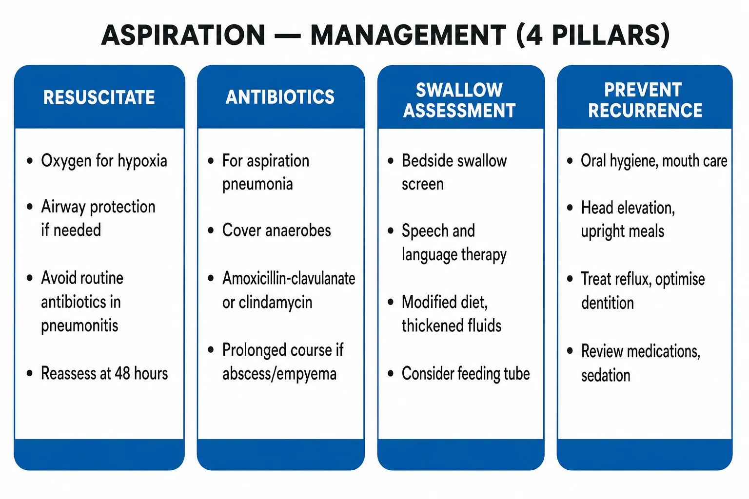
- Do NOT give prophylactic antibiotics routinely. In uncomplicated aspiration pneumonitis the injury is chemical and sterile; antibiotics on day one do not prevent infection and drive resistance, Clostridioides difficile, and cost. Give antibiotics only if features of secondary bacterial infection emerge at or beyond 48 hours (new or worsening fever, purulent sputum, rising inflammatory markers, progressive infiltrates, or clinical deterioration).[3]
Management — Definitive & Stepwise
Definitive management rests on four pillars: antibiotics (when indicated), treating the swallow deficit, preventive care, and addressing the underlying cause.[3][5]
Empiric antibiotics — drug, dose, route, rationale
The breadth of empiric cover is driven by setting (community vs hospital/nursing-home) and severity.[2][4][5]
| Setting / severity | Empiric regimen (drug, dose, route, rationale) |
|---|---|
| Community-acquired aspiration pneumonia, mild-moderate | Amoxicillin-clavulanate 875/125 mg orally three times daily (or 1.2 g IV for admitted patients) — covers anaerobes, S. pneumoniae, H. influenzae, oral aerobes. Penicillin-allergic: clindamycin 300 to 450 mg orally four times daily (600 mg IV three times daily) — excellent anaerobe and oral-flora cover, good lung penetration. |
| Community-acquired, severe / hospitalised | Piperacillin-tazobactam 4.5 g IV three times daily (or four times daily) — covers anaerobes plus Gram-negatives; OR ceftriaxone 2 g IV daily plus metronidazole 500 mg IV three times daily. Add azithromycin if atypical co-infection suspected. |
| Hospital / nursing-home / ICU acquisition | Broaden to piperacillin-tazobactam 4.5 g IV OR a carbapenem (meropenem 1 g IV three times daily); add vancomycin 15 to 20 mg/kg IV every 8 to 12 hours or linezolid 600 mg IV twice daily if MRSA is a risk. Cover Pseudomonas explicitly in neutropenic, bronchiectasis, or recent antibiotics. |
| Lung abscess / empyema | Prolong anaerobe cover to 4 to 6 weeks; drain empyema (chest tube) and large abscesses failing medical therapy. |
Why clindamycin historically leads for anaerobes: clindamycin has excellent oral-flora and anaerobic activity, high lung-tissue penetration, and a convenient oral formulation, which is why it has long been the textbook answer for aspiration pneumonia. Its limitations — emerging resistance in Bacteroides and a substantial C. difficile risk — mean it is increasingly paired with or replaced by beta-lactam/beta-lactamase inhibitors (amoxicillin-clavulanate, piperacillin-tazobactam). Metronidazole alone is inadequate because it lacks reliable activity against aerobic streptococci and microaerophilic oral flora and has poor activity against Actinomyces; it must be combined with an aerobic agent.[5]
Duration — typically 7 to 14 days for uncomplicated aspiration pneumonia, switching from IV to oral once the patient is haemodynamically stable, afebrile, and improving. Lung abscess and empyema require 4 to 6 weeks and may need drainage. Discharge when clinically stable, afebrile for 24 to 48 hours, tolerating oral intake (with a safe swallow), and with a safe social and swallow plan.[2]
ACE inhibitors — a pharmacological lever on salivary bacterial load
An elegant adjunct — frequently tested because it seems counterintuitive — is the use of angiotensin-converting enzyme (ACE) inhibitors in patients with post-stroke dysphagia and recurrent aspiration pneumonia. ACE is the enzyme that degrades substance P in the airways and salivary glands. ACE inhibitors raise substance P, which improves cough reflex sensitivity and swallow reflex coordination, and they also reduce the bacterial colonisation of saliva by lowering salivary proline-rich proteins that bacteria use as adhesion substrates. Small trials and a Cochrane systematic review suggest a reduced incidence of pneumonia in post-stroke patients on ACE inhibitors compared with other antihypertensives. Caveats: the cough itself can be a side-effect, the evidence is mainly from Japanese post-stroke cohorts, and ACE inhibitors are not given solely for pneumonia prevention — but the principle (a drug that treats hypertension can also reduce aspiration risk) is a high-yield viva answer. [1]
Tracheostomy — selected role in chronic aspiration
Tracheostomy is sometimes placed in patients with persistent severe aspiration (advanced neuromuscular disease, high brainstem stroke, prolonged weaning from ventilation) with the rationale that the cuffed tube prevents oral secretions from reaching the airway. The reality is more limited: a tracheostomy does not prevent aspiration of secretions that pool above the cuff (often leaking past it), it impairs laryngeal elevation and swallow coordination, it increases the risk of ventilator-associated pneumonia if ventilation continues, and it adds the complications of tracheal stenosis, malacia, bleeding, and infection. The current evidence supports tracheostomy primarily for prolonged mechanical ventilation rather than for aspiration prevention per se. Where chronic aspiration is the dominant problem, deflating the cuff for short periods under suction and using speaking valves may aid laryngeal function and swallow rehabilitation. [1]
Surgical and percutaneous drainage of lung abscess / empyema
Conservative therapy (prolonged antibiotics + postural drainage) resolves 70 to 90 percent of aspiration lung abscesses. Drainage is considered when the abscess is: [1]
- Larger than 4 to 6 cm — spontaneous drainage less likely;
- Failing medical therapy — persistent fever, leucocytosis, or non-resolving cavity after 6 to 8 weeks of antibiotics;
- Imminent rupture into the pleura — large peripheral abscess with pleural contact;
- Complicated by haemoptysis — bronchial artery embolisation is often preferred;
- Associated with empyema — drainage is mandatory; image-guided chest tube for simple empyema, surgical decortication for organised empyema. [1]
Percutaneous image-guided (CT or ultrasound) catheter drainage is the first-line intervention for non-resolving abscesses — smaller trauma than surgery, high success rate, allows serial imaging and irrigation. Surgical resection (lobectomy, segmentectomy, wedge resection) is reserved for failed percutaneous drainage, very large abscesses, suspicion of underlying malignancy, or massive haemoptysis. [1]
NG vs PEG — the practical decision in dysphagic stroke
The FOOD trial (2005) answered the most important practical question: in acute stroke with dysphagia, does the timing and route of tube feeding alter survival? Three arms were tested: early NG feeding, delayed NG feeding, and early PEG feeding. The findings: [1]
- Early tube feeding (within the first days) reduced mortality compared with no tube feeding (the strong message: feed the patient).
- Early PEG vs early NG showed a trend toward worse outcome with PEG, attributed to procedure-related complications, increased reflux, and delayed initiation. [1]
Implication: in acute stroke with unsafe swallow, place an NG tube first, start feeding promptly, and review the swallow weekly. Convert to PEG only if NG feeding is needed beyond 2 to 3 weeks, swallowing has not recovered, and the patient is medically stable. In patients with chronic dysphagia and reasonable prognosis (motor neurone disease with adequate life expectancy, head-and-neck cancer survivors, post-radiation stricture), PEG is appropriate after appropriate workup. [1]
Aspiration lung abscess — detailed management
An aspiration lung abscess develops over 1 to 2 weeks after the inciting aspiration event, typically in a dependent segment (posterior upper lobe, superior lower lobe, basal segments). The natural history is indolent fever, weight loss, night sweats, cough, foul sputum, sometimes with haemoptysis. Imaging shows a thick-walled cavity with an air-fluid level; CT may reveal an air crescent sign, multiple locules, or an underlying obstructing lesion. [1]
- Empiric therapy — same as aspiration pneumonia (amoxicillin-clavulanate or clindamycin); continue for 4 to 6 weeks, guided by clinical and radiographic response.
- Postural drainage — position the patient to drain the cavity by gravity (often lying prone or with the affected side up; SALT or physiotherapy input).
- Drainage — see criteria above.
- Investigate the underlying cause — every adult with an aspiration lung abscess deserves a bronchoscopy to exclude an obstructing tumour or foreign body; the cavity may be post-obstructive. [1]
Treatment-failure algorithm
When a patient is not improving at 48 to 72 hours, work through this in order: [1]
- Re-image — repeat CXR; CT if any concern. Look for new cavity, effusion, progression, mass.
- Re-culture — sputum if newly productive; blood cultures; consider bronchoscopy with BAL for quantitative microbiology, AFB, fungal, cytology, viral PCR.
- Check pleural fluid — if effusion present, thoracentesis with Light's criteria, pH, Gram stain, culture.
- Re-review the swallow — has the plan failed? Is the patient re-aspirating? Consider repeat VFSS / FEES.
- Re-review the drugs — adequate spectrum, dose, penetration, duration? Check renal/hepatic dose adjustment. Check drug interactions (for example clindamycin with non-depolarising neuromuscular blockers).
- Re-think the diagnosis — TB, lung cancer, vasculitis, eosinophilic pneumonia, cryptogenic organising pneumonia, pulmonary infarction, heart failure. [1]
Adjuncts and the rest of the bundle
Beyond chlorhexidine oral care and head-of-bed elevation, several adjuncts reduce recurrence: [1]
- Swallowing rehabilitation — exercises (chin-tuck, supraglottic swallow, effortful swallow, Mendelsohn manoeuvre), surface neuromuscular electrical stimulation in selected patients, McNeill dysphagia therapy programme in Parkinson's.
- Treatment of underlying cause — antiepileptics for seizures, dopaminergic therapy for Parkinson's, anti-reflux measures for GERD (lifestyle, PPIs, weight loss, head-of-bed elevation, fundoplication in selected severe cases), myotomy for achalasia, endoscopic diverticulotomy for Zenker, vocal-cord medialisation for unilateral paralysis.
- Positioning — upright for meals and 30 minutes after; chin-tuck during swallow; lateral recumbent positioning in selected patients.
- Pharmacology to reduce reflux — PPIs reduce acid reflux but raise gastric pH and may worsen bacterial overgrowth; H2 blockers less potent. Baclofen reduces transient lower-sphincter relaxations and may help reflux-driven aspiration. Prokinetics (metoclopramide, domperidone, erythromycin) help gastroparesis but with significant side-effect burden.
- Vaccination — annual influenza, pneumococcal vaccination (PCV20 or PCV15 followed by PPSV23), COVID-19 boosters; pertussis in those not immunised; oral hygiene as vaccine adjunct.
The prevention bundle — where outcome is really decided [1]
In aspiration pneumonia, prevention reduces recurrence more than any antibiotic. The bundle, applied to every patient: [1]
Prevention of aspiration pneumonia — HIDE BOLUS
HIDE BOLUS
Elevate to 30 to 45 degrees at all times, especially during and after feeds
VFSS or FEES when bedside screen fails; identify safe consistencies
Meticulous mouth care; the single highest-yield intervention in care homes
Upright for meals and 30 minutes after; chin-tuck manoeuvre if advised by SLT
Thickened fluids, texture-modified diet per SLT assessment
Treat gum disease, fit dentures, reduce oropharyngeal bacterial load
Review and reduce CNS-depressant drugs that blunt swallow and cough
Treat reflux, seizures, Parkinson, oesophageal disease; rehab swallow
Speech-language therapy; swallowing exercises and compensatory strategies
Oral hygiene / chlorhexidine has the strongest evidence of any single preventive measure in nursing-home and ICU populations, reducing the bacterial load of oropharyngeal secretions and thus the inoculum of any future aspiration.[7][8]
Feeding-tube decisions — the question that needs the most judgement
A feeding tube (NG or PEG) is considered when the swallow is unsafe and not expected to recover quickly (e.g. severe acute stroke, advanced dementia with recurrent aspiration, or critical illness). But a tube is not a cure for aspiration — it splints open the gastro-oesophageal sphincter, pools oropharyngeal secretions, and permits reflux and silent aspiration around the tube, so it reduces but does not abolish aspiration risk. The decision must weigh nutritional benefit against quality of life, comfort, and the evidence:[6]
- In acute stroke, the FOOD trial found no survival benefit from routine early PEG over nasogastric feeding in dysphagic stroke patients; NG feeding is preferred initially, with PEG reserved for those needing prolonged tube feeding (beyond 2 to 3 weeks).
- In advanced dementia, tube feeding does not meaningfully prevent aspiration, prolong survival, or improve comfort, and is associated with restraint and pressure-area complications — hand-feeding with careful oral care is often the kinder, evidence-based choice, framed within advance care planning.[6]
Specific Subtypes & Scenarios
Each clinical setting reframes both the prevention and the antibiotic breadth. [1]
- Aspiration pneumonitis in obstetric / anaesthetic practice (Mendelson syndrome) — pregnant women are at high risk because of delayed gastric emptying, increased intra-gastric pressure, and reduced lower-oesophageal sphincter tone. Prevention is procedural: fasting, rapid-sequence induction with pre-oxygenation and cricoid pressure (Sellick manoeuvre), and a cuffed endotracheal tube. If aspiration occurs, management is supportive (oxygen, ventilation) — no routine antibiotics.[3]
- Aspiration pneumonia in acute stroke — dysphagia affects roughly half of acute stroke patients and is the leading cause of stroke-associated pneumonia, which independently worsens outcome. A bedside swallow screen within four hours of admission and before any oral intake is mandatory; failed screen → nil by mouth, IV fluids, and escalation to VFSS/FEES.[5]
- Aspiration pneumonia in advanced dementia / end of life — recurrent aspiration is often a marker of disease progression. The decision to insert a feeding tube must be made with the family within the goals of care: tube feeding does not reliably prevent aspiration here, and hand-feeding, oral care, and comfort-focused care are often the better path.[6]
- Aspiration lung abscess — develops one to two weeks after a clinically evident aspiration, in a dependent segment, with a polymicrobial anaerobic flora. Treatment is prolonged antibiotics (4 to 6 weeks); percutaneous or surgical drainage is reserved for large abscesses, those failing medical therapy, or those that rupture into the pleura (empyema).
- Recurrent aspiration pneumonia — treat the cause: a swallow study (VFSS/FEES), oesophageal assessment, dental review, medication review, and where appropriate a feeding-tube or surgical strategy (e.g. fundoplication for severe reflux). Recurrence in a known dysphagic patient is a sign that the current feeding or oral-care plan is failing.
- Aspiration pneumonia in Parkinson's disease — Parkinson's produces a characteristic oropharyngeal bradykinesia with delayed swallow trigger, lingual pumping, fragmented pharyngeal transit, and incomplete laryngeal elevation. Aspiration occurs in up to half of advanced patients. Levodopa dosing timed around meals can dramatically improve swallow coordination; deep brain stimulation may help selected patients. The oral care bundle is essential because sialorrhoea and drooling raise aspiration risk.
- Aspiration in motor neurone disease (amyotrophic lateral sclerosis, ALS) — bulbar-onset ALS produces flaccid dysarthria, weak tongue movement, incomplete cricopharyngeal relaxation, and a weak ineffective cough. Aspiration becomes the dominant cause of death. Proactive PEG placement (ideally before forced vital capacity falls below 50 percent) preserves nutrition; non-invasive ventilation supports respiratory failure; the decision to proceed with long-term ventilation must be made early in the disease course within the patient's goals of care.
- Aspiration in myasthenia gravis — fatigable weakness worsens through the day and with sustained use. Aspiration risk peaks at the evening meal. Treatment of the underlying disease (pyridostigmine, immunosuppression, thymectomy, eculizumab, ravulizumab) is the primary prevention; edrophonium testing and acetylcholine-receptor antibody titres confirm the diagnosis; crisis requires plasma exchange or intravenous immunoglobulin.
- Aspiration in achalasia — failure of lower-oesophageal-sphincter relaxation with absent peristalsis causes megaoesophagus that retains food and saliva. The bird-beak appearance on barium swallow and high-resolution manometry confirm the diagnosis. Treatment is endoscopic pneumatic dilation, peroral endoscopic myotomy (POEM), or Heller myotomy; untreated disease produces chronic regurgitation and recurrent aspiration pneumonia.
- Aspiration in Zenker diverticulum — a posterior pharyngeal pouch through Killian's dehiscence traps food, which then spills into the airway during the swallow. The triad of dysphagia, regurgitation of undigested food, and halitosis is classic. Treatment is endoscopic stapled diverticulotomy or open diverticulectomy with cricopharyngeal myotomy; aspiration risk resolves in most patients.
- Aspiration around nasogastric tubes — large-bore NG tubes splint the gastro-oesophageal sphincter, increase reflux, and pool oropharyngeal secretions. Fine-bore tubes are preferred; regular oral hygiene and 30 to 45-degree head-of-bed elevation reduce aspiration risk. Continuous rather than bolus feeds may reduce aspiration in high-risk patients. PEG is not a guaranteed improvement.
- Aspiration in acute alcohol intoxication and chronic alcoholism — intoxication produces a witnessed or unwitnessed macro-aspiration of gastric contents during a drunken vomit; chronic alcohol use raises the risk of Klebsiella pneumoniae pneumonia (classically upper-lobe, "currant-jelly" sputum, cavitation), as well as aspiration driven by delirium tremens, alcohol-related seizures, and alcohol-related gastroparesis. Wernicke encephalopathy can itself impair swallow; always give thiamine before glucose in this population.
- Aspiration after bariatric or oesophageal surgery — Roux-en-Y gastric bypass, sleeve gastrectomy, and oesophagectomy all alter upper-gut anatomy; delayed gastric emptying, blind-loop syndrome, and anastomotic stricture can each present as recurrent aspiration pneumonia. Always take a surgical history in unexplained recurrent aspiration.
- Aspiration in critical illness and during procedural sedation — intubated and sedated patients aspirate around the cuff; subglottic secretion drainage tubes reduce ventilator-associated pneumonia. Procedural sedation (endoscopy, bronchoscopy, cardiac catheterisation, dental work) carries a small aspiration risk that justifies standard fasting intervals (2 hours for clear fluids, 6 hours for solids).
- Anaerobic lung abscess — diagnostic and management detail — an air crescent sign on CT, a putrid odour to the sputum, and dependent-segment distribution in a host with impaired consciousness or swallowing are the diagnostic triad. Treatment is 4 to 6 weeks of anaerobic cover (clindamycin or amoxicillin-clavulanate) with postural drainage; bronchoscopy is mandatory to exclude an obstructing tumour. Drainage (percutaneous first-line, surgical if that fails) is reserved for large or non-resolving cavities.
Complications & Pitfalls
Local complications of inadequately treated anaerobic infection include lung abscess, empyema (from rupture of an abscess or direct pleural spread), necrotising pneumonia, bronchopleural fistula, and — at the severe end — ARDS from overwhelming pneumonitis. Systemic complications include sepsis and septic shock, bacteraemia with metastatic infection (endocarditis, brain abscess, septic arthritis), and acute kidney injury.[1][5]
ARDS as the most severe pneumonitis complication — severe gastric-acid aspiration produces acute lung injury with a PaO2/FiO2 ratio under 200 and bilateral infiltrates within hours. Management is lung-protective ventilation (tidal volume 6 mL/kg ideal body weight, plateau pressure under 30 cm H2O, PEEP to recruit), prone positioning for moderate-to-severe ARDS (PaO2/FiO2 under 150), and consideration of neuromuscular blockade, inhaled nitric oxide, or ECMO in refractory cases. Corticosteroids are not given routinely for chemical pneumonitis; some centres use them for the late fibroproliferative phase, but evidence is weak. [1]
Empyema — frank pus in the pleural space, typically from rupture of a subpleural abscess or direct contiguous spread. Presents as pleuritic pain, fever, and a layering effusion on imaging. Diagnostic thoracentesis returns pH under 7.2, glucose under 40 mg/dL, LDH over 1000, positive Gram stain or culture, or frankly purulent fluid. Treatment is chest-tube drainage plus the underlying antibiotics (4 to 6 weeks total); intrapleural fibrinolytics (tissue plasminogen activator plus DNase) are now standard for loculated empyema based on the MIST-2 trial; VATS (video-assisted thoracoscopic surgery) decortication for organised empyema. [1]
Necrotising pneumonia — destruction of lung parenchyma with multiple small cavities, typically from Staphylococcus aureus, Klebsiella pneumoniae, or Streptococcus pyogenes, less commonly anaerobes. CT shows multiple thin-walled cavities in an area of consolidation with little surrounding inflammation. Treatment is prolonged antibiotics (3 to 4 weeks); surgery is rarely needed. [1]
Bronchopleural fistula — communication between the bronchial tree and the pleural space, presenting as persistent air leak after chest-tube drainage. Causes include necrotising pneumonia, abscess rupture, trauma, and barotrauma from positive-pressure ventilation. Management: drain the pleural space, decompress the fistula, consider bronchial stent or surgical repair. [1]
Metastatic infection — anaerobic bacteraemia can seed brain abscess (ring-enhancing lesion, headache, focal neurology), infective endocarditis (new murmur, embolic phenomena, vegetation on echo), septic arthritis (hot swollen joint), and vertebral osteomyelitis. Investigate any focal symptom; MRI brain and echocardiography are first-line. [1]
Sepsis and septic shock — the Surviving Sepsis hour-1 bundle (lactate, blood cultures, broad antibiotics within 1 hour, balanced crystalloid 30 mL/kg, vasopressors for refractory shock) applies verbatim. [1]
Recurrence and chronicity — the failure mode of aspiration care is the patient who is treated and discharged, then re-presents within weeks. Recurrence is a marker that the swallow plan, oral-care routine, dentition, medication burden, or reflux management has failed — and the next admission is the right time to escalate the workup and the bundle. [1]
The pitfalls are as important as the complications, because they cause the harm examiners test: [1]
Over-treating pneumonitis
Antibiotic harm
- Routine antibiotics for **uncomplicated chemical pneumonitis** drive **resistance, *C. difficile*, cost**
- Reassess at **48 hours**; add antibiotics **only if secondary infection** develops
Over-restricting diet
Nutritional harm
- Aggressive thickened-fluid / pureed diets cause **dehydration, malnutrition, and reduced quality of life**
- Tailor to the **instrumental swallow result**, not a blanket restriction
Assuming a PEG prevents aspiration
False security
- PEG tubes **reduce but do not abolish** aspiration — reflux, oropharyngeal pooling, and silent aspiration continue
- In **advanced dementia**, PEG does not improve survival or comfort and is often **not indicated**
Trusting the gag reflex
Assessment error
- An intact gag does **not** guarantee a safe swallow; use a **structured swallow screen**
Missing the underlying cause
Diagnostic error
- Always identify and treat the underlying cause — stroke, dementia, pregnancy, ICU, immunocompromise, alcohol misuse
- See detailed special-population sections below
Special Populations — Detailed
-
Acute stroke — a swallow screen within four hours of admission and before any oral intake; failed screen → nil by mouth, hydration by IV or NG, escalation to VFSS/FEES, and a speech-language-therapy-led diet reintroduction. Stroke-associated pneumonia independently worsens functional outcome.[5]
-
Elderly / dementia — atypical, blunted presentation (confusion, falls); lower threshold to investigate and admit; feeding-tube decisions framed within advance care planning and quality of life, not reflexively inserted.[6]
-
Pregnancy / obstetric anaesthesia — Mendelson syndrome is the prototypical peripartum aspiration; prevent with fasting, rapid-sequence induction, cricoid pressure, and a cuffed tube.[3]
-
ICU / intubated — ventilator-associated aspiration is reduced by head-of-bed elevation 30 to 45 degrees, subglottic secretion drainage, oral care with chlorhexidine, and daily sedation interruption.[8]
-
Immunocompromised — broaden the empiric differential to include Gram-negatives, Pseudomonas, and MRSA; consider opportunists and lower the threshold for bronchoalveolar-lavage microbiology.
-
Post-stroke dysphagia in detail — dysphagia affects roughly half of acute stroke patients, higher after brainstem and large hemispheric strokes. The bedside screen must be done before any oral intake — including medications. Failed screen → nil by mouth (NBM), hydration by IV or NG tube, daily review by SALT, escalation to VFSS or FEES within 24 to 48 hours. Diet reintroduction follows SALT recommendations: thin fluids, mildly thick, moderately thick, extremely thick (International Dysphagia Diet Standardisation Initiative, IDDSI levels 0 to 4). Stroke-associated pneumonia independently worsens 30-day and 1-year mortality and lengthens hospital stay; the prevention bundle (oral care, head-of-bed elevation, screening, treatment of reflux) cuts its incidence roughly in half. [1]
-
Elderly, frailty, and the atypical presentation — older patients may have no fever, no cough, no sputum. The presenting features are confusion, falls, new incontinence, anorexia, functional decline, fatigue. The single most sensitive vital sign is the respiratory rate (a rate above 25 carries an odds ratio of roughly 3 for pneumonia in frail elderly). Lower the threshold to investigate and admit; interpret CURB-65 with caution because age scores automatically — a low CURB-65 does not exclude severe disease in the elderly, and Clinical Frailty Scale (CFS) is increasingly used alongside CURB-65 for disposition decisions in this group. [1]
-
Pregnancy and peripartum — Mendelson syndrome was first described in obstetric patients under general anaesthesia for caesarean section. Pregnancy-specific risk factors: enlarged uterus raising intra-gastric pressure, progesterone relaxing the lower oesophageal sphincter, delayed gastric emptying, and the unpredictability of labour. Prevention is procedural: preoperative fasting (2 hours clear fluids, 6 hours solids), rapid-sequence induction with pre-oxygenation, cricoid pressure (Sellick manoeuvre) to occlude the upper oesophagus, and a cuffed endotracheal tube. Modern practice favours regional anaesthesia for caesarean section wherever possible. [1]
-
ICU and the ventilated patient — ventilator-associated pneumonia (VAP) is a distinct but overlapping entity. Prevention is the IHI ventilator bundle: head-of-bed elevation 30 to 45 degrees, daily sedation interruption with extubation readiness assessment, peptic-ulcer prophylaxis (controversial — increases gastric colonisation), deep-vein thrombosis prophylaxis, and oral care with chlorhexidine 0.12 percent every 6 hours. Subglottic secretion drainage endotracheal tubes reduce early VAP. Probiotics (Lactobacillus rhamnosus GG) may reduce VAP, but evidence is mixed. Selective decontamination of the digestive tract (SDD) with topical non-absorbable antibiotics plus a short course of systemic cephalosporin is effective in ICUs with low antibiotic resistance but rarely used outside the Netherlands. [1]
-
Immunocompromised hosts — neutropenic, transplant, HIV, and chronic-steroid patients may present with afebrile pneumonia. Broaden the differential to Gram-negatives, Pseudomonas, MRSA, Nocardia, Aspergillus, Pneumocystis jirovecii, mycobacteria, and CMV. Procalcitonin and galactomannan guide workup. Empiric piperacillin-tazobactam plus vancomycin is typical initial cover; escalate to meropenem, add antifungal (liposomal amphotericin B or voriconazole) and consider co-trimoxazole for Pneumocystis prophylaxis/treatment if CD4 under 200 or equivalent. Bronchoscopy with BAL is the diagnostic procedure of choice. [1]
-
Chronic alcohol misusers — chronic alcoholism is an independent risk factor for aspiration pneumonia via impaired consciousness, depressed cough and gag, alcohol-associated gastroparesis, and altered oropharyngeal flora. Klebsiella pneumoniae is classically over-represented. Always give thiamine before glucose to avoid precipitating Wernicke encephalopathy; provide folate, B12, and other nutritional support. Alcohol withdrawal can begin 6 to 24 hours after the last drink — monitor and use the Clinical Institute Withdrawal Assessment for Alcohol (CIWA-Ar) scale to titrate benzodiazepines. [1]
-
The home-ventilated patient with chronic aspiration — long-term home ventilation (via tracheostomy or non-invasive mask) is increasingly common in motor neurone disease, muscular dystrophy, and chronic respiratory failure. Aspiration remains the dominant risk; management is speech-language-therapy exercises, glossopharyngeal breathing training, mechanical insufflation-exsufflation to clear secretions, and a family-carer training programme for oral suctioning and postural drainage. [1]
Outcome is driven less by the radiographic extent of the opacity than by the underlying cause, the patient's frailty, the presence of sepsis, and whether aspiration recurs. Aspiration pneumonia in a frail, post-stroke, or demented patient carries a high 30-day mortality (around 10 to 20 percent), and recurrence is common unless the swallowing deficit and oral care are actively addressed.[1][5]
Uncomplicated aspiration pneumonitis usually resolves over days with supportive care; the prognosis is favourable if secondary infection is avoided. [1]
Discharge is safe when the patient is clinically stable, afebrile for 24 to 48 hours, tolerating oral intake with a confirmed safe swallow, has a completed swallow plan (diet consistency, posture, supervision), a reviewed medication list, and a safety-net to re-present if breathlessness, fever, or swallow deterioration recur. A follow-up chest X-ray at 6 to 8 weeks is indicated for persistent symptoms, smokers over 50, or any non-resolving opacity — to exclude an underlying malignancy (post-obstructive pneumonia behind a tumour).[2]
Special Populations
- Acute stroke — a swallow screen within four hours of admission and before any oral intake; failed screen → nil by mouth, hydration by IV or NG, escalation to VFSS/FEES, and a speech-language-therapy-led diet reintroduction. Stroke-associated pneumonia independently worsens functional outcome.[5]
- Elderly / dementia — atypical, blunted presentation (confusion, falls); lower threshold to investigate and admit; feeding-tube decisions framed within advance care planning and quality of life, not reflexively inserted.[6]
- Pregnancy / obstetric anaesthesia — Mendelson syndrome is the prototypical peripartum aspiration; prevent with fasting, rapid-sequence induction, cricoid pressure, and a cuffed tube.[3]
- ICU / intubated — ventilator-associated aspiration is reduced by head-of-bed elevation 30 to 45 degrees, subglottic secretion drainage, oral care with chlorhexidine, and daily sedation interruption.[8]
- Immunocompromised — broaden the empiric differential to include Gram-negatives, Pseudomonas, and MRSA; consider opportunists and lower the threshold for bronchoalveolar-lavage microbiology.
Evidence, Guidelines & Regional Differences
The ATS/IDSA 2019 CAP guideline retains aspiration pneumonia as a CAP entity and endorses empiric anaerobic coverage only where aspiration is a genuine risk factor (rather than routinely for all CAP). It retired the HCAP category, replacing blanket broad-spectrum therapy with risk-factor-based decisions — a patient from a nursing home no longer automatically receives anti-pseudomonal cover unless they carry individual risk factors (recent IV antibiotics, hospitalisation, dialysis, wound care).[2]
The Mandell 2007 IDSA/ATS guidelines (the predecessor) similarly recognised anaerobic cover for aspiration but seeded the move toward individualising empiric breadth.[4]
Oral hygiene / chlorhexidine is the best-evidenced single preventive measure in both nursing-home and ICU settings, reducing the oropharyngeal bacterial load and thus the inoculum of any future aspiration.[7][8]
The FOOD trial (2005) is the landmark evidence on feeding-tube timing in dysphagic stroke: it found no survival benefit from routine early PEG over NG feeding, supporting NG feeding first with PEG reserved for those needing prolonged tube feeding — a result that overturned the reflex to place PEGs early.[6]
Regional antibiotic deltas for aspiration pneumonia: [1]
| Region / guideline | First-line empiric for community aspiration |
|---|---|
| US (ATS/IDSA 2019) | Amoxicillin-clavulanate or clindamycin; broaden (piperacillin-tazobactam) for severity/hospital acquisition.[2] |
| UK (NICE) | Amoxicillin-clavulanate first-line; clindamycin or co-amoxiclav per local policy; anaerobe cover emphasised.[5] |
| India (ICMR/NCDC) | Amoxicillin-clavulanate + metronidazole, or clindamycin; in hospitalised/severe, piperacillin-tazobactam or a carbapenem per local antibiogram, given high community Gram-negative and ESBL prevalence. |
Exam Pearls
- The one-liner: aspiration pneumonitis = sterile gastric-acid chemical injury, no antibiotics initially; aspiration pneumonia = infected, anaerobes, antibiotics required.[3]
- Dependent-segment anatomy: posterior segment of the upper lobe and superior segment of the lower lobe in a recumbent patient; the right side is commoner (shorter, wider, more vertical right main bronchus).[1]
- Foul-smelling sputum = anaerobes = aspiration until proven otherwise.
- Anaerobes: Bacteroides, Prevotella, Fusobacterium, Peptostreptococcus, Actinomyces — usually missed on routine sputum culture.[5]
- Clindamycin is the textbook anti-anaerobic agent (good lung penetration); metronidazole alone is inadequate (poor aerobic streptococcal cover).[5]
- Mendelson syndrome = acidic gastric aspirate + impaired consciousness + peri-procedural (obstetric/anaesthetic) setting.
- An intact gag reflex does NOT guarantee a safe swallow. A swallow screen (3-oz water test / GUSS) is required.[5]
- The single highest-yield prevention measure in care homes and ICU is meticulous oral hygiene.[7][8]
- PEG does NOT reliably prevent aspiration — and in advanced dementia offers no survival or comfort benefit (FOOD trial framing).[6]
- Proton-pump inhibitors raise gastric pH, permitting bacterial overgrowth that can convert sterile pneumonitis into infected pneumonia.
- HCAP is retired (ATS/IDSA 2019) — base broad-spectrum cover on individual risk, not location.[2]
Exam application bank (NEET-PG / INICET)
One-line answer
Aspiration lung injury is not one disease but a spectrum. Aspiration pneumonitis (Mendelson syndrome) is an acute chemical burn of the lung from sterile, acidic gastric contents, typically after a witnessed large aspiration under anaesthesia or in impaired consciousness — usually no antibiotics initially, resolving over days if uncomplicated. Aspiration pneumonia is bacterial infection from aspiration of oropharyngeal secretions rich in anaerobes and mixed mouth flora, in a host with impaired swallowing or consciousness (stroke, dementia, seizures, alcohol, neuromuscular disease, nasogastric tubes, poor dentition), producing infiltrates in the dependent segments (posterior upper lobes, superior lower lobes). Aspiration pneumonia needs antibiotics covering anaerobes (amoxicillin-clavulanate or clindamycin). Prevention is the real treatment — swallow assessment, meticulous oral hygiene, he
Worked stems (answer without another resource)
Stem 1 — Classic presentation. Map symptoms to mechanism; name the first investigation and first treatment step with dose/route if drug therapy is standard. [1]
Stem 2 — Unstable / complicated. List red flags that force immediate resuscitation, theatre, ICU, antidote, or reperfusion — and what you do in the first 15 minutes. [1]
Stem 3 — Atypical group. Elderly, pregnancy, child, or immunocompromised: how presentation and thresholds change. [1]
Stem 4 — Differential trap. Name the three closest mimics and one discriminator for each. [1]
Stem 5 — Disposition. Who goes home with safety-netting, who is admitted, who needs HDU/ICU/theatre, and what follow-up is mandatory. [1]
Rapid viva checklist
- Definition + classification
- Pathophysiology chain
- Bedside signs / criteria
- Score with exact components (if any)
- Emergency bundle
- Definitive therapy with doses
- Complications of disease and of treatment
- Special populations
- Guideline/trial name if classic
- Three exam traps
Coverage self-check
If you cannot answer any stem above from this page alone, re-read the matching section — the page is intended to be self-sufficient for final-prof and NEET-PG/INICET questions on Aspiration Pneumonia & Pneumonitis.
References
- [1]Košutova P, Mikolka P, et al. Aspiration syndromes and associated lung injury: incidence, pathophysiology and management Physiol Res, 2021.PMID 35199544
- [2]Metlay JP, Waterer GW, Long AC, et al. Diagnosis and Treatment of Adults with Community-acquired Pneumonia. An Official Clinical Practice Guideline of the American Thoracic Society and Infectious Diseases Society of America Am J Respir Crit Care Med, 2019.PMID 31573350
- [3]Marik PE. Aspiration pneumonitis and aspiration pneumonia N Engl J Med, 2001.PMID 11228282
- [4]Mandell LA, Wunderink RG, Anzueto A, et al. Infectious Diseases Society of America/American Thoracic Society consensus guidelines on the management of community-acquired pneumonia in adults Clin Infect Dis, 2007.PMID 17278083
- [5]DiBardino DM, Wunderink RG. Aspiration pneumonia: a review of modern trends J Crit Care, 2015.PMID 25129577
- [6]Dennis MS, Lewis SC, Warlow C, FOOD Trial Collaboration. Effect of timing and method of enteral tube feeding for dysphagic stroke patients (FOOD): a multicentre randomised controlled trial Lancet, 2005.PMID 15733717
- [7]Liu C, Cao Y, Lin J, et al. Oral care measures for preventing nursing home-acquired pneumonia Cochrane Database Syst Rev, 2018.PMID 30264525
- [8]Hua F, Xie H, Worthington HV, et al. Oral hygiene care for critically ill patients to prevent ventilator-associated pneumonia Cochrane Database Syst Rev, 2016.PMID 27778318