Respiratory · Respiratory
Bronchiectasis (non-CF)
Also known as Bronchiectasis · Non-CF bronchiectasis · Adult bronchiectasis · Cylindrical/varicose/cystic bronchiectasis
Bronchiectasis is the permanent, abnormal dilation of one or more bronchi caused by destruction of the muscular and elastic wall components. It is not a single disease but the end-result of a vicious cycle of impaired mucociliary clearance, chronic infection and neutrophilic inflammation (Cole's hypothesis). The classic presentation is a chronic productive cough with daily mucopurulent sputum, recurrent exacerbations, coarse crackles, finger clubbing and, in advanced disease, haemoptysis and cor pulmonale. Diagnosis is clinical plus high-resolution CT (the signet-ring sign — bronchus wider than its accompanying artery — is the radiological hallmark). Commonest organisms are Haemophilus influenzae (early) and Pseudomonas aeruginosa (severe, accelerating decline). Management rests on four pillars: treat the underlying cause, airway-clearance physiotherapy, infection control (acute 14-day antibiotics plus long-term macrolides and/or inhaled antibiotics for frequent exacerbators), and prevention (vaccination, smoking cessation). Dornase alfa helps in CF but not in non-CF bronchiectasis (the O'Donnell trial).
On this page & tools
Your progress
Saved locally on this device.
Exam tags
Red flags
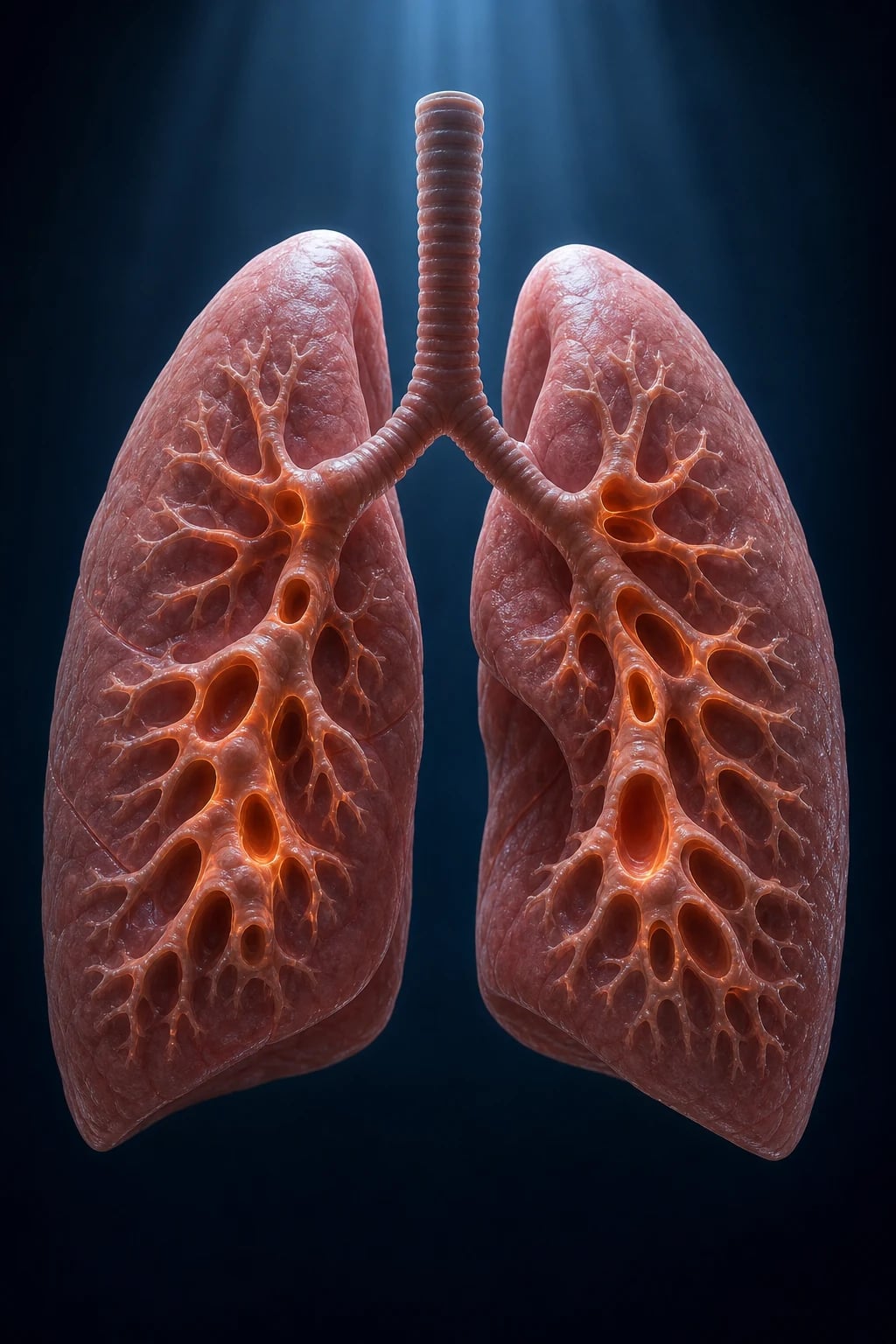
Overview & Definition
Bronchiectasis is the permanent, abnormal dilation of one or more bronchi, produced by destruction of the elastic and muscular components of the bronchial wall and sustained by a self-perpetuating cycle of impaired mucociliary clearance, chronic bacterial infection and neutrophil-driven inflammation.
[1]Historical context. The term was coined by Laennec in the early 19th century, and Reid's 1950 morphological classification (cylindrical, varicose, cystic) remains the basis of modern radiological reporting. Before HRCT, diagnosis relied on bronchography and was often missed. The recognition of bronchiectasis as a syndrome of treatable traits is the dominant modern paradigm, shifting focus from "the bronchi are dilated" to "why are they dilated and what can we treat?".
[1]It is not a single disease but a structural endpoint shared by many aetiologies, and it is now conceptualised as a syndrome of four treatable traits: infection, inflammation, impaired mucociliary clearance, and structural airway damage. The skill in bronchiectasis is therefore two-fold: (1) find and treat the underlying cause (a third remain idiopathic, but ABPA, immunodeficiency, post-TB and PCD are treatable); and (2) stratify severity with a multidimensional score (BSI or FACED) to decide who needs long-term suppressive therapy.
[1]Distinguish it sharply from chronic bronchitis (daily productive cough for at least 3 months in 2 successive years without HRCT dilation), COPD (predominantly small-airway and parenchymal disease, fixed airflow obstruction), and asthma (variable, eosinophilic, reversible). The HRCT is diagnostic and definitive.
[1]First presentation checklist. When a patient presents with suspected bronchiectasis, ask five questions: (1) Is the diagnosis confirmed on HRCT? (2) What is the extent and severity (BSI/FACED)? (3) What is the likely cause, and have I excluded treatable causes? (4) What is the microbiology (sputum culture, including NTM)? (5) What is the patient's functional status and comorbidity? Answering these questions ensures the patient is correctly stratified and entered into the appropriate management pathway.[1]
[1]Classification
Bronchiectasis is classified along three axes — morphology, distribution, and aetiology — because each axis drives different decisions (work-up, prognosis, therapy).
[1]By morphology (Reid's classification, now read off HRCT):
[1]Cylindrical (tubular)
- Mildest form — uniform, smooth dilation of bronchi
- Bronchi fail to taper and end squarely
- Common; potentially reversible components
- HRCT: parallel 'tram-track' walls
Varicose
- Intermediate severity — irregular beading
- Mixed dilated and constricted segments
- Suggests established wall destruction
- Often coexists with cylindrical areas
Cystic (saccular)
- Most severe — bronchi dilate into cysts/sacs toward the pleura
- Severe, often end-stage disease
- Highest risk of haemoptysis and infection
- HRCT: grape-like clusters; air-fluid levels
Reid's original pathological classification remains the backbone, but HRCT has replaced gross pathology in clinical practice. Cylindrical bronchiectasis can occasionally improve if the underlying cause is treated early (for example, removing a foreign body or starting immunoglobulin replacement), whereas cystic change is irreversible. The coexistence of varicose and cystic areas in the same patient is common and predicts worse outcomes.[1]
By distribution:
[1]- Localised (single lobe/segment) — usually post-infective (TB, pertussis, measles, adenovirus) or post-obstructive (foreign body, endobronchial tumour, node compression). Work-up should ask why that one lobe? — bronchoscopy if obstructive cause suspected.
- Diffuse (bilateral, multi-lobar) — usually systemic cause: immunodeficiency, PCD, CF, ABPA, rheumatoid arthritis, aspiration, or idiopathic.
Distribution predicts cause (a high-yield exam heuristic):
[1]| Distribution | Think of |
|---|---|
| Upper-lobe predominant | Post-TB, cystic fibrosis, ABPA, radiation, histoplasmosis |
| Lower-lobe predominant | Idiopathic, primary ciliary dyskinesia, rheumatoid arthritis, immunodeficiency, aspiration |
| Middle-lobe / lingula | Recurrent aspiration, atypical mycobacterial infection (Lady Windermere syndrome), middle-lobe syndrome |
| Central (proximal) upper-lobe | ABPA |
| Traction (peribronchial fibrosis) | Interstitial lung disease — different mechanism |
By aetiology — covered in depth under Epidemiology and Specific Subtypes. The modern approach is to identify a treatable cause in every patient, because the cause changes management and prognosis.
[1]The ERS 2017 and 2025 guidelines adopt the same four-pillar management framework and add structured thresholds for long-term macrolides (at least 3 exacerbations/year) and inhaled antibiotics (chronic Pseudomonas with exacerbations).[2][3]
Epidemiology & Risk Factors
[1]Bronchiectasis is commoner than once thought, and the recognised burden has risen sharply with routine thin-section CT. Prevalence estimates vary by country and age, but data from the UK and US suggest a prevalence of approximately 0.5–1.0% of adults, rising to over 5% in those over 75 years. There is a female preponderance in adults, possibly related to smaller airway calibre and delayed diagnosis. The burden is disproportionately high in South Asia where post-tuberculous and childhood-infection-related disease dominate.
[1]Occupational and environmental associations. While infection and host factors dominate, certain exposures increase risk or worsen outcomes. Biomass fuel smoke exposure is common in low-income settings and impairs mucociliary clearance; occupational dusts and fumes (silica, coal, cotton) cause chronic bronchitis and may coexist with bronchiectasis; ** aspiration** (alcohol, neurological disease, gastro-oesophageal reflux) is an under-recognised cause; and ** allergic bronchopulmonary aspergillosis** is both a consequence and cause of central bronchiectasis. Passive smoke exposure in childhood increases the risk of post-infectious bronchiectasis.
[1]Key epidemiology numbers:
[1]Bronchiectasis epidemiology
Aetiology by category (the structured way examiners want it):
[1]Interpreting the cause work-up. A systematic approach prevents missing treatable causes. All adults should have immunoglobulins, total IgE and Aspergillus-specific IgE/IgG, alpha-1-antitrypsin, and sputum including NTM culture. Selected patients need CFTR sweat chloride/genetics (young age, upper-lobe disease, male infertility, pancreatic symptoms, Staph aureus) or ciliary studies (neonatal respiratory distress, chronic rhinosinusitis, otitis, situs inversus, infertility). Rheumatology screen is indicated when joints, skin, or autoimmune features are present. HIV testing should be offered based on risk factors. Specific vaccine antibody responses identify functional antibody deficiency even when baseline immunoglobulins are normal.
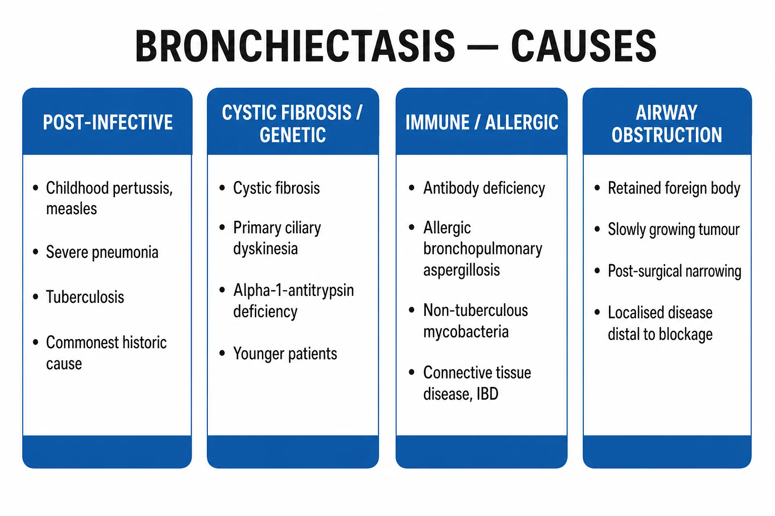
[1]Post-infective (commonest globally)
- Previous TB (esp. India/South Asia), measles, pertussis, adenovirus, severe bacterial pneumonia
- Often upper-lobe, localised
- Mucociliary damage plus residual scarring
Immunodeficiency
- Common variable immunodeficiency (CVID), specific antibody (functional IgG) deficiency, IgA deficiency, HIV
- Recurrent sinopulmonary infection → bronchiectasis
- Replacement immunoglobulin is disease-modifying
Ciliary / structural
- Primary ciliary dyskinesia (Kartagener = situs inversus + sinusitis + bronchiectasis), Young syndrome, cystic fibrosis (CFTR)
- Foreign body, endobronchial tumour, lymph node compression
- Aggressive work-up in children
Allergic / inflammatory
- Allergic bronchopulmonary aspergillosis (ABPA) — central upper-lobe bronchiectasis, asthma, eosinophilia, high IgE
- Rheumatoid arthritis (commonest connective-tissue cause)
- Inflammatory bowel disease, Sjogren
Idiopathic (up to ~30–50%)
- No cause found despite full work-up
- Diagnosis of exclusion
- Still managed with the four-pillar approach
Less common but examinable causes:
[1]- Alpha-1-antitrypsin deficiency — causes lower-zone panacinar emphysema in smokers, but can also be associated with bronchiectasis. Check serum alpha-1-antitrypsin level and phenotype/genotype (PiZZ, PiSZ, null alleles). Augmentation therapy is for emphysema, not bronchiectasis per se.
- Young syndrome — obstructive azoospermia and bronchiectasis; historically linked to mercury exposure but now considered a form of obstructive azoospermia with associated sinopulmonary disease. It is a rare cause but a classic named association.
- Yellow nail syndrome — triad of yellow nails, lymphedema, and pleural effusion, often with bronchiectasis. Lymphatic dysfunction is thought to underlie the lung and nail findings.
- Marfan syndrome and Ehlers-Danlos syndrome — connective-tissue defects may predispose to bronchial wall weakness and bronchiectasis.
- Inflammatory bowel disease — both Crohn's disease and ulcerative colitis are associated with bronchiectasis, sometimes preceding bowel diagnosis.
Microbiology of colonisation/infection (an examiner favourite): Haemophilus influenzae is the commonest organism overall in non-CF bronchiectasis, especially early/mild disease. Pseudomonas aeruginosa is the severity organism — it colonises more severe disease, accelerates FEV1 decline, doubles exacerbation frequency, drives hospitalisation and worsens quality of life and mortality. Streptococcus pneumoniae and Moraxella catarrhalis are common early colonisers; Staphylococcus aureus should prompt consideration of CF or ABPA (especially in children); non-tuberculous mycobacteria (NTM) — M. avium complex, M. abscessus — co-exist in roughly 8–10% and require specific work-up (multiple positive cultures) before treatment.[1][2]
Pathophysiology
The defining concept is Cole's vicious circle hypothesis (1986):[6]
- An initial insult (severe infection such as pertussis or TB, obstruction, or impaired host defence) impairs mucociliary clearance.
- Retained secretions become infected with bacterial pathogens (H. influenzae, Pseudomonas, S. pneumoniae).
- Infection triggers neutrophilic inflammation — neutrophils and macrophages release elastase, IL-8, TNF-alpha, MMP-8/9 and reactive oxygen species.
- Proteases destroy the elastin and muscularis of the bronchial wall; the weakened wall dilates under cough pressure, and the dilated bronchus clears secretions even more poorly — completing the circle.
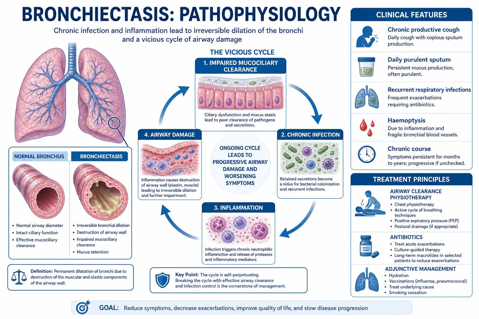
Why neutrophil elastase is the central villain: neutrophil elastase is a serine protease that (i) degrades elastin in the bronchial wall, (ii) stimulates goblet-cell hyperplasia and mucin (MUC5AC) secretion, (iii) slows ciliary beat frequency and strips ciliated epithelium, and (iv) inactivates complement and immunoglobulins and cleaves immune receptors — directly deepening the cycle. Sputum elastase activity correlates with exacerbation frequency and FEV1 decline.[6][2]
How dilated bronchi produce the clinical signs:
[1]- Pooled secretions → daily mucopurulent sputum and coarse crackles (often positional).
- Disruption of airway smooth muscle and wall oedema → wheeze and airflow obstruction (obstructive PFTs, not reversible to normal).
- Hypertrophy and proliferation of bronchial arteries under chronic inflammatory drive → haemoptysis, occasionally massive.
- Ventilation-perfusion mismatch and shunt across poorly ventilated but perfused dilated segments → hypoxaemia; in advanced disease, alveolar hypoventilation → type-2 respiratory failure and cor pulmonale.
- Chronic systemic inflammation → weight loss, anaemia of chronic disease, and secondary (AA) amyloidosis (which can cause nephrotic-range proteinuria).
Small-airway involvement. Bronchiectasis is not just large-airway disease. The tree-in-bud pattern on HRCT represents mucus and pus in centrilobular bronchioles. Small-airway obstruction and mucus plugging contribute to air trapping, ** ventilation–perfusion mismatch**, and exercise limitation. Expiratory HRCT can reveal air trapping that is not apparent on inspiratory images.
[1]The role of autoimmunity and systemic inflammation. In some patients, particularly those with rheumatoid arthritis or IBD-associated bronchiectasis, systemic immune dysregulation contributes to airway inflammation independent of infection. These patients often have more diffuse, lower-lobe disease and may respond better to immunomodulation of the underlying disease than to repeated antibiotics alone.[1]
Bronchiectasis — the numbers that matter
Why bronchiectasis develops — the three prerequisite concepts:
[1]- An initial insult damages mucociliary clearance. This may be an infection (pertussis, measles, adenovirus, severe pneumonia, TB), an obstruction (foreign body, tumour, lymph-node compression), or a host-defence defect (CVID, PCD, CF).
- A protease–antiprotease imbalance favours tissue destruction. Neutrophil elastase overwhelms endogenous inhibitors such as alpha-1-antitrypsin and secretory leucoprotease inhibitor, leading to unchecked elastolysis.
- Structural wall damage becomes self-perpetuating. Once dilation occurs, clearance worsens, infection persists, and the cycle continues even if the original insult is removed. This is why bronchiectasis is usually irreversible once established.
The cytokine cascade in more detail. Bacterial colonisation triggers IL-1β and TNF-α release from macrophages and airway epithelium. These cytokines drive neutrophil recruitment via IL-8 and CXCL-1/2/3. Neutrophils release elastase, proteinase-3, myeloperoxidase, and matrix metalloproteinases (MMP-8, MMP-9), which degrade elastin, collagen and proteoglycans. Simultaneously, elastase cleaves Toll-like receptors and opsonins, blunting bacterial clearance and deepening the inflammatory response. This dual action — tissue destruction and immune dysregulation — is what makes the cycle so hard to break.
[1]Airway wall changes beyond dilation. Histologically, bronchiectatic airways show loss of elastin and smooth muscle, mucosal oedema, ulceration, squamous metaplasia, and goblet-cell hyperplasia. Bronchial arteries proliferate and become tortuous, predisposing to haemoptysis. The surrounding parenchyma may show fibrosis, atelectasis or emphysema depending on the aetiology.
[1]Why Pseudomonas is uniquely damaging. P. aeruginosa forms a biofilm (quorum-sensing-regulated), shifts to a mucoid alginate-producing phenotype under selection pressure, and is intrinsically and acquired-resistant to many antibiotics. Its elastases (LasA/LasB) and proteases intensify the neutrophil response. Once mucoid Pseudomonas is established, eradication is rarely possible — management shifts from cure to suppression.[2]
How the vicious circle explains treatment. Every effective bronchiectasis therapy targets at least one step in Cole's circle. Airway clearance breaks the retention step. Antibiotics reduce the infection step. Macrolides dampen the neutrophilic inflammation step and also interfere with quorum sensing. Treating the cause (for example, removing a foreign body or replacing immunoglobulin) restores the initial defence step. Surgery removes the most destroyed segment and can interrupt the circle in localised disease. This mechanistic understanding helps candidates answer "why this treatment?" questions on exams.
[1]Inflammatory contrast (the exam question that distinguishes the chronic suppurative lung diseases): bronchiectasis is predominantly neutrophilic, asthma is eosinophilic/type-2, COPD is mixed. There is an increasingly recognised eosinophilic overlap endotype in bronchiectasis (blood eosinophils at least 300/μL) that is responsive to inhaled corticosteroids and — in severe cases — to anti-IL-5 biologics, but routine ICS is not recommended in non-CF bronchiectasis without an asthmatic/eosinophilic overlap.[1][3]
Clinical Presentation
Typical presentation is a chronic productive cough with mucopurulent or purulent sputum produced on most days for months or years, punctuated by acute exacerbations (increased sputum volume, purulence, dyspnoea, sometimes haemoptysis, fever, pleuritic pain). The classical clinical definition required cough and sputum on most days for at least 2 consecutive years, though HRCT now establishes the diagnosis.[12]
Exacerbations are defined (BTS) as a deterioration in at least one of three core symptoms: increased sputum volume, worsening sputum consistency/purulence, or increased breathlessness, often with increased cough, haemoptysis, fever, malaise or pleuritic pain. Exacerbation frequency (3 or more per year) is the trigger for long-term macrolide therapy.[1]
Physical signs:
[1]- Coarse inspiratory and/or expiratory crackles — often positional, over the affected segments.
- Wheeze (from airway obstruction/inflammation).
- Digital clubbing — present in up to a third; classic but not universal.
- Hyperinflation and use of accessory muscles in advanced disease.
- Weight loss and cachexia in end-stage disease.
- Cor pulmonale (raised JVP, ankle oedema, loud P2, hepatomegaly) in end-stage disease with pulmonary hypertension.
- Sinusitis and otitis (think PCD/CVID); nasal polyps (think CF or PCD); situs inversus (Kartagener).[1]
Atypical presentations examiners deliberately probe:
[1]- 'Dry' bronchiectasis — cough with little sputum, often upper-lobe and post-TB or ABPA; missed because the textbook description assumes wet sputum.
- Elderly patient with recurrent 'pneumonia' in the same segment — the same lobe recurring is bronchiectasis or obstruction until proven otherwise; HRCT and bronchoscopy.
- Child with failure to thrive and chronic productive cough — think CF or primary ciliary dyskinesia; sweat chloride, CFTR genetics, ciliary function testing.
- Immunocompromised host — atypical organisms (NTM, fungi, opportunists); lower threshold for bronchoscopy.
- Pregnant woman — stable disease usually tolerates pregnancy, but exacerbations may worsen; cough and dyspnoea can be mistaken for normal pregnancy.
- Diabetic patient — higher risk of Pseudomonas and NTM infection; TB may coexist.
- Patient presenting with nephrotic syndrome — long-standing bronchiectasis can cause secondary AA amyloidosis.
- Asymptomatic patient with incidental HRCT bronchiectasis — found on CT done for another reason; still requires cause work-up and monitoring.
- Patient with persistent 'cough-variant' presentation only — no sputum, normal examination except localised wheeze or crackles; HRCT is revealing.
- Smoker misdiagnosed with COPD — fixed airflow obstruction and cough attributed to smoking; HRCT shows bronchiectasis and changes management.
Differential Diagnosis
A chronic productive cough or recurrent chest infections is not always bronchiectasis. Distinguish each:
[1]Chronic bronchitis / COPD
- Smoker, chronic productive cough ≥3 mo in 2 successive years
- Fixed airflow obstruction on spirometry (FEV1/FVC <0.70 post-bronchodilator)
- NO bronchial dilation on HRCT — the decisive distinction
Asthma
- Variable symptoms, atopy, nocturnal, reversible airflow limitation
- Eosinophilic/type-2 inflammation; high response to bronchodilator
- Normal HRCT (or mild bronchial wall thickening)
Lung abscess
- Single cavity with air-fluid level, acute febrile illness
- Often post-aspiration or post-pneumonia
- Different radiology, different organism (anaerobes, S. aureus)
Pulmonary TB
- Upper-lobe, weight loss, night sweats, haemoptysis
- Positive sputum AFB / GeneXpert / NAAT
- Risk factors: contacts, diabetes, immunosuppression
Lung cancer
- Smoker, weight loss, mass on imaging
- Persistent non-resolving opacity; haemoptysis in older smoker
- CT + biopsy; bronchiectasis may be post-obstructive (the tumour)
Pulmonary fibrosis / ILD
- Fine 'Velcro' crackles, lower-zone, restrictive PFTs
- **Traction bronchiectasis** — airways 'pulled open' by peribronchial fibrosis (DIFFERENT mechanism)
- Management of the underlying ILD, not airway clearance
Congestive cardiac failure
- Orthopnoea, raised JVP, bibasal fine crackles, peripheral oedema
- B-type natriuretic peptide, echo
- No daily purulent sputum
Aspiration / recurrent pneumonia
- Dependent segments (posterior upper / lower lobes)
- Underlying swallow/neurological disease, reflux
- May itself cause bronchiectasis
Sputum character is diagnostically useful:
[1]- Purulent/mucopurulent — H. influenzae, Pseudomonas, S. pneumoniae.
- Foul-smelling — anaerobes/aspiration.
- Copious watery green — Pseudomonas aeruginosa.
- Blood-streaked — any cause; raises haemoptysis concern.
- Brownish plugs — ABPA (expectorated mucus plugs).
Key distinguishing features table:
[1]| Diagnosis | Clues that separate it from bronchiectasis | Decisive test |
|---|---|---|
| TB | Weight loss, night sweats, upper-lobe cavitary disease, contact history | Sputum AFB/GeneXpert |
| COPD | Smoker, fixed airflow obstruction, emphysema, no bronchial dilation | HRCT |
| ABPA | Asthma, eosinophilia, very high IgE, central upper-lobe bronchiectasis | Aspergillus-specific IgE/IgG |
| Aspiration | Dependent segments, neurological disease, reflux, anaerobic organisms | Videofluoroscopy swallow, bronchoscopy |
| ILD with traction bronchiectasis | Fine Velcro crackles, restrictive PFTs, honeycombing | HRCT pattern + PFTs |
| Lung cancer | Smoker, mass, rapid progression, unilateral hilar lymphadenopathy | CT + biopsy |
Chronic cough differential — when to think beyond bronchiectasis. A chronic cough can also be caused by upper-airway cough syndrome (post-nasal drip), gastro-oesophageal reflux disease, ACE-inhibitor therapy, non-asthmatic eosinophilic bronchitis, and airway hyperresponsiveness. The presence of purulent sputum, clubbing, localised crackles, and HRCT dilation separates bronchiectasis from these causes.
[1]Clinical & Bedside Assessment
Focused history — onset of cough/sputum; childhood triggers (pertussis, measles, recurrent infections, failure to thrive); TB exposure and treatment; chronic sinusitis, otitis, infertility (PCD); allergy, asthma, eggs/aspirin (ABPA); family history (CF, alpha-1-antitrypsin, PCD); smoking; connective-tissue disease (especially rheumatoid arthritis); immunisation history; growth chart in children.[1]
Focused examination — clubbing (a hallmark of chronic suppurative lung disease), pallor, cachexia, nasal polyps (CF, PCD), situs inversus (Kartagener), coarse crackles (often positional), wheeze, hyperinflation, cor pulmonale signs, and chronic sinus disease.
[1]Assess exacerbation severity — sputum volume, purulence and colour (the Murray/Stockley purulent-mucoid scale), fever, breathlessness, oxygen saturation, presence and volume of haemoptysis. Recognise the bedside signs that mandate urgent admission: respiratory distress, hypoxia, cyanosis, massive haemoptysis, confusion (type-2 failure), sepsis, or new pneumonic change.
[1]Bedside severity markers:
[1]| Marker | Mild (outpatient) | Moderate (consider admission) | Severe (admit) |
|---|---|---|---|
| Sputum | Increased volume/purulence | Markedly purulent, difficult to clear | Massive haemoptysis or respiratory distress |
| Temperature | Afebrile or low-grade | >38°C | >38.5°C with rigors |
| Breathlessness | Minimal | Moderate, affects ADLs | Severe, RR over 25, accessory muscle use |
| Oxygenation | SpO2 ≥94% | SpO2 90–93% | SpO2 under 90% or rising CO2 |
| Comorbidity | None | Some | Significant, or failure of outpatient therapy |
The Murray–Stockley sputum colour scale is a simple bedside tool: the more green/yellow the sputum, the higher the neutrophil load and the more likely the patient is having a bacterial exacerbation. Colour can be used to triage and to monitor response to antibiotics.
[1]Causes of bronchiectasis — 'AGRA IMMUNE'
AGRA IMMUNE
Allergic bronchopulmonary aspergillosis — central upper-lobe
CFTR (cystic fibrosis), alpha-1-antitrypsin
Rheumatoid arthritis, IBD, Sjogren — connective-tissue causes
Foreign body, tumour, node compression
Up to 30–50% after full work-up
Primary ciliary dyskinesia (Kartagener), Young syndrome
TB, pertussis, measles, adenovirus — commonest globally
CVID, specific antibody deficiency, IgA deficiency, HIV
Rare primary immune defects
Inhalational injury, toxic fumes
Investigations
The investigation strategy is (a) confirm the diagnosis, (b) define extent, severity and complications, and (c) find the underlying cause. The HRCT is central to (a) and (b); a structured cause work-up is central to (c).[1][2]
Confirm the diagnosis — high-resolution CT (HRCT):
[1]- Gold standard and diagnostic. Modern practice is thin-section volumetric CT (typically 1 mm slices) acquired in full inspiration (± expiratory cuts for air-trapping). The sensitivity and specificity of HRCT for bronchiectasis are very high when modern scanners and thin collimation are used.
- HRCT signs (reproduced verbatim):
- Signet-ring sign — the internal bronchial diameter exceeds the diameter of the accompanying pulmonary artery branch (the pathognomonic sign).
- Tram-track lines — thickened, parallel bronchial walls.
- Lack of normal bronchial tapering — bronchi remain the same calibre as they course peripherally.
- Varicose or cystic dilations, sometimes with air-fluid levels (cystic/saccular).
- Mucus plugging and 'tree-in-bud' appearance (small-airway impaction).
- Distribution is itself a clue — upper-lobe (post-TB, CF, ABPA, radiation) vs lower-lobe (idiopathic, PCD, RA, immunodeficiency, aspiration).
HRCT pitfall — traction bronchiectasis. In ILD, bronchi may appear dilated because surrounding fibrosis pulls them open. The key distinction is that traction bronchiectasis is accompanied by reticulation, honeycombing or architectural distortion and the patient has restrictive physiology. Management is of the underlying ILD, not airway clearance.
[1]When to order HRCT. BTS/ERS guidelines recommend HRCT in any adult with a consistent clinical syndrome: chronic productive cough, recurrent chest infections, or persistent haemoptysis, especially if examination shows coarse crackles or clubbing. Find the cause — structured blood and sputum work-up:
[1]The cause work-up is not optional. In a patient with newly diagnosed bronchiectasis, the BTS/ERS guidelines recommend a core panel for all adults, with additional tests guided by clinical phenotype.
[1]| Test | What it seeks |
|---|---|
| Full blood count + differential | Anaemia of chronic disease; eosinophilia (suggests ABPA) |
| Serum immunoglobulins (IgG, IgA, IgM) | CVID / specific antibody deficiency / IgA deficiency |
| Total IgE + Aspergillus-specific IgE and IgG | ABPA (along with skin-prick/precipitins) |
| Aspergillus precipitins / complement fixation | ABPA, aspergilloma |
| Alpha-1-antitrypsin level (± genotype) | AAT deficiency (esp. lower-zone emphysema) |
| Rheumatoid factor / ANA / anti-CCP | Rheumatoid arthritis and other connective-tissue disease |
| HIV test | HIV-related immunodeficiency |
| Sweat chloride ± CFTR genetics | Cystic fibrosis (esp. young, upper-lobe, Staph aureus, male infertility, pancreatic insufficiency) |
| Ciliary biopsy / nasal nitric oxide | Primary ciliary dyskinesia (Kartagener) |
| Sputum MC&S including AFB and NTM culture | Organism colonisation; exclude NTM co-infection (multiple positive cultures required) |
| Specific vaccine antibody responses | Functional antibody deficiency (response to pneumococcal vaccine) |
Spirometry — usually obstructive (reduced FEV1 and FEV1/FVC, often partial bronchodilator reversibility but not to normal). Spirometry is not diagnostic; its role is to grade severity and track progression by serial FEV1. A bronchodilator response may be seen because airway inflammation causes some reversible component, but FEV1 rarely returns to normal. Diffusing capacity (DLCO) is usually preserved unless there is coexisting emphysema or ILD. In severe disease, spirometry may show an obstructive–restrictive mixed pattern because of lung volume loss from atelectasis and fibrosis.
[1]Exercise and functional testing. The 6-minute walk test is useful for assessing functional capacity and exertional desaturation. A fall in SpO2 of 4% or more with walking, or a walk distance below predicted, indicates significant functional impairment and may prompt oxygen assessment or pulmonary rehabilitation.[1]
Multidimensional severity scores — reproduced verbatim
Two validated multidimensional scores stratify bronchiectasis into mild/moderate/severe and predict future mortality, exacerbations and hospitalisation. The BSI is more widely used and more prognostically accurate; FACED is simpler (only 5 inputs).[4][5][10]
Bronchiectasis Severity Index (BSI, Chalmers 2014):[4]
BSI components (weighted)
- Age (≥70 = 2; 50–69 = 1)
- FEV1 % predicted (<30 = 3; 30–49 = 2; 50–79 = 1)
- Prior hospitalisation for exacerbation (yes = 2)
- Exacerbations in past year (≥3 = 1; ≥1 needing IV antibiotics = 2)
- Sputum colonisation: Pseudomonas = 3; other potentially pathogenic organism = 1; none = 0
- mMRC dyspnoea ≥2 = 1
- Radiological extension (lobes involved, weighted)
BSI bands & risk
- Mild (0–4): low future exacerbation, low mortality
- Moderate (5–8): intermediate risk
- Severe (9–26): ~ up to 40% 10-year mortality, frequent exacerbations
- Predicts mortality, exacerbations, hospitalisation, quality of life
FACED score (Martinez-Garcia 2014):[5]
| Component | Points |
|---|---|
| FEV1 % predicted (≤50 = 2; 51–69 = 1) | 0–2 |
| Age (≥70 = 2; 50–69 = 1) | 0–2 |
| Chronic colonisation by Pseudomonas (yes = 1; any other = 0) | 0–1 |
| Extension (number of lobes involved; >2 lobes = 1) | 0–1 |
| Dyspnoea (mMRC ≥2 = 1) | 0–1 |
Total 0–5 — mild (0–2), moderate (3–4), severe (5); predicts 5-year all-cause mortality. FACED is easier to calculate at the bedside but is less comprehensive than BSI (it does not capture prior hospitalisation or exacerbation frequency).[13]
Other investigations:
[1]- Bronchoscopy — for suspected foreign body, endobronchial tumour, single-lobe disease, sampling for NTM/fungi in immunocompromised. Bronchoscopy also allows protected-brush sampling, bronchoalveolar lavage for microbiology, and localisation of bleeding in haemoptysis.
- Echocardiogram — to detect pulmonary hypertension / cor pulmonale in advanced disease.
- 6-minute walk test — functional capacity and oxygenation.
- Sputum colour/score (Murray–Stockley purulence scale) and computerised sputum analysis — useful for monitoring.
- CRP / ESR — non-specific inflammatory markers.
- 24-hour sputum culture — preferred for organism identification, sensitivities, and NTM.
- Urinalysis for proteinuria — to detect secondary AA amyloidosis in long-standing disease.
- Nitric oxide measurement — very low nasal nitric oxide supports PCD.[1]
Management — Resuscitation
The acute scenarios that need resuscitation are severe exacerbation with respiratory failure or sepsis, and massive haemoptysis.
[1]Severe exacerbation — ABCDE:
[1]- A/B — controlled oxygen to target SpO2 94–98% (or 88–92% in chronic CO2 retainers, by analogy with COPD).
- C — IV access, bloods (FBC, U&E, CRP), blood cultures if febrile/septic; treat sepsis with the Surviving Sepsis hour-1 bundle if septic.
- Empirical antibiotics started promptly after sputum culture (never delay antibiotics for tests in a septic patient): route and agent by severity and prior microbiology (see Definitive Management). Standard 14-day course.[1]
- NIV (BiPAP) if persisting hypercapnic type-2 respiratory failure despite medical therapy, by analogy with COPD.
- Physiotherapy to clear secretions as soon as the patient is stable.
Massive haemoptysis (covered in detail above and in Complications) — bleeding side down, protect the uninvolved lung, secure airway, bronchial artery embolisation first-line, surgical resection last resort.[1]
Massive haemoptysis — stepwise algorithm:
[1]- Call for help — anaesthetics, interventional radiology, thoracic surgery.
- Protect the good lung — place the patient bleeding side down (good lung up) to minimise soiling. Avoid lying flat.
- Airway — high-flow oxygen; if the airway is threatened or bleeding is torrential, secure it with a large-bore endotracheal tube and advance the tube to the main bronchus of the good lung, or use a dual-lumen tube if skilled.
- Breathing/Circulation — large-bore IV access ×2, bloods (FBC, coagulation, cross-match 4–6 units), group-and-save, correct coagulopathy and thrombocytopenia, resuscitate with crystalloid/blood as needed.
- Localise — rigid or flexible bronchoscopy once the patient is stable enough; identify the bleeding lobe/segment.
- Definitive control — urgent bronchial artery embolisation is first-line; it is highly effective and stops bleeding in most cases. Surgical resection is reserved for embolisation failure, localised disease, or recurrent life-threatening bleeding.
- Aftercare — ICU monitoring, watch for recurrent bleeding (bronchial artery collaterals can reconstitute), and plan definitive management of the underlying bronchiectasis once stable.
Remember: the commonest fatal mechanism is asphyxiation from blood flooding the uninvolved lung, not exsanguination. Positioning and protecting the good lung are therefore as important as stopping the bleeding.
[1]Emergency department disposition for acute exacerbation. Not every exacerbation needs admission. Admit patients who are toxic, hypoxaemic, unable to maintain oral intake, have respiratory failure, massive haemoptysis, or fail a short period of observation. For stable patients with milder symptoms and reliable follow-up, a 14-day oral antibiotic course with augmented airway clearance and a safety-net review is appropriate. Ensure a sputum sample is sent before antibiotics if possible, but do not delay antibiotics for a septic patient.
[1]Analgesia and antitussives. Pleuritic chest pain from inflammation can limit deep breathing and physiotherapy; simple analgesia helps. Sedative antitussives should be avoided because suppressing cough impairs clearance of infected secretions and can worsen infection.
[1]Management — Definitive & Stepwise
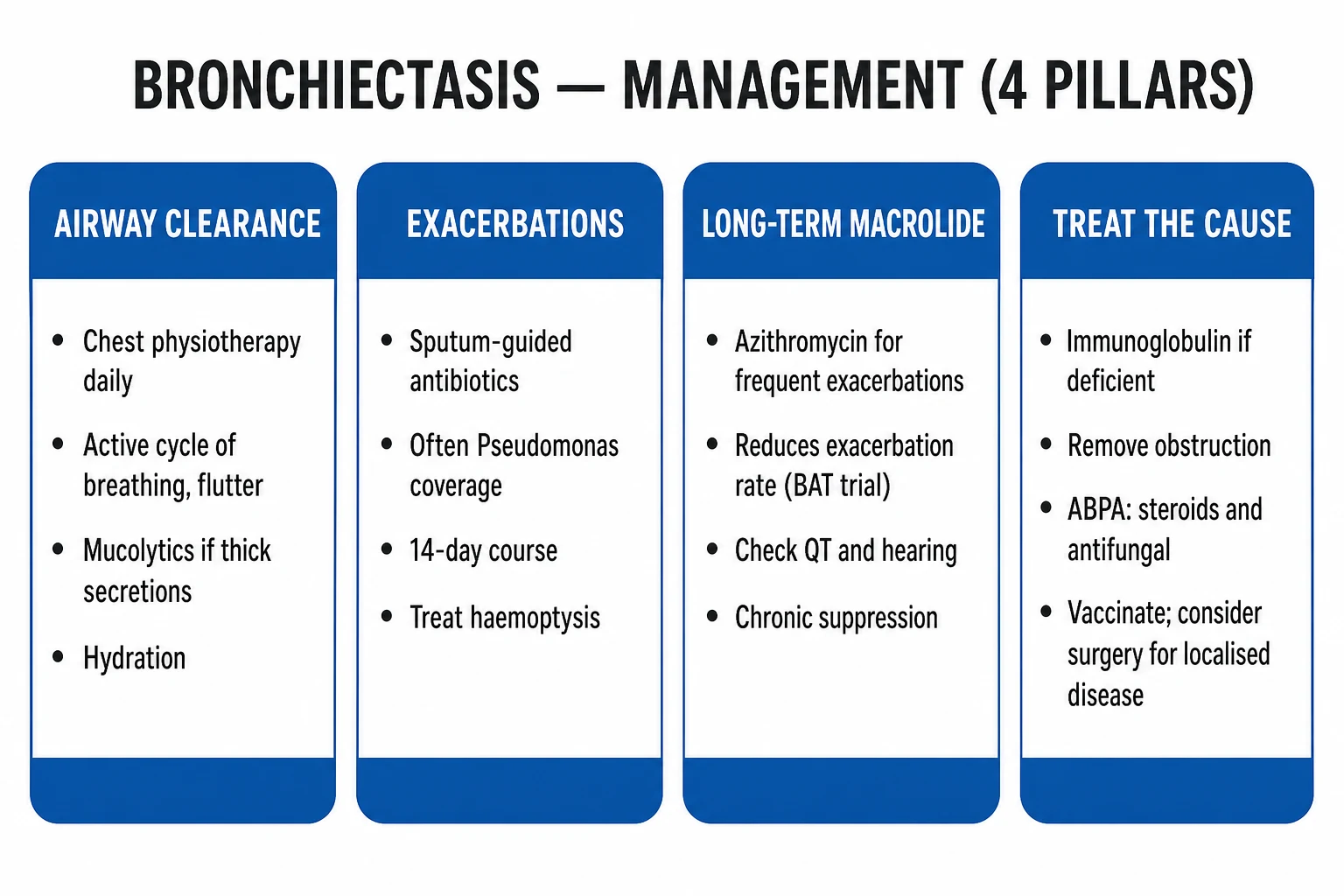
Long-term management rests on four pillars: (1) treat the underlying cause, (2) airway clearance (physiotherapy plus mucoactive drugs), (3) infection control (acute antibiotics plus long-term suppression), and (4) prevention of complications (vaccination, smoking cessation, pulmonary rehabilitation).[1][2]
[1]Pillar 1 — Treat the underlying cause
A treatable cause found must be treated on its own merits: ABPA (systemic corticosteroid ± itraconazole/voriconazole); immunodeficiency (replacement immunoglobulin for CVID/hypogammaglobulinaemia); foreign body or tumour (remove — bronchiectasis may regress); rheumatoid arthritis (treat the underlying disease); cystic fibrosis (CFTR modulators and CF-centre MDT); primary ciliary dyskinesia (multidisciplinary care).[1]
Pillar 2 — Airway clearance
Airway clearance — choosing the right technique. No single technique is superior for every patient. The choice depends on age, severity, lobar distribution, sputum volume, comorbidity, and patient preference. ACBT and autogenic drainage are first-line for most adults. PEP devices are useful when huffing alone is insufficient or when the patient has poor coordination. Oscillating PEP (Acapella, Flutter) adds vibration to loosen secretions and is particularly helpful for sticky mucus. High-frequency chest-wall oscillation vests are used in severe disease, CF, or patients who cannot perform active techniques. All techniques should be taught by a respiratory physiotherapist and reviewed regularly. Adjuncts include postural drainage (positioning to drain affected segments) and huff coughing to clear mobilised secretions without the airway closure of a forceful cough.
[1]Airway clearance in acute exacerbation. During an exacerbation, secretions increase and the patient may be too breathless for vigorous techniques. Start with gentle breathing exercises, thoracic expansion, and small huffs once pain and dyspnoea are controlled. Increase intensity as the patient improves. Early physiotherapy reduces sputum retention and may shorten hospital stay.
[1]Mucoactive pharmacotherapy:
[1]- Nebulised hypertonic saline (3–7%) — hydrates the periciliary fluid layer, reduces sputum viscosity, improves clearance; often preceded by a bronchodilator (bronchospasm risk). Randomised studies in non-CF bronchiectasis have shown improved lung function and quality of life.[15][16]
- Recombinant human DNase (dornase alfa) — effective in cystic fibrosis but NOT recommended in non-CF bronchiectasis: the O'Donnell randomised trial showed no benefit and possible harm (more exacerbations and greater FEV1 decline). This is one of the most frequently examined facts in the topic.[14][1]
- Inhaled mannitol (Bronchitol) — improves mucociliary clearance and quality of life in selected patients with acceptable tolerability; trial data support symptomatic benefit.[17][18]
- Carbocisteine and other oral mucolytics — modest benefit; trial in selected patients.
Pillar 3 — Infection control
Acute exacerbation — empirical antibiotic selection by severity and prior microbiology (BTS/ERS; standard 14-day course):[1]
Mild, home, no Pseudomonas
- **Amoxicillin 500 mg–1 g PO TDS** (high-dose)
- Alternative: doxycycline 100 mg OD–BD, or clarithromycin 500 mg BD (penicillin-allergic)
- Duration: 14 days
- Strengthen airway clearance; safety-net to review
Known Pseudomonas
- **Ciprofloxacin 500–750 mg PO BD**
- Add oral if mild; switch to IV if severe or no response
- Duration: 14 days
- Monitor for QTc, tendinopathy
Severe / hospitalised
- **IV ceftazidime 2 g TDS** OR **piperacillin-tazobactam 4.5 g TDS** (± aminoglycoside)
- Add IV anti-pseudomonal cover if known/likely Pseudomonas
- Tailor to sputum culture and sensitivities
- IV-to-oral switch once improving
Antibiotic selection in detail (adults):
[1]| Scenario | First-line agent | Dose and route | Duration | Notes |
|---|---|---|---|---|
| Mild exacerbation, no Pseudomonas | Amoxicillin | 500 mg–1 g PO TDS | 14 days | Use high dose; alternative: doxycycline 100 mg OD–BD or clarithromycin 500 mg BD |
| Mild exacerbation, penicillin allergy | Doxycycline or clarithromycin | 100 mg PO OD–BD or 500 mg PO BD | 14 days | Check local resistance |
| Known Pseudomonas, mild | Ciprofloxacin | 500–750 mg PO BD | 14 days | Watch QTc, tendons, neuropathy |
| Severe / hospitalised | Ceftazidime or piperacillin-tazobactam | 2 g IV TDS or 4.5 g IV TDS | 14 days | Add aminoglycoside if severe Pseudomonas |
| Pseudomonas, severe / IV | Ceftazidime or piperacillin-tazobactam ± tobramycin | 2 g IV TDS or 4.5 g IV TDS + tobramycin 5–7 mg/kg IV OD | 14 days | Tailor to sensitivities; monitor renal function and drug levels |
| NTM suspected | None empirically | — | — | Obtain cultures first; do not give macrolide monotherapy |
IV-to-oral switch criteria — improving clinically, able to absorb oral antibiotics, falling inflammatory markers, and an oral agent to which the organism is sensitive. The total course should be 14 days for most exacerbations; some clinicians extend to 21 days for severe Pseudomonas or slow responders.
[1]Pseudomonas eradication vs suppression. In newly isolated Pseudomonas, a trial of eradication with oral ciprofloxacin (or IV antipseudomonal therapy) may be attempted, but once chronic colonisation with a mucoid strain is established, eradication is rarely achieved and long-term suppression with inhaled antibiotics becomes the goal.
[1]Long-term suppressive antibiotic therapy:
[1]- Long-term macrolide (azithromycin 250 mg three times weekly or 500 mg three times weekly; or erythromycin 500 mg BD) for patients with 3 or more exacerbations per year despite optimised airway clearance. Mechanism is anti-inflammatory and anti-quorum-sensing, not antimicrobial. Before starting: baseline ECG (QTc) and LFTs, and exclude NTM co-infection (macrolide monotherapy drives macrolide resistance in NTM). Landmark trials — BAT (Altenburg, azithromycin, JAMA 2013), BLESS (Serisier, erythromycin, JAMA 2013), EMBRACE (Wong, azithromycin, Lancet 2012) — each showed a significant reduction in exacerbation frequency. Monitoring on therapy: repeat ECG if QTc prolonged, watch for hearing impairment, gastrointestinal upset, and hepatotoxicity.
- Inhaled antibiotic practical points. Inhaled antibiotics are usually reserved for patients with chronic Pseudomonas and frequent exacerbations despite macrolides and physiotherapy. Before starting, document chronic colonisation (typically 2 or more positive cultures at least 3 months apart) and exclude bronchospasm with a supervised first dose preceded by a bronchodilator. Common regimens include nebulised colistimethate 1–2 million units BD, inhaled tobramycin 300 mg BD in alternating months, aztreonam 75 mg TID, and liposomal amikacin 590 mg OD. Drug delivery depends on the nebuliser device; patients need training and adherence support. Side effects include cough, bronchospasm, wheeze, dyspnoea, and (with aminoglycosides) renal toxicity and ototoxicity.
Bronchodilators and inhaled corticosteroids
- Long-acting bronchodilators (LABA, LAMA) are used for breathlessness and airflow obstruction; useful in symptomatic patients with reversibility or coexisting COPD/asthma.
- Routine inhaled corticosteroids are NOT recommended in non-CF bronchiectasis without overlap. ICS is reserved for the asthmatic/eosinophilic overlap endotype (blood eosinophils at least 300/μL, wheeze, atopy).[1][3]
Pillar 4 — Prevention and rehabilitation
- Vaccination — annual influenza, pneumococcal (PCV/PPSV23 per age/risk schedule), COVID-19; reduces infective exacerbations. Ensure household contacts are vaccinated where possible to reduce transmission.
- Smoking cessation — the single most important modifiable factor accelerating decline.
- Pulmonary rehabilitation — improves exercise capacity and quality of life in breathless patients. Programmes include aerobic exercise, strength training, breathing techniques, airway-clearance education, and psychological support. Benefits are similar to those seen in COPD, even though bronchiectasis is less studied.
- Nutrition and weight management — underweight and malnutrition worsen outcomes. Dietary assessment, oral nutritional supplements, and treatment of micronutrient deficiencies (especially vitamin D) are important.
- Bone health — recurrent oral corticosteroid use (especially in ABPA) and chronic inflammation increase osteoporosis risk; consider bone protection with calcium/vitamin D and bisphosphonates when indicated.
- Patient education and written action plan — recognising exacerbations early, when to seek help, and adherence to physiotherapy.
- Travel and altitude — patients with severe disease or chronic hypoxaemia may need oxygen assessment before air travel or high-altitude exposure.
Surgery and transplantation
- Surgical resection (lobectomy/segmentectomy) for localised disease failing medical therapy, massive or recurrent haemoptysis (after embolisation failure), or a destroyed lobe; outcomes best when disease is truly localised.
- Lung transplantation for end-stage bilateral disease (especially CF) with respiratory failure.[1]
Specific Subtypes & Scenarios
- ABPA-related bronchiectasis — central (proximal), upper-lobe bronchiectasis in an asthmatic, with eosinophilia, markedly elevated total IgE (>1000 IU/mL), Aspergillus-specific IgE/IgG, and central bronchiectasis on HRCT. Treatment is systemic corticosteroid (prednisolone 0.5 mg/kg/day, taper) ± antifungal (itraconazole/voriconazole) for steroid-sparing. ABPA is a treatable cause — diagnose it and the trajectory changes.
ABPA diagnostic approach (high-yield overview): ABPA should be suspected in any asthmatic or CF patient with central bronchiectasis, eosinophilia, and high IgE. The classic diagnostic criteria include: (1) asthma or CF; (2) positive immediate or delayed skin reaction to Aspergillus; (3) elevated total IgE (typically >1000 IU/mL); (4) elevated Aspergillus-specific IgE and IgG; (5) peripheral blood eosinophilia; (6) central/proximal bronchiectasis on CT; and (7) fleeting or fixed pulmonary opacities. HRCT shows central bronchiectasis, mucus plugging (often high attenuation), and upper-lobe predominance. Serologically, Aspergillus fumigatus-specific IgE is the most sensitive marker, while precipitating antibodies support the diagnosis. Staging includes acute, remission, exacerbation, corticosteroid-dependent, and fibrotic phases. Treatment begins with prednisolone 0.5 mg/kg/day for 1–2 weeks, then taper over 6–8 weeks; itraconazole 200 mg BD or voriconazole 200 mg BD can be steroid-sparing. Monitoring relies on total IgE and eosinophil count — a rising IgE often heralds relapse.
[1]- Tuberculosis-associated bronchiectasis — post-TB upper-lobe, often 'dry', with mixed organisms; the commonest aetiology in India and other TB-endemic regions.[11] Cystic-fibrosis bronchiectasis — upper-lobe, Staphylococcus aureus then Burkholderia cepacia complex and Pseudomonas, CFTR modulators (elexacaftor–tezacaftor–ivacaftor in eligible genotypes), dornase alfa is indicated, sweat chloride and CFTR genetics. Distinct management under a CF-centre MDT. CF differs from non-CF bronchiectasis in several key ways: onset in childhood, upper-lobe predominance, pancreatic insufficiency and malabsorption, diabetes, male infertility, nasal polyps, and a specific requirement for CF-centre care. Dornase alfa, CFTR modulators, and higher-calorie nutrition are central to CF management but not to non-CF disease.
- Primary ciliary dyskinesia / Kartagener syndrome — situs inversus totalis + chronic sinusitis + bronchiectasis (Kartagener triad), with neonatal respiratory distress, chronic otitis media, and infertility (immotile/dyskinetic sperm and cilia). Ciliary biopsy / nasal nitric oxide confirms.
PCD diagnostic approach: PCD is a genetically heterogeneous disorder of ciliary ultrastructure and function. Key clinical clues include neonatal respiratory distress (often with unexplained lobar collapse), year-round nasal congestion and rhinorrhoea starting in infancy, chronic otitis media with glue ear, chronic wet cough from early childhood, situs inversus (in roughly 50% of cases — the Kartagener subgroup), and male infertility (reduced sperm motility). Initial screening is with nasal nitric oxide measurement — PCD patients typically have very low levels. Confirmatory testing requires ciliary ultrastructural analysis by transmission electron microscopy (looking for absent outer dynein arms, inner dynein arm defects, radial spoke defects, or microtubular disorganisation) and ciliary beat frequency/pattern analysis by high-speed video microscopy. Genetic testing for known PCD genes is increasingly available and is useful when biopsy is inconclusive. Management is primarily supportive and aggressive: airway clearance, prompt treatment of infections, hearing support, and genetic counselling. Immunodeficiency-related bronchiectasis. CVID is the most important acquired immunodeficiency to identify. Clues include recurrent bacterial sinopulmonary infections, chronic diarrhoea, autoimmune cytopenias, splenomegaly, and granulomatous-lymphocytic interstitial lung disease. Serum immunoglobulins show low IgG and low IgA or IgM; specific antibody responses to pneumococcal vaccine are impaired. Replacement immunoglobulin (IVIG or subcutaneous IgG) reduces infections and can slow or stabilise bronchiectasis. Specific antibody deficiency and IgA deficiency may also predispose; IgA deficiency alone is usually asymptomatic but can be associated with recurrent infections and coeliac disease. HIV causes bronchiectasis through repeated opportunistic infection and should be excluded in at-risk patients.
- Traction bronchiectasis (interstitial lung disease) — airways 'pulled open' by peribronchial fibrosis rather than destroyed by infection; different mechanism, different management (treat the underlying ILD).
- Post-obstructive bronchiectasis — single-lobe disease from foreign body, endobronchial tumour, or external compression; remove the obstruction and the bronchiectasis may improve.
- Aspiration-related bronchiectasis — dependent segments (posterior upper lobes, lower lobes), often with anaerobic organisms; requires swallow assessment, reflux management, and sometimes gastrostomy.
- NTM-associated bronchiectasis — M. avium complex classically produces right middle-lobe and lingula bronchiectasis in older, thin women with minimal cough (the "Lady Windermere syndrome" phenotype). Diagnosis requires multiple positive cultures or one positive culture with compatible CT findings. Management requires prolonged multidrug therapy and specialist input; macrolide monotherapy must be avoided.
Complications & Pitfalls
Disease complications:
[1]- Recurrent exacerbations and accelerated FEV1 decline. Each exacerbation drives further inflammation and wall damage; preventing them is a key treatment goal.
- Massive haemoptysis — the life-threatening emergency (above).
- Cor pulmonale and type-2 respiratory failure in end-stage disease.
- Secondary (AA) amyloidosis — long-standing neutrophilic inflammation drives serum-amyloid-A overproduction; presents as nephrotic-range proteinuria (renal failure); consider when a long-standing bronchiectasis patient develops oedema and proteinuria. Renal biopsy shows amyloid deposits derived from serum amyloid A.
- Chronic lung abscess, empyema, pneumothorax — more common in severe cystic disease.
- Metastatic infection — brain abscess (rare but classical in severe disease), septic emboli, and vertebral osteomyelitis.
- Severe impairment of quality of life — fatigue, breathlessness, depression, social isolation, and reduced exercise capacity.
- Osteoporosis, malnutrition, and cardiovascular comorbidity from chronic inflammation and repeated steroid exposure (especially in ABPA).
- Anxiety and depression — common and under-recognised; screening improves holistic care.
Classic pitfalls (examiner-favoured):
[1]- Missing a treatable cause — ABPA, immunodeficiency, foreign body. A systematic cause work-up is mandatory.
- Using dornase alfa in non-CF bronchiectasis — ineffective, potentially harmful (the O'Donnell trial).
- Failing to escalate to long-term macrolide / inhaled antibiotic in frequent exacerbators or chronic Pseudomonas.
- Under-recognising NTM co-infection — multiple positive cultures required; macrolide monotherapy drives resistance.
- Not involving a physiotherapist — airway clearance is the cornerstone, not an add-on.
- Confusing traction bronchiectasis (ILD) with infection-driven bronchiectasis — the management is entirely different.
- Prescribing routine ICS without an asthmatic/eosinophilic overlap — no benefit and potential harm.
- Using short antibiotic courses — the standard exacerbation course is 14 days in bronchiectasis.
- Ignoring sputum colour/volume trends — these are powerful early warning signs of exacerbation.
- Forgetting to protect the good lung in massive haemoptysis — bleeding side down, intubate the good lung if needed.
Prognosis & Disposition
Prognosis is highly variable and driven by severity scores (BSI/FACED), Pseudomonas colonisation, exacerbation frequency, FEV1 decline, radiological extension, and comorbidity. The BSI outperforms FEV1 alone for predicting mortality, exacerbations and hospitalisation; severe BSI predicts up to around 40% mortality at 10 years.
[1]Quality of life and outcomes. The St George's Respiratory Questionnaire (SGRQ) and the Quality of Life – Bronchiectasis (QoL-B) questionnaire are validated tools. QoL correlates closely with exacerbation frequency, sputum volume, and FEV1. Hospitalisations and exacerbations are the strongest drivers of impaired quality of life and are important trial endpoints.
[1]Disposition:
[1]- Most stable patients are managed in primary care/clinic with a respiratory physiotherapist and a written action plan.
- Admit for severe exacerbation, respiratory failure, sepsis, or massive haemoptysis.
- Tertiary referral for difficult/severe disease, recurrent hospitalisation, suspected rare cause (PCD, immunodeficiency), and transplant assessment in end-stage disease.
Key components of a bronchiectasis clinic. A well-functioning clinic includes: (1) a respiratory physician with expertise in bronchiectasis; (2) a specialist respiratory physiotherapist for airway clearance training; (3) a microbiology/laboratory service able to process sputum for bacteria, fungi and NTM; (4) access to bronchoscopy and interventional radiology; (5) links to immunology, rheumatology, CF and PCD specialists; (6) a nurse specialist for education and self-management support; and (7) smoking cessation, nutrition, and pulmonary rehabilitation services. MDT working is central to good outcomes, particularly in severe or complex disease.
[1]Referral thresholds. Refer to a respiratory specialist or bronchiectasis clinic when the diagnosis is new or uncertain, the patient has severe disease (BSI ≥5), chronic Pseudomonas colonisation, frequent exacerbations, massive haemoptysis, suspected rare cause, or failure of primary-care management.[1]
Follow-up and monitoring:
[1]- Regular spirometry (FEV1 trends) every 6–12 months in stable patients, more frequently if declining.
- Sputum cultures when stable (at least annually) and at every exacerbation.
- Review of exacerbation frequency, antibiotic use, and hospital admissions.
- Annual influenza vaccination; pneumococcal vaccination per local schedule.
- BSI/FACED recalculation if clinical status changes.
- Quality of life assessment (SGRQ or QoL-B) to guide supportive care.
- Screening for comorbidities: osteoporosis, cardiovascular disease, anxiety/depression, and sleep disturbance.
- Nutrition and bone health review, especially in patients on repeated corticosteroids.
Patient explanation. When explaining bronchiectasis to a patient, use plain language: the airways have been stretched and damaged, so they cannot clear mucus properly. Mucus pools, becomes infected, and causes inflammation, which further damages the airways. The goal of treatment is to break this cycle: clear mucus every day (physiotherapy), treat infections promptly (antibiotics), find and treat any underlying cause, and prevent future infections (vaccines, no smoking). Emphasise that bronchiectasis is usually a long-term condition, but with good management most people can lead active lives and keep exacerbations to a minimum.
[1]Prognosis counselling. Explain that severity varies widely. Some patients have mild disease with few exacerbations and near-normal life expectancy; others have severe disease with frequent hospitalisations. Severity scores (BSI/FACED), Pseudomonas status, exacerbation frequency, and FEV1 trend help estimate risk. Reassure that modern management — airway clearance, vaccines, targeted antibiotics, and treating the cause — has substantially improved outcomes compared with historical cohorts.
[1]Special Populations
- Children — suspect CF / PCD / immunodeficiency early; pursue sweat chloride and CFTR genetics, ciliary function, and immunoglobulins. Weight-based antibiotics; aggressive physiotherapy; MDT at a paediatric respiratory / CF centre. Growth and nutrition are central to management.
- Pregnancy — most women with stable bronchiectasis tolerate pregnancy well; optimise before conception. Azithromycin is generally avoided in the first trimester if possible; avoid aminoglycosides and tetracyclines; adjust antibiotic choice for fetal safety; intensify physiotherapy. Severe disease increases maternal and fetal risks.
Special situations in pregnancy:
[1]- Physiological changes — increased minute ventilation, reduced functional reserve, and immune adaptation can unmask previously stable bronchiectasis.
- Drugs to avoid or use cautiously — aminoglycosides (ototoxicity/fetal nephrotoxicity), tetracyclines (fetal bone and dental staining), fluoroquinolones (cartilage toxicity in animal models), and high-dose vitamin A analogues. Azithromycin is generally preferred when a macrolide is needed, but avoid if possible in the first trimester.
- Physiotherapy is safe and should be intensified; positional drainage may need modification in late pregnancy.
- Vaccination — influenza and pneumococcal vaccines are safe in pregnancy and should be given.
- Breastfeeding — most beta-lactams and macrolides are compatible; seek specialist advice for fluoroquinolones and aminoglycosides.
- Delivery planning — most patients can have vaginal delivery; epidural analgesia is safe. Supplemental oxygen and early physiotherapy postpartum help prevent atelectasis.
- Elderly — atypical/blunted presentation (often recurrent 'pneumonia' in one segment); comorbidity; aspiration risk; drug interactions and renal dosing; higher risk of macrolide QTc effects and fluoroquinolone tendon injury. In the elderly, consider silent aspiration, medication-induced cough (ACE inhibitors), and occult malignancy as alternative or contributing diagnoses.
- Immunocompromised — broader organism spectrum including NTM, fungi, opportunists; lower threshold for bronchoscopy and microbiological sampling; consider prophylaxis and earlier IV antibiotics.
- Patients with rheumatoid arthritis — RA is the commonest connective-tissue cause; bronchiectasis may predate or follow the arthritis; manage jointly with rheumatology. It is often lower-lobe and may be associated with Sjogren features or bronchiolitis obliterans.
- Post-transplant — immunosuppression increases risk of NTM and fungal bronchiectasis; specialist management required.
- Diabetes mellitus — diabetes predisposes to Pseudomonas and NTM infection; TB may coexist; glycaemic control improves infection outcomes.
Evidence, Guidelines & Regional Differences
The two foundational guidelines:
[1]- BTS 2019 (Hill et al.) — the UK standard; structures management around the four pillars + treatable traits, mandates a systematic cause work-up, defines exacerbations, and sets the 14-day antibiotic course and 3-exacerbation threshold for macrolide therapy.[1]
- ERS 2017 (Polverino et al.) and ERS 2025 (Chalmers et al.) — the European/global standard; same four-pillar framework, explicit thresholds for long-term macrolide and inhaled antibiotic, and structured multidimensional severity assessment.[2][3]
Landmark trials that built the evidence base:
[1]Landmark trials — what they changed
- BAT (Altenburg, JAMA 2013) — azithromycin 250 mg daily for 12 months reduced exacerbation frequency in adults with at least 3 exacerbations/year.[7]
- BLESS (Serisier, JAMA 2013) — erythromycin ethylsuccinate 400 mg BD reduced exacerbations and sputum neutrophil elastase.[9]
- EMBRACE (Wong, Lancet 2012) — azithromycin 500 mg three times weekly for 6 months reduced exacerbations in non-CF bronchiectasis.[8]
- O'Donnell (Chest 1998) — recombinant human DNase in stable idiopathic bronchiectasis was ineffective and potentially harmful, with more pulmonary exacerbations and greater FEV1 decline than placebo.[14]
- Barker (Am J Respir Crit Care Med 2000) — inhaled tobramycin reduced Pseudomonas sputum density and achieved eradication in 35% of patients at 4 weeks.[19]
- Kellett (Respir Med 2011) and Nicolson (Respir Med 2012) — nebulised hypertonic saline improved lung function and/or quality of life in non-CF bronchiectasis.[15][16]
- Bilton (Thorax 2014) — inhaled mannitol improved quality of life in selected non-CF bronchiectasis patients.[17]
The negative trial that defines practice:
[1]- Recombinant human DNase (dornase alfa) is effective in cystic fibrosis but NOT in non-CF bronchiectasis — the O'Donnell randomised trial showed no clinical benefit and a concerning signal toward increased exacerbations and decline. This is a near-guaranteed exam question.[14][1]
The ERS 2025 update reinforces several practical points:
[1]- Treat bronchiectasis as a multidimensional syndrome with four traits: infection, inflammation, impaired mucociliary clearance, and structural disease.
- Airway clearance should be individualised and supervised; no single technique is superior for all patients.
- Long-term macrolides are indicated for ≥3 exacerbations/year or ≥1 hospitalisation/year despite optimal airway clearance.
- Inhaled antibiotics are indicated for chronic Pseudomonas with recurrent exacerbations or severe disease; first dose should be given under medical observation because of bronchospasm risk.
- Inhaled corticosteroids should not be used routinely; reserve for eosinophilic/asthmatic overlap.
- Systematic cause work-up is recommended at diagnosis and when clinical course suggests a new aetiology.
Controversies and evolving areas. The optimal duration of antibiotic therapy for exacerbations (14 days is standard, but some advocate shorter courses for milder cases), the role of inhaled antibiotics in non-Pseudomonas disease, the place of anti-IL-5 or anti-IL-5R biologics in eosinophilic overlap, and the use of procalcitonin to guide antibiotic duration are active areas of research. ERS 2025 emphasises personalised therapy guided by microbiology, severity, and treatable traits rather than a one-size-fits-all approach.
[1]Regional differences beyond the UK and India:
[1]- United States — guidelines are less unified; the ATS/IDSA NTM guidelines and CF Foundation guidelines are often referenced. Inhaled antibiotics and macrolides are used similarly but access varies with insurance.
- Australia/New Zealand — similar to BTS/ERS; strong emphasis on physiotherapy and MDT care; TB is less common than in South Asia.
- Low- and middle-income countries — post-infectious and post-TB aetiology dominate; sputum bacteriology and local antibiograms guide empirical therapy; cost and availability limit macrolide and inhaled-antibiotic use.
In India and other TB-endemic regions, post-tuberculous aetiology dominates, Pseudomonas colonisation rates are high, drug-susceptibility-guided empirical therapy is essential (apply local antibiograms), and cost constrains the routine use of nebulised antibiotics and macrolides. The EMBARC-India registry documents this distinct epidemiology.[11]
Exam Pearls
Common exam stems to recognise:
[1]- "A 45-year-old woman with chronic productive cough and clubbing" — think bronchiectasis; order HRCT.
- "A child with bronchiectasis, sinusitis and situs inversus" — Kartagener / PCD.
- "Asthmatic with central upper-lobe bronchiectasis and very high IgE" — ABPA.
- "Patient with bronchiectasis improving on DNase" — the trick is that this is CF bronchiectasis, not non-CF.
- "Recurrent pneumonia in the same lobe" — think obstruction or bronchiectasis; bronchoscopy.
- "Elderly thin woman with right middle-lobe bronchiectasis" — NTM / Lady Windermere.
Key one-liners:
- Signet-ring sign on HRCT — bronchus wider than its accompanying pulmonary artery. The single most-tested imaging fact.
- Cole's vicious circle — impaired clearance → infection → neutrophilic inflammation → wall destruction → dilation → more impaired clearance. Name the four steps.
- Commonest organism in non-CF bronchiectasis = Haemophilus influenzae. Severity organism = Pseudomonas aeruginosa (mucoid, biofilm-forming, accelerates decline).
- Dornase alfa works in CF, not in non-CF bronchiectasis — the O'Donnell trial (no benefit, possible harm). The classic negative-trial question.
- Long-term macrolide = azithromycin; check QTc and LFTs; exclude NTM first. BAT, BLESS, EMBRACE.
- Massive haemoptysis — bleeding side down, protect the good lung, bronchial artery embolisation first-line, surgery last resort.
- Kartagener triad = situs inversus + chronic sinusitis + bronchiectasis (primary ciliary dyskinesia).
- Traction bronchiectasis = ILD — different mechanism (peribronchial fibrosis pulls airways open).
- BTS exacerbation definition: deterioration in at least one of sputum volume, consistency/purulence, or breathlessness. 14-day antibiotic course.
- FACED (5 inputs: FEV1, Age, Chronic colonisation, Extension, Dyspnoea) vs BSI (age, FEV1, prior hospitalisation, exacerbation frequency, colonisation, mMRC, radiology). BSI is more comprehensive and more prognostic.
- ABPA = central/proximal upper-lobe bronchiectasis + asthma + eosinophilia + high IgE + Aspergillus sensitisation; treat with steroids ± itraconazole.
- Clubbing is common (up to a third) in bronchiectasis — a chronic-suppurative-lung-disease sign.
- Secondary amyloidosis (AA) — long-standing neutrophilic inflammation → nephrotic proteinuria.
- Middle-lobe / lingula bronchiectasis — think aspiration, atypical mycobacteria (Lady Windermere), or middle-lobe syndrome.
- Inhaled antibiotics are for chronic Pseudomonas, not first-line for mild non-Pseudomonas disease.
- Airway clearance is the cornerstone, not antibiotics alone.
- The "Lady Windermere syndrome" — classic M. avium complex infection causing right middle-lobe and lingula bronchiectasis in elderly, thin women with minimal cough; named after the fastidious character in Oscar Wilde's play because patients suppress coughing.
- When not to intubate the bleeding lung — in massive haemoptysis, the aim is to intubate and ventilate the good lung, not the bleeding side, to protect the uninvolved lung from drowning in blood.
- The "cough and sputum for 2 years" definition is the older clinical definition; HRCT is now diagnostic.
- Don't treat bronchiectasis like COPD — bronchodilators help symptoms but do not replace airway clearance and infection control.
- Pseudomonas eradication is rarely achievable once chronic — shift from cure to suppression.
- Macrolides are not antibiotics in this context — their benefit is anti-inflammatory and anti-quorum-sensing.
- The differential of haemoptysis includes bronchiectasis, TB, lung cancer, pulmonary embolism, mitral stenosis, and vasculitis. In bronchiectasis, the source is almost always bronchial arteries (systemic circulation, high pressure), which is why embolisation works.
Exam application bank (NEET-PG / INICET)
One-line answer
Bronchiectasis is the permanent, abnormal dilation of one or more bronchi caused by destruction of the muscular and elastic wall components. It is not a single disease but the end-result of a vicious cycle of impaired mucociliary clearance, chronic infection and neutrophilic inflammation (Cole's hypothesis). The classic presentation is a chronic productive cough with daily mucopurulent sputum, recurrent exacerbations, coarse crackles, finger clubbing and, in advanced disease, haemoptysis and cor pulmonale. Diagnosis is clinical plus high-resolution CT (the signet-ring sign — bronchus wider than its accompanying artery — is the radiological hallmark). Commonest organisms are Haemophilus influenzae (early) and Pseudomonas aeruginosa (severe, accelerating decline). Management rests on four pillars: treat the underlying cause, airway-clearance physiotherapy, infection control (acute 14-d
Worked stems (answer without another resource)
Stem 1 — Classic presentation. Map symptoms to mechanism; name the first investigation and first treatment step with dose/route if drug therapy is standard. [1]
Stem 2 — Unstable / complicated. List red flags that force immediate resuscitation, theatre, ICU, antidote, or reperfusion — and what you do in the first 15 minutes. [1]
Stem 3 — Atypical group. Elderly, pregnancy, child, or immunocompromised: how presentation and thresholds change. [1]
Stem 4 — Differential trap. Name the three closest mimics and one discriminator for each. [1]
Stem 5 — Disposition. Who goes home with safety-netting, who is admitted, who needs HDU/ICU/theatre, and what follow-up is mandatory. [1]
Rapid viva checklist
- Definition + classification
- Pathophysiology chain
- Bedside signs / criteria
- Score with exact components (if any)
- Emergency bundle
- Definitive therapy with doses
- Complications of disease and of treatment
- Special populations
- Guideline/trial name if classic
- Three exam traps
Coverage self-check
If you cannot answer any stem above from this page alone, re-read the matching section — the page is intended to be self-sufficient for final-prof and NEET-PG/INICET questions on Bronchiectasis (non-CF).
References
- [1]Hill AT, Sullivan AL, Chalmers JD, et al. British Thoracic Society Guideline for bronchiectasis in adults Thorax, 2019.PMID 30545985
- [2]Polverino E, Goeminne PC, McDonnell MJ, et al. European Respiratory Society guidelines for the management of adult bronchiectasis Eur Respir J, 2017.PMID 28889110
- [3]Chalmers JD, Polverino E, Aksamit TR, et al. European Respiratory Society clinical practice guideline for the management of adult bronchiectasis Eur Respir J, 2025.PMID 41016738
- [4]Chalmers JD, Goeminne P, Aliberti S, et al. The bronchiectasis severity index. An international derivation and validation study Am J Respir Crit Care Med, 2014.PMID 24328736
- [5]Martinez-Garcia MA, de Granda J, Vendrell Relat M, et al. Multidimensional approach to non-cystic fibrosis bronchiectasis: the FACED score Eur Respir J, 2014.PMID 24232697
- [6]Cole PJ. Inflammation: a two-edged sword--the model of bronchiectasis Eur J Respir Dis Suppl, 1986.PMID 3533593
- [7]Altenburg J, de Graaff CS, Stienstra Y, et al. Effect of azithromycin maintenance treatment on infectious exacerbations among patients with non-cystic fibrosis bronchiectasis: the BAT randomized controlled trial JAMA, 2013.PMID 23532241
- [8]Wong C, Jayaram L, Karalus N, et al. Azithromycin for prevention of exacerbations in non-cystic fibrosis bronchiectasis (EMBRACE): a randomised, double-blind, placebo-controlled trial Lancet, 2012.PMID 22901887
- [9]Serisier DJ, Martin ML, McGuckin MA, et al. Effect of long-term, low-dose erythromycin on pulmonary exacerbations among patients with non-cystic fibrosis bronchiectasis: the BLESS randomized controlled trial JAMA, 2013.PMID 23532242
- [10]McDonnell MJ, Aliberti S, Goeminne PC, et al. Multidimensional severity assessment in bronchiectasis: an analysis of seven European cohorts Thorax, 2016.PMID 27516225
- [11]Dhar R, Singh S, Talwar D, et al. Bronchiectasis in India: results from the European Multicentre Bronchiectasis Audit and Research Collaboration (EMBARC) and Respiratory Research Network of India Registry Lancet Glob Health, 2019.PMID 31402007
- [12]Pasteur MC, Bilton D, Hill AT; British Thoracic Society Bronchiectasis (non-CF) Guideline Group. British Thoracic Society guideline for non-CF bronchiectasis Thorax, 2010.PMID 20627931
- [13]Minov J, Karadzinska-Bislimovska J, Petrovski T, et al. Assessment of the Non-Cystic Fibrosis Bronchiectasis Severity: The FACED Score vs the Bronchiectasis Severity Index Open Respir Med J, 2015.PMID 25893025
- [14]O'Donnell AE, Barker AF, Ilowite JS, et al.; rhDNase Study Group. Treatment of idiopathic bronchiectasis with aerosolized recombinant human DNase I. rhDNase Study Group Chest, 1998.PMID 9596315
- [15]Kellett F, Redfern J, Niven R. Nebulised 7% hypertonic saline improves lung function and quality of life in bronchiectasis Respir Med, 2011.PMID 22018993
- [16]Nicolson CH, Stirling RG, Chalmers JD. The long term effect of inhaled hypertonic saline 6% in non-cystic fibrosis bronchiectasis Respir Med, 2012.PMID 22349069
- [17]Bilton D, Tino G, Barker AF, et al. Inhaled mannitol for non-cystic fibrosis bronchiectasis: a randomised, controlled trial Thorax, 2014.PMID 25246664
- [18]Bilton D, Daviskas E, Anderson SD, et al. Phase 3 randomized study of the efficacy and safety of inhaled dry powder mannitol for the symptomatic treatment of non-cystic fibrosis bronchiectasis Chest, 2013.PMID 23429964
- [19]Barker AF, Couch L, Fiel SB, et al. Tobramycin solution for inhalation reduces sputum Pseudomonas aeruginosa density in bronchiectasis Am J Respir Crit Care Med, 2000.PMID 10934074
- [20]Drobnic ME, Sune P, Monso E, et al. Inhaled tobramycin in non-cystic fibrosis patients with bronchiectasis and chronic bronchial infection with Pseudomonas aeruginosa Ann Pharmacother, 2005.PMID 15562142