Respiratory · Respiratory
Community-Acquired Pneumonia
Also known as CAP · Pneumonia · Lobar pneumonia · Atypical pneumonia · Bronchopneumonia
Community-acquired pneumonia (CAP) is an acute infection of the lung parenchyma acquired outside hospital (or within the first 48 hours of admission). The commonest pathogen is Streptococcus pneumoniae; atypicals (Mycoplasma pneumoniae, Chlamydophila pneumoniae/psittaci, Legionella pneumophila), respiratory viruses (influenza, SARS-CoV-2, RSV), Haemophilus influenzae, Moraxella catarrhalis, and in selected hosts Staphylococcus aureus, Klebsiella pneumoniae and anaerobes. Typical CAP presents abruptly with fever, productive or rust-coloured sputum, dyspnoea, pleuritic chest pain and signs of consolidation; atypical CAP is insidious with a dry cough and prominent systemic features. Diagnosis is clinical plus chest X-ray; severity is graded with CURB-65 (Confusion, Urea over 7, RR 30 or more, systolic BP under 90 or diastolic 60 or less, age 65 or more). Treat with empirical antibiotics within 4 hours: low severity amoxicillin or doxycycline; moderate or severe a beta-lactam plus a macrolide; oxygen, fluids and sepsis care as needed. Admit if CURB-65 score is 2 or more; consider ICU if 3 or more. Prevention is vaccination (pneumococcal, influenza, COVID-19).
On this page & tools
Your progress
Saved locally on this device.
Practise this topic
Exam tags
Red flags
Overview & Definition
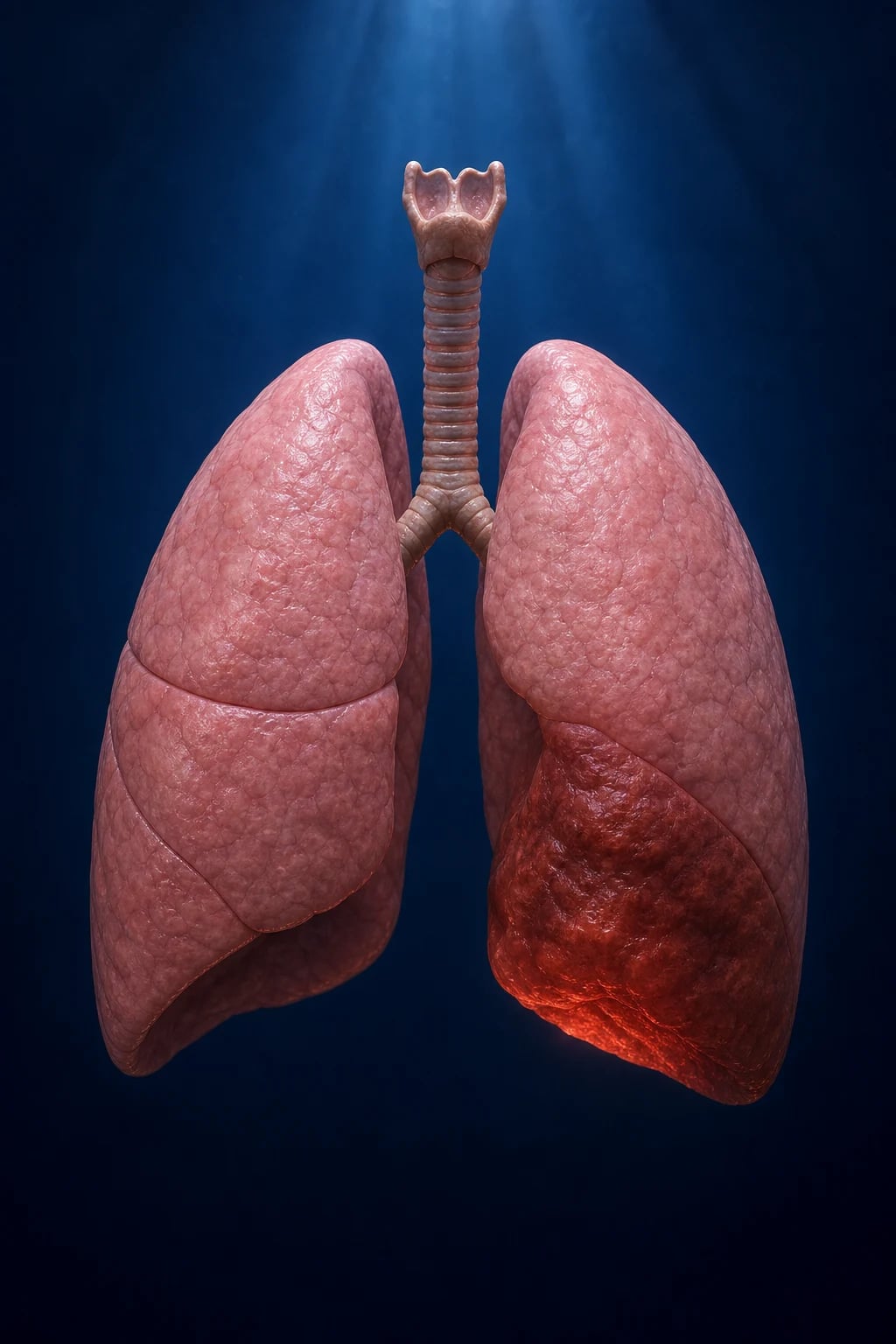
Pneumonia is an acute infection of the lung parenchyma — the alveoli, terminal bronchioles and the adjacent interstitium — that produces exudate and consolidation (usually visible on imaging) and impairs gas exchange.[2]
Community-acquired pneumonia (CAP) is pneumonia acquired outside hospital or developing within the first 48 hours of admission. The 48-hour rule is the operational boundary between CAP and hospital-acquired (nosocomial) pneumonia (HAP), because organisms causing early-onset inpatient pneumonia reflect the community flora and remain sensitive to standard empirical therapy, whereas pneumonia arising after 48 hours is driven by hospital flora (often multidrug-resistant Gram-negatives and Staphylococcus aureus) that demands broader cover. The distinction matters at the bedside: a patient who arrives at the emergency department septic and is found to have a new infiltrate has CAP and is treated with standard empirical cover, even if the formal microbiological confirmation comes back hours later. [1]
Three distinctions an examiner will probe:[2]
- Pneumonia versus acute bronchitis — bronchitis inflames the large airways, causes a cough (often productive) but produces no consolidation and no new infiltrate on chest X-ray; the patient is usually afebrile and not septic. Acute bronchitis is overwhelmingly viral and does not warrant antibiotics in the absence of specific indications. The chest X-ray is the decisive test.
- Pneumonia versus upper respiratory tract infection (URTI) — a URTI (common cold, pharyngitis) causes coryza, sore throat and malaise without dyspnoea, tachypnoea, focal chest signs or radiographic shadowing. A raised respiratory rate or new oxygen requirement points away from a URTI and towards a lower respiratory tract infection.
- Pneumonia versus atypical "walking pneumonia" — Mycoplasma and other atypicals cause a clinically milder, more insidious illness in which the patient looks better than the radiograph ("walking pneumonia"), but the parenchyma is genuinely infected. [1]
The clinical skill in CAP is not making the diagnosis (that is clinical plus chest X-ray) but recognising severity, choosing empirical antibiotics that cover typical AND atypical organisms, and remaining alert to viruses (COVID-19, influenza), aspiration, immunocompromised hosts, and complications (parapneumonic effusion, empyema, lung abscess, sepsis). The single largest process lever is early antibiotics — within 4 hours of admission for hospitalised patients, and within 1 hour if there is septic shock.[1][5]
Classification
By place of acquisition (the classification that drives empirical therapy):[1]
- Community-acquired (CAP) — onset outside hospital or within 48 hours of admission.
- Hospital-acquired (HAP) — onset 48 hours or more after admission, not incubating at admission. Caused by hospital flora (Gram-negative bacilli, S. aureus including MRSA).
- Ventilator-associated (VAP) — arising 48 hours or more after endotracheal intubation.
- (The older healthcare-associated pneumonia, HCAP, category was retired by ATS/IDSA 2019 because it did not reliably predict resistant organisms and led to over-use of broad-spectrum antibiotics.)[1]
By anatomy / radiology: [1]
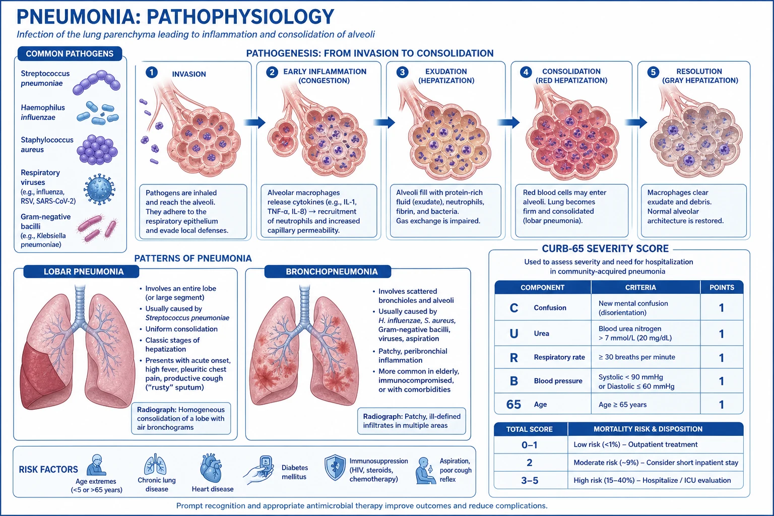
- Lobar pneumonia — consolidation confined to one lobe or segment; the typical bacterial pattern (S. pneumoniae, K. pneumoniae). Sharp lobar boundary, air bronchogram.
- Bronchopneumonia — patchy, peribronchial consolidation affecting multiple lobes bilaterally; S. aureus, H. influenzae, and terminal events.
- Interstitial pneumonia — inflammation of the interstitium rather than the alveolar space; atypicals and viruses; CXR shows diffuse reticular or reticulonodular shadowing. [1]
By aetiology — typical versus atypical (an examiner favourite):[2]
- Typical: Streptococcus pneumoniae (commonest), Haemophilus influenzae, Moraxella catarrhalis, Klebsiella pneumoniae, Staphylococcus aureus. Lobar consolidation, purulent or rust-coloured sputum, abrupt onset.
- Atypical: Mycoplasma pneumoniae, Chlamydophila pneumoniae/psittaci, Legionella pneumophila, Coxiella burnetii. Insidious, dry cough, prominent systemic features, interstitial CXR. [1]
By severity — see Investigations, where every named score (CURB-65, CRB-65, PSI/Pneumonia Severity Index, IDSA/ATS severe-CAP criteria) is reproduced verbatim.[3][4]
Typical (bacterial)
- S. pneumoniae, H. influenzae, Klebsiella, Staph aureus
- Abrupt onset, rigors, high fever
- Productive purulent or rust-coloured sputum
- Lobar consolidation on CXR
- Lobar exudate in alveolar space
- Responds to beta-lactams
- Organisms have a cell wall
- Blood cultures and sputum Gram stain useful
Atypical
- Mycoplasma, Chlamydophila, Legionella, Coxiella
- Insidious onset, low-grade fever
- Dry cough, headache, myalgia, sore throat
- Interstitial or patchy CXR, often looks worse than patient
- Organisms multiply in epithelium/interstitium or inside macrophages
- Need macrolide, tetracycline or fluoroquinolone
- Atypicals lack a cell wall or live inside cells
- Diagnosed by serology, urinary antigen or PCR
Epidemiology & Risk Factors
CAP is among the leading causes of infectious death worldwide, with the highest burden in the very young, the elderly, and the immunocompromised. The annual adult incidence in Europe and North America is around 5 to 11 per 1000 population, rising steeply with age to over 30 per 1000 in those over 75; in low- and middle-income countries the burden is substantially higher, driven by crowding, indoor biomass-fuel smoke, malnutrition, HIV and limited vaccination. There is a clear winter peak that overlaps with the influenza season, when secondary bacterial pneumonia (classically S. pneumoniae and S. aureus) follows viral infection.[1][2]
Streptococcus pneumoniae remains the single commonest identifiable pathogen in adults across all severity bands, accounting for roughly two-thirds of bacteraemic CAP, followed by the atypicals and respiratory viruses. In pathogens isolated from hospitalised adults, the order is roughly: S. pneumoniae, then H. influenzae and M. pneumoniae, Chlamydophila pneumoniae, Legionella, and the respiratory viruses (influenza, SARS-CoV-2, RSV). Importantly, in up to half of cases no organism is identified even with thorough microbiological work-up — which is exactly why empirical therapy must cover typical AND atypical organisms.[1]
Host and environmental risk factors: extremes of age, cigarette smoking (impairs mucociliary clearance and alveolar macrophage function), chronic lung disease (COPD, bronchiectasis, asthma), alcohol misuse (depresses macrophage function and raises aspiration risk), diabetes mellitus, chronic cardiac, renal or liver disease, immunocompromise (HIV, chemotherapy, splenectomy, hypogammaglobulinaemia, immunosuppressants including anti-TNF and corticosteroids), aspiration risk (stroke, dysphagia, reduced conscious level, seizures, anaesthesia, reflux), and — important in the developing world — malnutrition and indoor biomass-fuel smoke.[2]
Community-acquired pneumonia — epidemiology
Risk-factor to organism associations (high-yield for viva and MCQ): [1]
| Risk factor / host | Organism to consider |
|---|---|
| Young, otherwise well, school/college outbreak | Mycoplasma pneumoniae |
| COPD, bronchiectasis | Haemophilus influenzae, Moraxella catarrhalis, Pseudomonas |
| Alcoholism, diabetes | Klebsiella pneumoniae, S. pneumoniae |
| Post-influenza / post-viral | Staphylococcus aureus |
| Bird exposure (parrots, poultry) | Chlamydia psittaci (psittacosis) |
| Farm animals, parturient sheep/cattle/cats | Coxiella burnetii (Q fever) |
| Contaminated water, cooling towers, air-conditioning | Legionella pneumophila |
| Aspiration (stroke, seizures, alcohol) | Mixed oral flora including anaerobes |
| HIV or immunosuppressed, CD4 under 200 | Pneumocystis jirovecii |
| Structural lung disease, recent antibiotics, IV drug use | Pseudomonas aeruginosa, MRSA |
| Splenectomy, sickle cell, asplenia | Encapsulated organisms (S. pneumoniae, H. influenzae, N. meningitidis) — overwhelming post-splenectomy infection |
Pathophysiology
The lung distal to the vocal cords is normally sterile, defended by three layered mechanisms: the cough reflex (large-particle clearance), mucociliary clearance (the ascending mucus escalator of the airway epithelium, destroyed by smoking and viral injury), and alveolar macrophages (the final phagocytic sentinel). Pneumonia occurs when these defences are overwhelmed or bypassed.[2]
Routes by which pathogens reach the alveoli: [1]
- Microaspiration of oropharyngeal flora — the commonest mechanism in typical bacterial CAP. Even healthy people microaspirate during sleep; disease occurs when the inoculum is large or defences are impaired.
- Macroaspiration of gastric or oropharyngeal contents — stroke, reduced conscious level, seizures, anaesthesia, severe reflux.
- Inhalation of aerosolised droplets — Mycobacterium tuberculosis, Legionella, influenza, SARS-CoV-2, Histoplasma.
- Haematogenous spread — S. aureus from tricuspid endocarditis in injecting drug users; septic emboli.
- Direct extension — rarely, from a contiguous infection (empyema, mediastinitis) or following chest trauma. [1]
Once in the terminal airway, organisms multiply, triggering alveolar macrophage cytokine release (interleukin-1, tumour necrosis factor-alpha, interleukin-6). These cytokines recruit neutrophils and a protein-rich exudate into the alveolar space — producing consolidation. The exudate collapses alveolar units and floods gas-exchange surfaces, generating ventilation-perfusion mismatch and intrapulmonary shunt (perfused but unventilated alveoli) — the mechanism of hypoxaemia that does not correct with supplemental oxygen as readily as hypoxaemia from pure V/Q mismatch. The same cytokines spill into the systemic circulation, producing fever, tachycardia, tachypnoea and the systemic inflammatory response. Resolution follows as macrophages clear the exudate and type II pneumocytes regenerate surfactant.[2]
Why the hypoxaemia of consolidation is partly a shunt (examiner point): consolidated alveoli are perfused but not ventilated — blood traverses them without being oxygenated, a true right-to-left intrapulmonary shunt. Because shunted blood bypasses the gas-exchange surface entirely, raising the inspired oxygen only marginally improves arterial oxygenation (the oxygenated blood cannot "carry" the shunted blood's deficit). This contrasts with pure V/Q mismatch (e.g. mild COPD), which corrects well with supplemental oxygen. The widened alveolar-arterial (A-a) oxygen gradient is the bedside signature of shunt and pneumonia. [1]
Why "typical" and "atypical" differ mechanistically: typical organisms multiply extracellularly in the alveolar space (the pneumococcal polysaccharide capsule resists phagocytosis), generating a dense neutrophil-rich lobar exudate and a productive cough. Atypicals proliferate intracellularly or along the respiratory epithelium and interstitium — Mycoplasma adheres via the P1 adhesin to respiratory epithelium, Legionella multiplies inside alveolar macrophages (inhibiting phagolysosome fusion) and Chlamydophila replicates inside epithelial cells. The result is a less exudative, more interstitial process with disproportionate systemic upset and a non-productive cough. Because atypicals lack a cell wall (Mycoplasma) or hide inside cells (Legionella, Chlamydophila), beta-lactams are ineffective — a macrolide, tetracycline or respiratory fluoroquinolone is required. [1]
The four classic stages of lobar pneumonia (histological, now largely of historical/exam interest): (1) Congestion (first 24 hours) — vascular engorgement and intra-alveolar oedema; (2) Red hepatization (days 2 to 3) — exudate rich in erythrocytes and fibrin, lung resembles liver; (3) Grey hepatization (days 4 to 6) — fibrin and neutrophils dominate, grey-brown solid lung; (4) Resolution (after day 7) — enzymatic digestion of fibrin, macrophage clearance, restoration of normal architecture (no scar, unless necrosis occurred). Incomplete resolution may progress to organisation (fibroblast ingrowth) or suppuration (abscess formation).[2]
A normal blood pressure does not exclude severe illness or early sepsis — compensatory vasoconstriction maintains the pressure until late. Assess perfusion (capillary refill, lactate, urine output, conscious level), not the pressure alone.[2]
Clinical Presentation
Typical (bacterial) CAP — abrupt onset: fever with rigors, purulent or rust-coloured sputum (pneumococcus), dyspnoea and pleuritic chest pain (from pleural inflammation), tachypnoea. Examination reveals signs of consolidation (see below).[2]
Atypical CAP — insidious onset over days: dry cough, prominent headache, myalgia, fatigue, sore throat and gastrointestinal symptoms, low-grade fever, and few chest signs despite a symptomatic patient. The chest X-ray frequently looks worse than the patient (or, less often, the patient worse than the X-ray). [1]
Symptom-by-symptom, with the mechanism an examiner wants: the cough is the host attempt to clear the exudate; rust-coloured sputum is pneumococcal haemorrhagic exudate; pleuritic pain reflects inflammation of the parietal pleura (the visceral pleura has no pain fibres); dyspnoea and tachypnoea arise from hypoxaemia (shunt), stimulation of juxtacapillary (J) receptors by interstitial inflammation, and the metabolic acidosis of sepsis; rigors mark the abrupt cytokine-driven upward reset of the hypothalamic temperature set-point; confusion in the elderly reflects hypoxaemia, dehydration, sepsis-related delirium, or a combination. A raised respiratory rate is the single most sensitive sign of a lower respiratory tract infection and is the first to climb and the last to normalise.[2]
Organism-specific pointers (examiner favourites): [1]
- Legionella pneumophila — diarrhoea, abdominal pain, hyponatraemia, confusion, severe presentation; source is water systems, cooling towers, air-conditioning, spa pools; diagnose with urinary antigen (detects serogroup 1); also causes a mild Pontiac fever.
- Mycoplasma pneumoniae — young adults, dry cough, headache; extrapulmonary features are the exam twist — erythema multiforme / Stevens-Johnson syndrome, cold-agglutinin autoimmune haemolytic anaemia, Guillain-Barre syndrome, myocarditis, arthralgia, rash.
- Klebsiella pneumoniae (Friedlander) — alcoholics and diabetics; thick red-currant-jelly sputum; upper-lobe predilection with a bulging fissure; cavitates.
- Staphylococcus aureus — post-influenza (rapidly progressive, bilateral), injecting drug users (septic emboli from tricuspid endocarditis); causes cavitation and, in children, pneumatoceles.
- Chlamydia psittaci — psittacosis from bird (parrot, pigeon) exposure; splenomegaly, relative bradycardia.
- Coxiella burnetii — Q fever from farm animals; hepatitis and endocarditis.
- Viral — influenza, COVID-19 (often with anosmia/ageusia, bilateral ground-glass), RSV (elderly, infants).
- Pneumocystis jirovecii — HIV with CD4 under 200 cells/microL; insidious dyspnoea, dry cough, marked exertional desaturation, bilateral perihilar interstitial infiltrates, raised serum LDH. [1]
Which organism — rusty sputum, post-influenza, red-currant-jelly, bird exposure, CD4 under 200?
Rust-coloured / blood-tinged sputum = S. pneumoniae (the classic). Red-currant-jelly = Klebsiella pneumoniae (alcoholic, diabetic). Post-influenza, rapidly progressive, cavitation = Staphylococcus aureus. Bird exposure, splenomegaly, relative bradycardia = Chlamydia psittaci (psittacosis). HIV, CD4 under 200, raised LDH, exertional desaturation = Pneumocystis jirovecii. Cooling tower, diarrhoea, hyponatraemia = Legionella.
Atypical presentation in the elderly: confusion, falls, functional decline, anorexia, new incontinence — fever and cough may be absent. A lower threshold to admit, investigate and treat is essential. New-onset atrial fibrillation in an older patient with breathlessness is a classic CAP presentation. The immunocompromised may present with subtle dyspnoea and a normal examination but extensive radiographic disease — Pneumocystis classically causes marked exertional desaturation disproportionate to resting findings. [1]
Differential Diagnosis
An acute febrile illness with lung shadowing is not always CAP. Work through each with its distinguishing features:[2]
- Pulmonary embolism (PE) — pleuritic pain and dyspnoea out of proportion to signs; risk factors (immobility, malignancy, recent surgery, pregnancy); usually no fever or septic picture; wedge-shaped peripheral infarct or normal CXR; confirmed by CT pulmonary angiogram and raised D-dimer. CAP and PE co-exist more often than chance — keep both on the list.
- Pulmonary oedema (cardiogenic) — bilateral peri-hilar bat-wing shadowing, Kerley B lines, cardiomegaly, history of cardiac disease, elevated NT-proBNP, rapid response to diuretics; fever absent.
- Tuberculosis — subacute or chronic course, night sweats, weight loss, haemoptysis; upper-lobe infiltrate with cavitation; sputum acid-fast bacilli / GeneXpert / mantoux / IGRA positive; high-risk epidemiology.
- Lung cancer — post-obstructive pneumonia that is slow to resolve or recurs in the same lobe; weight loss, smoker over 50; bronchoscopy and follow-up imaging.
- Atelectasis / lobar collapse — volume loss signs (shifted trachea/mediastinum toward the lesion, raised hemidiaphragm); no fever.
- Acute bronchitis — cough with no consolidation and a normal chest X-ray; usually viral; no antibiotics unless specific indication.
- Viral pneumonitis / COVID-19 — bilateral ground-glass opacities, lymphopenia, viral prodrome; isolate and test.
- Interstitial lung disease acute exacerbation or organising pneumonia — subacute, characteristic HRCT pattern, sterile cultures.
- Pulmonary vasculitis (e.g., granulomatosis with polyangiitis) — cavitating nodules, haemoptysis, sinusitis, renal involvement.
- Aspiration pneumonitis (Mendelson) — chemical injury from sterile gastric contents, often follows anaesthesia or reduced consciousness; resolves over 24 to 48 hours unless secondarily infected. [1]
Always specifically consider TB and PE when the presentation, epidemiology or radiology is not a straightforward CAP.[2]
Clinical & Bedside Assessment
Vital signs drive severity — measure and record respiratory rate, oxygen saturation, blood pressure, temperature, heart rate, conscious level (GCS or confusion screen) and urine output. The single most sensitive marker of a lower respiratory tract infection is a raised respiratory rate, and it is the first to climb and the last to normalise — yet it is the vital sign most often omitted or guessed.[2]
Signs of consolidation on focused respiratory examination: reduced chest expansion on the affected side, dullness to percussion over solidified lung, bronchial (tubular) breath sounds, fine inspiratory crackles, increased vocal resonance and tactile fremitus, and a possible pleural rub. A silent hemithorax with dullness suggests a pleural effusion or complete collapse. [1]
Always assess for sepsis at the bedside using qSOFA (any two is concerning): respiratory rate 22 or more, altered mentation, systolic blood pressure 100 mmHg or less. Examine for complications — a parapneumonic effusion (stony dull, absent breath sounds), empyema (swinging fever with a dull effusion), and perioral cyanosis indicating hypoxaemia. Check for appendix-signs of metastatic infection (new murmur in endocarditis, neck stiffness, joint swelling) when bacteraemia is suspected. [1]
Signs of consolidation — POSTER
POSTER
Dullness to percussion over consolidated lung
Reduced chest wall movement on the affected side
Tubular bronchial breath sounds over the consolidation
Increased vocal resonance and tactile fremitus
Fine late inspiratory crackles
A pleural friction rub if the pleura is inflamed
Why hypoxaemia — and what to do
DOPES
Check the oxygen device is delivering flow and fits
Sputum plugging — consider suction, physiotherapy
Progressive consolidation or effusion — re-image
Sample any new pleural collection
True shunt does not correct with oxygen — escalate to ICU
Investigations
First-line investigations in admitted CAP:[1]
- Chest X-ray — mandatory to confirm consolidation; assess pattern (lobar, interstitial, multilobar, cavitation, effusion, pneumatocele). (A normal CXR does not fully exclude early pneumonia — if clinical suspicion is high, repeat at 24 to 48 hours or use ultrasound/CT.)
- Full blood count — leukocytosis (neutrophilia) is typical; leukopenia is a severe-CAP minor criterion.
- CRP and procalcitonin — elevated; procalcitonin can guide antibiotic initiation and duration (bacterial infection marker).
- Urea and electrolytes, creatinine — urea over 7 mmol/L is a CURB-65 point and reflects dehydration and severity; hyponatraemia points to Legionella or the syndrome of inappropriate antidiuretic hormone (SIADH).
- Liver function tests, glucose, albumin — low albumin predicts mortality.
- Arterial or venous blood gas — assess hypoxaemia, hypercapnia and acid-base (type 1 respiratory failure, metabolic acidosis in sepsis); lactate for tissue hypoperfusion.
- Blood cultures — drawn before antibiotics; positive in roughly 10 to 20 percent of hospitalised CAP (bacteraemia), S. pneumoniae most common.
- Sputum Gram stain and culture — in severe disease or with risk factors; a good sample has few epithelial cells and many neutrophils.
- Pneumococcal and Legionella urinary antigens — rapid, sensitive and specimen-independent; Legionella antigen detects serogroup 1 (the bulk of clinical disease).
- Respiratory viral PCR (including SARS-CoV-2, influenza A and B, RSV) — when viral CAP is suspected.
- Atypical serology / PCR (Mycoplasma, Chlamydophila) — paired sera (four-fold rise in IgG) or respiratory PCR; cold agglutinins (IgM anti-I) in Mycoplasma.
- HIV test — tuberculosis and Pneumocystis are AIDS-defining; CAP is an indication to test.
- ECG — arrhythmia (new atrial fibrillation) is common in the elderly. [1]
Chest X-ray patterns and what they imply: [1]
| CXR pattern | Implication |
|---|---|
| Lobar consolidation with air bronchogram | Typical bacterial — S. pneumoniae, Klebsiella |
| Bilateral patchy bronchopneumonia | S. aureus, H. influenzae, severe CAP |
| Interstitial reticulonodular shadowing | Atypicals, viruses, Pneumocystis |
| Cavitation | S. aureus, Klebsiella, anaerobes, tuberculosis, fungus |
| Pneumatocele (thin-walled cavity) | S. aureus (especially children) |
| Upper-lobe with bulging fissure | Klebsiella |
| Pleural effusion | Parapneumonic — sample it |
| Bilateral ground-glass | Viral pneumonitis / COVID-19 / Pneumocystis |
When and why each microbiological test: blood cultures and sputum are reserved for severe CAP or specific risk factors (ATS/IDSA 2019), not mild disease. The pneumococcal urinary antigen is rapid and roughly 70 to 90 percent sensitive in bacteraemic pneumococcal pneumonia and unaffected by prior antibiotics; the Legionella urinary antigen is highly specific for serogroup 1 (the cause of most sporadic and outbreak disease) but misses non-serogroup-1 strains. Procalcitonin rises with bacterial infection and can support both the decision to start antibiotics and the decision to stop them early; a low procalcitonin does not, however, exclude bacterial CAP in a clinically ill patient.[1][5]
Severity scores — reproduced verbatim
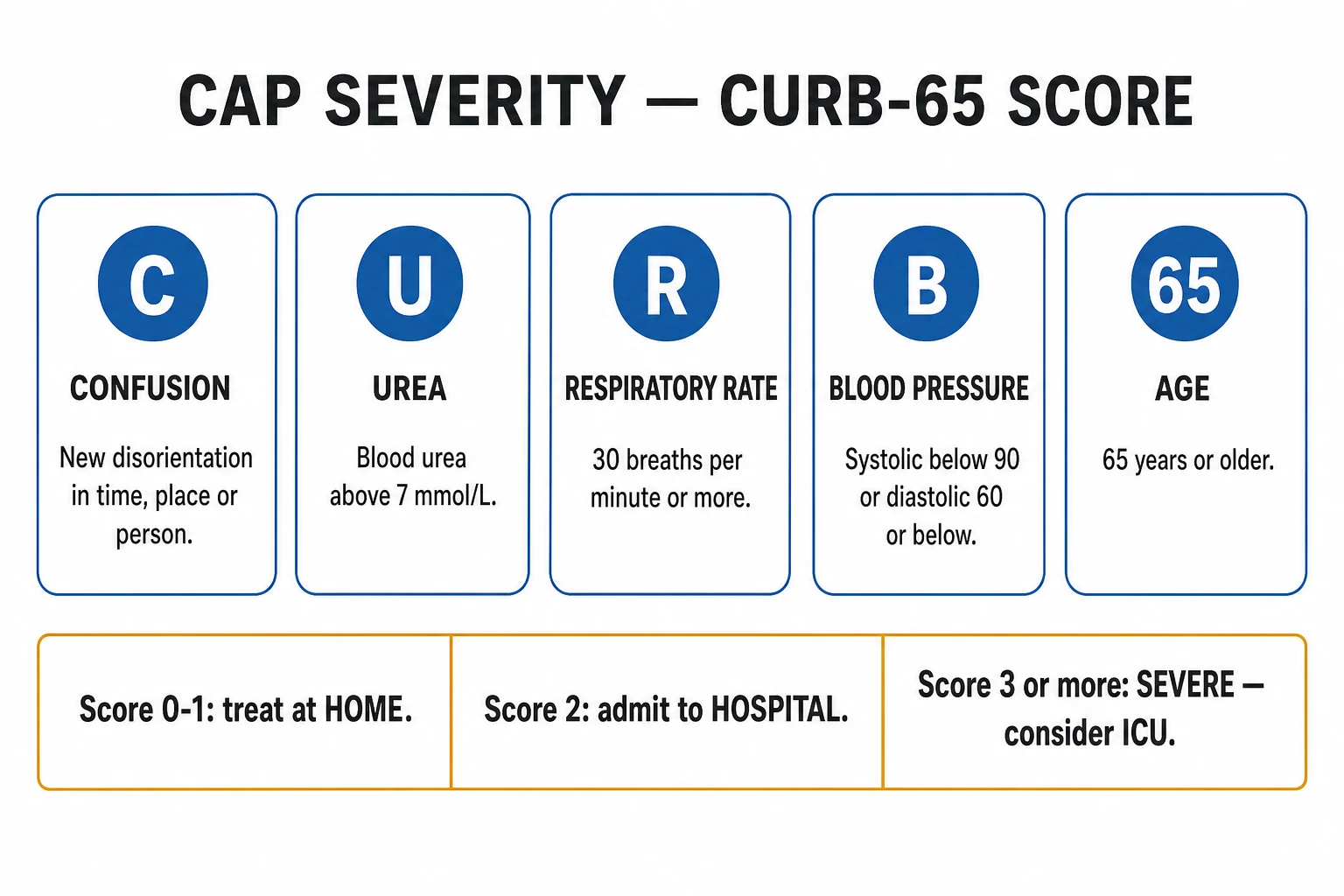
CURB-65 (one point each, maximum 5):[3][6]
- C — Confusion (new disorientation in time, place or person, using an abbreviated mental test score of 8 or less).
- U — Blood Urea nitrogen over 7 mmol/L (20 mg/dL).
- R — Respiratory Rate 30 breaths/min or more.
- B — Blood pressure: systolic under 90 mmHg OR diastolic 60 mmHg or less.
- 65 — age 65 years or over. [1]
CURB-65 severity and 30-day mortality
2
Hospital admission
Disposition and approximate 30-day mortality by CURB-65 score:[3]
| Score | Mortality (approx) | Disposition |
|---|---|---|
| 0 | 0.7 percent | Home treatment suitable |
| 1 | 2 to 3 percent | Home treatment usually suitable (consider comorbidity) |
| 2 | around 9 percent | Hospital admission |
| 3 | around 15 percent | Hospital; consider ICU |
| 4 | around 40 percent | ICU |
| 5 | around 57 percent | ICU |
(Where urea is unavailable, CRB-65 uses the same four clinical variables and adds nothing for urea — a CRB-65 of 0 may be suitable for home treatment, while a score of 1 or more warrants hospital assessment.)[3]
Pneumonia Severity Index (PSI / PORT)[4] — a 20-variable model (demographics, comorbidity, examination and laboratory findings) that stratifies patients into classes I to V. It is more accurate than CURB-65 at identifying low-risk patients safe for outpatient care, but it is slower and harder to use at the bedside because it requires laboratory results and arithmetic. Use PSI to confirm low risk; use CURB-65 for rapid bedside triage and to trigger the sepsis pathway.
IDSA/ATS 2019 severe-CAP criteria (define need for ICU admission):[1]
- At least 1 major criterion — invasive mechanical ventilation, OR septic shock requiring vasopressors.
- OR at least 3 of 9 minor criteria — respiratory rate 30 or more, PaO2/FiO2 ratio under 250, multilobar infiltrates, confusion/disorientation, blood urea nitrogen 20 mg/dL or more (urea over 7 mmol/L), white-cell count under 4 x 10^9/L, platelet count under 100 x 10^9/L, hypothermia (core temperature under 36 degrees C), hypotension requiring aggressive fluid resuscitation. [1]
Community-acquired pneumonia — key numbers
Management — Resuscitation
ABCDE assessment first. Secure the airway, give high-flow oxygen to a target SpO2 of 94 to 98 percent for previously well adults (or 88 to 92 percent in COPD or any patient at risk of hypercapnic respiratory failure). Escalate the oxygen delivery device by need: nasal cannula (low-flow, 24 to 40 percent), simple face mask (40 to 60 percent), non-rebreather reservoir mask (60 to 90 percent at 15 L/min), and high-flow nasal cannula or continuous positive airway pressure (CPAP) for refractory hypoxaemia. Re-check the arterial blood gas after 30 to 60 minutes.[1]
Apply the Surviving Sepsis Campaign hour-1 bundle to septic CAP (suspected sepsis or septic shock): measure lactate, draw blood cultures before antibiotics, give broad-spectrum antibiotics within 1 hour, give balanced crystalloid 30 mL/kg for hypotension or lactate 4 mmol/L or more, and start noradrenaline for fluid-refractory shock. Reassess fluid responsiveness (passive leg raise, IVC variability, pulse-pressure variation) before further boluses to avoid pulmonary oedema. The UK Sepsis Six is the bedside mnemonic: give oxygen, take blood cultures, give IV antibiotics, give IV fluids, measure lactate and a hourly urine output, and give high-flow oxygen.[1]
Give antibiotics within 4 hours of arrival for all hospitalised CAP (within 1 hour if septic shock) — do not delay antibiotics for investigations. A sputum sample or blood culture drawn a few minutes before the first dose is ideal, but a delay to obtain samples in a septic patient is not justified.[5]
Immediate management of severe CAP with sepsis
Airway, breathing: high-flow oxygen to target SpO2 94 to 98 percent (88 to 92 percent in COPD); assess work of breathing and ABG
Circulation: IV access; blood cultures before antibiotics; balanced crystalloid 30 mL/kg bolus if hypotensive or lactate 4 mmol/L or more; noradrenaline if fluid-refractory
Draw labs: FBC, CRP, U&E, LFTs, lactate, coagulation; urinary pneumococcal and Legionella antigens; respiratory viral PCR
Empirical IV antibiotics within 1 hour: co-amoxiclav (or ceftriaxone) PLUS a macrolide
Reassess at 1 hour: perfusion, lactate trend, MAP target 65 mmHg, urine output over 0.5 mL/kg/h
Escalate to ICU for invasive ventilation, vasopressor-resistant shock, or ATS/IDSA severe-CAP criteria
Management — Definitive & Stepwise
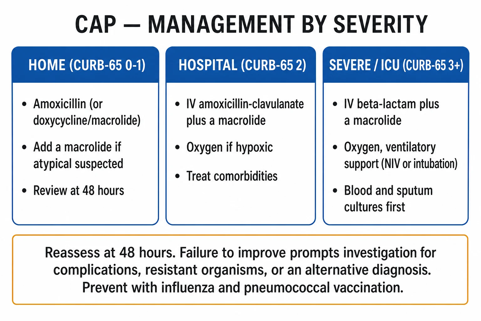
Empirical antibiotics are severity- and setting-driven and must cover both typical and atypical organisms in every hospitalised patient.[1][5]
[1]CAP antibiotic ladders at a glance
Duration of therapy: 5 to 7 days in the patient who is clinically improving and afebrile for 48 to 72 hours. Use longer courses for S. aureus pneumonia, Pseudomonas, complicated or meticillin-resistant infection, abscess, and Legionella (14 days); immunocompromised hosts are treated individually.[1]
IV-to-oral switch (step-down): once the patient is haemodynamically stable, improving clinically, afebrile, and able to swallow and absorb oral medication — typically within 2 to 4 days. Switch to the oral equivalent of the IV agent or a bioequivalent oral agent (e.g., IV amoxicillin to oral amoxicillin, IV clarithromycin to oral clarithromycin).[1]
Discharge criteria: the patient is clinically stable, afebrile for 24 to 48 hours, tolerating oral intake and oral antibiotics, with a safe social situation — and is given a safety-net to review or re-present if not improving within 48 hours. Arrange a follow-up chest X-ray at 6 to 8 weeks for persistent symptoms, smokers over 50, or any non-resolving opacity to exclude underlying malignancy.[1]
Adjunctive corticosteroids: a short course (e.g., hydrocortisone 200 mg/day IV or prednisolone 50 mg daily for 5 to 7 days) may modestly reduce mortality and time-to-clinical-stability in severe CAP with sepsis or septic shock; the benefit is small and the ATS/IDSA 2019 guideline does not recommend routine use — reserve for refractory septic shock or severe inflammation.[5]
Supportive care: venous thromboembolism prophylaxis (low-molecular-weight heparin) for immobile hospitalised patients; physiotherapy and mucolytics have limited evidence but assisted sputum clearance helps the weak or post-operative patient; glycaemic control (insulin sliding scale if needed); nutritional support; review of chronic medications; and reduction of aspiration risk (head-up positioning, swallow assessment) in the vulnerable elderly. [1]
Specific Subtypes & Scenarios
- Aspiration pneumonia — occurs in dependent segments: the posterior segments of the upper lobes and the apical segments of the lower lobes in the recumbent patient, and the right lower lobe in the upright patient (right main bronchus is wider, shorter and more vertical). Caused by mixed oral flora including anaerobes (Bacteroides, Prevotella, Fusobacterium, peptostreptococci). Cover with co-amoxiclav (or clindamycin plus a cephalosporin); address the underlying swallow, reflux or reduced conscious level. Foul-smelling sputum and anaerobic breath are classic.
- Atypical pneumonias — Mycoplasma (young, dry cough, cold agglutinins, extrapulmonary features; macrolide or doxycycline); Legionella (GI symptoms, hyponatraemia, confusion, severe; urinary antigen; macrolide or fluoroquinolone for 14 days); Chlamydia psittaci (bird exposure; tetracycline); Coxiella burnetii (Q fever, farm animals; tetracycline).
- Viral CAP / COVID-19 — influenza (give oseltamivir early during flu season), SARS-CoV-2 (bilateral ground-glass, lymphopenia; antivirals and immunomodulation per current guidance), RSV. Isolate and test; consider secondary bacterial superinfection (S. aureus, S. pneumoniae).
- Pneumocystis jirovecii pneumonia (PCP) — HIV with CD4 under 200; insidious dyspnoea, dry cough, exertional desaturation, raised LDH, bilateral perihilar interstitial infiltrates; treat with high-dose co-trimoxazole (trimethoprim-sulfamethoxazole, 120 mg/kg/day in divided doses) for 21 days, PLUS corticosteroids (prednisolone) if hypoxic (PaO2 under 70 mmHg).
- Lung abscess — a localised collection of pus in a cavity, often from aspiration, S. aureus, Klebsiella or anaerobes; air-fluid level on imaging; prolonged antibiotics (4 to 6 weeks), postural drainage, occasionally surgical resection. [1]
Complications & Pitfalls
Local: parapneumonic effusion, empyema (pus in the pleural space), lung abscess, necrotising pneumonia, pneumatocele (especially children with S. aureus), bronchopleural fistula.[2]
Systemic: sepsis and septic shock, acute respiratory distress syndrome (ARDS), acute kidney injury, metastatic infection (endocarditis, meningitis, septic arthritis, pericarditis), new atrial fibrillation (elderly), hyponatraemia (SIADH or adrenal in severe disease), disseminated intravascular coagulation, and multi-organ failure. The acute respiratory distress syndrome is defined by the Berlin criteria — acute onset within 1 week, bilateral opacities not fully explained by effusion or atelectasis, respiratory failure not fully explained by cardiac failure or fluid overload, and PaO2/FiO2 ratio under 300 mmHg with a PEEP of 5 cmH2O or more (mild under 300, moderate under 200, severe under 100).[2]
Parapneumonic effusion — classify exudate versus transudate with Light's criteria (pleural-fluid/serum protein ratio over 0.5, LDH ratio over 0.6, or fluid LDH over two-thirds the upper limit of normal for serum — any one makes it an exudate). Drain (chest tube) if complicated — i.e., purulent, or pH under 7.2, glucose under 2.2 mmol/L (40 mg/dL), LDH over 1000 IU/L, or positive culture or Gram stain. A loculated or large collection needs surgical drainage (video-assisted thoracoscopic surgery, VATS), and a delayed empyema may need surgical decortication.[2]
Parapneumonic effusion — the natural history
Classic pitfalls: under-treating severe CAP (missed ICU need or missed atypical cover); not covering atypicals in hospitalised patients; failing to consider TB or PE in the atypical case; delaying antibiotics for tests; over-investigating mild disease; missing bacteraemia (no repeat blood cultures); forgetting the follow-up CXR that unmasks an underlying lung cancer; and giving uncontrolled oxygen to a COPD patient precipitating CO2 narcosis. [1]
Approach to non-resolving (slowly resolving) pneumonia — a common and examiner-tested scenario. Pneumonia is expected to show radiographic improvement within 4 weeks and complete resolution by 8 to 12 weeks; failure to improve demands a structured re-think. Work through five questions:[2]
- Wrong diagnosis — is it TB, fungal infection, PE with infarct, organising pneumonia, vasculitis, or pulmonary oedema masquerading as CAP? Re-image with HRCT and re-sample.
- Wrong organism / wrong antibiotic — atypical not covered (no macrolide), Legionella, Mycoplasma, TB, or a resistant organism (MRSA, Pseudomonas); obtain sputum AFB / GeneXpert, atypical serology, and review the antibiogram.
- Impaired host defence — undiagnosed HIV, immunosuppression, diabetes, malignancy; test HIV and check immunoglobulins.
- Complication — parapneumonic effusion, empyema, lung abscess, bronchopleural fistula, ARDS; ultrasound and sample any pleural collection.
- Underlying structural lesion — post-obstructive lung cancer, inhaled foreign body, bronchiectasis; arrange bronchoscopy and a follow-up CXR at 6 to 8 weeks, pursuing any non-resolving opacity to exclude malignancy. [1]
The single highest-yield action in the older smoker with non-resolving pneumonia is bronchoscopy to exclude an obstructing lung cancer. [1]
Prognosis & Disposition
CURB-65 correlates with 30-day mortality (0 to 1 low, 2 moderate, 3 to 5 high) and drives disposition (home, ward, ICU). Overall 30-day mortality is 5 to 15 percent in hospitalised CAP and rises sharply with severity — from under 1 percent at CURB-65 0 to over 50 percent at CURB-65 5. Poor-outcome predictors include older age, comorbidity, multilobar disease, hypoxaemia, hyponatraemia, hypoalbuminaemia, bacteraemia, and septic shock.[3][6]
The PSI is better than CURB-65 at identifying genuinely low-risk patients safe for outpatient care (classes I and II), whereas CURB-65 is the better bedside triage tool for spotting severe disease and triggering the sepsis pathway. No score replaces clinical judgement — a socially isolated or frail patient, or one unable to take oral medication, may warrant admission even with a low score. [1]
Follow-up chest X-ray at 6 to 8 weeks is indicated for persistent symptoms, smokers over 50, and any non-resolving opacity — to exclude an underlying malignancy (post-obstructive pneumonia). A patient whose pneumonia "never quite resolved" may have a tumour behind it.[1]
Special Populations
- Elderly — blunted or atypical presentation (confusion, falls, anorexia); lower threshold to admit; beware aspiration, delirium and new atrial fibrillation; assess frailty and social support before discharge.
- Pregnancy — physiological immunosuppression of pregnancy (decreased cell-mediated immunity) and reduced functional residual capacity increase severity; influenza and varicella pneumonia are particularly dangerous. Treat promptly and fully; do not withhold beta-lactams (amoxicillin, ceftriaxone are safe); avoid tetracyclines and fluoroquinolones (fetal toxicity — tetracyclines discolour fetal teeth and bone; fluoroquinolones damage developing cartilage).
- Immunocompromised (HIV, transplant, chemotherapy, neutropenia) — broader organism list (Pneumocystis, CMV, aspergillus, Mycobacterium avium, Pseudomonas, MRSA); lower threshold to image with HRCT, perform bronchoalveolar lavage, and involve microbiology and infectious diseases early.
- Asplenic or hyposplenic (sickle cell, post-splenectomy, coeliac) — overwhelming post-splenectomy infection with encapsulated organisms; treat empirically for S. pneumoniae and give pneumococcal, meningococcal and Haemophilus vaccination plus lifelong penicillin prophylaxis (penicillin V 500 mg twice daily).
- COPD — H. influenzae, Moraxella, Pseudomonas; use co-amoxiclav or a cephalosporin and a macrolide; give controlled-oxygen (target 88 to 92 percent) to avoid CO2 narcosis; check a blood gas early.
- Children (weight-based) — high-risk under 5 years; S. pneumoniae, viruses (RSV), H. influenzae type b, S. aureus (pneumatoceles); first-line oral amoxicillin (or IV benzylpenicillin / ceftriaxone if severe); beware dehydration and rapid progression. Mycoplasma is common over 5 years of age. [1]
Evidence, Guidelines & Regional Differences
Key changes in the ATS/IDSA 2019 guideline:[1][5]
- Empirical therapy must cover atypical organisms in all hospitalised CAP (beta-lactam plus macrolide, OR a respiratory fluoroquinolone).
- HCAP was retired — do not automatically broaden therapy for "healthcare-associated" exposure; instead use validated risk factors for resistant pathogens (prior respiratory isolation of Pseudomonas, recent IV antibiotics, recent hospitalisation) to decide.
- Routine blood cultures and sputum culture are reserved for severe CAP or specific risk factors (not mild disease).
- Corticosteroids are NOT recommended routinely.
- Procalcitonin may help with antibiotic initiation and cessation decisions.
- Radiographic follow-up CXR at 6 to 8 weeks only if symptoms persist or for smokers over 50. [1]
Adjunctive corticosteroids in CAP — what the trials show
Pooled meta-analysis and CAPE COD / CAPO subgroup data
Population: Adults with severe CAP, with sepsis or septic shock
Key finding
Modest reduction in time-to-clinical-stability and, in the sickest subgroups (septic shock), a small mortality benefit
Prevention — vaccination (the intervention examiners reward):[1]
- Pneumococcal vaccination — conjugate vaccines (PCV13, PCV15, PCV20) and/or polysaccharide vaccine (PPSV23) per the age- and risk-based schedule; PCV20 simplifies the schedule.
- Annual influenza vaccination — reduces influenza and secondary bacterial pneumonia.
- COVID-19 vaccination — reduces severe COVID-19 pneumonitis.
- Smoking cessation, optimisation of chronic disease, oral hygiene (reduces aspiration pneumonia in care-home residents), and Hib and pertussis immunisation in children. [1]
Which vaccine schedule for a 70-year-old with COPD?
Give pneumococcal vaccination (PCV15 or PCV20, with PPSV23 a year later if PCV15 is used and not previously given), annual influenza vaccine, and COVID-19 vaccination as the core preventive bundle. Smoking cessation is the single most modifiable host risk factor.
Exam Pearls
- CURB-65: Confusion, Urea over 7, RR 30 or more, BP under 90/60, age 65 or more. Score 0 to 1 home, 2 hospital, 3 or more ICU.[3]
- S. pneumoniae is the commonest CAP organism; rust-coloured sputum.[2]
- Red-currant-jelly sputum = Klebsiella; foul-smelling = anaerobes/aspiration.
- Post-influenza cavitating pneumonia = Staph aureus.
- Legionella: GI symptoms, hyponatraemia, urinary antigen, intracellular — macrolide or fluoroquinolone for 14 days.
- Mycoplasma: cold agglutinins, erythema multiforme, Guillain-Barre; no cell wall so beta-lactams fail.
- Antibiotics within 4 hours; beta-lactam plus macrolide for admitted patients.
- Atypicals lack a cell wall (Mycoplasma) or hide inside cells (Legionella, Chlamydophila) — beta-lactams are ineffective.
- Drain parapneumonic effusion if pH under 7.2, glucose under 2.2 mmol/L, LDH over 1000, or purulent.
- Follow-up CXR at 6 to 8 weeks to exclude lung cancer in persistent symptoms or smokers over 50.[1]
- PSI is better than CURB-65 for identifying low-risk patients; CURB-65 is the better bedside triage tool.[4]
- Vaccinate: pneumococcal, influenza, COVID-19.
- Oxygen target 94 to 98 percent; 88 to 92 percent in COPD to avoid CO2 narcosis.
- Give antibiotics within 1 hour in septic shock (Surviving Sepsis hour-1 bundle).[5]
Exam application bank (NEET-PG / INICET)
One-line answer
Community-acquired pneumonia (CAP) is an acute infection of the lung parenchyma acquired outside hospital (or within the first 48 hours of admission). The commonest pathogen is Streptococcus pneumoniae; atypicals (Mycoplasma pneumoniae, Chlamydophila pneumoniae/psittaci, Legionella pneumophila), respiratory viruses (influenza, SARS-CoV-2, RSV), Haemophilus influenzae, Moraxella catarrhalis, and in selected hosts Staphylococcus aureus, Klebsiella pneumoniae and anaerobes. Typical CAP presents abruptly with fever, productive or rust-coloured sputum, dyspnoea, pleuritic chest pain and signs of consolidation; atypical CAP is insidious with a dry cough and prominent systemic features. Diagnosis is clinical plus chest X-ray; severity is graded with CURB-65 (Confusion, Urea over 7, RR 30 or more, systolic BP under 90 or diastolic 60 or less, age 65 or more). Treat with empirical antibiotics wit
Worked stems (answer without another resource)
Stem 1 — Classic presentation. Map symptoms to mechanism; name the first investigation and first treatment step with dose/route if drug therapy is standard. [1]
Stem 2 — Unstable / complicated. List red flags that force immediate resuscitation, theatre, ICU, antidote, or reperfusion — and what you do in the first 15 minutes. [1]
Stem 3 — Atypical group. Elderly, pregnancy, child, or immunocompromised: how presentation and thresholds change. [1]
Stem 4 — Differential trap. Name the three closest mimics and one discriminator for each. [1]
Stem 5 — Disposition. Who goes home with safety-netting, who is admitted, who needs HDU/ICU/theatre, and what follow-up is mandatory. [1]
Rapid viva checklist
- Definition + classification
- Pathophysiology chain
- Bedside signs / criteria
- Score with exact components (if any)
- Emergency bundle
- Definitive therapy with doses
- Complications of disease and of treatment
- Special populations
- Guideline/trial name if classic
- Three exam traps
Coverage self-check
If you cannot answer any stem above from this page alone, re-read the matching section — the page is intended to be self-sufficient for final-prof and NEET-PG/INICET questions on Community-Acquired Pneumonia.
References
- [1]Metlay JP, Waterer GW, Long AC, et al. Diagnosis and Treatment of Adults with Community-acquired Pneumonia. An Official Clinical Practice Guideline of the American Thoracic Society and Infectious Diseases Society of America Am J Respir Crit Care Med, 2019.PMID 31573350
- [2]Prina E, Ranzani OT, Torres A. Community-acquired pneumonia Lancet, 2015.PMID 26277247
- [3]Lim WS, van der Eerden MM, Laing R, et al. Defining community acquired pneumonia severity on presentation to hospital: an international derivation and validation study Thorax, 2003.PMID 12728155
- [4]Fine MJ, Auble TE, Yealy DM, et al. A prediction rule to identify low-risk patients with community-acquired pneumonia N Engl J Med, 1997.PMID 8995086
- [5]Metlay JP, Waterer GW. Update in adult community-acquired pneumonia: key points from the new American Thoracic Society/Infectious Diseases Society of America 2019 guideline Curr Opin Pulm Med, 2020.PMID 32084039
- [6]Patel S. Calculated decisions: CURB-65 score for pneumonia severity Emerg Med Pract, 2021.PMID 33529515