Respiratory · General Medicine
Chronic Obstructive Pulmonary Disease (COPD)
Also known as COPD · Chronic obstructive airway disease (COAD) · Chronic obstructive lung disease (COLD) · Emphysema · Chronic bronchitis with airflow limitation
COPD is a common, preventable and treatable disease characterised by persistent, progressive, not-fully-reversible airflow limitation driven by chronic inflammation from inhaled noxious particles (chiefly cigarette smoke). It comprises two overlapping phenotypes — chronic bronchitis (productive cough for at least 3 months in 2 successive years) and emphysema (destruction of alveolar parenchyma). Diagnosis is spirometric: a post-bronchodilator FEV1/FVC below 0.70. Severity is graded by FEV1 percent predicted (GOLD grades 1 to 4), while therapy is escalated by symptoms (mMRC/CAT) and exacerbation history (GOLD ABE). Smoking cessation and long-term oxygen therapy are the only interventions that change survival. An acute exacerbation is treated with controlled oxygen (SpO2 88 to 92 percent), nebulised bronchodilators, prednisolone 40 mg for 5 days, antibiotics when Anthonisen criteria are met, and non-invasive ventilation (BiPAP) for acute hypercapnic respiratory failure.
On this page & tools
Your progress
Saved locally on this device.
Practise this topic
Exam tags
Red flags
Overview & Definition
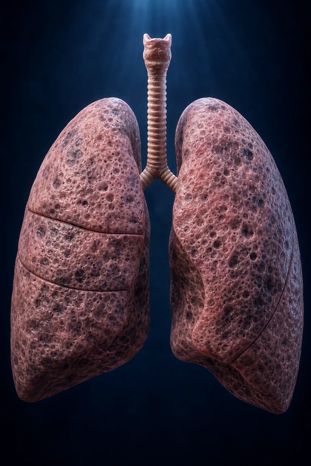
Chronic Obstructive Pulmonary Disease (COPD) is defined by GOLD as a common, preventable and treatable disease characterised by persistent respiratory symptoms and airflow limitation that is progressive and not fully reversible, caused by airway and/or alveolar abnormalities usually produced by significant exposure to noxious particles or gases.[1]
Three words deserve exam-level precision: [1]
- Persistent — the airflow obstruction is chronic and fixed; it does not resolve between attacks (contrast with the variable obstruction of asthma).
- Progressive — FEV1 declines faster than in health (smokers lose roughly 60 mL/year versus 30 mL/year in non-smokers); smoking cessation slows but does not halt this decline.
- Not fully reversible — there is some bronchodilator reversibility (up to 12 percent and 200 mL in FEV1 is allowed and still consistent with COPD), but it never returns to normal. [1]
The clinical skill in COPD is (i) recognising it early in the smoker over 40, (ii) confirming it objectively with spirometry, (iii) stratifying by symptoms and exacerbation risk (GOLD ABE) rather than FEV1 alone, (iv) escalating drug therapy by phenotype/eosinophils, and (v) recognising and managing the acute exacerbation — above all, avoiding CO2 narcosis from uncontrolled oxygen and starting NIV for hypercapnic failure.[1][1]
Classification
COPD is classified two overlapping ways — by phenotype and by GOLD spirometric grade + ABE group. [1]
Phenotypes (anatomical/clinical — they overlap)
| Phenotype | Definition | Classic picture |
|---|---|---|
| Chronic bronchitis | Productive cough for at least 3 months in 2 successive years, with airflow limitation | "Blue bloater" — obese, cyanosed, oedematous, chronic sputum, hypercapnia |
| Emphysema | Abnormal permanent enlargement of airspaces distal to the terminal bronchiole, with wall destruction, no obvious fibrosis | "Pink puffer" — thin, dyspnoeic, pursed-lip breathing, near-normal gases until late |
| Overlap | Most patients have both in varying proportion | The "pink puffer/blue bloater" dichotomy is an oversimplification — useful for exams, not for real life |
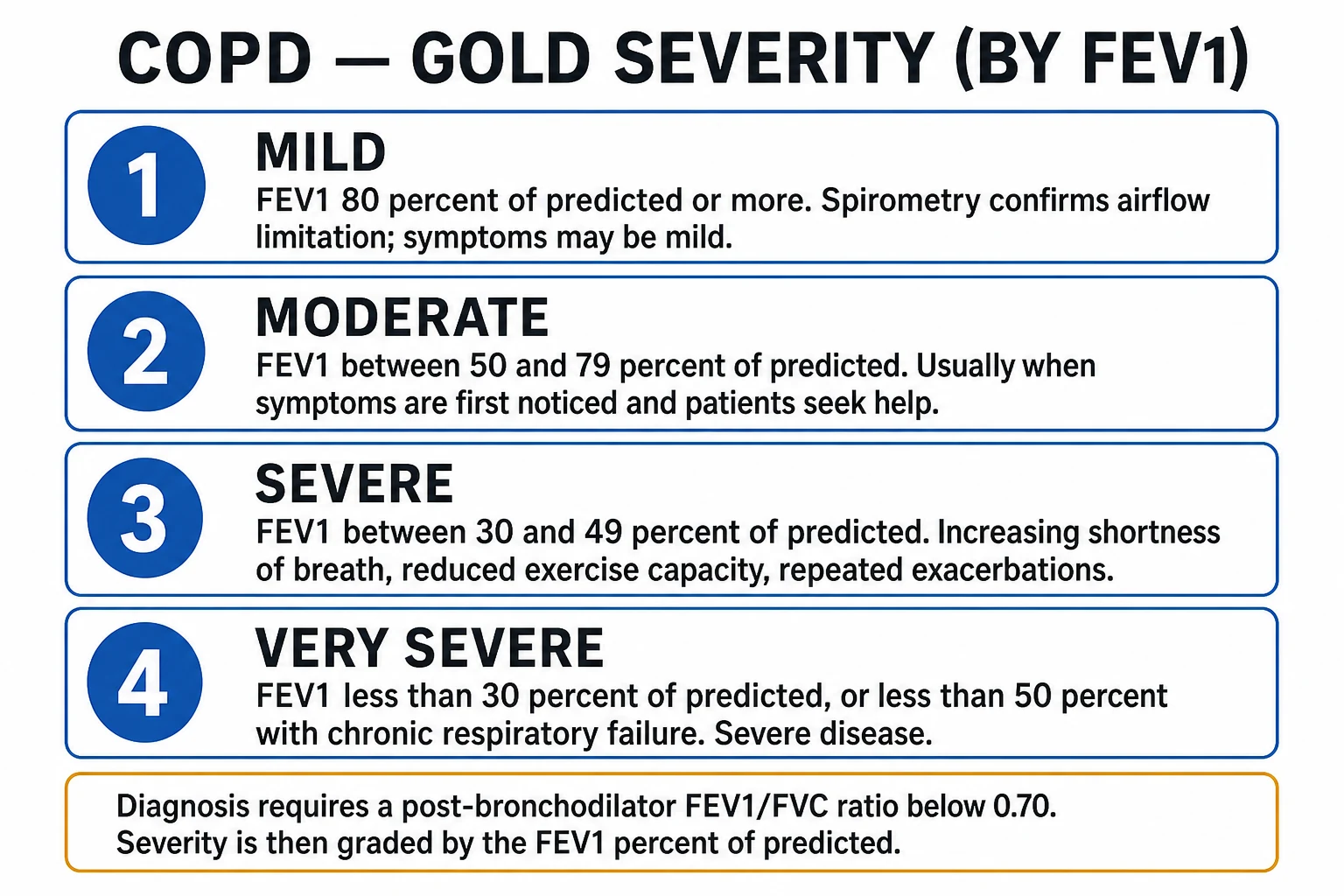
GOLD spirometric grades (severity — by FEV1 percent predicted, after the FEV1/FVC below 0.70 confirms the diagnosis)[1]
GOLD grades by post-bronchodilator FEV1 % predicted
GOLD ABE groups (therapy — by symptoms and exacerbations; the 2023 redesign merged old C and D into E, and removed FEV1 from grouping)[1]
An exacerbation here = a worsening of respiratory symptoms beyond day-to-day variation needing a treatment change. A moderate exacerbation is treated with antibiotics and/or systemic steroids in the community or emergency department; a severe exacerbation needs hospitalisation or a visit causing hospitalisation. [1]
Group A
- 0 or 1 moderate exacerbations, NOT leading to hospitalisation
- Low symptoms: mMRC 0 to 1 OR CAT below 10
- Therapy: any single bronchodilator (LABA or LAMA)
Group B
- 0 or 1 moderate exacerbations, NOT leading to hospitalisation
- High symptoms: mMRC at least 2 OR CAT at least 10
- Therapy: LABA + LAMA (dual bronchodilation preferred)
Group E
- At least 2 moderate exacerbations OR at least 1 leading to hospitalisation
- Any symptom level
- Therapy: LABA + LAMA; add ICS if blood eosinophils at least 300 (and consider if 100 to 299)
Definition of an acute exacerbation (AECOPD) and the Anthonisen criteria[11]
An acute exacerbation of COPD is a sustained worsening of the patient's respiratory symptoms beyond normal day-to-day variation, acute in onset, usually treated with a change in regular medication. The commonest triggers are respiratory infections (viral: rhinovirus, influenza, RSV; bacterial: Haemophilus influenzae, Streptococcus pneumoniae, Moraxella catarrhalis, Pseudomonas in severe disease) and environmental exposures; about one-third are of unknown cause. [1]
The Anthonisen criteria (1987) decide whether antibiotics will help, based on the three cardinal symptoms: [1]
- Increased dyspnoea
- Increased sputum volume
- Increased sputum purulence [1]
| Type | Criteria | Antibiotics |
|---|---|---|
| Type 1 | All 3 cardinal symptoms | Yes — strongest evidence of benefit |
| Type 2 | 2 of 3 cardinal symptoms | Yes, especially if increased sputum purulence is one of them |
| Type 3 | Only 1 cardinal symptom (or none) | No (unless another strong indication, e.g. pneumonia on CXR) |
Epidemiology & Risk Factors
COPD is a leading cause of morbidity and mortality worldwide — currently the third or fourth leading cause of death globally and a major cause of chronic disability. Prevalence is rising, driven by aging populations, the cumulative burden of smoking, and better survival from other diseases. Under-diagnosis is the rule, especially in low- and middle-income countries where spirometry is scarce.[1]
Principal risk factors: [1]
- Cigarette smoking — by far the commonest cause in high-income countries. Risk relates to total pack-years and is greater in those who started young; about 15 to 20 percent of smokers develop clinically significant COPD. Pipe, cigar, cannabis and water-pipe smoking also raise risk.
- Biomass fuel / indoor smoke — burning wood, dung or crop residues for cooking and heating in poorly ventilated homes; the dominant cause in women in the developing world and a key reason COPD is increasingly diagnosed in never-smokers.
- Occupational dusts and fumes — coal/mine dust, silica, cadmium, welding fumes, cotton and grain dusts. Silica exposure carries additional TB risk.
- Ambient (outdoor) air pollution — particulate matter (PM2.5), traffic and industrial emissions; worsens symptoms and exacerbations.
- Childhood respiratory events — severe childhood pneumonia, prematurity, recurrent childhood wheeze, and reduced maximal attained lung growth predispose to later COPD.
- Asthma — long-standing asthma is an independent risk factor (the asthma-COPD overlap).
- Genetics — alpha-1-antitrypsin deficiency (AATD) is the best-characterised genetic cause (see below); polygenic effects account for the remaining heritability.
- Socioeconomic status and nutrition — poverty, crowding, recurrent infections and low birth weight all increase risk. [1]
Alpha-1-antitrypsin deficiency (AATD)[1]
- Inheritance — autosomal co-dominant; the PiZZ genotype (serum level below 11 micromol/L, roughly 11 percent of normal) causes clinically significant disease. PiMZ is a milder carrier state. PiMM is normal.
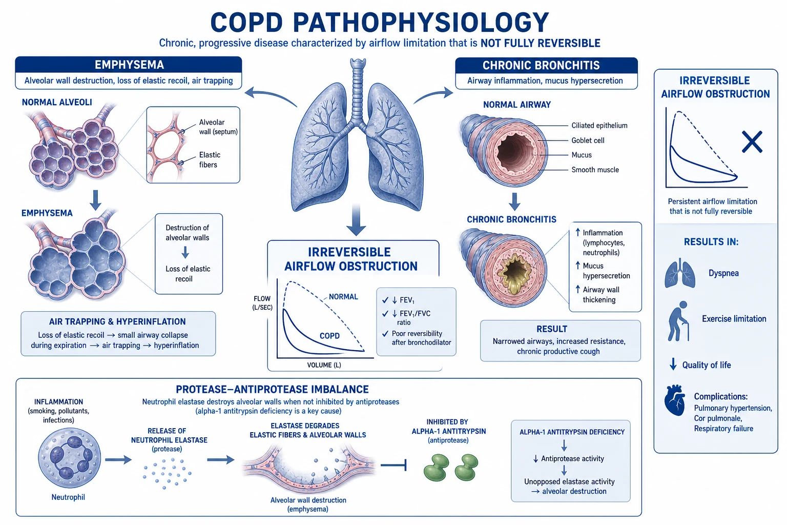
- Pathophysiology — alpha-1-antitrypsin is the main inhibitor of neutrophil elastase; its deficiency permits unopposed elastase-mediated proteolytic destruction of alveolar walls → panacinar (panlobular) emphysema predominantly in the lower zones.
- Clinical clues — COPD at young age (under 45), in a never-smoker or light smoker, with lower-zone emphysema, basal bullae, or a family history; coexistent liver disease (cholestatic hepatitis, cirrhosis, hepatocellular carcinoma) and panniculitis.
- Screen — measure serum alpha-1-antitrypsin level in all patients diagnosed with COPD (GOLD recommends universal once-in-a-lifetime screening), and confirm with phenotyping/genotyping if low. Counsel and screen family members.
- Treatment — weekly intravenous alpha-1-proteinase inhibitor augmentation (e.g. 60 mg/kg) slows lung-function decline in selected deficient patients; smoking avoidance is paramount; transplant for advanced disease. [1]
Pathophysiology
COPD is fundamentally an abnormal, exaggerated and persistent inflammatory response of the lung to inhaled noxious particles. That inflammation produces two anatomical lesions whose combined effect is airflow limitation, air trapping and gas-exchange failure.[1][1]
The inflammatory response
Inhaled particles activate epithelial cells and macrophages in the small airways and alveoli, which release a cytokine soup that recruits and sustains a chronic inflammatory infiltrate: [1]
- Neutrophils — dominant in the airway lumen; release neutrophil elastase, matrix metalloproteinases (MMP-9, MMP-12) and reactive oxygen species that digest alveolar walls and mucus-secreting epithelium.
- Macrophages — orchestrate the response, secreting tumour necrosis factor-alpha (TNF-alpha), interleukin-8 (IL-8/CXCL8) and leukotriene B4 (LT-B4), all of which are potent neutrophil chemoattractants.
- CD8+ T-cells — the characteristic lymphocyte of COPD (contrast with the CD4+/Th2 and eosinophilic pattern of asthma); release perforin and granzymes causing epithelial apoptosis.
- Oxidative stress — cigarette smoke and activated inflammatory cells generate free radicals that deplete antiproteases (notably inactivating alpha-1-antitrypsin) and amplify inflammation. [1]
The two anatomical lesions
- Chronic bronchiolitis (small-airway disease) — airways smaller than 2 mm bear the brunt: inflammation, goblet-cell hyperplasia, mucus hypersecretion, fibrosis, narrowing and eventual obliterative bronchiolitis. This is the major site of airflow limitation in COPD.
- Emphysematous destruction — loss of alveolar walls and their attachments to small airways (the alveolar attachments that normally tether the airway open) and loss of elastic recoil; both promote premature airway closure on expiration → air trapping. [1]
Centriacinar vs panacinar emphysema
Centriacinar (centrilobular)
- Affects respiratory bronchioles, SPARES alveolar ducts/sacs
- UPPER-zone predominance
- The classic SMOKING pattern
- Lesions centred on the bronchiole, surrounded by spared parenchyma
Panacinar (panlobular)
- Affects the ENTIRE acinus uniformly
- LOWER-zone predominance
- The classic ALPHA-1-ANTITRYPSIN DEFICIENCY pattern
- May produce prominent basal bullae; also seen with IV drug use ('talcosis')
Paraseptal
- Affects the distal acinus adjacent to pleura/fissures
- Upper-lobe and subpleural
- Associated with spontaneous pneumothorax in young men
- Often coexists with centriacinar disease
Why patients are breathless: dynamic hyperinflation
Loss of recoil plus narrowed airways means forced expiratory flow is reduced; the patient cannot exhale fully before the next breath begins, so air is trapped. End-expiratory lung volume rises (dynamic hyperinflation) and breathing occurs at a higher, less compliant lung volume — the diaphragm is flattened and at mechanical disadvantage, the work of breathing rises, and tidal volume encroaches on the relatively comfortable upper, steep portion of the pressure-volume curve. This is the chief mechanism of exertional dyspnoea and is what pursed-lip breathing (positive pressure in the airway, stenting it open longer) and bronchodilators partly relieve. [1]
Gas-exchange failure — type-1 vs type-2
- Ventilation-perfusion mismatch (low V/Q from narrowed, mucus-filled airways; high V/Q from destroyed capillary bed) is the predominant early mechanism of hypoxaemia, corrected partly by hypoxic pulmonary vasoconstriction.
- As the disease advances, fatigue and alveolar hypoventilation add CO2 retention — producing type-2 (hypercapnic) respiratory failure, typically with a compensated respiratory acidosis (raised serum bicarbonate from renal retention, normal or near-normal pH). [1]
Pulmonary hypertension and cor pulmonale
Loss of the alveolar capillary bed (pruning of the vascular tree), sustained hypoxic pulmonary vasoconstriction, and vascular remodelling together raise pulmonary vascular resistance → pulmonary hypertension → right ventricular hypertrophy and dilatation → cor pulmonale (right heart failure secondary to lung disease): raised JVP, hepatomegaly, ankle oedema, loud pulmonary component of the second heart sound (P2), and tricuspid regurgitation. [1]
Why uncontrolled high-flow oxygen is dangerous in a CO2-retainer[1]
Giving 100 percent oxygen to a chronic CO2-retainer commonly precipitates worsening hypercapnia, CO2 narcosis and death. Three mechanisms contribute (the traditional "loss of hypoxic drive" is the least important): [1]
- Ventilation-perfusion mismatch (V/Q effect) — high inspired oxygen abolishes hypoxic pulmonary vasoconstriction in poorly ventilated, low-V/Q units, redirecting blood flow to them and increasing dead-space ventilation, so CO2 clearance falls.
- The Haldane effect — oxygenation of haemoglobin reduces its affinity for CO2 (which is normally carried as carbamino compounds on deoxyhaemoglobin); more CO2 is therefore released and stays dissolved in plasma, raising PaCO2.
- Reduced ventilatory drive (the old theory) — a small fall in peripheral chemoreceptor stimulus contributes, but is minor compared with (1) and (2). [1]
This is why controlled oxygen — target SpO2 88 to 92 percent — and an early arterial blood gas are non-negotiable in the COPD exacerbation. [1]
Clinical Presentation
COPD is insidious. Symptoms usually begin after the age of 40 and many years of smoking; patients commonly attribute early breathlessness to "getting older" or being "unfit", and so present late.[1]
Classic presentation: [1]
- Chronic productive cough — often the earliest symptom, classically worse in the morning; mucoid sputum turning purulent during exacerbations.
- Progressive exertional dyspnoea — the cardinal symptom; gradually limiting activities over years, eventually present at rest.
- Wheeze and chest tightness — variable, may worsen on exertion, cold air, or exacerbations.
- Symptoms of advanced disease — weight loss and cachexia (emphysema phenotype), ankle swelling (cor pulmonale), morning headaches (CO2 retention), exertional syncope or chest pain (severe pulmonary hypertension). [1]
Examination — look for the signs of airflow obstruction and its consequences: [1]
- Inspection — barrel chest (increased anteroposterior diameter), pursed-lip breathing, use of accessory muscles (sternocleidomastoid, scalenes), intercostal indrawing, cyanosis, cachexia, tar-stained fingers; Hoover's sign (paradoxical inward movement of the lower rib cage on inspiration — highly specific for severe airflow obstruction and hyperinflation); tremor/asterixis and drowsiness/confusion in CO2 retention; pinpoint pupils/miosis (rare, in hypercapnia).
- Palpation — reduced chest expansion; apex beat displaced in hyperinflation or, if cor pulmonale, a right ventricular heave.
- Percussion — hyper-resonance bilaterally; reduced cardiac and liver dullness.
- Auscultation — prolonged expiratory phase, polyphonic wheeze, coarse crackles that may clear on coughing, distant vesicular (quiet) breath sounds; a loud P2 (pulmonary hypertension), tricuspid regurgitation murmur, or right-sided S3 in cor pulmonale.
- Cor pulmonale — raised JVP, ankle/sacral oedema, hepatomegaly (± hepatic pulsatility in tricuspid regurgitation), ascites. [1]
Pink puffer vs blue bloater — the exam-favourite (oversimplified) dichotomy
Pink puffer (emphysema)
- Thin, cachectic, barrel-chested
- Marked dyspnoea, pursed-lip breathing, accessory-muscle use
- Arterial gases NEAR-NORMAL until late (maintains ventilation)
- Hypoxaemia mild; PaCO2 normal or low
- Pink (well-oxygenated) skin
Blue bloater (chronic bronchitis)
- Overweight, oedematous, cyanosed
- Less dyspnoea but chronic sputum
- Chronically HYPERCAPNIC and HYPOXAEMIC
- Compensated respiratory acidosis (raised bicarbonate)
- Cor pulmonale and secondary polycythaemia early
- Blue (cyanosed), bloated (oedematous)
Atypical presentations
- Elderly — breathlessness attributed to ageing, heart failure, or deconditioning; cough passed off as "smoker's cough"; less wheeze; comorbidity (ischaemic heart disease, osteoporosis, depression) dominates the picture.
- Women with biomass-smoke exposure — may present younger, with less smoking history; chronic bronchitis phenotype is common; under-diagnosed.
- AATD in the young/never-smoker — basal panacinar emphysema, family history, liver disease.
- Diabetic with COPD — breathlessness may be misattributed; ketoacidosis can precipitate Kussmaul breathing misread as a COPD exacerbation.
- The decompensating exacerbation — increasing dyspnoea with drowsiness, confusion, asterixis/flap, falling GCS, rising PaCO2 and falling pH; a medical emergency. [1]
Differential Diagnosis
Chronic breathlessness with wheeze or cough is not always COPD. Distinguish:[1][1]
- Asthma — the cardinal differential. Earlier onset (often childhood), atopy (eczema, hayfever, allergic rhinitis), marked day-to-day and diurnal variability, good (often complete) bronchodilator reversibility (FEV1 rise at least 12 percent AND 200 mL; a rise over 400 mL is strongly suggestive of asthma), blood/sputum eosinophilia, family history of atopy/asthma. The asthma-COPD overlap (ACO) exists and is treated more aggressively with ICS.
- Chronic bronchitis WITHOUT airflow limitation — chronic productive cough meeting the epidemiological definition but normal spirometry; symptoms but no obstruction, so not "COPD" by GOLD (manage smoking cessation, bronchodilators for symptoms, vaccination).
- Bronchiectasis — large-volume purulent sputum daily, recurrent infections, haemoptysis, clubbing, coarse crackles; CT shows airway dilatation, signet-ring sign, tram-tracking.
- Tuberculosis — upper-zone infiltrate, cavitation, constitutional features (weight loss, night sweats, haemoptysis), risk factors (endemic exposure, HIV, diabetes); always send sputum for acid-fast bacilli when suspected.
- Congestive cardiac failure — orthopnoea, PND, bilateral crackles, raised JVP, gallop (S3), cardiomegaly, pleural effusion; BNP/NT-proBNP and echo distinguish.
- Lung cancer — new or changing cough, haemoptysis, weight loss, smoker over 50; mass, collapse, pleural effusion, or recurrent pneumonia in the same lobe on imaging; smoking is a shared risk factor, so always consider cancer in any change of symptoms.
- Interstitial lung disease — dry cough, progressive dyspnoea, Velcro-type fine bibasal crackles, clubbing, restrictive spirometry (low FVC, preserved or increased ratio), characteristic HRCT (reticulation, honeycombing, ground-glass).
- Anaemia — fatigue, dyspnoea out of proportion to chest signs; full blood count.
- Deconditioning / obesity — exertional dyspnoea without wheeze, normal spirometry and normal CXR.
- Pulmonary embolism — pleuritic pain, syncope, sudden dyspnoea; COPD patients have a higher baseline PE risk and PE can mimic exacerbation.
- Upper-airway obstruction / vocal-cord dysfunction — stridor or inspiratory wheeze, fixed extrathoracic obstruction on flow-volume loop. [1]
Symptoms/signs that mandate exclusion of another diagnosis: haemoptysis (cancer, TB, PE, bronchiectasis), marked day-to-day variability (asthma), onset under 40 or never-smoker (asthma, AATD), prominent constitutional features (TB, malignancy, ILD), and finger clubbing (NOT a feature of COPD — search for cancer, bronchiectasis, ILD, or right-to-left shunt). [1]
Clinical & Bedside Assessment
Focused history
Establish the smoking history in pack-years (1 pack-year = 20 cigarettes/day for 1 year), occupational and biomass exposure, age of onset and pace of symptoms, sputum (volume, colour, daily production), exacerbation frequency and hospitalisations (drives GOLD grouping), exercise capacity (stairs, distance on the flat), vaccination status (influenza, pneumococcal, COVID-19), comorbidity (ischaemic heart disease, hypertension, diabetes, osteoporosis, depression), and medications and inhaler technique. Screen for depression, anxiety and sleep disturbance. [1]
Focused examination — as above
Confirm airflow obstruction, hyperinflation, cor pulmonale, and the decompensating patient. Always measure respiratory rate, SpO2 on air, blood pressure, temperature and look for pneumonia, pneumothorax and heart failure as exacerbation triggers. [1]
Standardised symptom tools (reproduce verbatim)[1]
Modified MRC (mMRC) dyspnoea scale (0 to 4): [1]
| Grade | Description |
|---|---|
| 0 | Breathless only with strenuous exercise |
| 1 | Short of breath hurrying on the level or walking up a slight hill |
| 2 | Walks slower than people of the same age on the level because of breathlessness, or has to stop for breath when walking at own pace |
| 3 | Stops for breath after walking about 100 metres or after a few minutes on the level |
| 4 | Too breathless to leave the house, or breathless when dressing or undressing |
Threshold: mMRC at least 2 = more symptoms (drives GOLD B/E grouping). [1]
COPD Assessment Test (CAT), 0 to 40 — eight items each scored 0 to 5: cough, phlegm, chest tightness, climbing one flight of stairs, activity limitation at home, confidence leaving home, sleep, energy. CAT at least 10 = more symptoms. CAT correlates well with health status and is more sensitive than mMRC. [1]
Bedside signs of a life-threatening exacerbation
A patient with respiratory rate over 30, SpO2 below 88 percent on air, use of accessory muscles, unable to speak full sentences, silent chest (no wheeze because airflow is too low), cyanosis, GCS falling / asterixis / drowsiness, arrhythmia, or pH below 7.35 with rising PaCO2 needs immediate controlled oxygen, an arterial blood gas, nebulised bronchodilators, senior review and preparation for NIV/intubation. [1]
Investigations
Spirometry — the essential diagnostic test[1]
- Confirm COPD: a post-bronchodilator FEV1/FVC below 0.70 (after 400 mcg salbutamol or equivalent) confirms persistent airflow limitation. Do spirometry when the patient is clinically stable (not during an acute exacerbation).
- Grade severity by FEV1 percent predicted: GOLD 1 (at least 80), 2 (50 to 79), 3 (30 to 49), 4 (below 30, or below 50 with chronic respiratory failure).
- Bronchodilator reversibility — a rise of at least 12 percent AND at least 200 mL in FEV1 is "significant" but does not exclude COPD (it is common); a rise over 400 mL suggests asthma or ACO. Total lung capacity, residual volume and the inspiratory capacity/TLC ratio (on body plethysmography) show hyperinflation and air trapping; diffusing capacity for carbon monoxide (DLCO/TLCO) is reduced in emphysema. [1]
Chest X-ray
Findings of hyperinflation: flat, low hemidiaphragms, long, narrow heart shadow, increased retrosternal air space (over 2.5 cm on lateral), 6 or more anterior rib ends visible above the diaphragm, bullae (radiolucent areas), and paucity of peripheral vascular markings ("oligaemia"). CXR also excludes pneumonia, pneumothorax, pleural effusion, cardiomegaly and may reveal a mass. [1]
CT thorax
Shows distribution and type of emphysema (centrilobular upper-zone; panlobular lower-zone in AATD; paraseptal subpleural), bullae, coexisting bronchiectasis, pulmonary nodules/masses (lung cancer surveillance), and pulmonary artery dilatation (pulmonary hypertension). Low-dose CT lung-cancer screening is recommended in selected high-risk smokers (age 50 to 80, at least 20 pack-years). [1]
Arterial blood gas (ABG)
Indicated in severe exacerbation, SpO2 below 92 percent, clinical deterioration, signs of respiratory failure, and before starting or escalating oxygen/NIV. Report pH, PaO2, PaCO2, bicarbonate, base excess, lactate: [1]
- Type-1 respiratory failure — PaO2 below 60 mmHg (8 kPa) with normal or low PaCO2 (hypoxaemic, not retaining). Early/mild COPD, and many exacerbations.
- Type-2 respiratory failure — PaO2 below 60 mmHg (8 kPa) AND PaCO2 at least 45 mmHg (6 kPa) (hypercapnic). Severe/advanced COPD.
- Compensated respiratory acidosis — chronic CO2 retention has low pH with a markedly raised bicarbonate and base excess from renal compensation; an acute exacerbation on this baseline produces a falling pH with rising PaCO2.
- Repeat the ABG at 30 to 60 minutes after starting controlled oxygen or NIV. [1]
The BODE index — a multidimensional prognostic score[2]
The BODE index (Body-mass index, Obstruction, Dyspnoea, Exercise capacity) predicts mortality better than FEV1 alone: [1]
| Component | Variable | Score 0 | Score 1 | Score 2 | Score 3 |
|---|---|---|---|---|---|
| B | BMI (kg/m²) | over 21 | at most 21 | — | — |
| O | FEV1 % predicted | at least 65 | 50 to 64 | 36 to 49 | at most 35 |
| D | mMRC dyspnoea | 0 to 1 | 2 | 3 | 4 |
| E | 6-minute-walk distance (m) | at least 350 | 250 to 349 | 150 to 249 | at most 149 |
Total 0 to 10. Higher scores predict worse survival (a score of 7 to 10 carries roughly 80 percent 4-year mortality; a score of 0 to 2, about 20 percent). It captures the systemic nature of COPD (cachexia, deconditioning) that FEV1 misses. [1]
Other investigations
- Full blood count — secondary polycythaemia (raised haematocrit from chronic hypoxaemia), anaemia of chronic disease; eosinophil count (at least 300 cells/microL supports adding ICS).
- Alpha-1-antitrypsin level — screen once; if low, genotype.
- ECG — arrhythmias (AF, multifocal atrial tachycardia), right axis deviation, right ventricular hypertrophy, right bundle branch block, P pulmonale (tall P in lead II) — features of cor pulmonale.
- Echocardiogram — estimate pulmonary artery systolic pressure, assess right ventricular size/function, exclude left-heart disease.
- Sputum — Gram stain/culture and sensitivity in exacerbations with purulent sputum; AFB if TB suspected.
- 6-minute-walk test — exercise capacity, desaturation, used in BODE and for LTOT assessment.
- BNP/NT-proBNP, troponin — when cardiac causes overlap.
- Sleep study — if overlap syndrome (COPD + OSA) suspected. [1]
COPD — the numbers that matter
Management — Resuscitation (Acute Exacerbation)
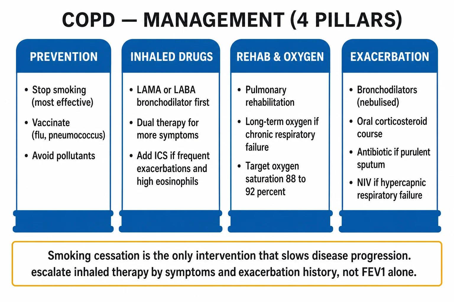
An acute exacerbation of COPD (AECOPD) is a common medical emergency. The immediate goals are (i) identify and treat the precipitant, (ii) correct hypoxaemia without causing CO2 narcosis, (iii) relieve bronchospasm, (iv) reduce airway inflammation, (v) treat infection, and (vi) support ventilation with NIV when needed.[1]
ABCDE assessment
- Airway — assess patency, position upright; clear secretions; anticipate the need for airway support.
- Breathing — rate, effort, accessory-muscle use, SpO2, chest expansion, wheeze, cyanosis, silent chest. Start controlled oxygen immediately.
- Circulation — IV access, fluid balance (overload worsens cor pulmonale; dehydration thickens secretions); treat arrhythmias.
- Disability — GCS, asterixis (a sign of CO2 narcosis), pupils; a falling GCS predicts the need for NIV.
- Exposure / Examination — full set, screen for pneumonia, pneumothorax, pulmonary embolism, heart failure as triggers. [1]
Controlled oxygen — the single most important resuscitation rule[1]
- Target SpO2 88 to 92 percent in all COPD exacerbations (and any patient at risk of CO2 retention). Use nasal cannulae at 1 to 2 L/min or a Venturi mask 24 percent to 28 percent titrated to the target.
- Check an ABG within 30 to 60 minutes of starting oxygen and again after any change in FiO2 or starting NIV. If the PaCO2 rises by more than about 8 mmHg or the pH falls, lower the FiO2 and consider NIV.
- Never give uncontrolled high-flow oxygen (e.g. non-rebreather at 15 L/min) to a known or suspected CO2-retainer unless intubated — the classical trap. [1]
The acute medical bundle (give together)
- Controlled oxygen as above.
- Nebulised bronchodilators — drive with air, not oxygen, in the CO2-retainer:
- Salbutamol 5 mg (or 2.5 mg) nebulised, every 4 to 6 hours (or continuously in severe bronchospasm); plus
- Ipratropium bromide 500 mcg nebulised, every 6 hours (often given combined with salbutamol as a "duoneb" every 4 to 6 h).
- In the very severe/acyanotic patient, nebulisers can be driven with oxygen while the ABG is awaited.
- Systemic corticosteroid — prednisolone 40 mg orally once daily for 5 days (REDUCE trial proved 5 days non-inferior to 14 days).[8] Use IV hydrocortisone 100 mg if the patient cannot take oral or is acutely ill. Do NOT prolong beyond 14 days. Prescribe bone protection (e.g. a PPI; calcium/vitamin D) only if a longer or recurrent course is given.
- Antibiotic when the Anthonisen criteria are met (Type 1 or Type 2), or if there is pneumonia on CXR or increased sputum purulence:[11]
- First-line oral: amoxicillin-clavulanate 500/125 mg three times daily (or 875/125 mg twice daily) for 5 to 7 days.
- Alternatives: doxycycline 100 mg twice daily (200 mg loading then 100 mg BD) for 5 to 7 days, or azithromycin 500 mg once daily for 3 to 5 days, or a macrolide (clarithromycin 500 mg BD).
- In severe disease / bronchiectasis / recent antibiotics / hospitalisation, broaden cover to include Pseudomonas — e.g. piperacillin-tazobactam 4.5 g IV every 8 hours or ciprofloxacin 750 mg orally twice daily per local policy.
- Send sputum culture (and blood cultures if septic) before the first dose where possible, but do not delay antibiotics in sepsis.
- Ventilatory support — non-invasive ventilation (NIV / BiPAP) — the game-changer.[10]
NIV (BiPAP) — indications, settings, escalation[1][10]
Indications (acute hypercapnic respiratory failure despite standard medical therapy for at least 30 to 60 minutes): [1]
- pH below 7.35 AND PaCO2 at least 45 mmHg (6 kPa) — start NIV; OR
- PaCO2 at least 60 mmHg (8 kPa) with pH 7.25 to 7.35 — start NIV. [1]
Settings (bilevel positive airway pressure): [1]
- IPAP (inspiratory positive airway pressure) 10 to 15 cmH2O, EPAP (expiratory) 4 to 5 cmH2O, titrate IPAP up by 2 cmH2O every 10 minutes toward 20 cmH2O to achieve a target tidal volume of 6 to 8 mL/kg and a falling PaCO2.
- FiO2 titrated to SpO2 88 to 92 percent.
- Use a well-fitting full-face mask initially. [1]
Repeat ABG at 1 hour — look for rising pH and falling PaCO2. Continue NIV with breaks for meals, nebulisers and physiotherapy; wean as the patient recovers. [1]
Escalate to invasive mechanical ventilation (intubation) when: [1]
- NIV fails — worsening pH/PaCO2 at 1 to 4 hours, or worsening consciousness, agitation, or respiratory arrest.
- Absolute contraindications to NIV — respiratory arrest, inability to protect airway (reduced GCS, copious secretions), haemodynamic instability, severe encephalopathy/coma, facial trauma/deformity, fixed upper-airway obstruction, active pneumothorax (drain first), or patient refusal.
- Intubate with a low tidal volume (6 to 8 mL/kg ideal body weight), permissive hypercapnia, and minimise positive end-expiratory pressure; extubate onto NIV ("NIV-assisted weaning") to reduce ventilator days. [1]
Management — Definitive & Stepwise (Stable COPD)
Stable-disease management aims to reduce symptoms, reduce exacerbations, improve exercise tolerance and health status, slow disease progression and reduce mortality. Therapy is built on non-pharmacological foundations and then escalated by the GOLD ABE group and the blood eosinophil count.[1]
The only disease-modifying intervention: smoking cessation + vaccination
- Smoking cessation is the single most effective and cost-effective intervention — it slows the rate of FEV1 decline toward that of a never-smoker and reduces mortality. Offer behavioural support plus pharmacotherapy to every smoker at every visit:
- Nicotine replacement therapy (NRT) — patches (15 to 21 mg/24 h) plus a short-acting form (gum, lozenge, inhalator, nasal spray) for craving; 8 to 12 weeks.
- Varenicline — partial alpha-4-beta-2 nicotinic agonist; 0.5 mg once daily for 3 days, then 0.5 mg twice daily for 4 days, then 1 mg twice daily for 12 weeks. Most effective single agent; reduce dose in CKD.
- Bupropion — 150 mg once daily for 6 days then 150 mg twice daily for 7 to 9 weeks; avoid in epilepsy/eating disorder.
- Vaccination — annual influenza; pneumococcal (conjugate PCV15/PCV20 and/or polysaccharide PPSV23 per local schedule); COVID-19; consider pertussis, RSV, herpes zoster where indicated. Vaccines reduce exacerbations and pneumonia. [1]
GOLD ABE pharmacologic ladder[1][5][6][7]
| Group | First-line | Escalation |
|---|---|---|
| A | A single bronchodilator — LABA or LAMA | If persistent symptoms, step up to dual |
| B | LABA + LAMA (dual bronchodilation) preferred over monotherapy | If persistent dyspnoea, switch device/molecule or add triple |
| E | LABA + LAMA (dual) | Add ICS if blood eosinophils at least 300/microL (consider at 100 to 299 if very frequent exacerbations); consider roflumilast and/or azithromycin for persistent exacerbations |
Why dual bronchodilation first in B/E? LABA + LAMA combinations outperform either monotherapy for lung function and symptoms, and — crucially — adding ICS to low-eosinophil COPD brings pneumonia risk without benefit.[7]
Common drug doses and routes: [1]
- Short-acting relievers (always co-prescribed for breakthrough): salbutamol 100 to 200 mcg (1 to 2 puffs) PRN via MDI; ipratropium 20 mcg (2 puffs) up to four times daily; nebulised salbutamol 2.5 to 5 mg, ipratropium 500 mcg.
- LAMA: tiotropium 18 mcg once daily (HandiHaler, dry powder) or 5 mcg once daily (Respimat, soft mist); umeclidinium 62.5 mcg once daily; glycopyrronium 44 mcg once daily; aclidinium 400 mcg twice daily.
- LABA: salmeterol 50 mcg twice daily; formoterol 12 mcg twice daily; indacaterol 150 mcg once daily (75 mcg if combined with glycopyrronium); olodaterol 5 mcg once daily.
- LABA + LAMA fixed-dose (single inhaler): tiotropium + olodaterol (5/5 mcg) once daily; umeclidinium + vilanterol (62.5/25 mcg) once daily; indacaterol + glycopyrronium (110/50 mcg) once daily; formoterol + aclidinium (400/12 mcg) twice daily.
- ICS: fluticasone propionate 100 to 500 mcg twice daily; budesonide 200 to 400 mcg twice daily; beclometasone 100 to 200 mcg twice daily; fluticasone furoate 100 or 200 mcg once daily.
- LABA + ICS combinations: salmeterol/fluticasone (50/250 or 50/500 mcg) twice daily; formoterol/budesonide (4.5/160 or 6/200 mcg) twice daily.
- Single-inhaler triple therapy (ICS + LABA + LAMA): fluticasone furoate/umeclidinium/vilanterol (100/62.5/25 mcg) once daily (IMPACT trial — reduced exacerbations and mortality versus dual).[7]
- Phosphodiesterase-4 inhibitor — roflumilast 500 mcg once daily — for severe COPD (GOLD 3 to 4) with chronic bronchitis and a history of exacerbations, especially in the frequent-exacerbator phenotype; main adverse effects nausea, diarrhoea, weight loss, mood disturbance.
- Macrolide prophylaxis — azithromycin 250 mg once daily (or 500 mg three times weekly) for one year in former smokers with frequent exacerbations despite optimal inhaled therapy; reduces exacerbation frequency (Albert 2011).[9] Screen for QT prolongation and macrolide resistance first, and counsel on hearing and hepatic monitoring.
Pulmonary rehabilitation
A 6- to 8-week structured programme of exercise training, education, behaviour change and nutritional support. Improves exercise capacity, dyspnoea and quality of life, reduces anxiety/depression and hospital readmissions after an exacerbation. Recommended for any COPD patient with mMRC at least 2 or CAT at least 10, including immediately after a hospitalising exacerbation. [1]
Long-term oxygen therapy (LTOT)[3][4]
LTOT is one of only two interventions that improve survival in COPD (the other is smoking cessation). The landmark Nocturnal Oxygen Therapy Trial (NOTT) and MRC LTOT trial established the criteria: [1]
Prescribe LTOT when (on two ABGs at least 3 weeks apart in a clinically stable patient): [1]
- PaO2 at most 55 mmHg (7.3 kPa), or SaO2 at most 88 percent; OR
- PaO2 56 to 59 mmHg (7.4 to 7.8 kPa) / SaO2 89 percent with evidence of pulmonary hypertension, cor pulmonale, peripheral oedema from right-heart failure, or polycythaemia (haematocrit above 55 percent). [1]
Use for at least 15 hours per day (target 24 h; overnight is essential), titrated to PaO2 at least 60 mmHg (8 kPa) / SaO2 at least 90 percent. Counsel on smoking cessation (absolute requirement — fire risk) and arrange home oxygen risk assessment. Ambulatory oxygen helps those who desaturate on exertion. [1]
Advanced therapies
- Lung-volume-reduction surgery (LVRS) / endobronchial valves — for selected patients with severe upper-lobe-predominant emphysema and low exercise capacity; endobronchial one-way valves achieve lobar atelectasis by blocking collateral ventilation. Best in heterogeneous (upper-lobe) emphysema with intact interlobar fissures; contraindicated in homogeneous disease, DLCO below 20 percent, or severe pulmonary hypertension.
- Lung transplantation — for selected patients under about 65 with very severe COPD (BODE 7 to 10, or FEV1 below 25 percent, or chronic hypercapnia, or severe pulmonary hypertension) without major contraindication; bilateral sequential is usual.
- Bullectomy — for giant bullae compressing functional lung. [1]
Acute COPD exacerbation bundle
OBASIS
Target SpO2 88 to 92 percent; drive nebs with air
Nebulised salbutamol 5 mg + ipratropium 500 mcg
If Anthonisen criteria met — amoxiclav/doxy/azithromycin
Prednisolone 40 mg PO for 5 days (REDUCE trial)
ABG at 30 to 60 min; CXR; FBC, CRP, U&E, cultures
BiPAP if pH below 7.35 with PaCO2 at least 45 mmHg; escalate to intubation if fails
Specific Subtypes & Scenarios
- Acute hypercapnic respiratory failure — the prototype scenario: controlled oxygen (88 to 92 percent), early NIV (BiPAP), nebulised bronchodilators, prednisolone, antibiotics if Anthonisen-positive. NIV reduces intubation rate, mortality, length of stay and complications versus invasive ventilation in suitable patients.[10]
- Frequent-exacerbator phenotype (at least 2 moderate or at least 1 hospitalising exacerbation/year) — escalate to LABA + LAMA, add ICS if eosinophils at least 300, add roflumilast 500 mcg once daily (severe chronic-bronchitis pattern), and azithromycin 250 mg once daily (after ECG and LFT checks). Verify inhaler technique and adherence before declaring failure.
- Asthma-COPD overlap (ACO) — features of both (persistent airflow obstruction plus significant reversibility, eosinophilia, atopy). Earlier and more aggressive ICS (often triple therapy) because the eosinophilic/Th2 component responds.
- Alpha-1-antitrypsin deficiency — screen, counsel family, augmentation therapy (weekly IV alpha-1-proteinase inhibitor 60 mg/kg) in selected patients, avoid smoking absolutely, refer for transplant in advanced disease.
- COPD with cor pulmonale / chronic respiratory failure — LTOT (the survival intervention), diuretics (furosemide 20 to 40 mg) for fluid overload (caution over-diuresis, metabolic alkalosis that suppresses ventilation), treat secondary polycythaemia by oxygenation (venesection only for symptomatic hyperviscosity, haematocrit above 56 percent), and work up pulmonary hypertension for targeted vasodilators under specialist care.
- COPD with pneumothorax (ruptured bulla) — high index of suspicion: sudden worsening dyspnoea, reduced breath sounds, hyper-resonance on one side. CXR to confirm. Treat with small-bore chest drain / aspiration; in known large bullae, beware a misdiagnosis of pneumothorax (CT distinguishes). Avoid over-oxygenation.
- COPD with pneumonia — common; broaden antibiotics, target SpO2 88 to 92 percent, watch PaCO2.
- COPD with suspected PE — COPD patients are pro-thrombotic; D-dimer and CTPA (the CXR is already abnormal so a V/Q scan is harder to interpret). Anticoagulate per risk.
- Overlap syndrome (COPD + OSA) — daytime hypercapnia, nocturnal desaturation, morning headaches, cor pulmonale out of proportion; treat with CPAP/NIV plus usual COPD therapy.
Complications & Pitfalls
Complications: [1]
- Acute and chronic respiratory failure — hypoxaemic and/or hypercapnic.
- Cor pulmonale (right-heart failure secondary to pulmonary hypertension).
- Pneumothorax from ruptured bullae.
- Secondary polycythaemia from chronic hypoxaemia (raises viscosity, thrombosis risk).
- Pulmonary embolism — increased baseline risk; can mimic exacerbation.
- Recurrent chest infections / pneumonia — including ICS-associated pneumonia in low-eosinophil COPD.
- Osteoporosis (smoking, steroids, inactivity, vitamin D deficiency).
- Skeletal-muscle wasting, cachexia, sarcopenia.
- Depression and anxiety (common, under-recognised, worsens outcomes).
- Lung cancer (shared smoking risk — the commonest cause of death in mild COPD).
- Arrhythmias — atrial fibrillation, multifocal atrial tachycardia.
- Respiratory acidosis and CO2 narcosis (iatrogenic from over-oxygenation). [1]
Classic pitfalls (the things examiners trap you with): [1]
- Diagnosing COPD without spirometry — symptoms plus smoking are not enough; the FEV1/FVC below 0.70 (post-bronchodilator) is mandatory.
- Giving high-flow oxygen to a CO2-retainer, precipitating CO2 narcosis.
- Missing an alternative diagnosis — a pneumothorax, PE, pneumonia, heart failure, or lung cancer as the trigger for "an exacerbation".
- Overusing ICS in low-eosinophil COPD — adds pneumonia risk without benefit; check blood eosinophils before adding ICS.
- Failing to start NIV when the ABG shows acidosis — NIV within 60 minutes is the standard of care.
- Forgetting smoking cessation and vaccination — the only disease-modifier and the cheap effective prevention.
- Confusing asthma with COPD — reversibility, age, atopy, eosinophilia.
- Mislabeling a normal CXR "emphysema" without spirometric confirmation.
- Under-treating comorbidity — cardiac disease, osteoporosis, depression. [1]
Prognosis & Disposition
COPD is progressive but highly modifiable by smoking cessation, vaccination, pulmonary rehabilitation, appropriate inhaled therapy, and early treatment of exacerbations. Prognosis worsens with low FEV1, high exacerbation frequency (≥2/year or ≥1 hospitalisation), severe dyspnoea, low BMI (cachexia), cor pulmonale, and comorbidities (CVD, lung cancer, osteoporosis, depression). [1]
BODE index components (prognostic — reproduce)
BMI, Obstruction (FEV1 %), Dyspnoea (mMRC), Exercise (6-minute walk distance). Higher BODE → higher mortality; used more for prognosis/transplant discussion than daily prescribing.
GOLD ABE assessment (stable COPD — reproduce for exams)
After confirming FEV1/FVC <0.7 post-bronchodilator:
- Spirometric grade GOLD 1–4 by FEV1 % predicted (1 ≥80%, 2 50–79%, 3 30–49%, 4 <30%)
- Symptom/exacerbation group:
- Group A: mMRC 0–1 and CAT <10, and 0–1 moderate exacerbations (not leading to hospital)
- Group B: mMRC ≥2 or CAT ≥10, and 0–1 moderate exacerbations
- Group E: ≥2 moderate exacerbations or ≥1 hospitalisation (exacerbation pathway) — symptoms then guide dual vs triple therapy intensity
Acute exacerbation disposition
- Home: mild AECOPD, SpO2 at baseline, no acidosis, good support, steroids ± antibiotics as indicated, early review
- Ward: significant dyspnoea, hypoxaemia needing controlled O2, inability to cope at home
- HDU/ICU / NIV unit: pH <7.35 with PaCO2 rise (acute hypercapnic respiratory failure) — NIV is first-line unless contraindicated; invasive ventilation if NIV fails, coma, or contraindications
Controlled oxygen targets
In patients at risk of hypercapnia, target SpO2 88–92% with controlled O2 (24–28% Venturi) until ABG known — then titrate. Uncontrolled high-flow O2 can worsen hypercapnia and is a classic exam trap.
Special Populations
Asthma–COPD overlap (ACO)
Features of both: significant smoking history with fixed obstruction plus substantial bronchodilator reversibility, eosinophilia, or prior asthma. Often needs ICS earlier than pure COPD because exacerbation benefit tracks eosinophilic inflammation. Still vaccinate and rehabilitate like COPD.
Alpha-1 antitrypsin deficiency
Suspect in early-onset emphysema (<45–50 years), basilar-predominant panacinar emphysema, family history, or COPD never/light smoker. Check serum AAT level and genotype. Augmentation therapy is region-specific; smoking cessation is mandatory. Screen relatives.
Elderly and multimorbid
Inhaler technique and strength matter more than molecule choice. Prefer once-daily LAMA/LABA devices they can use. Review cardiac drugs, osteoporosis (ICS risk), glaucoma/BPH (anticholinergic caution), and aspiration.
Pulmonary hypertension / cor pulmonale
Long-term hypoxaemia → hypoxic vasoconstriction → pulmonary hypertension → right heart failure. Signs: loud P2, TR murmur, raised JVP, peripheral oedema, RV heave. Long-term oxygen therapy (LTOT) when criteria met improves survival in severe resting hypoxaemia. Diuretics for oedema; do not treat as left-HF with aggressive afterload reduction alone.
LTOT criteria (classic teaching numbers)
Consider LTOT in stable patients (clinically stable ~8 weeks) with:
- PaO2 ≤7.3 kPa (≤55 mmHg) on air, or
- PaO2 7.3–8.0 kPa with pulmonary hypertension, secondary polycythaemia, or nocturnal hypoxaemia / cor pulmonale features Use ≥15 hours/day including night. Smoking while on oxygen is a fire risk and usually precludes LTOT until cessation.
Surgery / lung volume reduction / transplant
LVRS or endobronchial valves for heterogeneous upper-lobe emphysema with hyperinflation in selected patients; transplant referral for advanced disease meeting centre criteria (BODE, exacerbation frequency, PaCO2, PAH).
Evidence, Guidelines & Regional Differences
Landmark trials (know the acronym, the comparator, and the result)
COPD landmark trials
OUTRAGE
NOTT (1980, PMID 6776858) and MRC (1981, PMID 6110912): long-term oxygen at least 15 h/day improves survival in chronic hypoxaemia
Tashkin 2008 (PMID 18836213): 4 years of tiotropium reduced exacerbations and improved lung function vs placebo
Calverley 2007 (PMID 17314337): salmeterol/fluticasone reduced COPD mortality (borderline) and exacerbations vs placebo
Leuppi 2013 (PMID 23695200): 5 days of prednisolone non-inferior to 14 days in AECOPD
Albert 2011 (PMID 21864166): daily azithromycin for 1 year reduced exacerbation frequency in frequent exacerbators
Global strategy (2024 Report) — ABE re-grouping and eosinophil-guided ICS
Plant 2000 (PMID 10859037): early NIV on general wards reduced intubation, mortality and length of stay
And the IMPACT trial (Lipson 2018, PMID 29668352): single-inhaler triple therapy (FF/UMEC/VI) reduced moderate/severe exacerbations and all-cause mortality versus dual LABA/ICS and LABA/LAMA in symptomatic exacerbating COPD.[7] The BODE index was derived and validated by Celli (NEJM 2004).[2]
The ICS controversy — eosinophil-guided therapy
ICS reduce exacerbations in eosinophilic COPD but carry a dose-related pneumonia risk (TORCH, INSPIRE, SUMMIT). Current guidance therefore targets ICS by blood eosinophil count: benefit outweighs risk when eosinophils are at least 300/microL (a 30 percent or greater exacerbation reduction); an intermediate effect at 100 to 299; and no benefit with clear pneumonia harm below 100. Consider ICS withdrawal in patients with no exacerbation history who develop pneumonia on ICS. [1]
Regional deltas
[1]- GOLD (global) — the international reference (2024 Report); drives the ABE grouping and the eosinophil-guided approach.
- India (NMC/ICMR/NCDC) — faces high biomass-fuel exposure and tuberculosis overlap; standard empirical antibiotics for AECOPD often include amoxicillin-clavulanate or doxycycline, broadened to antipseudomonal cover in severe/bronchiectatic disease; spirometry access is limited so under-diagnosis dominates; tobacco cessation and pneumococcal/influenza vaccination are promoted in national programmes.[1]
AECOPD Drug Doses Exam Table & Worked Stems
AECOPD drug doses exam table
| Intervention | Adult exam dose / target | Notes |
|---|---|---|
| Controlled O2 | SpO2 88–92% if hypercapnia risk | ABG after 30–60 min |
| Bronchodilators | Salbutamol 2.5–5 mg neb ± ipratropium 500 mcg | Repeat as needed; then back to inhalers |
| Steroid | Prednisolone 30–40 mg oral × 5 days | Nebulised steroids not preferred if oral possible |
| Antibiotics | When purulent sputum / infection signs — e.g. amoxicillin 500 mg TDS or doxycycline 100 mg BD (local resistance matters) | 5 days typical; broader if severe/Pseudomonas risk |
| NIV | For pH 7.25–7.35 with raised PaCO2 after medical therapy (thresholds vary slightly by guideline) | Contraindications: severe vomiting, fixed obstruction, facial trauma, patient refusal, extreme instability |
| Methylxanthines | Not routine | Toxicity |
Worked stem — oxygen trap
A COPD patient arrives SpO2 84% on air; ambulance gave 15 L O2; now drowsy, SpO2 99%, ABG pH 7.22, PaCO2 9.5 kPa. Action: reduce to controlled O2 targeting 88–92%, senior review, urgent NIV pathway if pH remains low with hypercapnia, reverse reversible triggers, do not simply increase O2 further.
Worked stem — GOLD Group E
Patient with FEV1 45%, CAT 18, three exacerbations last year including one admission. Group E — start dual long-acting bronchodilation (LAMA+LABA); add ICS if blood eos high (often ≥300 cells/µL strongly favours ICS; ≥100 may support if frequent exacerbations) forming triple therapy when indicated. Refer pulmonary rehab; check inhaler technique; vaccinate.
Worked NEET-PG Stems — COPD
- SpO2 target in CO2 retainer → 88–92% controlled oxygen.
- pH 7.28, high PaCO2 after nebs/steroids → NIV first-line if no contraindication.
- GOLD Group E → dual bronchodilators; ICS if eos high / frequent exacerbations.
- PaO2 7.0 kPa stable → LTOT assessment (≥15 h/day).
- Early emphysema never-smoker → alpha-1 antitrypsin level.
- AECOPD steroid course → prednisolone 30–40 mg for 5 days typical. [1]
Exam Pearls
- Post-bronchodilator FEV1/FVC below 0.70 confirms COPD; severity graded by FEV1 percent predicted (GOLD 1 to 4); therapy escalated by symptoms and exacerbations (GOLD ABE), not by FEV1.
- GOLD ABE (2023 redesign): A = 0 to 1 moderate exacerbations + low symptoms; B = 0 to 1 moderate + high symptoms (LABA + LAMA); E = at least 2 moderate or at least 1 hospitalising exacerbation (LABA + LAMA; add ICS if eosinophils at least 300) — old C and D collapsed into E.
- Smoking cessation is the only disease-modifier; LTOT (at least 15 h/day) is one of only two survival interventions.
- Controlled oxygen target SpO2 88 to 92 percent; NIV (BiPAP) for pH below 7.35 with PaCO2 at least 45 mmHg (6 kPa) within 60 minutes.
- Prednisolone 40 mg for 5 days (REDUCE — non-inferior to 14 days).
- Antibiotics when Anthonisen criteria met (Type 1 = all 3 of increased dyspnoea, sputum volume, sputum purulence; Type 2 = 2 of 3).
- BODE index (Body mass, Obstruction, Dyspnoea, Exercise) predicts survival better than FEV1 alone.
- mMRC at least 2 or CAT at least 10 = "more symptoms" — drives B/E grouping.
- Centriacinar emphysema = upper-zone, smoker; Panacinar = lower-zone, alpha-1-antitrypsin deficiency.
- ICS in low-eosinophil COPD = pneumonia risk without benefit; check blood eosinophils before adding ICS.
- Pink puffer (emphysema, thin, near-normal gases) vs blue bloater (chronic bronchitis, cyanosed, hypercapnic, cor pulmonale) — exam favourite, but an oversimplification.
- CO2 narcosis from uncontrolled oxygen — V/Q redistribution (loss of hypoxic vasoconstriction) and the Haldane effect dominate; the old "loss of hypoxic drive" is minor.
- Alpha-1-antitrypsin deficiency — young, lower-zone panacinar emphysema, liver disease, screen once.
- COPD exacerbation triggers to exclude: pneumonia, pneumothorax (ruptured bulla), pulmonary embolism, heart failure, pneumonic cancer.
- Clubbing is NOT a feature of COPD — if present, look for cancer, bronchiectasis, ILD or right-to-left shunt. [1]
Exam application bank (NEET-PG / INICET)
One-line answer
COPD is a common, preventable and treatable disease characterised by persistent, progressive, not-fully-reversible airflow limitation driven by chronic inflammation from inhaled noxious particles (chiefly cigarette smoke). It comprises two overlapping phenotypes — chronic bronchitis (productive cough for at least 3 months in 2 successive years) and emphysema (destruction of alveolar parenchyma). Diagnosis is spirometric: a post-bronchodilator FEV1/FVC below 0.70. Severity is graded by FEV1 percent predicted (GOLD grades 1 to 4), while therapy is escalated by symptoms (mMRC/CAT) and exacerbation history (GOLD ABE). Smoking cessation and long-term oxygen therapy are the only interventions that change survival. An acute exacerbation is treated with controlled oxygen (SpO2 88 to 92 percent), nebulised bronchodilators, prednisolone 40 mg for 5 days, antibiotics when Anthonisen criteria are me [1]
Worked stems (answer without another resource)
Stem 1 — Classic presentation. Map symptoms to mechanism; name the first investigation and first treatment step with dose/route if drug therapy is standard. [1]
Stem 2 — Unstable / complicated. List red flags that force immediate resuscitation, theatre, ICU, antidote, or reperfusion — and what you do in the first 15 minutes. [1]
Stem 3 — Atypical group. Elderly, pregnancy, child, or immunocompromised: how presentation and thresholds change. [1]
Stem 4 — Differential trap. Name the three closest mimics and one discriminator for each. [1]
Stem 5 — Disposition. Who goes home with safety-netting, who is admitted, who needs HDU/ICU/theatre, and what follow-up is mandatory. [1]
Rapid viva checklist
- Definition + classification
- Pathophysiology chain
- Bedside signs / criteria
- Score with exact components (if any)
- Emergency bundle
- Definitive therapy with doses
- Complications of disease and of treatment
- Special populations
- Guideline/trial name if classic
- Three exam traps
Coverage self-check
If you cannot answer any stem above from this page alone, re-read the matching section — the page is intended to be self-sufficient for final-prof and NEET-PG/INICET questions on Chronic Obstructive Pulmonary Disease (COPD).
References
- [1]Decramer M, Janssens W, Miravitlles M. Chronic obstructive pulmonary disease Lancet, 2012.PMID 22314182
- [2]Celli BR, Cote CG, Marin JM, et al. The body-mass index, airflow obstruction, dyspnea, and exercise capacity index in chronic obstructive pulmonary disease N Engl J Med, 2004.PMID 14999112
- [3]Nocturnal Oxygen Therapy Trial Group. Continuous or nocturnal oxygen therapy in hypoxemic chronic obstructive lung disease: a clinical trial. Nocturnal Oxygen Therapy Trial Group Ann Intern Med, 1980.PMID 6776858
- [4]Medical Research Council Working Party. Long term domiciliary oxygen therapy in chronic hypoxic cor pulmonale complicating chronic bronchitis and emphysema. Report of the Medical Research Council Working Party Lancet, 1981.PMID 6110912
- [5]Calverley PM, Anderson JA, Celli B, et al. (TORCH). Salmeterol and fluticasone propionate and survival in chronic obstructive pulmonary disease N Engl J Med, 2007.PMID 17314337
- [6]Tashkin DP, Celli B, Senn S, et al. (UPLIFT). A 4-year trial of tiotropium in chronic obstructive pulmonary disease N Engl J Med, 2008.PMID 18836213
- [7]Lipson DA, Barnhart F, Brealey N, et al. (IMPACT). Once-Daily Single-Inhaler Triple versus Dual Therapy in Patients with COPD N Engl J Med, 2018.PMID 29668352
- [8]Leuppi JD, Schuetz P, Bingisser R, et al. (REDUCE). Short-term vs conventional glucocorticoid therapy in acute exacerbations of chronic obstructive pulmonary disease: the REDUCE randomized clinical trial JAMA, 2013.PMID 23695200
- [9]Albert RK, Connett J, Bailey WC, et al. Azithromycin for prevention of exacerbations of COPD N Engl J Med, 2011.PMID 21864166
- [10]Plant PK, Owen JL, Elliott MW. Early use of non-invasive ventilation for acute exacerbations of chronic obstructive pulmonary disease on general respiratory wards: a multicentre randomised controlled trial Lancet, 2000.PMID 10859037
- [11]Anthonisen NR, Manfreda J, Warren CP, Hershfield ES, Harding GK, Nelson NA. Antibiotic therapy in exacerbations of chronic obstructive pulmonary disease Ann Intern Med, 1987.PMID 3492164