Respiratory · General Medicine
Cor Pulmonale
Also known as Cor pulmonale · Pulmonary heart disease · Right heart failure from lung disease · Chronic hypoxic cor pulmonale
Cor pulmonale is right ventricular hypertrophy, dilatation and failure caused by lung disease (NOT by a primary cardiac problem), via pulmonary hypertension driven by chronic hypoxia and loss of the pulmonary vascular bed. The commonest cause is COPD; others include interstitial lung disease, obstructive sleep apnoea/obesity hypoventilation, chronic thromboembolic disease and restrictive chest-wall disease. Patients show the features of the underlying lung disease plus right-heart failure — raised JVP, peripheral oedema, hepatomegaly, a loud pulmonary second sound (P2), a parasternal right-ventricular heave and tricuspid regurgitation. Echocardiography demonstrates RV hypertrophy/dilatation and raised estimated pulmonary pressures (right heart catheterisation is the gold standard). Management is to treat the underlying lung disease, give long-term oxygen therapy (which improves survival, per the MRC and NOTT trials), optimise ventilation (NIV/CPAP), and diurese cautiously; pulmonary vasodilators are NOT routinely used in COPD/ILD, and chronic thromboembolic PH is the one cause that can be cured by pulmonary endarterectomy.
On this page & tools
Your progress
Saved locally on this device.
Exam tags
Red flags
Overview & Definition
Cor pulmonale is defined as a structural and functional alteration of the right ventricle caused by pulmonary hypertension that itself arises from disease of the lungs, the pulmonary vasculature, or disorders of ventilatory control — explicitly in the absence of left-heart disease or congenital heart disease. The WHO (1963) original definition was "hypertrophy of the right ventricle resulting from diseases affecting the function and/or structure of the lung, except when these alterations are the result of diseases that primarily affect the left side of the heart." The mechanism is chronic alveolar hypoxia-driven pulmonary vasoconstriction plus loss of the pulmonary vascular bed, both of which raise pulmonary vascular resistance (PVR) and load the right ventricle. [1]
Within the current WHO clinical classification of pulmonary hypertension, cor pulmonale sits in Group 3 — pulmonary hypertension due to lung disease and/or hypoxia.[1]
[1]The clinically important distinctions are: [1]
- Acute cor pulmonale — sudden RV pressure overload, classically from a massive pulmonary embolism (or ARDS); the RV acutely dilates and fails. This is a different entity from chronic cor pulmonale and is managed as acute PE.
- Chronic cor pulmonale — the progressive, exam-favourite form; arises insidiously in advanced COPD, ILD, OSA/OHS, CTEPH, or kyphoscoliosis. The remainder of this chapter concerns chronic cor pulmonale.
- Compensated vs decompensated — compensated cor pulmonale is RV hypertrophy without systemic congestion; decompensated cor pulmonale is overt RV failure with raised JVP, oedema and hepatomegaly. [1]
Pulmonary hypertension vs cor pulmonale — the haemodynamic anchor
These terms are often confused. Pulmonary hypertension (PH) is a haemodynamic state; cor pulmonale is a structural RV state. The 2022 ESC/ERS Guidelines lowered the diagnostic threshold and introduced a PVR cut-off:[1]
- PH is present when mean pulmonary artery pressure (mPAP) is over 20 mmHg at rest (previously over 25 mmHg).
- Pre-capillary PH (the form that causes cor pulmonale) — mPAP over 20 mmHg, pulmonary capillary wedge pressure (PCWP) under 15 mmHg, PVR over 2 Wood units.
- Isolated post-capillary PH (left-heart disease) — mPAP over 20 mmHg, PCWP over 15 mmHg, PVR under 2 WU. [1]
Cor pulmonale — the numbers an examiner wants
Classification & Causes
Cor pulmonale inherits the five WHO groups of PH; the causes that produce cor pulmonale cluster in Group 3, but a surgically curable cause hides in Group 4 (CTEPH) and must be excluded in every case. [1]
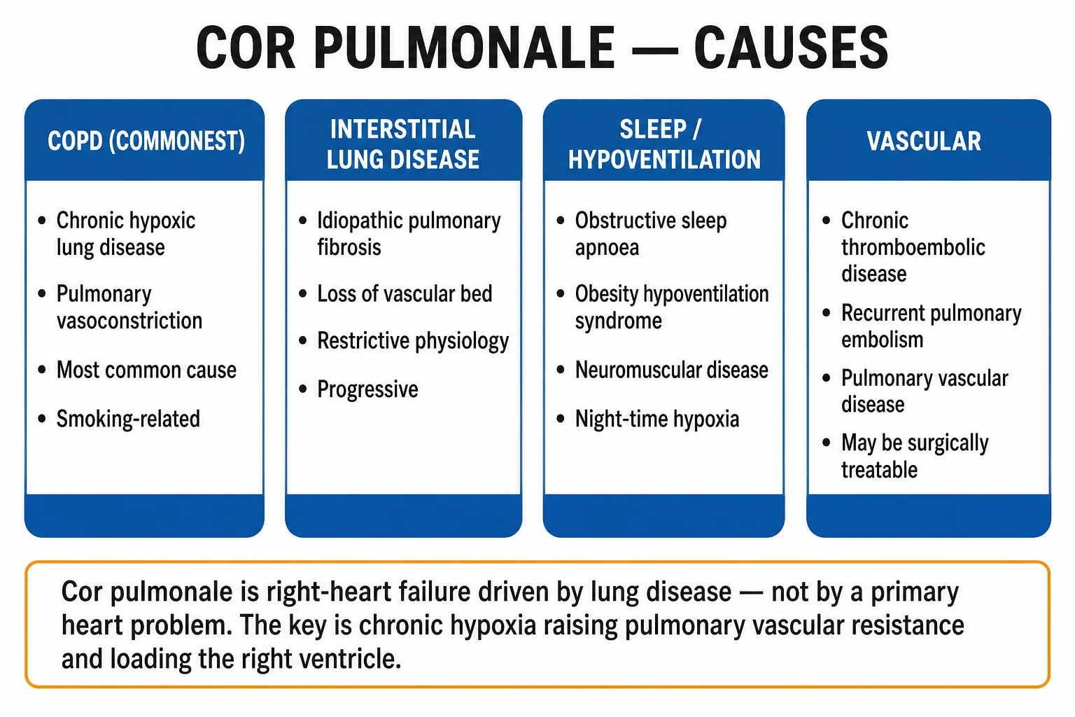
WHO Group 3 (lung disease / hypoxia) — the cor-pulmonale causes
| Mechanism | Specific causes | Comment |
|---|---|---|
| Obstructive airways disease | COPD (commonest), asthma with chronic fixed obstruction, bronchiectasis, cystic fibrosis | COPD accounts for the majority of cor pulmonale cases |
| Restrictive parenchymal disease | Idiopathic pulmonary fibrosis (IPF), connective-tissue disease-associated ILD, sarcoidosis, pneumoconiosis | Severe PH in IPF carries a poor prognosis |
| Disorders of ventilatory control | Obstructive sleep apnoea, obesity hypoventilation syndrome (OHS), central hypoventilation, high-altitude dwelling | Nocturnal hypoxia drives PH; often reversible with CPAP/NIV and weight loss |
| Restrictive chest-wall disease | Severe kyphoscoliosis, thoracoplasty, pleural disease | Long-term NIV is the treatment |
| Neuromuscular disease | Muscular dystrophy, motor neuron disease, myasthenia (chronic) | Long-term NIV |
| Pulmonary vascular (Group 4) | Chronic thromboembolic PH (CTEPH), recurrent PE, tumour emboli | The only surgically curable cause — pulmonary endarterectomy |
The complete WHO 5-group classification of pulmonary hypertension
The 2022 ESC/ERS Guidelines organise PH into five clinical groups based on the underlying mechanism. Cor pulmonale is, by definition, the structural RV consequence of Groups 1, 3, 4 and 5 (anything pre-capillary), but the term is used most strictly for Group 3. Every patient with suspected cor pulmonale must be placed in the correct group, because treatment is group-specific.[1]
The five WHO groups of pulmonary hypertension
Group 1 — PAH
- Pulmonary arterial hypertension: pre-capillary, idiopathic, heritable (BMPR2 mutation)
- Drug/toxin (anorexigens, methamphetamines)
- Associated with CTD (systemic sclerosis), portal hypertension, congenital heart disease, HIV
- Treat WITH pulmonary vasodilators (ERA, PDE5i, prostacyclin, sGC)
- Cor pulmonale if untreated
Group 2 — LHD
- Left-heart disease (LV systolic/diastolic, mitral/aortic valve)
- Post-capillary (PCWP over 15 mmHg)
- NOT cor pulmonale — treat the LV
- Vasodilators not indicated
Group 3 — lung disease / hypoxia
- COPD, ILD, OSA/OHS, kyphoscoliosis, neuromuscular, CF, high altitude
- Pre-capillary from hypoxic vasoconstriction + vascular loss
- THE cor-pulmonale group
- Treat lungs + LTOT + NIV; vasodilators NOT routine
Group 4 — CTEPH
- Chronic thromboembolic PH after PE
- The ONLY surgically curable cause (pulmonary endarterectomy)
- V/Q scan mandatory in every unexplained PH
- Riociguat, BPA if inoperable
Group 5 — miscellaneous
- Haematological (myeloproliferative, sickle cell), systemic (sarcoid, vasculitis), metabolic, chronic renal failure
- Pre-capillary, multifactorial
- Treat the underlying cause + specialist PH referral
Epidemiology & Risk Factors
- Cor pulmonale complicates a significant proportion of advanced COPD; the prevalence of PH in COPD rises with GOLD stage, reaching a majority of patients with very severe (GOLD 4) disease. Longstanding COPD with chronic hypoxaemia is the prototypical substrate.
- In interstitial lung disease (especially IPF), PH develops in a large fraction of advanced cases and is a major cause of death.
- Obesity hypoventilation syndrome (OHS) is almost always accompanied by some degree of PH, and the rising prevalence of obesity has made this an increasingly common cause.
- Chronic thromboembolic PH (CTEPH) complicates roughly 0.1–9% of survivors of acute PE within 2 years; it is under-diagnosed because it is missed without a V/Q scan. [1]
Principal risk factors
Heavy smoking (for COPD), longstanding COPD with chronic hypoxaemia, severe ILD (IPF), morbid obesity, uncontrolled OSA, recurrent pulmonary embolism or prior DVT, high-altitude dwelling, kyphoscoliosis, neuromuscular disease, and occupational dust/fume exposure. [1]
Key physiological question — why does chronic hypoxia produce cor pulmonale? Because sustained alveolar hypoxia (PaO2 under 55 mmHg) produces diffuse hypoxic pulmonary vasoconstriction, which — combined with loss of the capillary bed — chronically raises PVR, loads the RV and (when hypoxia is corrected by LTOT) is partly reversible.[1][2]
Pathophysiology
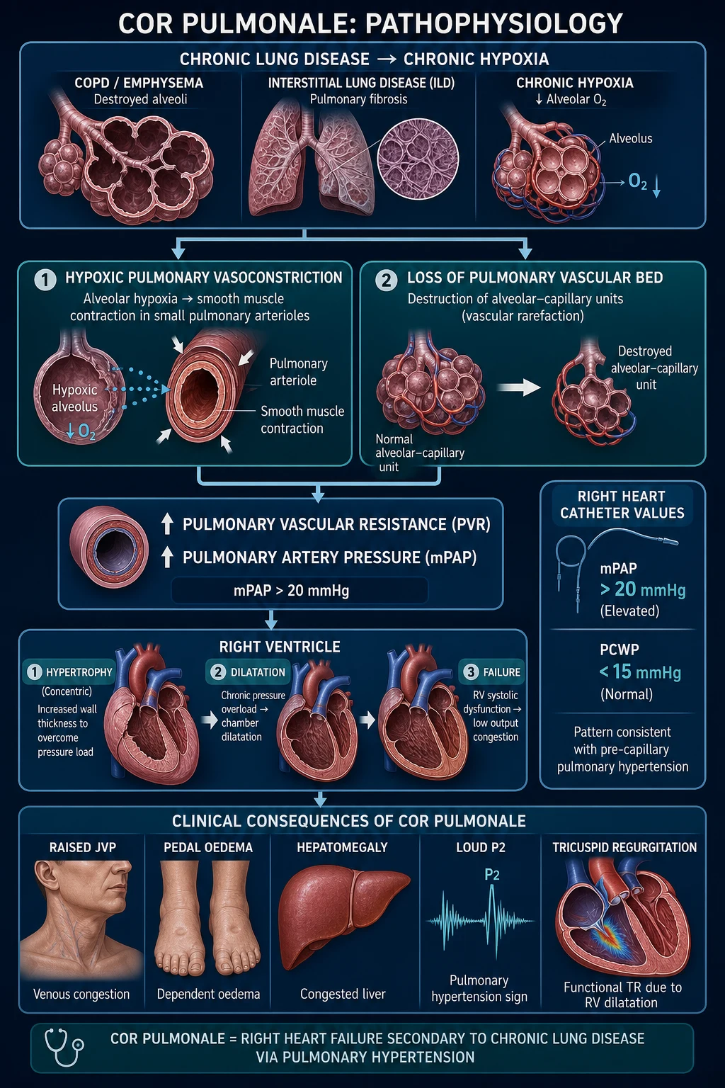
The mechanism has three stages — (A) the rise in PVR, (B) the RV response, and (C) the vicious cycle of decompensation. [1]
A. Why pulmonary vascular resistance rises
(i) Hypoxic pulmonary vasoconstriction (Euler-Liljestrand reflex). Locally, when an alveolus is poorly ventilated, the low alveolar oxygen tension inhibits voltage-gated potassium (KV) channels on the smooth muscle of the adjacent pulmonary arteriole. The membrane depolarises, voltage-gated calcium channels open, intracellular calcium rises, and the smooth muscle contracts — diverting blood away from poorly ventilated alveoli toward well-ventilated ones (a local V/Q matching mechanism). When the hypoxia is focal this is adaptive; when it is global and chronic (as in COPD, ILD, sleep apnoea, high altitude) it produces diffuse vasoconstriction, raising PVR throughout the lung. [1]
(ii) Loss of the pulmonary vascular bed. In emphysema the destruction of alveolar septa obliterates the capillaries that ran through them; in ILD, fibrosis obliterates small vessels. Because PVR depends on the total cross-sectional area of the pulmonary circulation, a smaller vascular bed inevitably raises PVR — even at a given blood flow. The equation governing this is: [1]
- PVR = (mPAP − PCWP) / CO (Wood units)
- mPAP = PVR × CO + PCWP [1]
(iii) Secondary vascular remodelling. Chronic hypoxia and shear stress stimulate medial hypertrophy (smooth muscle thickening), intimal fibrosis, in-situ microthrombus and (rarely in Group 3, more in Group 1) plexiform lesions. These changes fix the high PVR even after oxygen is corrected — which is why prevention (early LTOT) is more effective than reversal. [1]
(iv) Secondary polycythaemia. Chronic hypoxaemia stimulates erythropoietin release from the kidney, raising haematocrit. Polycythaemia increases blood viscosity, which raises PVR (and thrombotic risk), further loading the RV. [1]
B. The right ventricular response — hypertrophy, then dilatation, then failure
The RV is a thin-walled, high-compliance, low-pressure chamber adapted to a low-pressure pulmonary circulation (normal mPAP 14 ± 3 mmHg). It generates forward flow largely by filling (volume change), unlike the LV which does so by contraction (pressure change). Under a chronic pressure load: [1]
- Concentric hypertrophy — increased RV wall thickness, an adaptive response to raised afterload (compensated cor pulmonale).
- Dilatation — as PVR continues to rise and exceeds the RV's compensatory capacity, the chamber dilates, the tricuspid annulus stretches, and tricuspid regurgitation develops (now a volume as well as a pressure load).
- Failure — RV stroke output falls; systemic venous pressure rises (raised JVP, hepatomegaly, ascites, peripheral oedema) and cardiac output falls (fatigue, exertional syncope, cool peripheries). [1]
The RV is also uniquely vulnerable to ischaemia: it is perfused during both systole and diastole, but at high PVR the systemic blood pressure may not exceed the rising pulmonary pressure, reducing RV coronary perfusion and producing RV ischaemia that worsens failure. [1]
C. The vicious cycle of decompensation
Once RV output falls, tissue hypoxia worsens (driving more pulmonary vasoconstriction), renal perfusion falls (activating RAAS, retaining sodium and water, worsening congestion and oedema), and tachyarrhythmias (especially atrial flutter and fibrillation) — poorly tolerated in a stiff RV — develop. Each step reinforces the next, which is why an acute decompensation is a medical emergency. [1]
Right-heart failure: cor pulmonale vs left-heart failure
Cor pulmonale (Group 3)
- Cause = lung disease (COPD, ILD, OSA, CTEPH)
- Pre-capillary PH (PCWP under 15 mmHg)
- Systemic congestion dominant (JVP, hepatomegaly, oedema)
- Little/no pulmonary oedema
- Treat lungs + oxygen + ventilation
- Pulmonary vasodilators NOT routine
Left-heart failure (Group 2)
- Cause = LV systolic/diastolic disease, mitral/valve disease
- Post-capillary PH (PCWP over 15 mmHg)
- Pulmonary congestion dominant (orthopnoea, PND, crackles)
- Bilateral pulmonary oedema on imaging
- Treat LV (ACEi/ARB/ARNI, beta-blocker, MRA, SGLT2i, diuretics)
- Vasodilators/afterload reduction central
Clinical Presentation
The presentation blends the underlying lung disease with right-heart failure. The classic patient is a middle-aged or older smoker with known COPD who develops ankle swelling, increasing dyspnoea and fatigue over months. [1]
Symptoms
- Progressive exertional dyspnoea — the most common symptom, often the first sign that PH has developed on top of the lung disease.
- Fatigue, low exercise tolerance — from low cardiac output.
- Ankle and lower-limb swelling — from systemic venous congestion; may progress to sacral oedema, ascites.
- Right-upper-quadrant discomfort — from hepatic congestion (capsular stretch).
- Abdominal distension — hepatomegaly ± ascites.
- Syncope on exertion — an important red flag indicating severe PH and an inability to raise cardiac output on exercise.
- Cough, sputum, wheeze — from the underlying lung disease. [1]
Signs of right-heart failure
- Raised JVP — at rest, with prominent a waves (right atrial hypertrophy) and v waves (tricuspid regurgitation). A prominent v wave with a systolic liver pulse indicates significant TR.
- Ankle / sacral oedema.
- Hepatomegaly — smooth, tender, pulsatile if TR; a positive hepatojugular reflux.
- Ascites in advanced disease.
- Cool peripheries, low-volume pulse — from low cardiac output. [1]
Signs of pulmonary hypertension (the auscultatory cluster)
- Loud, palpable P2 (pulmonary component of the second heart sound) at the upper left sternal edge.
- Pulmonary ejection click and a pulmonary flow murmur (raised PA pressure).
- Graham Steell murmur — a high-pitched early diastolic murmur of pulmonary regurgitation (from dilation of the pulmonary valve ring).
- Right-sided S3 (RV failure) and S4 (RV hypertrophy).
- Tricuspid regurgitation — a pansystolic murmur at the lower left sternal edge, louder on inspiration (Carvallo's sign), with giant v waves.
- Parasternal right-ventricular heave — sustained impulse felt at the left sternal border.
- Palpable pulmonary impulse in the second left intercostal space. [1]
Signs of the underlying lung disease
In COPD: cyanosis, barrel chest, prolonged expiration, pursed-lip breathing, use of accessory muscles, Hoover's sign (paradoxical inward movement of the lower rib margin on inspiration), biphasic wheeze, coarse basal crackles, asterixis in CO2 retention. In ILD: finger clubbing, fine Velcro-like basal crackles, reduced lung volumes. In OSA/OHS: obesity, large neck circumference, daytime somnolence. [1]
Atypical presentations (high-yield)
- The elderly patient with known COPD presenting with only unexplained ankle oedema — cor pulmonale must be considered.
- OSA / obesity hypoventilation — the patient presents with morning headache, daytime hypersomnolence, snoring, and cor pulmonale rather than the classic COPD picture.
- CTEPH — progressive dyspnoea with disproportionate exertional syncope and a prior PE/DVT; the lung fields may look normal.
- Decompensating cor pulmonale — drowsiness, asterixis, hypoxaemia, rising PaCO2, oliguria, hypotension, cool peripheries. [1]
Differential Diagnosis
Cor pulmonale produces right-heart failure — but right-heart failure has many causes, and the diagnosis of cor pulmonale requires that the cause is the lung, not the left heart. [1]
| Differential | Distinguishing features | Key tests |
|---|---|---|
| Left-heart failure (Group 2 PH) | Pulmonary congestion (orthopnoea, PND, bilateral crackles), raised PCWP on echo/RHC, raised BNP/NT-proBNP, LV dysfunction on echo | Echo (LV, diastolic dysfunction), RHC (PCWP over 15 mmHg) |
| Idiopathic pulmonary arterial hypertension (Group 1) | Younger patient, no intrinsic lung disease, normal PFTs, normal PCWP, raised mPAP; family history (BMPR2) | RHC, exclude other causes |
| Chronic thromboembolic PH (Group 4) | Prior PE/DVT, exertional syncope, segmental perfusion defects on V/Q | V/Q scan (most sensitive), then CTPA/pulmonary angiography |
| Chronic venous insufficiency | No raised JVP, no hepatomegaly, varicose veins/stasis dermatitis, normal heart/lungs | Clinical; echo normal |
| Constrictive pericarditis | Raised JVP with Kussmaul's sign, pericardial knock, equal ventricular diastolic pressures, pericardial calcification on CT | Echo, cardiac CT/MRI, RHC (dip-and-plateau) |
| Right-ventricular infarction | Acute presentation with inferior MI, raised troponin, ECG changes, normally low PVR | ECG (inferior ST changes, right-sided leads V4R), troponin |
| Cirrhosis with ascites (hepatopulmonary/cardiac) | Signs of chronic liver disease (palmar erythema, spider naevi, low albumin), no raised JVP | LFTs, synthetic function, imaging |
| Tricuspid valve disease (primary) | Isolated TR/TS without PH or lung disease; giant v waves, prominent c-v waves | Echo |
Mandatory exclusion: every patient with unexplained PH needs a V/Q scan to exclude surgically curable CTEPH — relying on echo alone misses this diagnosis.[1]
Clinical & Bedside Assessment
Focused history
- Smoking pack-years and current status; biomass-fuel exposure (a major cause in women in developing nations); occupational dusts/fumes.
- Known COPD/ILD/OSA; prior PE/DVT; current inhalers, oxygen or NIV.
- Exacerbation frequency and hospitalisations; morning headache/somnolence (OSA/OHS); syncope on exertion (severe PH).
- Vaccination history (influenza, pneumococcal); comorbidity. [1]
Focused examination
- Cardiovascular — JVP (height, waveform, prominent a/v waves), parasternal RV heave, palpable P2, auscultation (loud P2, pulmonary click, Graham Steell, TR murmur, RV S3/S4), hepatojugular reflux, peripheral oedema.
- Respiratory — cyanosis, clubbing (ILD), barrel chest, pursed-lip breathing, Hoover's sign, wheeze, crackles, prolonged expiration.
- Abdominal — hepatomegaly (pulsatile if TR), ascites.
- Neurological — asterixis (CO2 retention), conscious level in decompensation. [1]
Functional class (WHO/NYHA, reproduced verbatim)
| Class | Definition |
|---|---|
| I | No limitation of physical activity; ordinary activity does not cause undue dyspnoea, fatigue, chest pain or near-syncope |
| II | Slight limitation of physical activity; comfortable at rest; ordinary activity causes dyspnoea, fatigue, chest pain or near-syncope |
| III | Marked limitation of physical activity; less-than-ordinary activity causes symptoms; comfortable only at rest |
| IV | Unable to carry out any physical activity without symptoms; symptoms at rest; signs of right-heart failure |
The WHO/NYHA functional class is a key prognostic and follow-up marker — patients in class IV have markedly worse survival. [1]
Investigations
1. Echocardiography — the key first-line test
- RV hypertrophy and dilatation, a flattened/bowing interventricular septum (D-shaped LV in parasternal short axis — a sign of RV pressure overload), tricuspid regurgitation.
- Estimated RV systolic pressure (RVSP) from the peak TR jet velocity using the simplified Bernoulli equation (RVSP = 4 × v² + right atrial pressure).
- TAPSE (tricuspid annular plane systolic excursion) — a measure of RV longitudinal systolic function (under 17 mm indicates RV dysfunction).
- Excludes left-heart disease (LV systolic/diastolic function, valve disease, raised left atrial pressure).
- Cannot definitively diagnose PH — echocardiographic estimates of mPAP are imprecise. [1]
2. Right heart catheterisation — the GOLD STANDARD
- Confirms PH (mPAP over 20 mmHg), classifies pre-capillary (PCWP under 15 mmHg, PVR over 2 WU) vs post-capillary, and measures cardiac index, right atrial pressure and mixed venous O2 saturation.
- Required before committing to advanced PH therapy, and in CTEPH workup.
- Measures pulmonary vascular resistance (PVR) = (mPAP − PCWP) / cardiac output, in Wood units (WU); PVR over 2 WU defines a pre-capillary component. Also allows acute vasodilator testing (inhaled nitric oxide or IV epoprostenol) — a positive response (fall in mPAP over 10 mmHg to a normal range) identifies the small subset of Group 1 PAH patients who benefit from calcium-channel blockers. No patient in Group 3 is vasoreactive.
- Indications for RHC: (a) confirm PH and define the group when echo is suggestive; (b) exclude post-capillary (left-heart) PH before starting any PH-specific drug; (c) baseline before advanced therapy; (d) CTEPH workup and transplant assessment. [1]
The haemodynamic profiles that separate the groups at the catheter are reproduced below — the PCWP value is the single most important number, because it tells you whether the left heart is the problem. [1]
Right-heart-catheterisation haemodynamic profiles (2022 ESC/ERS)
Pre-capillary PH (cor pulmonale)
- mPAP over 20 mmHg
- PCWP under 15 mmHg
- PVR over 2 WU
- Groups 1, 3, 4, 5 — lung/vasculature is the cause
- Treat the lungs + oxygen + ventilation
Isolated post-capillary PH
- mPAP over 20 mmHg
- PCWP over 15 mmHg
- PVR under 2 WU
- Group 2 — left-heart disease
- Treat the LV; NOT cor pulmonale
Combined pre- and post-capillary
- mPAP over 20 mmHg
- PCWP over 15 mmHg
- PVR over 2 WU
- Mixed — e.g. advanced LV failure with secondary vascular remodelling
- Treat both; specialist input
2b. Cardiac magnetic resonance (CMR)
- The reference standard for measuring right-ventricular mass, volume and ejection fraction — more accurate than echo for the thin-walled, crescentic RV, which is geometrically hard to assess.
- Demonstrates RV hypertrophy, dilatation, late gadolinium enhancement of the RV insertion points (a marker of chronic pressure overload), and interventricular septal flattening.
- Used for baseline and serial follow-up in specialist PH centres, and to assess right-to-left shunts (phase-contrast flow) that may contribute to PH. [1]
2c. Cardiopulmonary exercise testing (CPET)
- A maximal, incremental cycle test that pinpoints the limiting organ system (lung, heart, muscle) by measuring peak VO2, anaerobic threshold and the ventilatory equivalent for CO2. In PH the peak VO2 is reduced and the VE/VCO2 slope is steepened (inefficient ventilation), helping separate cardiovascular from respiratory limitation when both are present. [1]
3. ECG
- Right-axis deviation (QRS axis over +90°).
- P-pulmonale — tall peaked P waves (over 2.5 mm) in inferior leads II, III, aVF (right atrial enlargement).
- Right bundle branch block (complete or incomplete).
- Dominant R wave in V1 (R/S ratio over 1) — RV hypertrophy criterion.
- Poor R-wave progression in anterior precordial leads.
- Right ventricular strain — T-wave inversion in V1–V3.
- S1Q3T3 pattern (in acute PE; insensitive in chronic).
- Atrial arrhythmia — atrial flutter/fibrillation, which is poorly tolerated. [1]
4. Chest X-ray
- Enlarged central pulmonary arteries with peripheral pruning (loss of the normal tapering).
- Right-ventricular prominence (cardiomegaly with a lifted apex).
- Features of the underlying lung disease (hyperinflation, flat hemidiaphragms, bullae in COPD; reticulonodular changes, reduced volumes in ILD). [1]
5. Arterial blood gases
- Type 2 respiratory failure — low PaO2 (under 60 mmHg), raised PaCO2 (over 45 mmHg).
- Compensated respiratory acidosis — pH low-normal (7.35–7.40), raised bicarbonate (over 28 mmol/L) from renal compensation.
- The degree of hypoxaemia and hypercapnia correlates with prognosis and LTOT need. [1]
6. V/Q scan (ventilation-perfusion scintigraphy)
- Most sensitive test for CTEPH — segmental perfusion defects with preserved ventilation (mismatch).
- Mandatory in any unexplained PH, because CTEPH is the surgically curable cause. [1]
7. CT pulmonary angiogram and high-resolution CT
- CTPA — excludes acute PE; defines central vascular anatomy for surgical planning; less sensitive than V/Q for chronic thromboembolic disease.
- HRCT — characterises the underlying lung disease (emphysema distribution, fibrosis, bronchiectasis), assesses for vascular pruning in PH, excludes lung cancer. [1]
8. Pulmonary function tests
- Confirms and grades the underlying lung disease (COPD: post-bronchodilator FEV1/FVC under 0.70; ILD: reduced FVC and TLCO).
- Reduced diffusing capacity (TLCO) is a sensitive marker of PH complicating lung disease. [1]
9. 6-minute walk test (6MWT)
- Distance walked and SpO2 desaturation are functional and prognostic markers; a drop in SpO2 under 88% or a walk distance under 332 m suggests significant PH. [1]
10. Blood tests
- NT-proBNP — a useful non-invasive screening marker for PH in chronic lung disease (raised in RV stretch).
- Full blood count — secondary polycythaemia (raised haematocrit) from chronic hypoxia; anaemia of chronic disease.
- U&E, LFTs — renal/hepatic congestion, hyponatraemia of advanced heart failure.
- Thyroid function, HIV, connective-tissue screen — exclude secondary causes of PH. [1]
11. Polysomnography
- Mandatory when OSA/OHS is suspected — confirms sleep-disordered breathing and guides CPAP/BiPAP. [1]
Management — Resuscitation (Acute Decompensation)
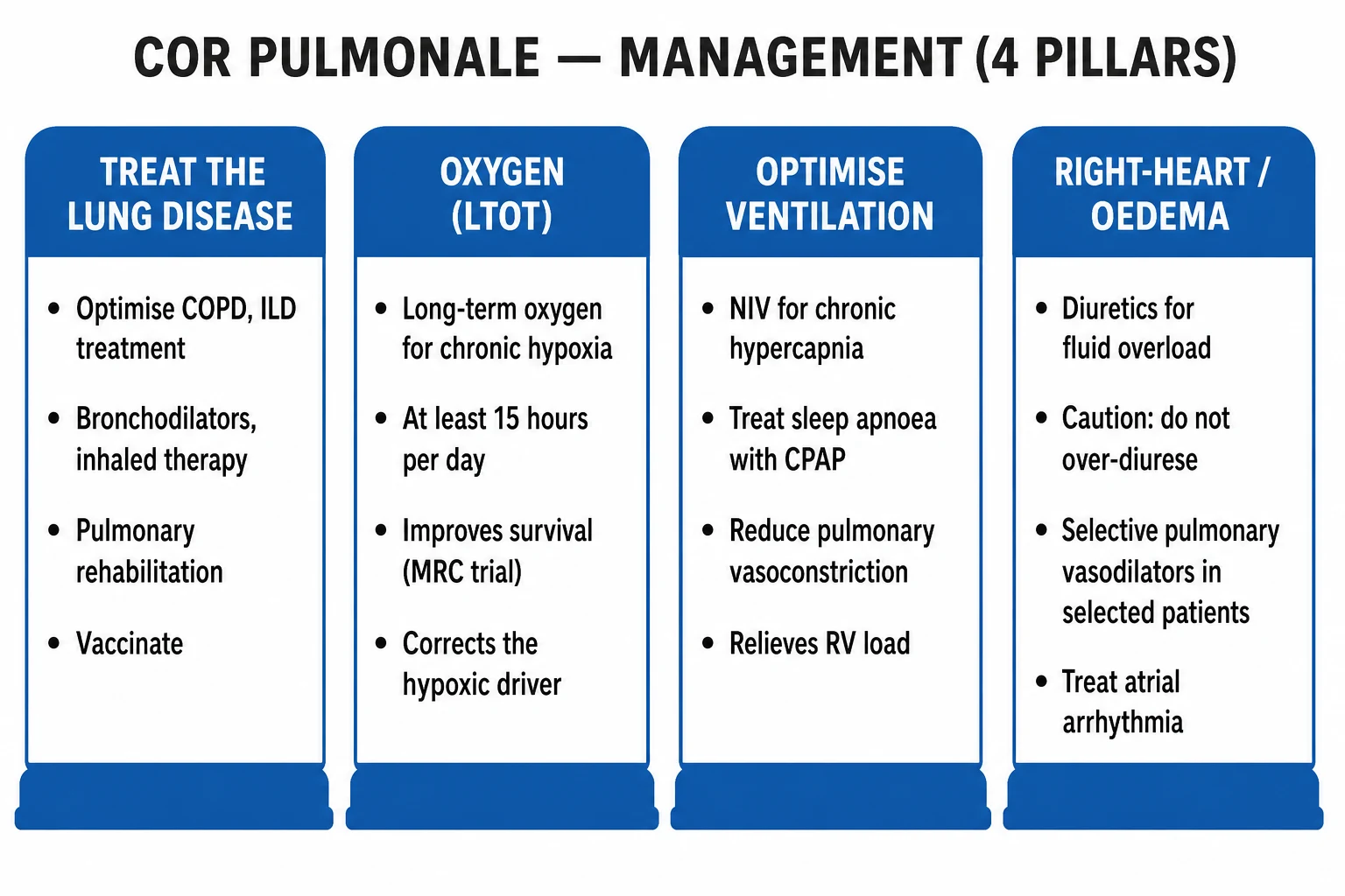
An acutely decompensating cor pulmonale patient (worsening hypoxaemia, rising PaCO2, drowsiness, oliguria, hypotension) is a medical emergency managed with the ABCDE approach: [1]
- Airway / Breathing — assess and secure.
- Controlled oxygen — target SpO2 88–92% in chronic CO2 retainers (see Pitfalls below). Use a Venturi mask (24% or 28%) for a predictable inspired oxygen concentration.
- Repeat ABG at 30–60 minutes after any change in oxygen or ventilation.
- Non-invasive ventilation (BiPAP) — start for acute hypercapnic respiratory failure (pH under 7.35 with PaCO2 over 45 mmHg despite controlled oxygen). Settings: IPAP 10–15 cmH2O, EPAP 4–5 cmH2O, titrate upwards.
- Intravenous diuretics — furosemide 20–40 mg IV for volume overload; cautious (see Pitfalls).
- Treat reversible triggers — pneumonia, exacerbation, arrhythmia, PE.
- Treat poorly-tolerated atrial flutter/fibrillation — rate-control (digoxin, cautious calcium-channel blocker), anticoagulation, consideration of cardioversion.
- Invasive mechanical ventilation — last resort; the raised intrathoracic pressure worsens RV afterload and may precipitate cardiovascular collapse. Use lung-protective ventilation, avoid hyperinflation, ensure adequate preload. [1]
The classic iatrogenic pitfall — CO2 narcosis from excessive oxygen
In chronic CO2 retainers, giving high-flow oxygen to target normoxia risks CO2 narcosis via two mechanisms: (1) V/Q mismatch — oxygen taken up by previously hypoxic-vessels-constricted alveoli increases perfusion of low-V/Q units; (2) the Haldane effect — oxygen binding to haemoglobin displaces CO2 from carbamino groups and reduces CO2 carriage as bicarbonate, releasing CO2 into venous blood. Always use controlled oxygen (target SpO2 88–92%) in suspected CO2 retainers. [1]
Management — Definitive & Stepwise
Pillar 1 — Treat the underlying lung disease
- COPD — dual bronchodilation (LAMA + LABA; e.g. tiotropium 18 microgram once daily + a LABA such as formoterol); add ICS only if blood eosinophils over 300 or frequent exacerbations; pulmonary rehabilitation; influenza and pneumococcal vaccination; smoking cessation (the only disease-modifying intervention — nicotine replacement, varenicline 0.5 mg daily titrated to 1 mg twice daily, bupropion 150 mg daily).
- ILD — disease-specific therapy (e.g. antifibrotics pirfenidone and nintedanib for IPF), immunosuppression for inflammatory ILD; refer for transplant in advanced disease.
- OSA / obesity hypoventilation — CPAP (or BiPAP for daytime hypercapnia) and weight reduction (bariatric surgery in selected OHS). [1]
Pillar 2 — Long-term oxygen therapy (LTOT)
- Criteria (per the MRC and NOTT trials): PaO2 under 55 mmHg (under 7.3 kPa) on room air on two stable occasions, OR PaO2 under 59 mmHg (under 7.8 kPa) with evidence of cor pulmonale or secondary polycythaemia.
- Prescription — oxygen for at least 15 hours/day (ideally approaching 24 h), via concentrator, at a flow that raises PaO2 to at least 60 mmHg (over 8 kPa) without worsening CO2.
- Survival benefit — the MRC trial (1981, PMID 6110912) showed domiciliary oxygen for about 15 h/day reduced mortality versus no oxygen in chronic hypoxic cor pulmonale; the NOTT trial (1980, PMID 6776858) showed continuous oxygen (about 24 h/day) halved mortality versus nocturnal-only (12 h).[2][3]
- Mechanism of benefit — corrects the hypoxic pulmonary vasoconstriction driving the RV overload; partially reverses PH and polycythaemia; reduces the afterload on the RV.
Pillar 3 — Optimise ventilation
- NIV (BiPAP) — for chronic hypercapnia (PaCO2 over 50 mmHg, especially with nocturnal hypoventilation), OHS, neuromuscular disease, kyphoscoliosis. Reduces PVR, lowers RV load, improves sleep quality and survival.
- CPAP — for obstructive sleep apnoea. Abolishes nocturnal hypoxia, often reversing the PH if treated early.
- Both are part of the treat-the-lungs philosophy that distinguishes cor pulmonale management from left-heart failure. [1]
Pillar 4 — Right-heart failure / oedema / arrhythmia
- Cautious diuretics — furosemide 20–40 mg orally once daily, titrated to clinical euvolaemia. Monitor renal function and blood pressure; the RV is preload-dependent — over-diuresis lowers cardiac output.
- No role for ACE inhibitors / beta-blockers as routine therapy in cor pulmonale (unlike LV failure); they may worsen hypotension and have no proven survival benefit.
- Atrial fibrillation/flutter — rate-control (digoxin, cautious beta-blocker/calcium-channel blocker), rhythm-control, anticoagulation with CHA2DS2-VASc.
- Secondary polycythaemia — venesection only if symptomatic hyperviscosity (haematocrit over 56%) — not routinely.
- Pulmonary vasodilators in Group 3 PH — generally NOT recommended in COPD/ILD (e.g. bosentan, sildenafil, riociguat) — they worsen V/Q mismatch and hypoxaemia without proven benefit; considered only in selected, severe cases under a specialist PH centre.
- Calcium-channel blockers — NOT useful in Group 3 PH (no acute vasodilator response, unlike idiopathic PAH). [1]
Pillar 4b — Pulmonary vasodilator / PH-specific therapy: the full drug ladder
An examiner will ask you to name the classes of pulmonary hypertension drug. The 2022 ESC/ERS Guidelines define a tiered pharmacology built almost entirely for Group 1 PAH; these agents are the standard of care in PAH but are generally NOT used in Group 3 (lung disease) PH, because non-selective pulmonary vasodilation worsens intrapulmonary V/Q matching and systemic hypoxaemia without proven benefit.[1] They are summarised here so you can answer the "name the drug class and its target" question, and to mark the narrow exceptions where a specialist centre does use them in Group 3.
| Class | Example agents | Mechanism | Indication / caveat |
|---|---|---|---|
| Calcium-channel blockers | Nifedipine, amlodipine, diltiazem | Block L-type Ca²⁺ channels in pulmonary vascular smooth muscle | Only the small subset of Group 1 PAH with a positive acute vasodilator test at RHC (about 6 to 15 per cent of IPAH). Never in Group 3 — no vasoreactivity, and risk systemic hypotension |
| Endothelin receptor antagonists (ERA) | Bosentan (dual ETA/ETB), ambrisentan (ETA-selective), macitentan | Block endothelin-1 mediated vasoconstriction and proliferation | First-line oral in Group 1 PAH. Monitor LFTs monthly (bosentan hepatotoxicity); teratogenic — reliable contraception |
| PDE5 inhibitors | Sildenafil 20 mg TDS, tadalafil 40 mg OD | Inhibit PDE5 → raise cGMP → pulmonary vasodilation | First-line oral in Group 1 PAH. Caution with nitrates/alpha-blockers (hypotension) |
| sGC stimulator | Riociguat 1 mg TDS, titrated | Stimulates soluble guanylate cyclase → cGMP (independent of, and synergistic with, NO) | Group 1 PAH and Group 4 CTEPH (inoperable/persistent). Not combined with PDE5i (both raise cGMP — hypotension) |
| Prostacyclin analogues | Epoprostenol (continuous IV), iloprost (inhaled), treprostinil (SC/IV/inhaled/oral) | Prostacyclin (PGI2) receptor agonism → vasodilation, anti-platelet, anti-proliferation | Advanced Group 1 PAH; IV epoprostenol improves survival. High logistical complexity (continuous infusion, line infection risk) |
| IP receptor agonist | Selexipag (oral) | Selective prostacyclin IP receptor agonist | Oral, for Group 1 PAH; delays progression. Less complex than parenteral prostacyclin |
Why these are NOT routine in cor pulmonale (Group 3): in COPD and ILD the hypoxia is the driver and the vascular bed is structurally damaged. Diffuse vasodilation opens up poorly ventilated units (worsened low-V/Q shunt), lowering PaO2, while the fixed vascular loss is unaffected — so symptoms and prognosis do not improve. The RISE-IIP (riociguat in ILD-PH) and earlier sildenafil trials gave mixed or negative results; current evidence supports their use in Group 3 only in highly selected, severe, progressive cases under a specialist PH centre. The single exception that an examiner rewards: riociguat is licensed for inoperable or persistent CTEPH (Group 4), and pulmonary endarterectomy can cure it. [1]
Pillar 5 — Lung transplantation
- Considered in end-stage cor pulmonale from ILD (IPF), COPD, or cystic fibrosis when medical therapy fails and the RV failure is still potentially reversible — the transplanted lung(s) remove the hypoxic/vascular load and the RV recovers.
- A pre-transplant RHC is mandatory; severely fixed RV failure with multi-organ dysfunction may need heart-lung transplantation rather than bilateral lung alone.
- RV reverse remodelling after lung transplantation is often dramatic, which is why transplantation is offered before irreversible RV failure sets in. [1]
Specific treatment of CTEPH — the surgically curable cause
- Lifelong anticoagulation (warfarin or DOAC).
- Pulmonary endarterectomy (PEA) — the potentially curative operation for operable, accessible organised thrombus in the main/lobar/segmental pulmonary arteries; performed at a specialist centre.
- Riociguat (soluble guanylate cyclase stimulator) — for non-operable or persistent CTEPH; 1 mg three times daily, titrated.
- Balloon pulmonary angioplasty (BPA) — for inoperable distal disease.
- Operability is decided at a specialist CTEPH multidisciplinary team — depends on thrombus accessibility (proximal main/lobar is operable; distal segmental/subsegmental is not), the haemodynamic burden, and surgical fitness. A pre-operative pulmonary angiogram (not CTPA) maps the organised thrombus. PEA mortality in expert centres is under 5 per cent, and patients who survive can be functionally cured with near-normal pulmonary pressures.
- Residual/persistent PH after PEA or inoperable disease is treated with riociguat and staged BPA (balloon pulmonary angioplasty — serial catheter dilatations of organised thrombotic stenoses, typically over multiple sessions). Anticoagulation continues lifelong; the optimal agent in CTEPH remains debated (warfarin historically preferred; DOACs increasingly used). [1]
Cor pulmonale — the four pillars (OLAT)
OLAT
LTOT for at least 15 h/day when PaO2 under 55 mmHg — improves survival (MRC, NOTT)
Treat the underlying COPD/ILD — bronchodilators, ICS by eosinophils, rehab, vaccines, smoking cessation
Optimise ventilation — NIV/BiPAP for hypercapnia, CPAP for OSA
Cautious diuretics (RV is preload-dependent), treat atrial arrhythmia, exclude CTEPH with V/Q
Specific Subtypes & Scenarios
- COPD with cor pulmonale — the prototype. LTOT, NIV if hypercapnic, optimise inhalers, treat exacerbations aggressively, smoking cessation.
- Cor pulmonale in ILD (IPF) — restrictive, hypoxic; often severe PH in advanced disease. Vasodilators generally avoided (worsen hypoxaemia); refer early for lung transplantation.
- Cor pulmonale in OSA / obesity hypoventilation syndrome — CPAP/BiPAP plus weight loss; PH is often reversible if treated early.
- Cor pulmonale in CTEPH — the only surgically curable cause; V/Q mandatory in every unexplained PH; PEA, riociguat or BPA.
- Cor pulmonale in kyphoscoliosis / neuromuscular disease — restrictive chest-wall disease; long-term NIV.
- Acute cor pulmonale from massive PE — a different entity (sudden RV overload); anticoagulation, thrombolysis, embolectomy.
- High-altitude cor pulmonale (chronic mountain sickness / Monge's disease) — chronic hypoxia at altitude; descent or supplemental oxygen; venesection for polycythaemia. [1]
Complications & Pitfalls
Complications
- Worsening right-heart failure — refractory oedema, ascites, anasarca.
- Atrial arrhythmia — atrial flutter/fibrillation, poorly tolerated.
- Secondary polycythaemia — from chronic hypoxia; raises viscosity and thrombotic risk.
- Venous thromboembolism and pulmonary embolism — increased risk from immobility and polycythaemia.
- Cardiorenal syndrome — type 2 (chronic), rising creatinine from renal congestion and low cardiac output.
- Hepatorenal dysfunction — congestive hepatopathy ("nutmeg liver"), fibrosis, coagulopathy.
- Death — once severe RV failure is established, prognosis is poor without intervention. [1]
Classic iatrogenic pitfalls (high-yield)
- CO2 narcosis from high-flow oxygen in a chronic CO2 retainer — always target SpO2 88–92%.
- Over-diuresis — lowers preload, reduces RV stroke output (RV is preload-dependent), worsens renal function.
- Indiscriminate pulmonary vasodilators in COPD/ILD — worsen V/Q mismatch and hypoxaemia.
- Missing CTEPH — a surgically curable cause overlooked if only an echo is done without a V/Q scan.
- Calcium-channel blockers without an acute vasodilator study — useless in Group 3 PH and may cause hypotension.
- Diagnosing cor pulmonale without excluding left-heart disease — post-capillary PH (Group 2) is treated very differently. [1]
Exam application bank (NEET-PG / INICET)
One-line answer
Cor pulmonale is right ventricular hypertrophy, dilatation and failure caused by lung disease (NOT by a primary cardiac problem), via pulmonary hypertension driven by chronic hypoxia and loss of the pulmonary vascular bed. The commonest cause is COPD; others include interstitial lung disease, obstructive sleep apnoea/obesity hypoventilation, chronic thromboembolic disease and restrictive chest-wall disease. Patients show the features of the underlying lung disease plus right-heart failure — raised JVP, peripheral oedema, hepatomegaly, a loud pulmonary second sound (P2), a parasternal right-ventricular heave and tricuspid regurgitation. Echocardiography demonstrates RV hypertrophy/dilatation and raised estimated pulmonary pressures (right heart catheterisation is the gold standard). Management is to treat the underlying lung disease, give long-term oxygen therapy (which improves survival,
Worked stems (answer without another resource)
Stem 1 — Classic presentation. Map symptoms to mechanism; name the first investigation and first treatment step with dose/route if drug therapy is standard. [1]
Stem 2 — Unstable / complicated. List red flags that force immediate resuscitation, theatre, ICU, antidote, or reperfusion — and what you do in the first 15 minutes. [1]
Stem 3 — Atypical group. Elderly, pregnancy, child, or immunocompromised: how presentation and thresholds change. [1]
Stem 4 — Differential trap. Name the three closest mimics and one discriminator for each. [1]
Stem 5 — Disposition. Who goes home with safety-netting, who is admitted, who needs HDU/ICU/theatre, and what follow-up is mandatory. [1]
Rapid viva checklist
- Definition + classification
- Pathophysiology chain
- Bedside signs / criteria
- Score with exact components (if any)
- Emergency bundle
- Definitive therapy with doses
- Complications of disease and of treatment
- Special populations
- Guideline/trial name if classic
- Three exam traps
Coverage self-check
If you cannot answer any stem above from this page alone, re-read the matching section — the page is intended to be self-sufficient for final-prof and NEET-PG/INICET questions on Cor Pulmonale.
Prognosis & Disposition
Prognostic markers
- WHO/NYHA functional class — class IV markedly worse.
- 6-minute-walk distance — under 332 m suggests significant disease.
- Right-ventricular function — TAPSE under 17 mm, RV dilatation, pericardial effusion (poor prognostic markers).
- NT-proBNP — higher is worse.
- Haemodynamics (RHC) — right atrial pressure over 8 mmHg, cardiac index under 2.5 L/min/m², mixed venous O2 saturation under 60%.
- Underlying disease — COPD with chronic hypoxaemia, severe IPF, CTEPH. [1]
Survival and what improves it
- 5-year survival in PH complicating COPD/ILD is poor once RV failure is established.
- LTOT (MRC, NOTT) and optimised ventilation (NIV/CPAP) improve survival.
- Pulmonary endarterectomy can cure CTEPH.
- Smoking cessation slows the underlying COPD. [1]
Follow-up
Multidisciplinary respiratory / PH clinic; serial echocardiography and NT-proBNP; repeat 6MWT; optimise underlying disease; vaccination; written action plan for decompensation. [1]
Special Populations
- Elderly — multimorbidity, polypharmacy; may present with only ankle oedema; beware over-diuresis and drug interactions.
- Pregnancy — pulmonary hypertension carries a high maternal mortality (30–56% historically); counsel against pregnancy; manage in a specialist PH-obstetric clinic if pregnancy occurs.
- Obesity hypoventilation syndrome / OSA — BMI over 30, daytime hypercapnia, sleep-disordered breathing; CPAP/BiPAP and weight loss; PH is often reversible.
- Children — bronchopulmonary dysplasia, cystic fibrosis-related PH, congenital lung disease.
- Patients already on anticoagulants — balance bleeding vs thrombosis in CTEPH; lifelong anticoagulation is needed regardless.
- Perioperative — patients with cor pulmonale carry high perioperative risk; optimise oxygenation and ventilation, avoid hypoxia/hypercapnia and over-sedation; consider invasive monitoring. [1]
Evidence, Guidelines & Regional Differences
- 2022 ESC/ERS PH Guidelines (Humbert et al., PMID 36017548) — current global reference. Revised the diagnostic threshold (mPAP over 20 mmHg, down from over 25 mmHg) and introduced PVR over 2 WU for pre-capillary PH. Stratifies PH into the five WHO groups.[1]
- MRC trial (1981, PMID 6110912) — established that domiciliary oxygen for about 15 h/day improved survival in chronic hypoxic cor pulmonale complicating chronic bronchitis/emphysema.[2]
- NOTT (1980, PMID 6776858) — showed continuous oxygen (about 24 h/day) halved mortality compared with nocturnal-only (12 h/day) in hypoxaemic COPD.[3]
- NICE NG115 (COPD) and GOLD 2024 — recommend LTOT, NIV, and the treat-the-lung approach; reaffirm the controlled-oxygen target SpO2 88–92%.
- BTS home oxygen guidance — practical delivery of LTOT in the community.
- Regional deltas — drug availability and naming differ (e.g. riociguat widely available; roflumilast for COPD exacerbators; access to pulmonary endarterectomy is centralised).
- Controversy — pulmonary vasodilators in Group 3 PH. Current evidence does NOT support routine use in COPD/ILD; trials (e.g. RISE-IIP in ILD-PH, INCREASE in ILD-PH) are mixed. Specialist PH centres may use them in selected, severe, progressive cases.
- Controversy — the 2022 mPAP threshold change. Lowering the threshold from over 25 to over 20 mmHg and adding the PVR cut-off reclassified many patients; some clinicians worry about over-diagnosis, but the 2022 guidance is now the global standard.
Exam Pearls
- Cor pulmonale = RV hypertrophy then failure from lung disease via pulmonary hypertension (WHO Group 3 PH).
- Commonest cause is COPD; other big causes — ILD, OSA/OHS, CTEPH, restrictive chest-wall disease.
- Mechanism: hypoxic pulmonary vasoconstriction (Euler-Liljestrand reflex — KV channel inhibition, Ca²⁺ influx) plus loss of the vascular bed → raised PVR.
- Bedside cluster: loud P2, RV heave, raised JVP, TR murmur (Carvallo's sign), hepatomegaly, ankle oedema.
- ECG cluster: right-axis deviation, P-pulmonale, RBBB, dominant R in V1, poor R-wave progression.
- Echo: RV hypertrophy/dilatation, D-shaped septum, TR, raised RVSP; right heart catheterisation is the gold standard.
- Haemodynamics (2022 ESC/ERS): PH = mPAP over 20 mmHg; pre-capillary = PCWP under 15 mmHg + PVR over 2 WU.
- LTOT for at least 15 h/day improves survival when PaO2 under 55 mmHg — MRC and NOTT trials.
- Controlled oxygen target SpO2 88–92% in CO2 retainers (avoid CO2 narcosis — Haldane effect, V/Q mismatch).
- Every unexplained PH needs a V/Q scan to find surgically curable CTEPH — pulmonary endarterectomy.
- Avoid over-diuresis (RV is preload-dependent); avoid routine vasodilators/CCBs in COPD/ILD Group 3 PH (worsen V/Q mismatch).
- NT-proBNP is a useful non-invasive screening marker for PH in chronic lung disease.
- Pregnancy in PH carries high maternal mortality — counsel against. [1]
References
- [1]Humbert M, Kovacs G, Hoeper MM, et al. 2022 ESC/ERS Guidelines for the diagnosis and treatment of pulmonary hypertension Eur Heart J, 2022.PMID 36017548
- [2]Medical Research Council Working Party. Long term domiciliary oxygen therapy in chronic hypoxic cor pulmonale complicating chronic bronchitis and emphysema. Report of the Medical Research Council Working Party Lancet, 1981.PMID 6110912
- [3]Nocturnal Oxygen Therapy Trial Group. Continuous or nocturnal oxygen therapy in hypoxemic chronic obstructive lung disease: a clinical trial. Nocturnal Oxygen Therapy Trial Group Ann Intern Med, 1980.PMID 6776858