Respiratory · Respiratory
Haemoptysis
Also known as Haemoptysis · Hemoptysis · Coughing up blood · Massive haemoptysis · Life-threatening haemoptysis · Diffuse alveolar haemorrhage
Haemoptysis is the coughing up of blood that originates from the lower respiratory tract (below the vocal cords). It ranges from blood-streaked sputum to massive, life-threatening haemoptysis. Worldwide the commonest cause is tuberculosis; in developed countries bronchial carcinoma, bronchiectasis and pneumonia predominate. Massive haemoptysis (any volume threatening the airway or gas exchange, classically over 100 to 240 mL/24h) kills by asphyxia, not exsanguination — because over 90 percent of such bleeds arise from the high-pressure bronchial arteries. Management is stepwise: protect the airway, oxygenate, put the BLEEDING LUNG DOWN, reverse anticoagulation, then rigid bronchoscopy with tamponade and bronchial artery embolisation (BAE) first-line; surgery is last resort. Always exclude PE, TB, carcinoma and diffuse alveolar haemorrhage (ANCA / anti-GBM).
On this page & tools
Your progress
Saved locally on this device.
Exam tags
Red flags
Overview & Definition
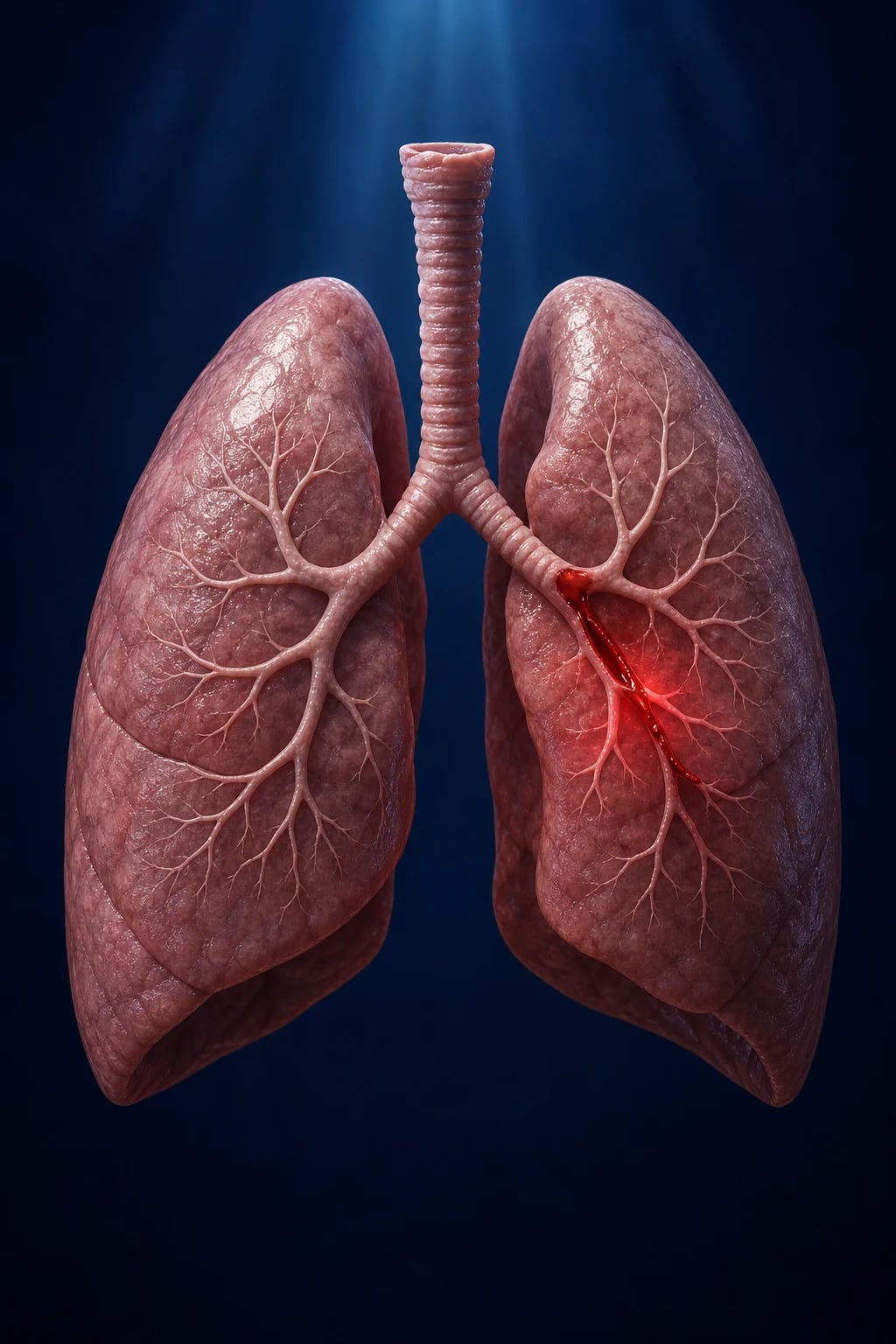
Haemoptysis is the expectoration of blood that originates from the respiratory tract below the vocal cords — i.e. from the trachea, bronchi, bronchioles or lung parenchyma.[3] The word itself (haima, blood + ptysis, to spit) names the act: blood that is coughed from the airway, not vomited from the stomach or swallowed from the nose. Defining the source as below the vocal cords is what separates true haemoptysis from its mimics, and it is the single most important bedside distinction to make before any investigation is ordered.
It must be distinguished at the bedside from two mimics: [1]
- Haematemesis — vomiting blood from the upper GI tract. Haemoptysis is bright red, frothy, alkaline and coughed up; it is mixed with sputum and may continue over several days. Haematemesis is dark ('coffee-ground'), acidic and vomited, often with a history of liver disease, peptic ulcer or dyspepsia. (In practice, aspiration of haematemesis and swallowing of haemoptysis blur the picture — a careful history of the act — cough vs vomit — is decisive.)
- Pseudohaemoptysis — blood from the upper airway (epistaxis, gingival disease, pharyngeal or laryngeal lesion) or factitious (food dye, Munchausen). Always inspect the nose, oropharynx and upper airway before concluding the blood is pulmonary.[3]
The clinical skill in haemoptysis is triage: deciding who is stable for an outpatient work-up (mild, self-limiting, no red flags) versus who needs emergency airway protection, bronchoscopy and BAE (massive/life-threatening). Volume alone is a poor discriminator — a small bleed in a patient with a poor cough or limited respiratory reserve can kill.[1][4]
Classification
Haemoptysis is classified by volume/severity (which drives the urgency and site of care) and by source (bronchial arterial vs pulmonary capillary). Volume thresholds have historically varied widely, which is why the modern trend is toward a functional definition.[3]
Scant / mild
- Blood-streaked sputum or under 30 mL/24h
- Stable; no airway threat
- Most common presentation
- Outpatient work-up: CXR, consider CT, sputum, bronchoscopy if indicated
- Underlying cause often benign (bronchitis, post-infective) but EXCLUDE malignancy in smokers over 40
Moderate (sub-massive)
- 30 to 200 mL/24h, repeated small clots
- Monitor closely on respiratory ward
- CXR + CT thorax/CTPA
- Flexible bronchoscopy once stable
- Crossmatch 2 to 4 units; correct coagulopathy
- Risk of escalation — observe for airway compromise
Massive / life-threatening
- Any volume threatening the airway, gas exchange or haemodynamics (classically over 100 to 240 mL/24h, or over 50 mL single episode)
- Airway compromise, hypoxia, shock
- ITU/HDU; airway first
- Rigid bronchoscopy + BAE first-line
- Death is from ASPHYXIA, not exsanguination
- Mortality historically 50 to 80 percent, now 7 to 20 percent
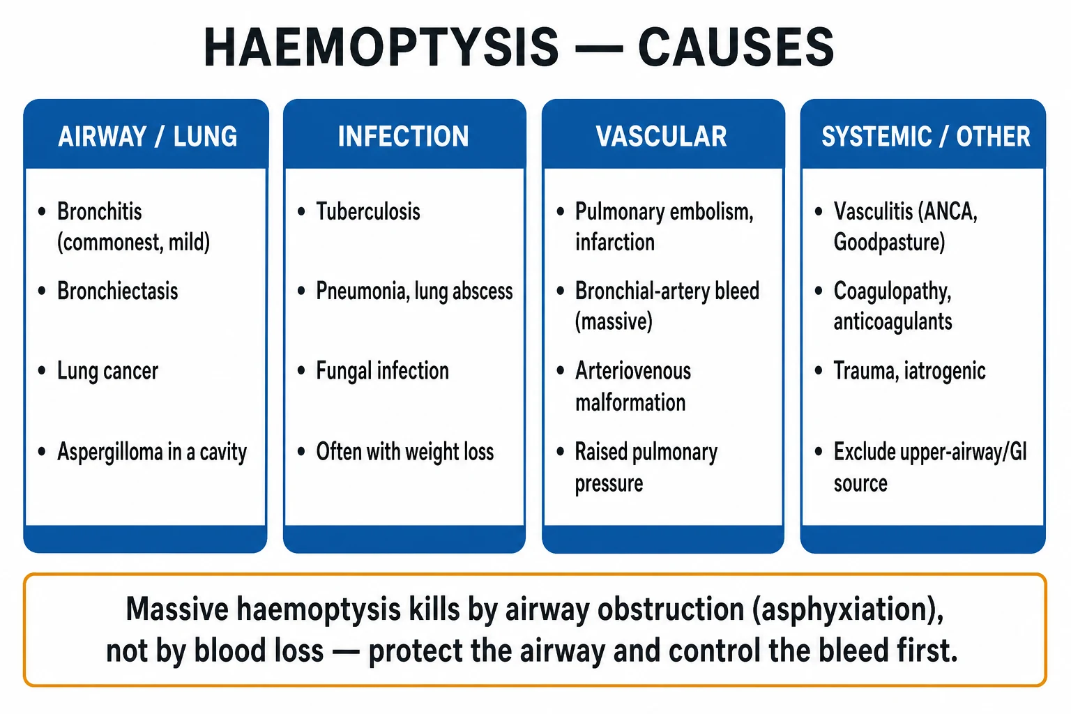
In TB-endemic regions (India, South Asia, sub-Saharan Africa) the lower threshold (over 100 mL/24h) is widely taught, because post-TB bronchiectasis and aspergilloma make massive bleeds common. The functional definition is universally adopted for management decisions regardless of the volume quoted on an exam.
Epidemiology & Risk Factors
Haemoptysis accounts for a substantial minority of respiratory presentations — roughly one in a thousand emergency attendances, and it is the presenting feature in a significant fraction of lung cancer and TB diagnoses. The aetiology is geography-dependent:[3]
- Worldwide / TB-endemic regions (India, South Asia, sub-Saharan Africa): tuberculosis is the commonest cause — both active disease (cavitary, endobronchial) and the late sequelae Rasmussen aneurysm, post-TB bronchiectasis and aspergilloma colonising an old TB cavity.
- Developed countries: bronchial carcinoma, bronchiectasis, pneumonia/lung abscess, pulmonary embolism; chronic bronchitis in smokers.
- After a full work-up, 7 to 25 percent remain cryptogenic (idiopathic) — these require follow-up imaging as a small proportion harbour an occult malignancy that declares itself over months.[3]
Cryptogenic haemoptysis deserves emphasis: when CXR, CT and bronchoscopy are all normal, 6 to 10 percent of these "idiopathic" cases turn out to harbour a bronchial carcinoma that declares itself on follow-up — which is why repeat imaging at 6 to 8 weeks (and again at 6 months) is mandatory in smokers and older patients, and why "cryptogenic" is a diagnosis of exclusion made only after a complete work-up, never a reassurance.[3]
Causes of MASSIVE (life-threatening) haemoptysis differ in rank from mild haemoptysis: the leading sources are bronchiectasis (including cystic fibrosis), lung carcinoma, active TB / mycetoma (aspergilloma), chronic necrotising aspergillosis, and pulmonary arteriovenous malformations (Osler–Weber–Rendu). Mild haemoptysis, by contrast, is most often bronchitis (smoker's) or a post-infective cough. This distinction — that bronchitis heads the list for mild bleeds while bronchiectasis heads the list for massive bleeds — is a favourite examiner question.[1][2]
Haemoptysis — high-yield numbers
Risk factors (high-yield associations): [1]
- Smoking, age over 40 — bronchial carcinoma (the single most important red-flag combination).
- Chronic lung disease — bronchiectasis (incl. cystic fibrosis), COPD, previous TB cavity (aspergilloma).
- Immunosuppression — invasive aspergillosis, TB reactivation, CMV, thrombocytopenia.
- Anticoagulation / antiplatelet therapy / coagulopathy — these unmask an occult lesion rather than cause bleed de novo; never assume the drug is the sole cause.
- Recent procedure — bronchoscopy with biopsy, transthoracic needle biopsy, pulmonary artery catheter (Swan–Ganz) — risk of iatrogenic PA rupture.
- Structural heart disease — mitral stenosis (rare but classic), left-heart failure.
- Vascular / hereditary — hereditary haemorrhagic telangiectasia (Osler–Weber–Rendu) with pulmonary AVM.
- Connective tissue / vasculitis — ANCA-associated vasculitis, anti-GBM disease, SLE, IgA nephropathy, antiphospholipid syndrome.
- Recreational drugs — cocaine (parenchymal damage), and occupational (trimellitic anhydride). [1]
Pathophysiology
The lung has a dual blood supply, and understanding it is the key to haemoptysis:[1][4]
- Pulmonary circulation — the low-pressure (mean about 15 mmHg) system carrying the entire cardiac output through the pulmonary capillary bed for gas exchange. Bleeding here tends to be diffuse (diffuse alveolar haemorrhage) and low-pressure, so it oozes rather than spurts.
- Bronchial circulation — the high-pressure systemic supply (aorta, mean about 100 mmHg) that nourishes the airways and is responsible for over 90 percent of massive haemoptysis. When a bronchial artery erodes into a bronchus (carcinoma, bronchiectasis, mycetoma, TB cavity), the high pressure produces brisk, voluminous bleeding that floods the airway. [1]
This dual anatomy is also why bronchial artery embolisation works: the culprit vessel is a systemic artery reachable from the aorta by an interventional radiologist, not the fragile pulmonary capillary bed. [1]
- Bronchiectasis / chronic infection — chronic airway inflammation causes hypertrophy and neo-vascularisation of the bronchial arteries (angiogenesis driven by chronic inflammation); the enlarged, tortuous, high-pressure vessels lie submucosally and rupture into the lumen. This is the commonest cause of massive haemoptysis in many series.
- Bronchogenic carcinoma — direct tumour invasion of bronchial vessels, tumour neo-angiogenesis, and necrosis of a vascular tumour bed.
- Tuberculosis — endobronchial disease, cavitary erosion, and the Rasmussen aneurysm (a pseudoaneurysm of a pulmonary artery branch in the wall of a chronic TB cavity that ruptures into the cavity and airway).
- Aspergilloma (mycetoma) — the fungus ball and its surrounding granulation tissue erode the highly vascular cavity wall; massive, recurrent haemoptysis is characteristic.
- Pulmonary embolism — infarction of a segment with haemorrhagic necrosis produces blood-tinged sputum (usually modest volume).
- Diffuse alveolar haemorrhage — capillaritis (ANCA, anti-GBM) or bland bleeding (coagulopathy, mitral stenosis) floods alveoli; haemoptysis may be absent because the blood stays in the alveolar space and is reabsorbed, leaving the patient anaemic with bilateral infiltrates but no coughed blood. [1]
Diffuse alveolar haemorrhage in more detail: DAH arises from injury to the pulmonary capillary bed. In capillaritis (the commoner, more dangerous form) neutrophils attack the capillary wall in ANCA-associated vasculitis (GPA, MPA, EGPA), anti-GBM disease (Goodpasture) and immune-complex disease (SLE, IgA nephropathy, antiphospholipid syndrome) — blood leaks into the alveolar space and the patient develops a pulmonary–renal syndrome (alveolar bleeding plus rapidly progressive glomerulonephritis). In bland DAH there is no inflammation — the capillaries simply ooze because of coagulopathy, mitral stenosis (raised pulmonary venous pressure), drugs (propylthiouracil, penicillamine) or toxins (trimellitic anhydride, cocaine). Bronchoalveolar lavage is diagnostic: progressively bloodier returns from sequential aliquots confirm ongoing alveolar bleeding.[4]
Why the source matters for treatment: because over 90 percent of massive bleeds come from the high-pressure bronchial arteries, the definitive therapy targets that circulation — bronchial artery embolisation from the aorta. The pulmonary capillary bleeds (DAH) are instead treated by immunosuppression (steroids, cyclophosphamide, plasma exchange), not embolisation. Identifying the source on CT angiography therefore changes the entire management pathway.[1]
Why massive haemoptysis kills: the airway is a low-volume space. As little as 150 to 200 mL of blood filling a segmental bronchus or one lung can divert ventilation, collapse alveoli and flood the contralateral lung if aspirated — causing asphyxia and hypoxaemic cardiac arrest long before hypovolaemic shock ensues. Airway protection, not volume resuscitation, is therefore the first priority.[1][5]
Causes by Category
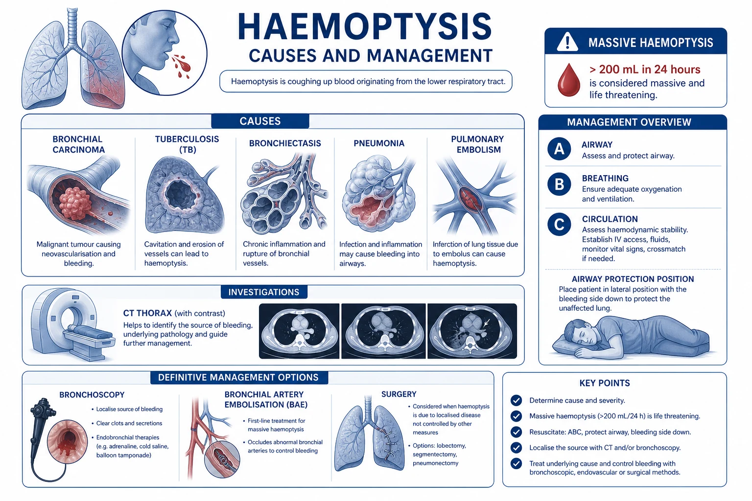
The examiner expects the causes organised by anatomical source, because the source determines the investigation pathway and the intervention. Work through the categories in this order, and decide the most likely from the clinical picture.[2][3][4]
Bronchial (commonest)
- Bronchitis — the commonest cause of MILD haemoptysis overall (smoker's bronchitis)
- Bronchiectasis — the commonest cause of MASSIVE haemoptysis (CF, post-infective)
- Bronchogenic carcinoma — smoker over 40, central or peripheral mass
- Bronchial carcinoid (adenoma) — young, highly vascular, recurrent bleed
- Foreign body lodged in a bronchus — children, aspiration history
Pulmonary parenchymal
- Tuberculosis — worldwide number one; apical cavity, sputum AFB
- Pneumonia / lung abscess — fever, purulent sputum, consolidation
- Aspergilloma (mycetoma) — fungus ball in old cavity, massive bleed
- Invasive aspergillosis — immunocompromised, angioinvasion
- Pulmonary embolism with infarction — dyspnoea, pleuritic pain, wedge
Vascular
- Pulmonary AVM / Osler–Weber–Rendu (HHT) — telangiectasia, family history
- Rasmussen aneurysm — PA pseudoaneurysm in chronic TB cavity
- Pulmonary artery aneurysm (Behçet, Hughes–Stovin)
- Dieulafoy lesion / bronchial artery aneurysm
- Iatrogenic PA rupture — Swan–Ganz catheter
Autoimmune / DAH
- ANCA vasculitis (GPA/MPA/EGPA) — ENT + renal
- Anti-GBM / Goodpasture — lung + rapidly progressive GN
- SLE, IgA nephropathy, antiphospholipid syndrome
- Often NO frank haemoptysis — anaemia + bilateral infiltrates + AKI
Iatrogenic
- Bronchoscopic biopsy / transbronchial biopsy
- Transthoracic needle biopsy
- Pulmonary artery catheter (Swan–Ganz) — PA rupture
- Anticoagulation / antiplatelet therapy unmasking a lesion
Tracheal / upper-airway (exclude first)
- Epistaxis, gingival bleeding, pharyngeal/laryngeal lesion
- Tracheal trauma, foreign body, tumour
- Factitious — food dye, Munchausen
- Inspect nose, oropharynx, larynx BEFORE concluding pulmonary source
Three-way differentiation the examiner always wants:[3]
- Bronchial carcinoma — smoker over 40, weight loss, clubbing, central or peripheral mass on imaging, persistent haemoptysis; cytology/biopsy diagnostic.
- Pulmonary TB — younger, fever/night sweats, apical cavity, sputum AFB-positive, high local prevalence.
- Bronchiectasis — years of chronic purulent sputum, clubbing, tram-track/signet-ring on HRCT.
- Pulmonary embolism — sudden dyspnoea and pleuritic pain, clear CXR (or wedge infarct), risk factors; CTPA filling defect.
- Mitral stenosis — rare but classic — mid-diastolic murmur, AF, orthopnoea, normal CXR or pulmonary oedema. [1]
Clinical Presentation
History — establish the act (cough vs vomit), the volume, the colour (bright red/fresh vs dark), whether it is mixed with sputum or pure blood, the duration and frequency (one episode vs recurrent), and associated symptoms. Bright red, frothy, alkaline blood mixed with sputum that is coughed = true haemoptysis.[3]
Associated features that localise the cause: [1]
- Fever, night sweats, weight loss, chronic cough, apical infiltrate — TB.
- Smoker over 40, weight loss, clubbing, haemoptysis out of proportion to symptoms, new mass — bronchial carcinoma.
- Years of chronic daily purulent sputum, recurrent infections, clubbing — bronchiectasis.
- Sudden dyspnoea, pleuritic chest pain, clear CXR, risk factors (immobility, malignancy, pregnancy, recent surgery) — PE.
- Dyspnoea, bilateral infiltrates, acute kidney injury, haematuria — diffuse alveolar haemorrhage (ANCA, anti-GBM).
- Orthopnoea, mitral facies, mid-diastolic murmur, AF — mitral stenosis.
- Epistaxis, telangiectasia, family history — Osler–Weber–Rendu (pulmonary AVM).
- Recent bronchoscopy/biopsy, PA catheter in situ, anticoagulants — iatrogenic.
- Recurrent oral aphthae, genital ulcers, uveitis — Behçet disease (pulmonary artery aneurysm). [1]
Atypical presentation:[4]
- Elderly — blunted cough reflex (blood pools, aspiration risk higher), comorbidity, frequently on anticoagulants; a small bleed can be life-threatening.
- Immunocompromised — opportunistic infection (invasive aspergillosis, TB, CMV) and thrombocytopenia; may present with little blood but extensive infiltrates.
- Anticoagulated — an occult lesion (carcinoma, AVM) unmasked; never assume the anticoagulant is the sole cause.
- Cystic fibrosis — recurrent episodes; baseline lung function is poor, so even moderate bleeds are dangerous. [1]
Massive haemoptysis: rapid onset, brassy cough, large volume of bright-red blood and clots, dyspnoea, tachypnoea, and signs of airway compromise (stridor, falling SpO2, inability to clear secretions, agitation then obtundation). Shock is late and indicates the bleed is enormous.[1]
Differential Diagnosis
Always work through the causes by category (above) and decide the most likely from the clinical picture. The can't-miss diagnoses that must be actively excluded in every patient are bronchial carcinoma, TB, PE and diffuse alveolar haemorrhage.[3][4]
The mimics to exclude at the bedside are the non-pulmonary sources: epistaxis (swallowed then coughed), gingival bleeding, haematemesis (aspirated), and factitious causes (food dye, Munchausen). A careful upper-airway inspection and a precise history of the act (cough vs vomit) resolve most. [1]
The rare-but-classic causes that win viva marks: mitral stenosis (mid-diastolic murmur), Behçet disease and Hughes–Stovin syndrome (pulmonary artery aneurysm), Goodpasture/anti-GBM (pulmonary–renal syndrome), and the Rasmussen aneurysm (PA pseudoaneurysm in a chronic TB cavity). [1]
The four causes examiners contrast head-to-head — memorise the discriminator for each:[3][4]
- Carcinoma vs TB vs bronchiectasis vs PE — a smoker over 40 with weight loss and a mass is carcinoma; a young patient with fever, night sweats and an apical cavity is TB; years of daily purulent sputum with tram-track on HRCT is bronchiectasis; sudden dyspnoea and pleuritic pain with a clear CXR (or wedge) and risk factors is PE.
- Pulmonary–renal syndromes — if haemoptysis coexists with acute kidney injury, haematuria and bilateral infiltrates, think ANCA vasculitis (GPA, MPA) or anti-GBM disease (Goodpasture); the discriminator is the ANCA pattern (PR3/c-ANCA in GPA, MPO/p-ANCA in MPA) and anti-GBM antibodies, confirmed by renal biopsy.
- Drug-induced — anticoagulants, cocaine (parenchymal damage), propylthiouracil and penicillamine (DAH), and trimellitic anhydride (occupational). [1]
Clinical & Bedside Assessment
ABCDE first. In massive haemoptysis the airway takes priority — assess airway patency, ability to cough and clear blood, SpO2, RR, BP, conscious level.[1][4]
Upper-airway inspection (ENT): inspect the nose, oropharynx, gums and (if possible) larynx to exclude epistaxis or pharyngeal bleeding before concluding the source is pulmonary. This is the first step and is frequently forgotten.[3]
Respiratory examination to localise the bleeding lung: [1]
- Reduced air entry / breath sounds on the bleeding side (bronchial obstruction by blood or clot).
- Crackles, rhonchi over the bleeding lobe.
- Signs of consolidation (pneumonia, infarction), dullness (effusion/haemothorax), cavity (TB, abscess, mycetoma).
- A silent, dull hemithorax suggests collapse or effusion. [1]
General examination — high-yield pointers: [1]
- Finger clubbing — carcinoma, bronchiectasis, idiopathic pulmonary fibrosis, lung abscess.
- Cachexia, supraclavicular (Virchow) lymphadenopathy — malignancy.
- Telangiectasia of lips/tongue/fingers — Osler–Weber–Rendu (pulmonary AVM).
- Mitral facies, loud S1, mid-diastolic rumble, AF — mitral stenosis.
- Purpura, petechiae, mucosal bleeding — coagulopathy/thrombocytopenia.
- Nasal septum perforation, saddle nose, oral ulcers — granulomatosis with polyangiitis (GPA).
- Peripheral oedema, haematuria on dipstick — DAH with renal involvement (pulmonary–renal syndrome).
- Leg swelling/tenderness — DVT/PE. [1]
Triage decision — the skill that matters: the bedside assessment culminates in a single decision — is this patient safe for an outpatient work-up, or do they need the massive-haemoptysis pathway? The answer hinges on volume per unit time, airway patency, oxygenation, haemodynamics, and respiratory reserve (FEV1, baseline exercise tolerance, age). A smoker over 40 with even a small volume of blood, weight loss, or a mass on CXR is not an outpatient — they need urgent CT and bronchoscopy to exclude carcinoma. Conversely, a young non-smoker with a single episode of blood-streaked sputum, a normal CXR, and no red flags can be safely investigated as an outpatient.[3]
Investigations
First-line (in parallel with resuscitation):[2][3]
- FBC — haemoglobin (baseline + trend), platelets, MCV.
- Coagulation — PT/INR, APTT, fibrinogen; identify the anticoagulant and its reversal agent.
- U&E, LFT, bone profile — renal function (DAH/pulmonary–renal syndrome), hepatic disease (coagulopathy).
- Group & screen / crossmatch 2 to 4 units (massive).
- ABG / venous blood gas — hypoxaemia, hypercapnia (fatigue), lactate (perfusion).
- CRP — infection/inflammation; D-dimer if PE plausible; troponin if cardiac.
- Urinalysis + U&E — whenever DAH/pulmonary–renal syndrome is plausible (haematuria, proteinuria, red-cell casts).
- ECG — arrhythmia, right-heart strain (PE). [1]
Imaging: [1]
- Chest X-ray — first-line; localises the bleed in roughly 60 to 80 percent but is normal in 20 to 30 percent (PE, early carcinoma, central lesion, DAH early). Patterns: apical cavity (TB, mycetoma), mass (carcinoma), consolidation (pneumonia, infarct), bilateral infiltrates (DAH, pulmonary oedema), tram-track (bronchiectasis), wedge (PE infarct), meniscus (effusion).[3]
- CT thorax (often CT pulmonary angiography, CTPA) — the gold-standard / most sensitive test for localising the source and cause; defines bronchial artery anatomy for embolisation, masses, cavities, AVMs, DAH, PE. Perform once the patient is stable; in massive haemoptysis do not delay airway control for CT.[4]
- Bronchoscopy — flexible (submassive, awake patient — diagnosis + topical therapy) vs rigid (massive, allows ventilation + large-bore suction + tamponade). Diagnostic AND therapeutic. Timing is debated — early bronchoscopy (within 24 h) improves localisation; in massive bleeding the bleeding side is hard to see, so CTA often guides.[4]
Sputum (when TB/infection suspected): AFB microscopy and mycobacterial culture (TB), GeneXpert/CBNAAT (rapid TB PCR + rifampicin resistance), Gram stain and culture (pneumonia/abscess), cytology (malignancy). Send three early-morning samples for AFB.[3]
Autoimmune panel if DAH suspected: ANCA (PR3/c-ANCA points to GPA; MPO/p-ANCA points to MPA/EGPA), anti-GBM (Goodpasture), ANA, dsDNA, complement, and renal biopsy.[4]
Chest X-ray and CT patterns that name the cause — a high-yield viva area:[3][4]
- Apical cavity with an air-crescent / intracavitary mass — aspergilloma (fungus ball in an old TB cavity); the "monod sign".
- Apical cavity without fungus ball, fibrosis, volume loss — post-TB bronchiectasis or active cavitary TB.
- Spiculated / coin lesion, hilar or mediastinal nodes — bronchogenic carcinoma.
- Bilateral, perihilar airspace opacification that spares the apices — diffuse alveolar haemorrhage (or pulmonary oedema).
- Peripheral wedge-shaped density — pulmonary infarct (PE).
- Tram-track, signet-ring, varicose bronchi — bronchiectasis on HRCT.
- Tortuous, dilated enhancing vessels — pulmonary AVM (Osler–Weber–Rendu); confirmed by CT angiography.
- Enhancing nodule in a cavity wall — Rasmussen aneurysm (pulmonary artery pseudoaneurysm).
- Normal CXR — think PE, early/small carcinoma, bronchial carcinoid, DAH early, tracheal/bronchial lesion not yet visible. [1]
Bronchoscopy timing — the controversy: early bronchoscopy (within 24 h of a non-massive bleed) improves the localisation yield because active bleeding points the endoscopist to the source. In massive haemoptysis, however, the airway is flooded and the bleeding side is impossible to see — here CT angiography (CTA) is more useful to map the culprit bronchial artery before embolisation. The modern sequence for massive bleeds is therefore secure the airway, then CTA, then BAE, with rigid bronchoscopy reserved for airway control and tamponade. Flexible bronchoscopy is best suited to submassive bleeds in the stable, awake patient.[4] Named thresholds — reproduced verbatim (a favourite exam area):
- "Massive" haemoptysis — classical volume thresholds: any of — over 100 mL/24h (a frequently cited lower threshold), over 200 to 240 mL/24h, over 50 mL in a single episode, over 600 mL/24h, or over 1000 mL/24h. The modern functional definition: any volume threatening the airway, gas exchange or haemodynamics.[1][2]
- Bronchial artery embolisation (BAE): immediate clinical success 70 to 99 percent; recurrence 10 to 50 percent over months; spinal-cord ischaemia (anterior spinal artery / artery of Adamkiewicz) is the feared complication.[2][6]
Management — Massive Haemoptysis Algorithm
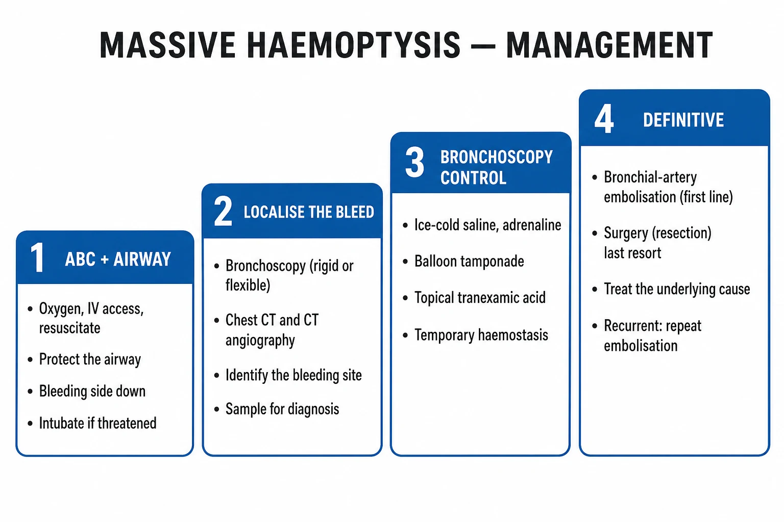
In massive (life-threatening) haemoptysis, resuscitation and airway control come first, with simultaneous activation of the bronchoscopy, interventional radiology (IR), and surgical teams.[1][4]
Step 1 — Airway and breathing
- High-flow oxygen — target SpO2 94 to 98 percent, or 88 to 92 percent in COPD/CO2-retainers.[1]
- Position the BLEEDING LUNG DOWN (dependent, lateral decubitus) to protect the good lung from aspiration of blood. Sit up if mild and stable. Placing the good lung down drowns the patient — a classic and lethal error.
- Vigorous large-bore suction to keep the airway clear of blood and clot.
Step 2 — Circulation and coagulation
- Two large-bore (14 to 16 G) IV cannulae, send bloods + crossmatch 2 to 4 units, balanced crystalloid and blood transfusion for shock; monitor SpO2, HR, BP, urine output, GCS.
- Reverse anticoagulation — stop anticoagulants/antiplatelets:[4]
- Warfarin → vitamin K 5 to 10 mg IV + prothrombin complex concentrate (PCC) 25 to 50 IU/kg (faster than FFP) or FFP 15 mL/kg.
- Dabigatran → idarucizumab 5 g IV (2 vials of 2.5 g).
- Rivaroxaban/apixaban → andexanet alfa.
- Heparin → protamine sulfate 1 mg per 100 IU heparin.
- Antiplatelets — platelet transfusion if life-threatening.
Step 3 — Secure the airway if threatened
- Endotracheal intubation with a large single-lumen tube (at least 8 mm internal diameter) to allow passage of a bronchoscope and large-bore suction catheter, OR a double-lumen tube / bronchial blocker for selective lung ventilation (isolate the bleeding lung). Transfer to ITU. Never intubate with a small tube — you cannot pass a scope or suction clots through it. [1]
Escalation trigger: any patient with airway compromise, falling SpO2, inability to clear blood, or shock must be intubated and moved to ITU immediately.[4]
Management — Definitive & Stepwise
Once the airway is secure, the goal is to localise and control the source.[1][2][4]
Bronchoscopic control (rigid preferred for massive — ventilation + tamponade + working channel):[4]
- Iced saline lavage of the bleeding bronchus (vasoconstriction).
- Topical adrenaline 1:20,000 (or 1:10,000) instilled via the scope.
- Balloon tamponade — a Fogarty catheter or bronchial blocker inflated in the bleeding segment to occlude it; leave up to 24 to 48 h.
- Topical thrombin / fibrinogen-thrombin glue.
- Argon plasma coagulation (APC), Nd:YAG laser, cryotherapy — to coagulate or debulk a visible bleeding lesion (carcinoid, carcinoma). [1]
Rigid vs flexible bronchoscopy — when each: the rigid scope is the instrument of choice for massive haemoptysis because it provides a large working channel (large-bore suction, tamponade balloons, rigid forceps for clot extraction), controlled ventilation, and the mechanical ability to tamponade a bleeding segment by packing with gauze or balloon. Its limitation is that it reaches only the central airways. The flexible scope is ideal for submassive bleeds in the awake, stable patient: it reaches segmental and subsegmental bronchi, allows diagnosis and topical therapy, and can be passed through a rigid scope or large single-lumen tube when both central and peripheral access are needed.[4]
Practical sequence at the scope: once the bleeding side is identified, isolate it with a bronchial blocker or double-lumen tube, suction the clot, then apply iced-saline lavage (50 mL aliquots — the cold vasoconstricts the bleeding vessel), instil topical adrenaline, and if bleeding persists deploy a balloon tamponade left inflated for 24 to 48 h before graded deflation. If a discrete bleeding lesion is visible (carcinoid, vascular tumour, Dieulafoy lesion), APC or laser coagulates it directly. [1]
Bronchial artery embolisation (BAE) — first-line intervention
BAE is the first-line intervention for massive haemoptysis when bronchoscopy cannot control the bleed, and has displaced surgery as the default definitive therapy.[2][6]
Technique: [1]
- Pre-procedure CT angiogram (CTA) maps the bronchial arteries and any abnormal systemic collaterals (intercostal, subclavian, internal mammary, inferior phrenic) that may be feeding the bleed.
- Under fluoroscopy, the interventional radiologist catheterises the culprit bronchial artery from a femoral arterial access, usually with a coaxial microcatheter system.
- Embolisation with polyvinyl alcohol (PVA) particles, gelatin sponge (Gelfoam), or microspheres (tris-acryl gelatin) until flow is abolished.
- All feeding arteries are embolised to prevent early rebleed from collaterals. [1]
Bronchial artery embolisation (BAE) — the numbers
Caveats: the artery of Adamkiewicz (a spinal feeder arising from a bronchial or intercostal artery) must be identified on CTA before embolisation — inadvertent embolisation causes anterior spinal artery syndrome and paraplegia. Non-target embolisation to the coronary, cerebral or mesenteric circulation causes ischaemia and stroke. Contrast-induced nephropathy and recurrence (10 to 50 percent) are the main drawbacks; recurrence is highest in aspergilloma and active TB.[2][6]
Surgery — last resort
Surgery is last resort, reserved for uncontrolled bleeding that is localised and surgically resectable (single lobe, good lung function, fit patient). Lobectomy (or pneumonectomy) in the emergency setting carries peri-operative mortality up to 40 percent. Selective-lung ventilation and stabilisation before resection improve outcome.[5]
Pharmacological and supportive: tranexamic acid 1 g IV three times daily (modest evidence, widely used), vasopressin in selected centres, broad-spectrum antibiotics for infection; treat the underlying cause (TB therapy, chemotherapy, vasculitis immunosuppression, antifungals). [1]
Non-massive haemoptysis management: investigate the cause (CXR + CT + bronchoscopy), treat the underlying disease, and discharge only if stable with small volume, no red flags, and a normal or explained CXR. Arrange follow-up CXR at 6 to 8 weeks in smokers over 50 to exclude an occult malignancy that was too small to see initially.[3]
Specific Subtypes & Scenarios
- Diffuse alveolar haemorrhage (DAH) — bilateral infiltrates, anaemia, dyspnoea; haemoptysis may be absent. Capillaritis (ANCA, anti-GBM, immune-complex) vs bland (coagulopathy, mitral stenosis, drugs). Treat the underlying cause: high-dose methylprednisolone 1 g IV daily for 3 days, cyclophosphamide, plasma exchange for anti-GBM. Bronchoalveolar lavage shows progressively bloodier returns.[4]
- Aspergilloma (mycetoma) — a fungus ball in a pre-existing cavity (post-TB, sarcoid); massive, recurrent haemoptysis. Surgical resection is definitive; BAE as a bridge or in unfit patients; long-term antifungals (itraconazole/voriconazole) have limited role. With its high rebleed and mortality, surgical referral is warranted in fit patients.
- Iatrogenic pulmonary artery rupture (from a PA/Swan–Ganz catheter) — sudden haemoptysis in the catheterised, often anticoagulated patient. Advance the catheter and inflate the balloon to tamponade, isolate the bleeding lung (bronchial blocker / double-lumen tube), reverse coagulopathy, urgent thoracic surgery/IR; mortality is high.
- Cystic fibrosis with recurrent/massive haemoptysis — BAE first-line; vitamin K; cessation of NSAIDs; repeat BAE for recurrence; bilateral disease makes surgery hazardous — refer for transplant assessment in advanced disease.
- Bronchial carcinoid — a highly vascular tumour (often central, in younger non-smokers); recurrent bleeds; surgical resection (sleeve lobectomy) is both diagnostic and curative; bronchoscopic debulking/APC for palliation.
- Rasmussen aneurysm — pulmonary artery pseudoaneurysm in the wall of a chronic TB cavity; massive haemoptysis; treated by PA coil embolisation or surgery; underlying anti-TB therapy.
- Pulmonary AVM (Osler–Weber–Rendu / HHT) — dyspnoea, cyanosis, clubbing, paradoxical emboli/stroke; recurrent minor or massive haemoptysis; treated by coil or vascular plug embolisation; screen the family.
- Pregnancy with haemoptysis — PE is the leading cause (pregnancy is a 5-fold prothrombotic state); TB in endemic regions. Use chest X-ray with shielding and CTPA judiciously (the radiation risk to the fetus is low and outweighed by the risk of a missed PE); BAE is feasible; avoid warfarin (teratogenic; use LMWH).[3]
Complications & Pitfalls
- Asphyxia and death from aspiration of blood — the principal mode of death.
- Hypovolaemic / haemorrhagic shock (less common; indicates an enormous bleed).
- Aspiration pneumonia, atelectasis (clot obstruction), airway obstruction by clot, ARDS.
- After BAE: recurrence (10 to 50 percent), contrast-induced nephropathy, anterior spinal artery syndrome / paraplegia (Adamkiewicz), stroke and ischaemia from non-target embolisation (e.g. broncho-oesophageal artery).
- After emergency surgery: bronchopleural fistula, empyema, respiratory failure, high mortality. [1]
Classic pitfalls: [1]
- Mistaking haematemesis for haemoptysis (or vice versa) — leading to wrong investigations and the wrong team.
- Failing to localise the bleeding lung before positioning — placing the good lung down drowns the patient.
- Over-sedating the patient for bronchoscopy — depressing the cough and airway reflexes and accelerating aspiration.
- Intubating with a small tube — cannot pass a bronchoscope or suction large clots; use at least 8 mm.
- Delaying BAE while awaiting CT in an unstable patient — airway and embolisation first.
- Assuming the anticoagulant is the sole cause — always look for an underlying lesion.
- Missing diffuse alveolar haemorrhage — bilateral infiltrates with anaemia and renal failure; haemoptysis may be absent.
- Forgetting PE in the patient with haemoptysis, dyspnoea and a clear CXR. [1]
Prognosis & Disposition
- Mild haemoptysis — usually benign once serious causes are excluded; outpatient work-up (CXR, consider CT, sputum, bronchoscopy if indicated, follow-up CXR at 6 to 8 weeks in smokers over 50 to exclude occult malignancy).[3]
- Massive haemoptysis mortality — historically 50 to 80 percent, now 7 to 20 percent with modern BAE and ICU.[1][2]
- Poor-prognosis predictors: volume over 1000 mL/24h (mortality 50 to 80 percent), malignancy, aspergilloma, need for mechanical ventilation, comorbidity, aspiration into the good lung.
- Disposition: massive → ITU/HDU with airway secured and IR/surgery on standby; moderate → respiratory ward with close monitoring and crossmatched blood; mild → outpatient if stable with no red flags and a normal CXR.[4]
Prevention of recurrence is cause-specific:[2][4]
- Treat the underlying disease to completion — full anti-TB therapy, eradication therapy for bronchiectasis, oncological treatment for carcinoma, immunosuppression for vasculitis.
- Control modifiable factors — smoking cessation (the single most effective lung-cancer and chronic-bronchitis prevention measure), optimise airway clearance in bronchiectasis/CF (physiotherapy, mucolytics, prophylactic antibiotics), and anticoagulation review in patients with a known pulmonary lesion.
- In aspergilloma, because rebleed is the rule, surgical resection in fit patients is preventive; repeat BAE is offered to those unfit for surgery.
- In pulmonary AVM (HHT), embolisation of all feeding arteries plus family screening (autosomal dominant) prevents both rebleed and paradoxical embolism.
- After BAE, surveillance for recurrence (rebleed in 10 to 50 percent) — educate the patient to return promptly with any fresh bleed. [1]
Special Populations
- Children — haemoptysis is uncommon. Causes: foreign-body aspiration (a leading cause in toddlers), infection (pneumonia, TB), bronchiectasis (CF), vascular malformation, tracheo-oesophageal fistula. A small airway means a small volume can obstruct; rigid bronchoscopy under GA is both diagnostic and therapeutic.
- Pregnancy — PE is the leading cause (5-fold prothrombotic state); TB in endemic regions. Shielded CXR and CTPA are acceptable; BAE is feasible; avoid warfarin (use LMWH).[3]
- Elderly — blunted cough, comorbidity, anticoagulants and high malignancy risk; lower threshold for hospital admission, imaging and bronchoscopy.
- Immunocompromised — invasive aspergillosis (angioinvasive), TB, CMV, thrombocytopenia; broad differential, lower threshold for bronchoscopy/BAL.
- Anticoagulated / coagulopathic — reverse the agent; exclude an underlying lesion (do not attribute the bleed to the anticoagulant alone).
- Cystic fibrosis — recurrent episodes; BAE first-line; consider transplant referral for advanced disease.
Evidence, Guidelines & Regional Differences
Landmark evidence and reviews:[1][2][4][5][6]
- Jean-Baptiste (Crit Care Med 2000) — established the modern approach to massive haemoptysis (airway, bronchoscopy, BAE).
- Davidson & Shojaee (Chest 2020) and Deshwal & Mehta (Semin Respir Crit Care Med 2021) — comprehensive stepwise algorithms confirming airway-first and BAE first-line.[1][4]
- Fan et al (BMC Pulm Med 2024) — systematic review and meta-analysis supporting BAE over conservative management.[6]
- Tranexamic acid (the FOCAL trial and smaller studies) shows modest benefit in non-massive haemoptysis; evidence in massive haemoptysis remains weak and the drug is used as an adjunct, not a definitive therapy.
Modern consensus shift — away from rigid volume thresholds toward the functional definition of life-threatening haemoptysis, recognising that a small bleed can kill a patient with poor reserve.[1][2]
[1] [1]Causes of haemoptysis — 'BATTLE CAMP'
BATTLE
the two big 'B' causes in the West
vascular
the worldwide number one
iatrogenic
infection
with clear CXR and pleuritic pain
Massive haemoptysis management — 'AIR BLEED'
AIRBLEED
protect and oxygenate; large-bore or double-lumen tube
position BLEEDING LUNG DOWN
PCC/vitamin K; idarucizumab/andexanet
tamponade, iced saline, adrenaline, APC
CXR then CT angiogram
first-line intervention, 70 to 99 percent success
surgery (lobectomy) if BAE fails
pharmacological adjunct
Exam Pearls
- Commonest cause worldwide = TB; in the West = bronchial carcinoma; of mild haemoptysis overall = bronchitis (smoker's); of massive haemoptysis = bronchiectasis.[3]
- Over 90 percent of massive haemoptysis arises from the BRONCHIAL (high-pressure systemic) arteries — that is why the bleed is brisk and why BAE works.[1]
- Death is from ASPHYXIA, not exsanguination — protect the airway first; only 150 to 200 mL in a bronchus can be fatal.[5]
- Position the BLEEDING LUNG DOWN to protect the good lung.[1]
- BAE is first-line for massive haemoptysis; surgery (lobectomy) is last resort (emergency mortality up to 40 percent).
- Rigid (not flexible) bronchoscopy for massive haemoptysis — ventilation + tamponade.
- Beware the artery of Adamkiewicz during BAE — anterior spinal artery syndrome / paraplegia.[2]
- Haemoptysis vs haematemesis: bright red, frothy, alkaline, coughed vs dark coffee-ground, acidic, vomited.
- Aspergilloma (fungus ball) + massive haemoptysis → surgical resection is definitive (BAE as bridge).[4]
- Diffuse alveolar haemorrhage may have NO haemoptysis — anaemia + bilateral infiltrates + renal failure → think ANCA / anti-GBM (Goodpasture); treat with methylprednisolone, cyclophosphamide, plasma exchange.
- Rasmussen aneurysm = pulmonary artery pseudoaneurysm in a chronic TB cavity causing massive haemoptysis.
- Clear CXR + dyspnoea + pleuritic pain = PE — do not be reassured by a normal film.[3]
- Mitral stenosis — rare but classic; look for the mid-diastolic murmur.
- Massive = any volume threatening airway/gas exchange (functional definition) — the modern threshold, not a rigid mL cut-off.[1][2]
Exam application bank (NEET-PG / INICET)
One-line answer
Haemoptysis is the coughing up of blood that originates from the lower respiratory tract (below the vocal cords). It ranges from blood-streaked sputum to massive, life-threatening haemoptysis. Worldwide the commonest cause is tuberculosis; in developed countries bronchial carcinoma, bronchiectasis and pneumonia predominate. Massive haemoptysis (any volume threatening the airway or gas exchange, classically over 100 to 240 mL/24h) kills by asphyxia, not exsanguination — because over 90 percent of such bleeds arise from the high-pressure bronchial arteries. Management is stepwise: protect the airway, oxygenate, put the BLEEDING LUNG DOWN, reverse anticoagulation, then rigid bronchoscopy with tamponade and bronchial artery embolisation (BAE) first-line; surgery is last resort. Always exclude PE, TB, carcinoma and diffuse alveolar haemorrhage (ANCA / anti-GBM). [1]
Worked stems (answer without another resource)
Stem 1 — Classic presentation. Map symptoms to mechanism; name the first investigation and first treatment step with dose/route if drug therapy is standard. [1]
Stem 2 — Unstable / complicated. List red flags that force immediate resuscitation, theatre, ICU, antidote, or reperfusion — and what you do in the first 15 minutes. [1]
Stem 3 — Atypical group. Elderly, pregnancy, child, or immunocompromised: how presentation and thresholds change. [1]
Stem 4 — Differential trap. Name the three closest mimics and one discriminator for each. [1]
Stem 5 — Disposition. Who goes home with safety-netting, who is admitted, who needs HDU/ICU/theatre, and what follow-up is mandatory. [1]
Rapid viva checklist
- Definition + classification
- Pathophysiology chain
- Bedside signs / criteria
- Score with exact components (if any)
- Emergency bundle
- Definitive therapy with doses
- Complications of disease and of treatment
- Special populations
- Guideline/trial name if classic
- Three exam traps
Coverage self-check
If you cannot answer any stem above from this page alone, re-read the matching section — the page is intended to be self-sufficient for final-prof and NEET-PG/INICET questions on Haemoptysis.
References
- [1]Davidson K, Shojaee S. Managing Massive Hemoptysis Chest, 2020.PMID 31374211
- [2]Prey B, Francis A, Williams J, Krishnadasan B. Evaluation and Treatment of Massive Hemoptysis Surg Clin North Am, 2022.PMID 35671767
- [3]Ittrich H, Bockhorn M, Klose H, Simon M. The Diagnosis and Treatment of Hemoptysis Dtsch Arztebl Int, 2017.PMID 28625277
- [4]Deshwal H, Sinha A, Mehta AC. Life-Threatening Hemoptysis Semin Respir Crit Care Med, 2021.PMID 32862418
- [5]Yendamuri S. Massive Airway Hemorrhage Thorac Surg Clin, 2015.PMID 26210922
- [6]Fan S, Cheng X, Wang X, Liu Y, He W, Chen H. Bronchial artery embolization versus conservative treatment for hemoptysis: a systematic review and meta-analysis BMC Pulm Med, 2024.PMID 39215233