Respiratory · General Medicine
Influenza (Seasonal & Pandemic)
Also known as Influenza · Flu · Seasonal influenza · Pandemic influenza · H1N1 · Swine flu · Avian influenza · H5N1
Influenza is an acute, highly contagious respiratory infection caused by orthomyxoviruses of types A, B and (rarely) C. Type A is subtyped by two surface glycoproteins — haemagglutinin (H, 18 subtypes) and neuraminidase (N, 11 subtypes); current human strains are A/H1N1pdm09 and A/H3N2; type B is divided into Victoria and Yamagata lineages. Antigenic drift (point mutations in H and N) drives annual seasonal epidemics; antigenic shift (reassortment of genome segments when two viruses co-infect a host, e.g. swine as a 'mixing vessel') produces a novel subtype → pandemic (1918 H1N1 'Spanish flu', 1957 H2N2 'Asian flu', 1968 H3N2 'Hong Kong flu', 2009 H1N1pdm09 'swine flu'). Transmission is by respiratory droplets and aerosols with an incubation period of 1–4 days. Clinically: abrupt high fever, rigors, headache, myalgia, dry cough, sore throat, extreme prostration — distinguished from the common cold by sudden onset and severity of systemic symptoms. Diagnosis is clinical in season; RT-PCR (nasopharyngeal swab) is the gold standard; RIDTs give a result in 15 min but have moderate sensitivity (50–70%) and high specificity (90–95%), so a negative RIDT does NOT exclude influenza. Treat with oral oseltamivir 75 mg twice daily for 5 days, started within 48 hours of onset (reduces duration by ~1 day and prevents complications); always treat hospitalised, pregnant, immunocompromised, and high-risk patients regardless of delay. Annual quadrivalent vaccine (2 A + 2 B strains) is the cornerstone of prevention — recommended for all over 6 months, with priority for elderly, pregnant, children under 5, chronic disease, immunocompromise, healthcare workers. Complications: primary viral pneumonia, secondary bacterial pneumonia (Strep pneumoniae, Staph aureus — classically post-influenza), bronchiolitis, otitis media (children), myocarditis, encephalitis, Guillain–Barre syndrome, Reye syndrome (children given aspirin), rhabdomyolysis, ARDS. Global mortality: 290,000–650,000 respiratory deaths and up to 1 million deaths annually.
On this page & tools
Your progress
Saved locally on this device.
Exam tags
Red flags
Overview & Definition
Influenza is an acute, febrile, highly contagious viral infection of the respiratory tract caused by influenza viruses A, B and (rarely) C, members of the Orthomyxoviridae family. Influenza A and B cause the substantial burden of human seasonal disease; influenza C produces only mild, sporadic upper-respiratory illness and is not included in routine vaccines.[2]
What makes influenza a perpetual global threat — and a perpetual exam favourite — is the antigenic mutability of its surface glycoproteins: minor point mutations (antigenic drift) accumulate continuously and force annual vaccine reformulation and seasonal epidemics, while major gene-segment reassortment (antigenic shift) periodically produces a wholly novel subtype to which the population has little immunity, igniting a pandemic. The 1918 'Spanish flu' (H1N1) killed an estimated 50–100 million people worldwide; the 2009 H1N1pdm09 pandemic was the first influenza pandemic of the 21st century.[9]
The clinical task is three-fold: (1) recognise influenza early in the febrile patient during season, (2) identify who needs antivirals and who needs admission (the high-risk groups for whom influenza is lethal), and (3) anticipate the complications — viral pneumonitis, secondary bacterial pneumonia, myocarditis, encephalitis, and the multi-organ syndromes that follow.[1]
Classification
Influenza viruses are classified by type, by subtype (A only), and — at the public-health level — by lineage (B) and clade. The classification is not merely taxonomic: it dictates vaccine composition, antiviral susceptibility, and pandemic risk.[2][4]
By type: [1]
- Influenza A — infects humans, pigs, horses, birds (the natural reservoir, especially aquatic wild birds). Subtyped by H (1–18) and N (1–11). Causes pandemics (because of its broad animal reservoir allowing reassortment). Currently circulating human strains: A/H1N1pdm09 and A/H3N2.
- Influenza B — almost exclusively human. No subtypes; divided into two antigenically distinct lineages — Victoria and Yamagata (Yamagata not detected globally since 2020, likely extinct). Causes seasonal epidemics but not pandemics (no animal reservoir for reassortment).
- Influenza C — causes mild sporadic illness; not vaccine-targeted.
- Influenza D — primarily cattle; not a human pathogen of consequence. [1]
Subtyping of influenza A — the H and N nomenclature (high-yield):[2]
- Haemagglutinin (H or HA) — 18 subtypes; mediates receptor binding and viral entry by binding sialic-acid receptors on respiratory epithelium. Antibody to HA is neutralising and protective. The haemagglutinin is the basis of subtyping.
- Neuraminidase (N or NA) — 11 subtypes; cleaves sialic acid to release progeny virions from the host cell (otherwise they remain tethered). The drug class neuraminidase inhibitors — oseltamivir, zanamivir, peramivir, laninamivir — target N.
- Only a small subset of H and N combinations circulate in humans: H1N1, H2N2 (1957–68), H3N2. Avian subtypes H5N1, H5N6, H7N9, H9N2 cause zoonotic human disease with very high mortality but very limited human-to-human transmission. [1]
Antigenic DRIFT
- Minor point mutations in H and N from RNA-polymerase error (no proof-reading)
- Occurs in BOTH A and B
- Drives ANNUAL seasonal epidemics and vaccine reformulation
- Pandemic potential: NONE — population still has partial immunity
- Example: gradual antigenic change in H3N2 each year
Antigenic SHIFT
- Major reassortment of the 8 gene segments when TWO viruses co-infect a single host cell
- Occurs ONLY in influenza A (needs animal reservoir — the swine 'mixing vessel')
- Produces a NOVEL H or N subtype — population has NO immunity
- Drives PANDEMICS (1918 H1N1, 1957 H2N2, 1968 H3N2, 2009 H1N1pdm09)
- Example: 2009 swine flu = triple-reassortant of avian, swine and human genes
Epidemiology & Risk Factors
Influenza is one of the most significant infectious diseases of humans. Globally, modelling studies estimate that seasonal influenza causes approximately 1 billion infections, 3–5 million severe cases, and 290,000–650,000 respiratory deaths annually, the vast majority in adults aged over 65 and in low- and middle-income countries.[10]
Seasonality: in temperate climates, influenza circulates in discrete winter epidemics (December–March in the Northern Hemisphere, May–August in the Southern). In tropical and subtropical regions (including much of India), influenza can circulate year-round or with semi-annual peaks linked to monsoon — this regional pattern influences both vaccination timing and the differential diagnosis of a febrile illness. [1]
Pandemic epidemiology — landmarks every candidate must know:[9]
| Pandemic | Year | Subtype | Common name | Estimated deaths |
|---|---|---|---|---|
| 1st measurable | 1918 | H1N1 | Spanish flu | 50–100 million |
| Asian flu | 1957 | H2N2 | Asian flu | 1–2 million |
| Hong Kong flu | 1968 | H3N2 | Hong Kong flu | ~1 million |
| Swine flu | 2009 | H1N1pdm09 | Swine flu | 200,000–500,000 |
Risk groups for severe or complicated influenza (these define who gets priority vaccination, who gets antivirals regardless of duration of illness, and who needs a low threshold to admit):[1][5]
- Age — children under 5 years (especially under 2) and adults aged 65 and over (blunted immunity, declining respiratory reserve).
- Pregnancy (and up to 2 weeks postpartum) — particularly the 2nd and 3rd trimesters; physiological immune modulation and reduced functional residual capacity.
- Chronic respiratory disease — COPD, asthma, cystic fibrosis, bronchiectasis.
- Cardiovascular disease — heart failure, ischaemic heart disease (influenza triggers acute coronary events).
- Chronic metabolic disease — diabetes mellitus, chronic kidney disease, chronic liver disease.
- Immunocompromise — HIV, haematological malignancy, chemotherapy, transplant, splenectomy, prolonged corticosteroid.
- Neurological/neuromuscular disease (often overlooked) — cerebral palsy, stroke, muscular dystrophy; impair swallowing and cough.
- Extreme obesity (BMI 40 or more) — emerged as a risk factor in 2009 H1N1; restrictive lung physiology and chronic inflammation.
- Residents of long-term care / nursing homes — explosive outbreaks.
- Healthcare workers — both at risk and a vector for vulnerable patients.
- Indigenous and certain ethnic populations (e.g. American Indian, Australian Aboriginal) — disproportionate mortality. [1]
Key zoonotic avian influenza (high-yield exam point): H5N1 (highly pathogenic avian influenza) has a case fatality rate approaching 60% in confirmed human cases, but sustained human-to-human transmission has not occurred. Risk factor: direct contact with sick or dead poultry.[8] H7N9 (China) and H5N6 are other notable zoonotic threats. All suspected avian influenza must be notified to public health.
Pathophysiology
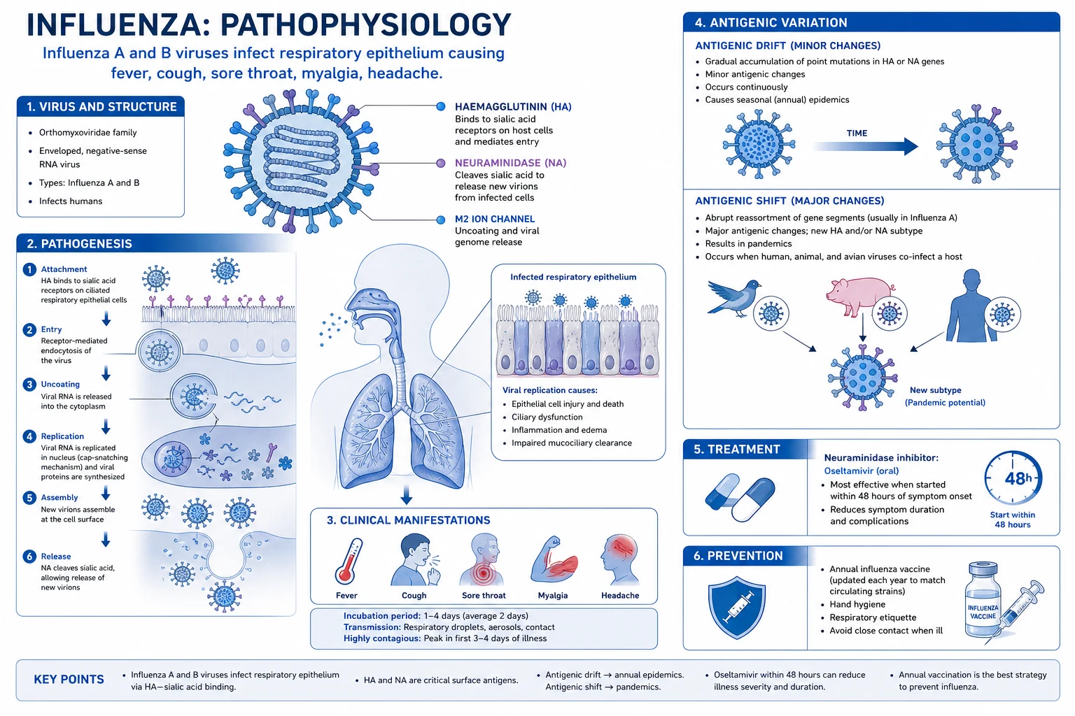
Influenza virus is transmitted from person to person mainly by large respiratory droplets (over 5 micrometres, travel up to 1–2 metres) generated by coughing and sneezing, and to a lesser extent by aerosols (smaller particles that travel further and linger) and by contact with contaminated surfaces (fomites) followed by self-inoculation of mucous membranes.[2]
Incubation period is 1–4 days (average 2). Patients are contagious from 1 day before symptoms until 5–7 days after onset (longer in children and the immunocompromised). [1]
The molecular cascade of infection and inflammation: [1]
- Receptor binding and entry. The viral haemagglutinin (HA) binds to sialic-acid receptors (alpha-2,6-linked in humans; alpha-2,3-linked in birds — the receptor difference explains species tropism and the swine 'mixing vessel', which has both linkage types) on ciliated columnar epithelial cells of the nose, trachea and bronchi. The virus is internalised by receptor-mediated endocytosis.
- Replication. The virus is an enveloped, negative-sense, single-stranded RNA virus with a genome of 8 segments (each encoding one or two proteins). The viral RNA-dependent RNA polymerase (PB1, PB2, PA) — which lacks proof-reading and so accumulates errors (drift) — replicates in the host-cell nucleus. Newly synthesised HA and NA are inserted into the host cell membrane.
- Release. Neuraminidase cleaves sialic-acid residues on the cell surface, releasing new virions. This is the drug target of oseltamivir/zanamivir — without NA, virions remain tethered to the dying cell and cannot spread.
- Cellular destruction and inflammation. Viral replication lyses the ciliated epithelial cells, stripping the mucociliary escalator and exposing the basal epithelium. Release of virions and intracellular contents activates macrophages and dendritic cells, which release a cytokine storm (interferon-alpha/beta, TNF-alpha, IL-1, IL-6, IL-8, CXCL10). These cytokines — not the virus itself — produce the systemic 'flu' symptoms of fever, headache, myalgia, fatigue and anorexia.
- Impaired defence and secondary invasion. Loss of the mucociliary barrier plus impaired neutrophil/macrophage function (transient post-influenzal immune paralysis) creates a fertile field for secondary bacterial infection — classically Strep pneumoniae, Staphylococcus aureus (especially the post-influenza necrotising pneumonia) and Haemophilus influenzae. This is why the second-spike of fever a few days into recovery is the cardinal sign of secondary bacterial pneumonia.
- Resolution. In the uncomplicated case, cytotoxic CD8+ T cells clear infected cells, and HA-specific neutralising IgA (mucosal) and IgG (systemic) antibody develops, providing strain-specific immunity that lasts years against the same strain but does not protect against drifted strains — the rationale for annual vaccination. [1]
Why pandemics occur (mechanism of shift): when a host cell is co-infected by two different influenza A viruses (e.g. an avian and a human strain, often in a pig that has both alpha-2,3 and alpha-2,6 sialic-acid receptors), the 8 genome segments can reassort like a deck of cards, producing a progeny virus with a novel combination of H and N to which the human population has no antibody. This reassortment, plus the segmented genome, is the molecular basis of pandemics.[2][4]
Clinical Presentation
The hallmark of influenza is its suddenness. Patients can usually state the exact hour the illness began — this is the single feature that most reliably distinguishes influenza from the common cold.[1]
Classic uncomplicated influenza (adult): abrupt onset of: [1]
- Fever (typically 38–40 degC) with rigors or chills — near-universal; may be absent in the elderly.
- Headache — often severe, frontal or retro-orbital.
- Diffuse myalgia — most severe in back and legs; incapacitating.
- Marked fatigue and weakness — out of proportion to the fever; can persist for 2–3 weeks.
- Dry (non-productive) cough — usually develops early and worsens.
- Sore throat.
- Anorexia, malaise.
- Nasal congestion, sneezing, lacrimation — present but milder than in the cold.
- Sometimes N/V and diarrhoea — more common in children and in H1N1pdm09 / avian strains than in classic seasonal influenza. [1]
The classic triad of "flu" = fever + cough + acute onset. Sensitivity of this triad in season for influenza approaches 80%; specificity around 60–80%. [1]
Differentiating influenza from the common cold (high-yield bedside exam):[1]
| Feature | Influenza | Common cold |
|---|---|---|
| Onset | Abrupt (hours) | Gradual (days) |
| Fever | High (38–40 degC), with rigors | Mild or absent |
| Myalgia | Severe | Mild |
| Headache | Severe | Mild |
| Fatigue, prostration | Severe, can persist weeks | Mild |
| Cough | Dry, prominent | Mild, often productive |
| Sore throat | Mild–moderate | Common, prominent |
| Nasal symptoms | Mild | Prominent (sneezing, rhinorrhoea) |
| Sneezing | Occasional | Common |
| Season | Winter epidemic | Year-round |
Signs on examination (uncomplicated): febrile, flushed, conjunctival injection, mild pharyngeal erythema, cervical lymphadenopathy, and a clear chest or a few scattered wheezes/ronchi. Chest signs beyond this in the first 48 hours suggest viral pneumonitis or bacterial co-infection. [1]
Influenza in specific populations — the atypical presentations every candidate must know: [1]
- Elderly (over 65): atypical presentation — fever may be absent or low-grade; presenting features may be confusion, falls, anorexia, weakness, failure to cope, or functional decline. A high index of suspicion in winter is essential; morbidity and mortality are markedly higher.
- Neonates and infants: may present as sepsis-like illness, apnoea, poor feeding, irritability, or febrile seizures; cough may be absent.
- Young children (under 5): high fever, croup (laryngotracheobronchitis), bronchiolitis, otitis media; influenza is a leading cause of paediatric hospitalisation. Reye syndrome is the feared complication in children given aspirin.
- Pregnant women: particularly in the 2nd/3rd trimester and up to 2 weeks postpartum, influenza carries a markedly increased risk of severe pneumonitis, ARDS, ICU admission, miscarriage, preterm delivery, and fetal demise; the 2009 pandemic demonstrated this dramatically.[7]
- Immunocompromised: may have prolonged viral shedding (weeks), atypical or absent fever, and higher rates of lower-respiratory involvement and resistance.
- Severe / fulminant influenza: rapid progression to primary viral pneumonia — dyspnoea, hypoxia, bilateral infiltrates, ARDS within 24–72 hours of onset; this is most common in pregnancy, immunocompromise, and extreme obesity.
Differential Diagnosis
A wide range of pathogens produce an influenza-like illness (ILI). The WHO defines ILI as measured fever of 38 degC or over AND cough, with onset within the last 10 days. The task is to recognise when it is NOT influenza and when to escalate.[1]
Common cold (rhinovirus, RSV mild, coronaviruses)
- Gradual onset over 1–3 days
- Nasal symptoms (rhinorrhoea, sneezing) dominant
- Little/no fever, mild myalgia
- Patient remains functional
COVID-19 (SARS-CoV-2)
- Cannot distinguish clinically from influenza — TEST both
- Anosmia/ageusia (loss of smell/taste) more suggestive early in pandemic
- May have prominent GI symptoms
- Co-infection with influenza possible and worsens prognosis
Pneumonia (bacterial)
- Productive purulent or rust-coloured sputum
- Pleuritic chest pain, signs of consolidation
- CXR shows lobar consolidation
- Commonest organisms post-influenza: Strep pneumoniae, Staph aureus
Mycoplasma / atypical pneumonia
- Insidious dry cough, prominent headache, malaise
- Walking pneumonia — CXR worse than patient
- Cold agglutinins, extra-pulmonary features (rash, bullous myringitis)
Infectious mononucleosis (EBV)
- Insidious onset, prolonged fatigue
- Pharyngitis prominent, posterior cervical lymphadenopathy
- Hepatosplenomegaly, atypical lymphocytes on film
- Young adult; avoid ampicillin/amoxicillin (rash)
Dengue (in tropical regions)
- High fever, severe headache, retro-orbital pain, myalgia
- Leukopenia, thrombocytopenia, positive tourniquet test
- Plasma leakage around defervescence (day 3–7) — watch for warning signs
- In India and SE Asia both influenza and dengue circulate together
Malaria (in endemic regions)
- Paroxysmal fever with rigors, diurnal periodicity
- Splenomegaly, anaemia, thrombocytopenia
- Travel/exposure history
- Blood film / rapid diagnostic test essential
Meningitis / encephalitis
- Severe headache with neck stiffness, photophobia, altered sensorium
- Petechial rash (meningococcaemia)
- Lumbar puncture mandatory
Septicaemia (any source)
- Hypotension, tachycardia, altered mentation, mottled skin
- qSOFA, lactate, blood cultures
- Source (urinary, biliary, abdominal) often clinically apparent
Acute HIV seroconversion illness
- Mononucleosis-like illness with mucocutaneous ulcers
- Lymphadenopathy, rash, oral ulcers
- Recent exposure — HIV RNA (viral load) over HIV antibody
The most dangerous pitfall is the febrile patient returning from an endemic area: in tropical and subtropical India, dengue, malaria, leptospirosis, scrub typhus and enteric fever must be considered alongside influenza in any acute undifferentiated fever. [1]
Clinical & Bedside Assessment
ABCDE first. Vital signs drive severity and disposition:[1]
- Respiratory rate — the most sensitive single sign of lower-respiratory compromise; RR over 30/min suggests pneumonitis, pneumonia or ARDS.
- Oxygen saturation — target SpO2 94–98% (88–92% in COPD/CO2 retainers); hypoxia is a red flag.
- Temperature — high fever is expected, but hypothermia in the elderly is ominous.
- Heart rate, blood pressure — tachycardia, hypotension, and cool peripheries / prolonged capillary refill suggest sepsis.
- Conscious level — confusion, drowsiness, or seizures suggest hypoxia, encephalitis, or septic/metabolic encephalopathy.
- Hydration, urine output. [1]
Focused respiratory examination — most uncomplicated influenza has a clear chest; the finding of focal crackles, bronchial breath sounds, dullness to percussion, or pleural rub indicates viral pneumonitis or secondary bacterial pneumonia and warrants urgent CXR. [1]
Bedside tests: [1]
- Pulse oximetry — for every patient.
- Capillary glucose — in pregnancy, diabetes, the elderly, and any drowsy patient.
- Urinalysis — to exclude UTI as the source of fever. [1]
Assess for complications: otitis media (otoscopy in children), sinus tenderness, new murmur (myocarditis/endocarditis), rash (petechial — consider meningococcaemia), muscle tenderness with dark urine (rhabdomyolysis). [1]
Investigations
The decision to test depends on whether the result will change management. In a healthy ambulatory patient during a confirmed influenza season, the diagnosis is clinical. Testing is recommended for hospitalised patients, immunocompromised, pregnant, and any patient in whom the result will alter antiviral use, infection control, or antibiotic stewardship.[1]
Diagnostic tests, in order of utility:[1][2]
| Test | Specimen | Sensitivity | Specificity | Time | Comment |
|---|---|---|---|---|---|
| RT-PCR (molecular) — GOLD STANDARD | Nasopharyngeal / throat swab, BAL | 95–98% | over 99% | 1–8 hours | Detects A/B; can subtype; multiplex with RSV, SARS-CoV-2 |
| RT-PCR point-of-care (e.g. Cepheid Xpert, Cobas) | NP swab | 95–98% | over 99% | 20–60 min | Sample-to-answer; ideal for ED/ward |
| Rapid influenza diagnostic test (RIDT, immunochromatographic) | NP swab | 50–70% | 90–95% | 10–15 min | A negative RIDT does NOT exclude influenza — confirm with PCR if clinical suspicion high |
| Rapid molecular (e.g. ID NOW, RT-LAMP) | NP swab | 90–95% | 95–99% | 13–15 min | Better than RIDT; POC |
| Viral culture | NP swab, BAL | high (when +) | high | 3–10 days | Retrospective; useful for surveillance/resistance |
| Direct fluorescent antibody (DFA) | NP aspirate | 70–90% | 95% | 1–2 hours | Largely superseded by PCR |
| Serology (paired HI antibody, acute vs convalescent) | Blood | retrospective | high | 2–4 weeks | Acute and 2–4 weeks later; 4-fold rise is diagnostic but retrospective |
| Nucleoprotein / antigen detection | NP swab | varies | varies | 15–30 min | Older; replaced by molecular |
Sampling pearl: the nasopharyngeal swab (inserted through the nostril into the nasopharynx, rotated, withdrawn) is the specimen of choice; throat swabs have lower yield. Lower respiratory samples (BAL, sputum) should be sent in intubated patients or those with pneumonitis — upper-airway samples may become negative as disease descends. [1]
Admission bloods (when hospitalised or severe): [1]
- Full blood count — usually leukopenia with relative lymphopenia; a neutrophilic leucocytosis suggests secondary bacterial infection; thrombocytopenia in severe disease.
- CRP — often mildly to moderately raised; very high CRP suggests bacterial co-infection.
- U&E, LFTs — AKI, transaminitis (mild) common in severe influenza; raised CK suggests myositis/rhabdomyolysis.
- Lactate — for sepsis; elevated lactate with hypoxia is a poor sign.
- Chest X-ray — for any hypoxic, dyspnoeic, or focal-sign patient; bilateral interstitial or patchy infiltrates suggest viral pneumonitis; lobar consolidation suggests bacterial; multilobar is severe.
- ABG — if hypoxic, for the P/F ratio to grade ARDS severity.
- Blood cultures, sputum culture and Gram stain — before antibiotics in suspected secondary bacterial pneumonia.
- ECG — myocarditis, pericarditis; troponin if suspected.
- LP / neuroimaging — for any patient with altered mentation, seizure, or focal neurology (exclude influenza encephalitis, aseptic meningitis). [1]
Severity stratification for influenza (no single named score as for pneumonia — apply clinical judgement with these triggers): [1]
- Admit if: hypoxia (SpO2 under 92%), RR over 30, dehydration, altered mentation, haemodynamic instability, suspected complication, OR any high-risk group with significant symptoms (pregnancy, immunocompromise, chronic disease, age over 65).
- ICU if: ARDS (P/F ratio under 300), septic shock, respiratory failure needing ventilatory support, multi-organ failure. [1]
Management — Resuscitation
ABCDE. Most uncomplicated influenza is managed at home with supportive care. The resuscitation priorities apply to the severe or deteriorating patient.[1]
- Oxygen to target SpO2 94–98% (88–92% in COPD).
- Fluid resuscitation — cautious, because influenza pneumonitis causes non-cardiogenic pulmonary oedema/ARDS; use balanced crystalloid, boluses of 250–500 mL, reassess (passive leg raise, IVC) before each further bolus.
- Paracetamol 1 g PO/IV four times daily (max 4 g/day; 60 mg/kg/day in children) or ibuprofen 400 mg three times daily for fever and myalgia. AVOID aspirin in children and teenagers (Reye syndrome).
- Antipyretic hydration and rest — supportive bedrock.
- Isolation and infection control — droplet precautions (single room or cohort; surgical mask within 1 m, eye protection on entry); for aerosol-generating procedures use FFP3/N95 + negative pressure.
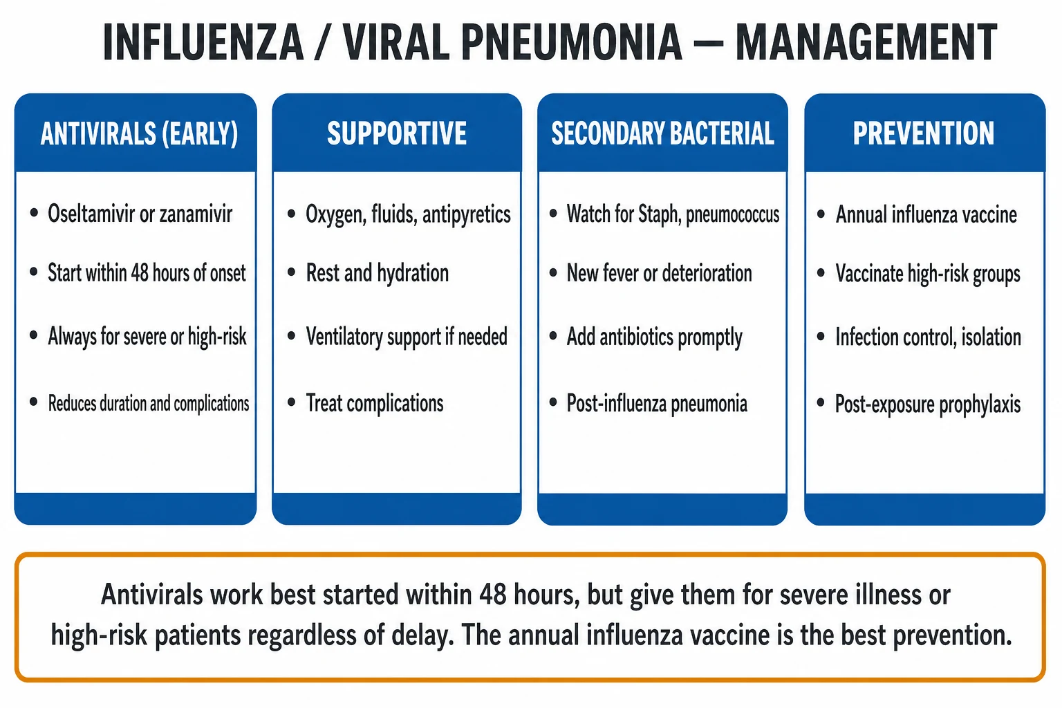
- Empirical antiviral therapy — START IMMEDIATELY in any hospitalised patient with suspected influenza; do NOT wait for test results.[1]
- Surviving Sepsis hour-1 bundle if septic: oxygen, blood cultures, lactate, broad-spectrum antibiotics within 1 hour, balanced crystalloid 30 mL/kg, noradrenaline for fluid-refractory shock.
Management — Definitive & Stepwise
The two pillars are (1) antiviral therapy for those who need it, and (2) prevention by vaccination. Antibiotics are added only for documented or suspected secondary bacterial infection — never routinely.[1][5]
Antiviral therapy — neuraminidase inhibitors (first line)
Oseltamivir (Tamiflu) — oral; the workhorse in adults and children. [1]
| Indication | Adult dose | Paediatric dose | Duration |
|---|---|---|---|
| Treatment (uncomplicated) | 75 mg PO twice daily | Weight-based: under 1 yr — 3 mg/kg twice daily; 1 yr+ — see schedule below | 5 days |
| Treatment (severe / hospitalised) | 75 mg PO twice daily (or 75 mg NG; up to 150 mg twice daily in critically ill / immunocompromised with high viral load) | As weight-based | Minimum 5 days; extend until clinical recovery + negative PCR in immunocompromised |
| Prophylaxis (post-exposure) | 75 mg PO once daily | Weight-based once daily | 7 days (10 days in outbreaks) |
Paediatric oseltamivir treatment doses (twice daily, 5 days):[1]
- under 1 yr: 3 mg/kg/dose twice daily.
- 1 yr or over, up to 15 kg: 30 mg twice daily.
- over 15 to 23 kg: 45 mg twice daily.
- over 23 to 40 kg: 60 mg twice daily.
- over 40 kg: 75 mg twice daily (adult dose). [1]
Timing: best within 48 hours of symptom onset for uncomplicated disease — reduces duration by about 1 day and reduces complications/hospitalisation.[3] For hospitalised, immunocompromised, pregnant, or severe disease — start at ANY time after onset, even beyond 48 hours; benefit persists and mortality is reduced.
Other neuraminidase inhibitors: [1]
- Zanamivir (Relenza) — inhaled (Diskhaler), 10 mg (2 inhalations) twice daily for 5 days; for prophylaxis 10 mg once daily for 10 days. Avoid in underlying airways disease (asthma/COPD — bronchospasm reported). Limited to those unable to take oseltamivir.
- Peramivir (Rapivab) — intravenous, single dose 600 mg IV (adults), 12 mg/kg IV (children over 6 months) — useful when PO/inhaled impossible (intubated, vomiting).
- Laninamivir (Inavir) — inhaled, single long-acting dose; not available everywhere. [1]
Cap-dependent endonuclease inhibitor (cap-snatch blocker)
Baloxavir marboxil (Xofluza) — first-in-class; blocks the cap-dependent endonuclease (PA subunit) of the viral polymerase, preventing 'cap-snatching' and viral mRNA synthesis. Single oral dose: adults and children 12 yr+ weighing 40–80 kg = 40 mg; over 80 kg = 80 mg. Comparable to oseltamivir for symptom alleviation; faster viral clearance and reduces duration of viral shedding.[6][11] Useful in high-risk outpatients; not preferred in pregnancy or severe disease pending more data.
M2 ion-channel inhibitors — historical and now obsolete
Amantadine and rimantadine block the M2 ion channel of influenza A (only). Resistance in circulating A/H1N1pdm09 and A/H3N2 is essentially universal — they are NOT recommended for treatment or prophylaxis. Mention only as an exam footnote and for the mechanism (block uncoating). [1]
Adjunctive therapy in severe influenza
- Corticosteroids — NOT recommended routinely; associated with increased mortality, prolonged viral shedding, secondary infection in influenza. Consider ONLY for the concomitant indication: septic shock (hydrocortisone 200 mg/day), or asthma/COPD exacerbation. Avoid high-dose steroids for viral pneumonitis.[1]
- Antibiotics for secondary bacterial pneumonia — guided by culture; empirical IV co-amoxiclav 1.2 g three times daily OR ceftriaxone 2 g once daily, adding flucloxacillin 2 g six times daily or vancomycin if Staphylococcus aureus (including MRSA) is suspected — classically post-influenza.
- Respiratory support escalation — high-flow nasal cannula → non-invasive ventilation (caution: aerosol generation) → intubation and lung-protective mechanical ventilation (tidal volume 6 mL/kg ideal body weight, plateau pressure under 30 cm water) → proning for moderate-severe ARDS (P/F under 150) → ECMO in refractory hypoxaemia at a referral centre.
- Convalescent plasma, immunoglobulin — considered in severe immunocompromised with refractory disease; not routine.
Infection control and public-health actions
- Droplet precautions for the duration of illness (until afebrile and at least 24 hours after symptom resolution; longer if immunocompromised).
- Notify the public-health authority of any suspected novel / avian / pandemic influenza (H5N1, H7N9, novel reassortant), and any influenza death in a pregnant woman or child.
- Antiviral post-exposure prophylaxis (PEP) for high-risk close contacts within 48 hours of exposure: oseltamivir 75 mg PO once daily for 7 days (10 days in institutional outbreaks). PEP is NOT a substitute for vaccination. [1]
Stepwise care ladder (escalation triggers)
- Mild uncomplicated influenza, low-risk patient — home, supportive care, paracetamol, fluids, safety-net. Antiviral if within 48 h or high-risk.
- Symptomatic high-risk or within 48 h — oseltamivir 75 mg BD x 5 days (or baloxavir single dose), home with close review.
- Hypoxia, dehydration, dyspnoea, or any red flag — admit, oxygen, IV fluids, antivirals, CXR, bloods.
- Severe disease / pneumonitis / ARDS / shock — ICU, antivirals (consider dual oseltamivir + baloxavir), lung-protective ventilation, proning, vasopressors, ECMO referral if refractory.
- Immunocompromised / persistent shedding — extended antiviral course, monitor for oseltamivir resistance (H275Y mutation), switch to IV zanamivir if resistance emerges. [1]
Specific Subtypes & Scenarios
- Seasonal H1N1pdm09 — descendant of the 2009 pandemic strain; now a regular seasonal strain. Younger adults disproportionately affected (less pre-existing immunity). Higher rate of viral pneumonitis in pregnancy, obesity, and immunocompromise.[7]
- H3N2 seasons — cause the most severe seasonal epidemics in the elderly, with excess mortality and excess nursing-home outbreaks; antigenic drift in H3N2 is faster than H1N1, so vaccine effectiveness against H3N2 is consistently the lowest.
- Influenza B — primarily affects children; less severe in adults but can cause significant outbreaks in schools and residential care. Two lineages (Victoria, Yamagata) — mismatch of vaccine B lineage to circulating strain (a 'B-mismatch') causes moderate epidemics.
- Avian influenza A/H5N1 — zoonotic from infected poultry; case fatality approximately 60%; presents with rapidly progressive viral pneumonia, ARDS, multi-organ failure; treat with high-dose oseltamivir (150 mg BD); notify public health.[8]
- Avian influenza A/H7N9 (China) — also high mortality, often with bilateral pneumonia and ARDS.
- Influenza + SARS-CoV-2 co-infection — coinfection is well described and worsens prognosis; test for both in any patient with ILI in season; manage both with their respective protocols.
- Institutional outbreak (nursing home, dormitory, military barracks) — defines an outbreak: 2 or more cases of ILI within 72 hours in the same unit. Response: isolate, offer testing, antiviral treatment of cases, antiviral prophylaxis of all residents and staff regardless of vaccination status for at least 14 days, and continue 7 days after the last case.[1]
- Immunocompromised host — prolonged viral shedding (weeks to months), risk of emergence of antiviral resistance (oseltamivir H275Y mutation), atypical presentation; consider IV zanamivir for resistance; prolonged or combination therapy may be needed.
Complications & Pitfalls
Pulmonary: [1]
- Primary viral pneumonia — direct viral damage; bilateral interstitial infiltrates, rapid hypoxia, ARDS; highest in pregnancy, immunocompromise, obesity, native populations.
- Secondary bacterial pneumonia — second fever spike after initial improvement; purulent sputum, lobar consolidation. Strep pneumoniae, Staph aureus (necrotising), Haemophilus influenzae. Post-influenza Staph aureus pneumonia is the classic exam association.
- Exacerbation of underlying COPD/asthma/cystic fibrosis — the most common complication of influenza in chronic lung disease.
- Bronchiolitis (children), croup, otitis media (especially paediatric), sinusitis, epiglottitis (rare). [1]
Cardiac: [1]
- Myocarditis — chest pain, dyspnoea, raised troponin, new arrhythmia, cardiogenic shock; can be fulminant.
- Pericarditis.
- Exacerbation of heart failure and acute coronary syndrome — influenza triggers plaque rupture; vaccination reduces major adverse cardiac events. [1]
Neurological: [1]
- Influenza-associated encephalitis/encephalopathy (IAE) — especially in children (H1N1pdm09); altered consciousness, seizures, focal neurology; MRI T2/FLAIR hyperintensities; high mortality.
- Guillain–Barre syndrome (GBS) — within 1–6 weeks of influenza; classically ascending flaccid paralysis with areflexia; treat with IVIG or plasmapheresis. (Also historically associated with the 1976 swine flu vaccine — the basis of GBS surveillance for every influenza vaccine campaign.)
- Transverse myelitis, ADEM, febrile seizures. [1]
Musculoskeletal: [1]
- Myositis and rhabdomyolysis — calf pain, raised serum CK, dark (myoglobinuric) urine, AKI; more common in children and H1N1/H3N2.
- Reactive arthritis (rare). [1]
Renal: AKI from rhabdomyolysis, sepsis, or myocarditis. [1]
Haematological: thrombocytopenia, disseminated intravascular coagulation (rare), haemophagocytic lymphohistiocytosis (HLH). [1]
Metabolic / hepatic: [1]
- Reye syndrome — classically in children given aspirin during a viral (influenza, VZV) illness — mitochondrial hepatopathy: vomiting, hepatomegaly, hypoglycaemia, altered consciousness, microvesicular steatosis, raised ammonia with normal/mild LFTs. NEVER give aspirin to a child with a fever — this is the single exam point. Mortality 20–40%. [1]
Toxic shock from superantigen-producing Staph/Strep co-infection. [1]
Classic pitfalls: [1]
- Over-reliance on a negative RIDT — repeat with PCR or treat empirically if clinical suspicion is high.
- Delaying antivirals waiting for confirmation in a hospitalised or high-risk patient.
- Failing to recognise the atypical presentation in the elderly (no fever; presenting as confusion or falls).
- Missing pregnancy as a high-risk group.
- Giving aspirin to a febrile child.
- Routine corticosteroids for influenza pneumonitis (harm).
- Forgetting the second fever spike = secondary bacterial pneumonia.
- Not testing for SARS-CoV-2 in any ILI during COVID circulation. [1]
Prognosis & Disposition
Most uncomplicated influenza resolves in 3–7 days, though cough and fatigue may persist for 2–3 weeks. The cough and post-viral asthenia are the patient's main complaints.[1]
Predictors of poor outcome: age over 65, pregnancy, BMI 40 or more, immunocompromise, chronic cardiopulmonary/renal/hepatic/metabolic disease, delayed antiviral therapy, viral pneumonitis/ARDS, bacterial co-infection, MMT/ICU admission, neutrophilia, raised lactate, raised CK. [1]
Global mortality: seasonal influenza causes an estimated 290,000–650,000 respiratory deaths and up to 1 million total deaths annually; over 90% occur in adults over 65.[10]
Disposition: [1]
- Home — uncomplicated, low-risk, improving; safety-net for return if breathless, drowsy, unable to keep fluids down, or fever recurs.
- Hospital (ward) — hypoxia, dehydration, secondary pneumonia, pregnancy with significant symptoms, immunocompromise, any high-risk group not improving.
- ICU — ARDS, septic shock, respiratory failure, multi-organ failure.
- Follow-up — review at 48–72 hours; follow-up CXR not routinely needed unless complicated. [1]
Special Populations
- Pregnancy (and up to 2 weeks postpartum). Influenza is markedly more severe — particularly H1N1pdm09 — with excess ICU admission, ARDS, miscarriage, preterm birth, and fetal death.[7] Start oseltamivir immediately at any gestation (75 mg BD x 5 days; do NOT wait for results); inactivated influenza vaccine at any stage of pregnancy is recommended and protects both mother and infant for the first 6 months of life (transplacental IgG). Avoid baloxavir, live-attenuated vaccine, and zanamivir if asthma.
- Children (under 5, especially under 2). Higher rates of hospitalisation, otitis media, bronchiolitis, croup, febrile seizures. Oseltamivir weight-based (see dose table). AVOID aspirin (Reye syndrome); AVOID cough and cold remedies containing antitussives/decongestants in under 6 yr. Live attenuated intranasal vaccine (LAIV) available for 2–17 yr in some countries; contraindicated in immunocompromise, severe asthma, salicylate therapy, pregnancy.
- Elderly (over 65). Atypical presentation (no fever; confusion, falls); higher-dose or adjuvanted vaccines (e.g. high-dose quadrivalent, adjuvanted MF59, recombinant HA vaccines) preferred to overcome immunosenescence.
- Immunocompromised (transplant, chemotherapy, HIV with low CD4). Prolonged shedding, atypical presentation, antiviral resistance; consider prolonged/combination antiviral therapy; inactivated vaccine only (LAIV contraindicated); antibody response is blunted.
- Chronic respiratory/cardiac/renal/hepatic disease, diabetes, severe obesity (BMI 40+). All meet criteria for annual vaccination and early antivirals; vaccinate household contacts.
- Healthcare workers. Mandatory annual influenza vaccination in many institutions; protects both the worker and vulnerable patients (reduces nosocomial outbreaks and patient mortality).
- Anticoagulated patients. No specific influenza–anticoagulant interaction; continue anticoagulation, but balance bleeding risk against the markedly increased VTE risk in severe influenza/ARDS (consider prophylactic LMWH in all hospitalised adults).
Evidence, Guidelines & Regional Differences
Landmark evidence
- Dobson 2015 (Lancet, individual-patient-data meta-analysis of 9 RCTs) — oseltamivir in adults reduced symptom duration by about 1 day (from 122 to 98 h) and halved hospitalisations in high-risk adults.[3]
- Hayden 2018 NEJM (CAPSTONE-1) — baloxavir marboxil was non-inferior to oseltamivir for symptom alleviation and reduced viral shedding faster.[6]
- Ison 2020 (CAPSTONE-2, Lancet Infect Dis) — baloxavir in high-risk adolescents and adults reduced complications and time to alleviation.[11]
- Iuliano 2018 (Lancet, global mortality model) — redefined the global burden at 290,000–650,000 respiratory deaths/year.[10]
- Taubenberger 2020 (CSH Perspect Med) — definitive account of the 1918 pandemic and its scientific legacy.[9]
Guidelines
- IDSA 2018 (Uyeki et al.) — the global reference for diagnosis, treatment, chemoprophylaxis and outbreak management.[1]
- CDC ACIP — annual updates to vaccination recommendations, strains, and indication list.[5]
- WHO Global Influenza Surveillance and Response System (GISRS) — global strain surveillance and the body that announces twice-yearly vaccine composition (February for Northern Hemisphere, September for Southern).
Regional differences
[1] [1] [1]Controversies
- Cochrane reviews (Jefferson/Demicheli) on oseltamivir — questioned the magnitude of benefit and highlighted publication bias; mainstream guidance still endorses oseltamivir for high-risk and severe disease based on individual-patient data.
- Routine corticosteroids for influenza pneumonitis — harm in observational data, but still used in some settings for septic shock; guideline: avoid unless indicated for another reason.
- Universal influenza vaccine — the holy grail; targets the conserved stalk of HA, the M2e ion channel, or the conserved NA; would eliminate the need for annual reformulation and seasonal strain prediction.[4]
Prevention — vaccination (cornerstone)
Annual quadrivalent inactivated influenza vaccine (QIV) — the single most important intervention. The composition is updated twice yearly by WHO; a typical Northern Hemisphere season includes A/Victoria/H1N1pdm09-like, A/Darwin/H3N2-like, B/Victoria-lineage, and B/Yamagata-lineage strains.[4]
Indications (CDC/ACIP: everyone 6 months and over; high-priority groups):[5]
- All children 6 months to 5 yr (and their household contacts).
- All adults over 50 or 65 (depends on country; US over 50, UK over 65).
- Pregnant women at any gestation.
- Chronic pulmonary, cardiac (excluding isolated hypertension), renal, hepatic, neurological, haematological, metabolic (including diabetes) disease.
- Immunocompromised (including HIV).
- Residents of long-term care / nursing homes.
- Healthcare workers and household contacts of high-risk persons.
- Extreme obesity (BMI 40 or more).
- Children 6 months to 18 yr on long-term aspirin (to prevent Reye syndrome if they acquire influenza). [1]
Doses: single annual IM dose (deltoid); children 6 months–8 yr receiving influenza vaccine for the first time need TWO doses, 4 weeks apart. High-dose, adjuvanted (MF59), or recombinant vaccines are preferred for adults over 65 (improve immunogenicity). [1]
Contraindications/precautions: severe allergic reaction (e.g. anaphylaxis) to a previous dose or component; severe egg allergy (recombinant cell-based vaccines are egg-free; in most modern guidelines mild egg allergy is no longer a contraindication — observe for 30 min); history of GBS within 6 weeks of a previous influenza vaccine (precaution); moderate-to-severe acute illness (defer). Live attenuated intranasal vaccine (LAIV) has additional contraindications: immunocompromise, pregnancy, severe asthma, salicylate therapy in children, age under 2 or over 49. [1]
Vaccine effectiveness (VE): typically 40–60% in the well-matched years; lower (10–40%) against H3N2 due to rapid drift, egg-adaptation mutations, and immunosenescence. Even when VE against infection is low, vaccination reduces severe disease, hospitalisation, ICU admission, and death. [1]
Exam Pearls
- Influenza = sudden onset (can date to the hour). Common cold = gradual.
- Surface antigens: HA (18) and NA (11). HA = entry (binds sialic acid); NA = release (cleaves sialic acid — drug target of oseltamivir).
- Drift vs shift: drift = point mutations in BOTH A and B → annual epidemics; shift = reassortment of genome segments in A ONLY → pandemics (swine = mixing vessel).
- 8 RNA segments, no proof-reading — basis of drift.
- Receptor binding: HA binds alpha-2,6 sialic acid (human upper airway); avian HA prefers alpha-2,3 (lower airway/bird gut) — explains tropism and severity of H5N1 (binds deep lung).
- Antivirals — oseltamivir 75 mg BD x 5 days (within 48 h for uncomplicated; any time for severe/pregnant/immunocompromised). Weight-based in children.
- Baloxavir = single-dose cap-dependent endonuclease inhibitor (blocks PA subunit).
- M2 inhibitors (amantadine, rimantadine) = universally resistant; NOT used.
- Pandemic mnemonic — 'SpHAN': Spanish 1918 H1N1; Hong Kong 1968 H3N2; Asian 1957 H2N2; Novel 2009 H1N1pdm09.
- Quadrivalent vaccine = 2 A + 2 B strains. Updated annually by WHO.
- Children under 9 first time = TWO doses, 4 weeks apart.
- NO aspirin in children with viral fever → Reye syndrome (mitochondrial hepatopathy, raised ammonia, microvesicular steatosis).
- Post-influenza Staph aureus pneumonia = the classic secondary pneumonia.
- Second fever spike in recovery = secondary bacterial pneumonia (Strep pneumoniae, Staph aureus, H. influenzae).
- Influenza + SARS-CoV-2 co-infection is possible — TEST BOTH.
- Pregnancy, especially 2nd/3rd trimester, is high-risk — start oseltamivir immediately; vaccinate at any gestation.
- H5N1 (avian) — CFR about 60%; from infected poultry; high-dose oseltamivir.
- H1N1pdm09 = triple reassortment (avian + swine + human) — first 21st-century pandemic.
- Negative RIDT does NOT exclude influenza — repeat with PCR. [1]
Influenza — the numbers an examiner wants
Antigenic DRIFT vs SHIFT — the rule
DRIFT
point mutations in H and N; both A and B; annual epidemics; no pandemic
= SHIFT; needs 2 viruses in same cell; only A; pandemics; swine = mixing vessel
H (1–18) and N (1–11); human: H1N1, H2N2, H3N2
oseltamivir window for uncomplicated flu; treat ANY time if severe/high-risk
Victoria + Yamagata; quadrivalent vaccine covers both
Exam application bank (NEET-PG / INICET)
One-line answer
Influenza is an acute, highly contagious respiratory infection caused by orthomyxoviruses of types A, B and (rarely) C. Type A is subtyped by two surface glycoproteins — haemagglutinin (H, 18 subtypes) and neuraminidase (N, 11 subtypes); current human strains are A/H1N1pdm09 and A/H3N2; type B is divided into Victoria and Yamagata lineages. Antigenic drift (point mutations in H and N) drives annual seasonal epidemics; antigenic shift (reassortment of genome segments when two viruses co-infect a host, e.g. swine as a 'mixing vessel') produces a novel subtype → pandemic (1918 H1N1 'Spanish flu', 1957 H2N2 'Asian flu', 1968 H3N2 'Hong Kong flu', 2009 H1N1pdm09 'swine flu'). Transmission is by respiratory droplets and aerosols with an incubation period of 1–4 days. Clinically: abrupt high fever, rigors, headache, myalgia, dry cough, sore throat, extreme prostration — distinguished from the c
Worked stems (answer without another resource)
Stem 1 — Classic presentation. Map symptoms to mechanism; name the first investigation and first treatment step with dose/route if drug therapy is standard. [1]
Stem 2 — Unstable / complicated. List red flags that force immediate resuscitation, theatre, ICU, antidote, or reperfusion — and what you do in the first 15 minutes. [1]
Stem 3 — Atypical group. Elderly, pregnancy, child, or immunocompromised: how presentation and thresholds change. [1]
Stem 4 — Differential trap. Name the three closest mimics and one discriminator for each. [1]
Stem 5 — Disposition. Who goes home with safety-netting, who is admitted, who needs HDU/ICU/theatre, and what follow-up is mandatory. [1]
Rapid viva checklist
- Definition + classification
- Pathophysiology chain
- Bedside signs / criteria
- Score with exact components (if any)
- Emergency bundle
- Definitive therapy with doses
- Complications of disease and of treatment
- Special populations
- Guideline/trial name if classic
- Three exam traps
Coverage self-check
If you cannot answer any stem above from this page alone, re-read the matching section — the page is intended to be self-sufficient for final-prof and NEET-PG/INICET questions on Influenza (Seasonal & Pandemic).
References
- [1]Uyeki TM, Bernstein HH, Bradley JS, Englund JA, File TM, Fry AM, et al. Clinical Practice Guidelines by the Infectious Diseases Society of America: 2018 Update on Diagnosis, Treatment, Chemoprophylaxis, and Institutional Outbreak Management of Seasonal Influenzaa Clin Infect Dis, 2019.PMID 30566567
- [2]Krammer F, Smith GJD, Fouchier RAM, Peiris M, Kedzierska K, Doherty PC, et al. Influenza Nat Rev Dis Primers, 2018.PMID 29955068
- [3]Dobson J, Whitley RJ, Pocock S, Monto AS. Oseltamivir treatment for influenza in adults: a meta-analysis of randomised controlled trials Lancet, 2015.PMID 25640810
- [4]Paules C, Subbarao K. Chasing Seasonal Influenza - The Need for a Universal Influenza Vaccine N Engl J Med, 2018.PMID 29185857
- [5]Fiore AE, Fry A, Shay D, Gubareva L, Bresee JS, Uyeki TM, et al. (CDC ACIP). Antiviral agents for the treatment and chemoprophylaxis of influenza --- recommendations of the Advisory Committee on Immunization Practices (ACIP) MMWR Recomm Rep, 2011.PMID 21248682
- [6]Hayden FG, Sugaya N, Hirotsu N, Lee N, de Jong MD, Hurt AC, et al. Baloxavir Marboxil for Uncomplicated Influenza in Adults and Adolescents N Engl J Med, 2018.PMID 30184455
- [7]Jamieson DJ, Honein MA, Rasmussen SA, Williams JL, Swerdlow DL, Biggerstaff MS, et al. H1N1 2009 influenza virus infection during pregnancy in the USA Lancet, 2009.PMID 19643469
- [8]Kandun IN, Tresnaningsih E, Purba WH, Lee V, Samaan G, Harun S, et al. Factors associated with case fatality of human H5N1 virus infections in Indonesia: a case series Lancet, 2008.PMID 18706688
- [9]Taubenberger JK, Morens DM. The 1918 Influenza Pandemic and Its Legacy Cold Spring Harb Perspect Med, 2020.PMID 31871232
- [10]Iuliano AD, Roguski KM, Chang HH, Muscatello DJ, Palekar R, Tempia S, et al. Estimates of global seasonal influenza-associated respiratory mortality: a modelling study Lancet, 2018.PMID 29248255
- [11]Ison MG, Portsmouth S, Yoshida Y, Shishido T, Mitchener M, Tsuchiya K, et al. Early treatment with baloxavir marboxil in high-risk adolescent and adult outpatients with uncomplicated influenza (CAPSTONE-2): a randomised, placebo-controlled, phase 3 trial Lancet Infect Dis, 2020.PMID 32526195