Respiratory · General Medicine
Interstitial Lung Disease
Also known as Diffuse parenchymal lung disease · Pulmonary fibrosis · Cryptogenic fibrosing alveolitis · Idiopathic interstitial pneumonia · Pneumoconiosis · Hypersensitivity pneumonitis
Interstitial lung disease (ILD), also called diffuse parenchymal lung disease (DPLD), is a heterogeneous group of over 200 disorders that share inflammation and/or fibrosis of the lung interstitium (alveolar walls, septa, peribronchovascular and perilymphatic spaces), producing a restrictive ventilatory defect (low total lung capacity, low FVC, low DLCO with preserved or raised FEV1/FVC) and a diffuse abnormality on imaging. The clinical archetype is idiopathic pulmonary fibrosis (IPF) — chronic, progressive fibrosis of older adults with a usual interstitial pneumonia (UIP) pattern on HRCT (basal, subpleural reticulation, honeycombing, traction bronchiectasis, with little ground-glass) and a median survival of three to five years. Diagnosis is multidisciplinary, requires exclusion of known causes (connective tissue disease, drugs, occupation, hypersensitivity), and is anchored by thin-section HRCT. Management of the fibrotic phenotype is pirfenidone or nintedanib (antifibrotics that slow — not reverse — decline), long-term oxygen, pulmonary rehabilitation, and lung transplantation for selected patients.
On this page & tools
Your progress
Saved locally on this device.
Practise this topic
Exam tags
Red flags
Overview & Definition
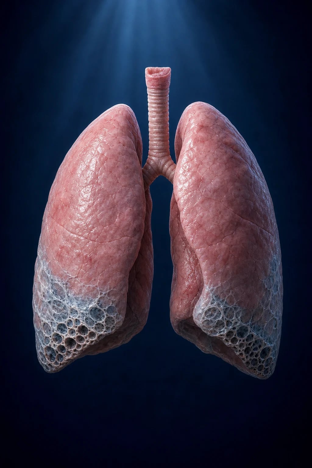
Interstitial lung disease (ILD) — interchangeably called diffuse parenchymal lung disease (DPLD) — is an umbrella term for more than 200 entities that share inflammation and/or fibrosis of the lung interstitium. The "interstitium" here is the entire gas-exchanging apparatus: alveolar epithelium and basement membrane, interstitial space, capillary endothelium, plus the peribronchovascular, perilymphatic and septal connective tissue that scaffolds the lung.[6]
Three operational features define an ILD: [1]
- A restrictive ventilatory defect on pulmonary function testing (reduced total lung capacity [TLC] and forced vital capacity [FVC] with a preserved or raised FEV1/FVC ratio), accompanied by a reduced diffusion capacity (DLCO).
- A diffuse abnormality on chest imaging (chest X-ray and especially thin-section HRCT).
- Exclusion of infection, malignancy, and overt cardiac failure as the primary explanation. [1]
ILDs are not a single disease. The clinical archetype, idiopathic pulmonary fibrosis (IPF) — historically called cryptogenic fibrosing alveolitis (CFA) — is a chronic, progressive, fibrosing interstitial pneumonia of older adults, limited to the lungs, with a usual interstitial pneumonia (UIP) pattern on surgical lung biopsy or HRCT and no identifiable cause.[1] Median survival from diagnosis is three to five years, worse than many common cancers.
Classification
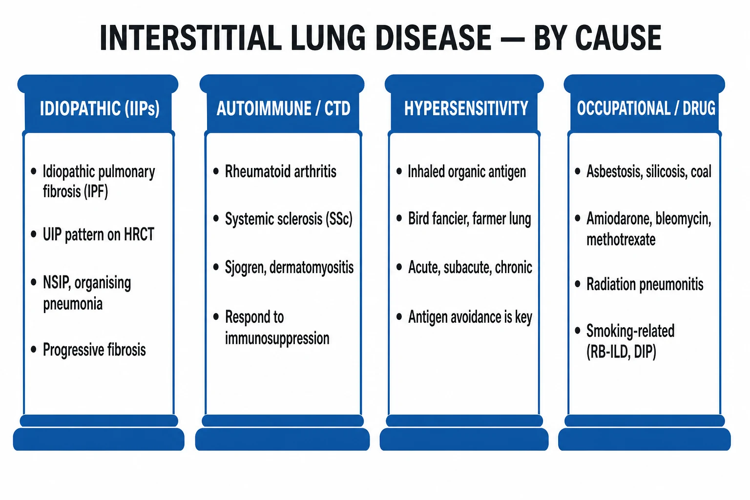
The clinically useful classification groups ILDs by aetiology, beginning with the single most important question — is there a known cause? [1]
Known cause
- Autoimmune / connective tissue disease — systemic sclerosis (most common and most aggressive), rheumatoid arthritis, polymyositis/dermatomyositis (antisynthetase, anti-MDA5), Sjögren, SLE, MCTD
- Drug / iatrogenic — amiodarone, bleomycin, methotrexate, nitrofurantoin, sulfasalazine, gold, hydralazine, checkpoint inhibitors, radiation
- Occupational / environmental — asbestos, silica, coal dust, beryllium, hard metal, bird antigens, mouldy hay (farmer's lung)
- Hypersensitivity pneumonitis (HP) — bird fancier's, farmer's, humidifier, hot-tub, mushroom worker
Idiopathic interstitial pneumonias (IIPs)
- Idiopathic pulmonary fibrosis (IPF) — UIP pattern; commonest IIP; worst prognosis
- Non-specific interstitial pneumonia (NSIP) — cellular and fibrotic subtypes; common in CTD
- Cryptogenic organising pneumonia (COP / BOOP) — steroid responsive, patchy consolidation
- Respiratory bronchiolitis-associated ILD (RB-ILD) and desquamative interstitial pneumonia (DIP) — smoking-related
- Acute interstitial pneumonia (AIP / Hamman-Rich) — diffuse alveolar damage, ARDS-like
- Lymphoid interstitial pneumonia (LIP) — Sjögren, HIV, CVID
Granulomatous
- Sarcoidosis — non-caseating granulomas; bilateral hilar lymphadenopathy; multi-system
- Hypersensitivity pneumonitis (chronic) — poorly formed granulomas, bronchiolocentric
- Berylliosis — non-caseating granulomas, sarcoid mimic
Rare / other
- Lymphangioleiomyomatosis (LAM) — cystic, women, sirolimus
- Pulmonary Langerhans cell histiocytosis (LCH) — smoking, upper-zone nodules + cysts, CD1a/CD207/S100 positive
- Pulmonary alveolar proteinosis — crazy-paving, anti-GM-CSF, whole-lung lavage
- Eosinophilic lung diseases — acute/chronic eosinophilic pneumonia
- Pulmonary alveolar microlithiasis, amyloidosis, light-chain deposition
The ATS/ERS 2013 Travis multidisciplinary classification (updated 2022) reorganises the idiopathic interstitial pneumonias by clinical–radiological–pathological pattern rather than histology alone — the diagnosis is made by an ILD multidisciplinary team (MDT) and may change as new data emerge.[6]
Epidemiology & Risk Factors
The epidemiology of the fibrotic ILDs, and especially IPF, dominates exam questions: [1]
- Prevalence of IPF is 14–43 per 100 000 population; annual incidence 7–11 per 100 000 in North America and Europe.[1]
- Age — IPF is a disease of older adults; mean age at diagnosis 65–70 years, rare under 50.
- Sex — men affected roughly 2:1 over women.
- Smoking — current or former smokers carry a 2–3-fold increased risk of IPF; smoking is also the principal driver of RB-ILD, DIP, and pulmonary Langerhans cell histiocytosis.
- Occupational — metal and wood dust, farming, hairdressing, and stone cutting are over-represented in ILD registries.
- Genetics — familial pulmonary fibrosis (autosomal dominant, incomplete penetrance) accounts for 3–20% of cases. Key genes: MUC5B promoter polymorphism (rs35705950) — strongest common risk allele; telomerase complex mutations (TERT, TERT promoter, TERC, RTEL1, PARN); surfactant protein mutations (SFTPC, SFTPA2, ABCA3). Short telomere length predicts worse prognosis and informs family screening.
Pathophysiology
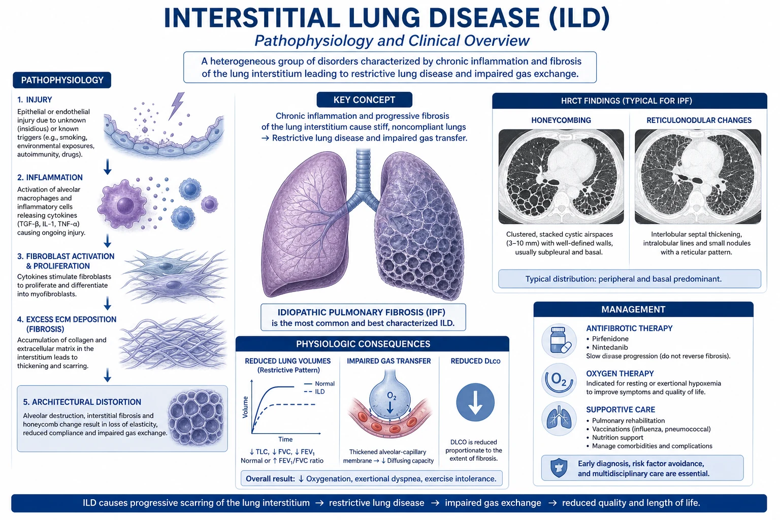
The modern paradigm for IPF — the prototype fibrotic ILD — is recurrent alveolar epithelial micro-injury with aberrant wound healing, NOT a primarily inflammatory process. The old "inflammation precedes fibrosis" model was discarded after anti-inflammatory therapy failed to alter the disease course.[1]
The wound-healing cascade proceeds in five linked steps: [1]
- Epithelial injury and apoptosis — repetitive, subclinical injury (smoke, viral, gastro-oesophageal reflux, oxidative stress, genetic susceptibility) to type II alveolar epithelial cells.
- Impaired re-epithelialisation — failure of epithelial regeneration and basement membrane repair. In telomerase-mutant IPF, stem cell exhaustion accelerates this step.
- Fibroblast recruitment and proliferation — epithelium secretes TGF-β1 (the master pro-fibrotic cytokine), PDGF (mitogen/chemoattractant), CTGF, FGF-2, and IL-13. Fibrocytes are recruited from the bone marrow.
- Myofibroblast differentiation and matrix deposition — fibroblasts differentiate into contractile α-smooth muscle actin-positive myofibroblasts that pile up in fibroblastic foci (the histological hallmark of active UIP) and deposit disordered type I and type III collagen, fibronectin and proteoglycans.
- Architectural destruction — alveolar collapse and traction give the honeycomb lung: cystic spaces lined by bronchiolar epithelium embedded in dense fibrous tissue. [1]
The result is a stiff lung with reduced compliance. Pulmonary vascular pruning and secondary vascular remodelling produce group 3 pulmonary hypertension in roughly a third of advanced cases. [1]
Acute exacerbation of IPF is the catastrophic clinical event: diffuse alveolar damage (the histological pattern of ARDS) is superimposed on underlying UIP, with rapid gas-exchange failure and high mortality. [1]
The other IIPs follow different mechanistic programmes: NSIP is a more uniform, temporally homogeneous process; COP is granulation tissue polyps occluding distal airways (a defect of resolution of injury rather than perpetuation); HP is a type III + type IV hypersensitivity reaction to inhaled antigen with poorly formed granulomas. [1]
Clinical Presentation
The classical symptom triad of fibrotic ILD (especially IPF): [1]
- Progressive exertional dyspnoea — the cardinal symptom; insidious onset over months; worse on inclines and stairs; eventually present at rest.
- Chronic dry, persistent cough — often the most distressing symptom; mechanical (stretch receptors in fibrotic parenchyma) and reflux-related.
- Fatigue and weight loss — frequently out of proportion to objective lung function. [1]
Key examination findings: [1]
- Velcro crackles — fine, late-inspiratory, end-inspiratory, non-clearing crackles best heard posteriorly at the lung bases and in the axillae with the diaphragm of the stethoscope. The name derives from the sound of opening the hook-and-loop fastener. Highly suggestive of UIP/IPF.
- Digital clubbing — present in roughly half of IPF patients; if present, predicts worse prognosis.
- Cyanosis and hypoxaemia — initially on exercise (diffusion limitation + low V/Q), later at rest.
- Tachypnoea and use of accessory muscles.
- Pulmonary hypertension and cor pulmonale — loud pulmonary component of S2 (P2), right ventricular heave, tricuspid regurgitation murmur, raised JVP, hepatomegaly, ankle oedema.
- Signs of underlying cause — sclerodactyly, digital pitting scars, telangiectasia and perioral furrowing (systemic sclerosis); heliotrope rash, Gottron papules, mechanic's hands, proximal muscle weakness (dermatomyositis/antisynthetase); Raynaud phenomenon, sicca symptoms, parotid enlargement (Sjögren); erythema nodosum, uveitis (sarcoid). [1]
Atypical presentations
- Elderly — IPF may present as isolated unexplained dyspnoea dismissed as "ageing", "heart failure with preserved ejection fraction", or "mild COPD". Auscultation of Velcro crackles is the discriminating bedside finding.
- Diabetic / immunosuppressed — drug-induced ILD (methotrexate, checkpoint inhibitors) and opportunistic infection may overlap; BAL is critical.
- Pregnancy — most ILDs are rare in pregnancy; pirfenidone and nintedanib are NOT recommended (insufficient teratogenicity data).
- Hypersensitivity pneumonitis phenotype — acute HP: fever, chills, cough, dyspnoea 4–12 h after antigen exposure (bird droppings, mouldy hay); subacute/chronic HP: insidious cough, weight loss, progressive dyspnoea, basal crackles ± wheeze.
- CTD-ILD phenotype — ILD may precede the CTD by years ("lung-dominant CTD"); always ask for Raynaud, arthralgia, sicca and rashes. [1]
High-yield numbers in ILD
Differential Diagnosis
A comprehensive ILD differential is built around the clinical question "what looks like ILD but isn't?" — and "which ILD is it?" [1]
Mimics of ILD
- COPD — obstructive pattern (↑TLC, ↓FEV1/FVC), rhonchi, smoking history; PFTs discriminate
- Left heart failure — raised BNP/NT-proBNP, septal lines and pleural effusion on CT, no honeycombing, response to diuresis
- Atypical infection — PJP, viral, mycoplasma; subacute fever, BAL diagnostic
- Miliary tuberculosis — random miliary nodules, fever, TB risk factors, AFB positive
- Lymphangitis carcinomatosa — nodular septal thickening, known primary, hilar adenopathy
- Pulmonary alveolar proteinosis — crazy-paving, milky BAL, anti-GM-CSF
- Diffuse alveolar haemorrhage — anaemia, haemoptysis, hemosiderin-laden macrophages
Within fibrotic ILD — distinguish on HRCT
- IPF (UIP) — basal, subpleural, honeycombing, traction bronchiectasis, little ground-glass
- NSIP — symmetric basal ground-glass + fine reticulation, spares extreme base, no honeycombing
- Chronic HP — mid/upper-zone, centrilobular nodules, mosaic attenuation, air trapping
- Sarcoidosis — bilateral hilar and mediastinal lymphadenopathy, perilymphatic nodules, upper-zone fibrosis
- Asbestosis — lower-lobe fibrosis + pleural plaques
- Silicosis — upper-zone conglomerate masses, eggshell-calcified hilar nodes
The single most useful discriminating test is thin-section HRCT, interpreted within the clinical context. When in doubt, the ILD MDT adjudicates. [1]
Clinical & Bedside Assessment
A focused examination closes the diagnostic loop and is heavily examined. [1]
Auscultation: how to elicit Velcro crackles
- Use the diaphragm of the stethoscope.
- Listen posteriorly at the lung bases and in the axillae with the patient sitting forward and arms crossed.
- Crackles are fine, late-inspiratory, end-inspiratory (the "Velcro" tear), and do not clear with coughing (unlike those of secretions in bronchiectasis/COPD).
- Bilateral, symmetrical basal distribution favours UIP. [1]
Schamroth window test for clubbing
Place the dorsal surfaces of the distal phalanges of identical fingers nail-to-nail. A normal diamond-shaped window disappears with clubbing (loss of the profile angle greater than 180°, spongy nail bed, watch-glass deformity). [1]
Look for clues to underlying CTD
- Systemic sclerosis — sclerodactyly, digital pitting scars, telangiectasia, perioral furrowing, salt-and-pepper pigmentation (CREST subset: Calcinosis, Raynaud, oEsophageal dysmotility, Sclerodactyly, Telangiectasia).
- Dermatomyositis/antisynthetase — heliotrope (violaceous periorbital) rash, Gottron papules (over MCP/PIP joints), mechanic's hands (hyperkeratotic lateral fingers), proximal muscle weakness.
- Sjögren — sicca (dry eyes/mouth), parotid enlargement.
- Rheumatoid arthritis — symmetrical small-joint deformity, rheumatoid nodules.
- Sarcoidosis — erythema nodosum, uveitis, lacrimal/salivary enlargement. [1]
Examine for pulmonary hypertension and cor pulmonale
Right ventricular heave at the left sternal edge, parasternal lift, palpable P2, loud P2, tricuspid regurgitation murmur (pansystolic, lower left sternal edge, worse on inspiration), raised JVP with prominent a and v waves, tender hepatomegaly, and dependent oedema. [1]
Six-minute walk test (6MWT)
Standardised functional walk that measures distance walked, lowest SpO₂, symptoms, and Borg dyspnoea index. A fall in SpO₂ of 4% or more to a value below 88% is a significant desaturation and an independent predictor of mortality in IPF.[5]
The modified MRC (mMRC) dyspnoea scale — reproduce verbatim
| Grade | Description |
|---|---|
| 0 | Breathless with strenuous exercise |
| 1 | Short of breath hurrying on level ground or walking up a slight hill |
| 2 | Walks slower than people of the same age on level ground because of breathlessness, or has to stop for breath when walking at own pace |
| 3 | Stops for breath after walking about 100 metres or after a few minutes on level ground |
| 4 | Too breathless to leave the house, or breathless when dressing or undressing |
Investigations
Investigation in ILD proceeds from least to most invasive, driven by the HRCT pattern. [1]
Pulmonary function tests (PFTs)
- Spirometry and lung volumes — restrictive pattern: TLC below 80% predicted (or below the lower limit of normal), FVC reduced, FEV1 reduced proportionally, FEV1/FVC preserved or increased (often greater than 0.8). Lung volumes measured by body plethysmography or helium dilution.
- Diffusion capacity (DLCO) — reduced disproportionately; the earliest and most sensitive abnormality. Reflects loss of gas-exchanging surface and alveolar–capillary membrane thickening.
- KCO (DLCO corrected for alveolar volume) — helps separate ILD (low KCO) from extra-parenchymal restriction (extrapulmonary restriction such as kyphosis or neuromuscular disease raises KCO).
- Arterial blood gas — mild hypoxaemia with a normal or low PaCO₂ (hyperventilation); type 1 respiratory failure in advanced disease. [1]
Imaging — HRCT is the cornerstone
- Plain chest X-ray — bilateral basal reticulonodular opacities, reduced lung volumes, occasionally honeycombing; non-specific.
- Thin-section HRCT (1–3 mm collimation, prone + supine, inspiratory + expiratory). The four UIP criteria (ATS/ERS/JRS/ALAT 2018/2022):[1]
- Basal and subpleural predominance
- Reticular abnormality (intralobular and interlobular septal thickening)
- Honeycombing (clustered cystic airspaces, typically 3–10 mm, with well-defined walls)
- Traction bronchiectasis and bronchiolectasis
- In addition, absence of features inconsistent with UIP (listed below).
- Features inconsistent with UIP — upper-zone or peribronchovascular predominance; extensive ground-glass exceeding reticulation; profuse micronodules; multiple cysts away from areas of honeycombing; diffuse mosaic attenuation / air trapping; consolidation.
- HRCT diagnostic levels (2018/2022 ATS/ERS/JRS/ALAT):
- UIP — all four criteria met (no biopsy needed)
- Probable UIP — clearly basal subpleural reticulation and traction bronchiectasis but definitive honeycombing absent
- Indeterminate for UIP — features of fibrosis but pattern does not fit UIP
- Alternative diagnosis — features suggesting a non-UIP entity (e.g. extensive ground-glass, nodules)
- Bronchiectasis of the varicose subtype within areas of fibrosis is highly characteristic of UIP.
Bronchoalveolar lavage (BAL)
Used to exclude infection and to support a non-IPF diagnosis. Performed via flexible bronchoscope in a subsegment of the most radiologically affected lobe. [1]
- Hypersensitivity pneumonitis — lymphocytic alveolitis (lymphocytes 30–70%), CD4/CD8 ratio reduced (often below 1).
- Sarcoidosis — lymphocytic alveolitis with CD4/CD8 ratio above 3.5.
- Diffuse alveolar damage / acute exacerbation — bloody, neutrophilic, hyaline membrane fragments.
- Diffuse alveolar haemorrhage — increasingly bloody return, hemosiderin-laden macrophages exceeding 20%.
- Eosinophilic pneumonia — eosinophils above 25%.
- Infection — PJP stain/PCR, AFB, fungal stains, viral PCR. [1]
Tissue diagnosis
- Transbronchial lung biopsy (TBB) — useful for granulomatous disease (sarcoid, HP, berylliosis) and infection; usually too small to diagnose UIP.
- Transbronchial lung cryobiopsy — larger samples, rising evidence, lower morbidity than surgical biopsy.
- Surgical lung biopsy (video-assisted thoracoscopic surgery, VATS) — the gold-standard tissue sample when HRCT is indeterminate and MDT considers a non-IPF diagnosis plausible. Multiple lobes biopsied. Contraindicated in advanced disease, severe hypoxaemia, or significant comorbidity. [1]
Bloods and immunology
- Routine — FBC (anaemia of chronic disease), ESR/CRP (inflammation), U&E (renal involvement in CTD), LFT (methotrexate, azathioprine), CK and aldolase (myositis).
- Connective tissue serology — ANA (HEp-2 with pattern), ENA panel including anti-Scl-70 (topoisomerase I), anti-centromere, anti-Ro52 (poor prognostic in CTD-ILD), rheumatoid factor and anti-CCP, ANCA (c-ANCA/PR3, p-ANCA/MPO).
- Antisynthetase panel — anti-Jo-1 (histidyl-tRNA synthetase), anti-PL-7, PL-12, EJ, OJ, KS, Zo.
- Anti-MDA5 — rapidly progressive ILD, high mortality; dermatomyositis subtype.
- ACE (sarcoid — supportive, not diagnostic), serum Ig (CVID, IgG4-related disease), HIV.
- Hypersensitivity panels — avian precipitins, Aspergillus, Trichosporon, Thermoactinomyces.
- Tumour markers — where lymphangitis or organising pneumonia as paraneoplastic phenomenon suspected. [1]
Other investigations
- Echocardiogram — screen for pulmonary hypertension (tricuspid regurgitant jet velocity, RV size and function).
- Right heart catheterisation — confirms pulmonary hypertension (mean PAP 25 mmHg or above at rest with PCWP 15 mmHg or below = pre-capillary, WHO group 3).
- 24-hour oesophageal pH / impedance — strong association between IPF and gastro-oesophageal reflux; reflux is a putative driver of micro-aspiration.
- 6-minute walk test — functional and prognostic (desaturation).
- Lung cancer screening — IPF carries a 3–7-fold increased risk of lung cancer; consider low-dose CT in eligible smokers. [1]
Management — Resuscitation
Most ILDs are chronic, but acute presentations — respiratory failure, acute exacerbation of IPF, severe hypoxaemia, pulmonary hypertensive crisis — demand time-critical bundles. [1]
- Oxygen — titrate to maintain SpO₂ at or above 88–92% (target PaO₂ at or above 60 mmHg / 8 kPa). High-flow nasal cannula may bridge selected patients to transplant.
- Acute exacerbation of IPF (AE-IPF) — defined by idiopathic acute worsening of dyspnoea within 30 days, new bilateral ground-glass/consolidation on HRCT not fully explained by cardiac failure or fluid overload. Management is largely supportive:
- Empiric broad-spectrum antibiotics pending cultures (e.g. piperacillin-tazobactam 4.5 g IV every 8 hours ± macrolide).
- High-dose corticosteroid — methylprednisolone 0.5–1 g IV daily for three days then tapered (evidence low quality; common practice).
- Treat precipitating factors — infection, aspiration, drug toxicity, surgery.
- Mechanical ventilation in advanced IPF is generally unhelpful (in-hospital mortality greater than 80%); consider only as bridge to transplant in transplant-eligible patients.
- Pulmonary hypertensive crisis — supplementary oxygen, cautious diuresis (furosemide 20–40 mg IV), pulmonary vasodilator (sildenafil 20 mg orally every 8 hours), and ICU support.
- Massive haemoptysis (rare in ILD; occurs in LAM, diffuse alveolar haemorrhage) — airway protection, lateral decubitus affected side down, urgent bronchial artery embolisation. [1]
Management — Definitive & Stepwise
The fibrotic ILDs (especially IPF) demand a disease-modifying, symptom-relieving, and end-stage plan run by an ILD MDT.[1]
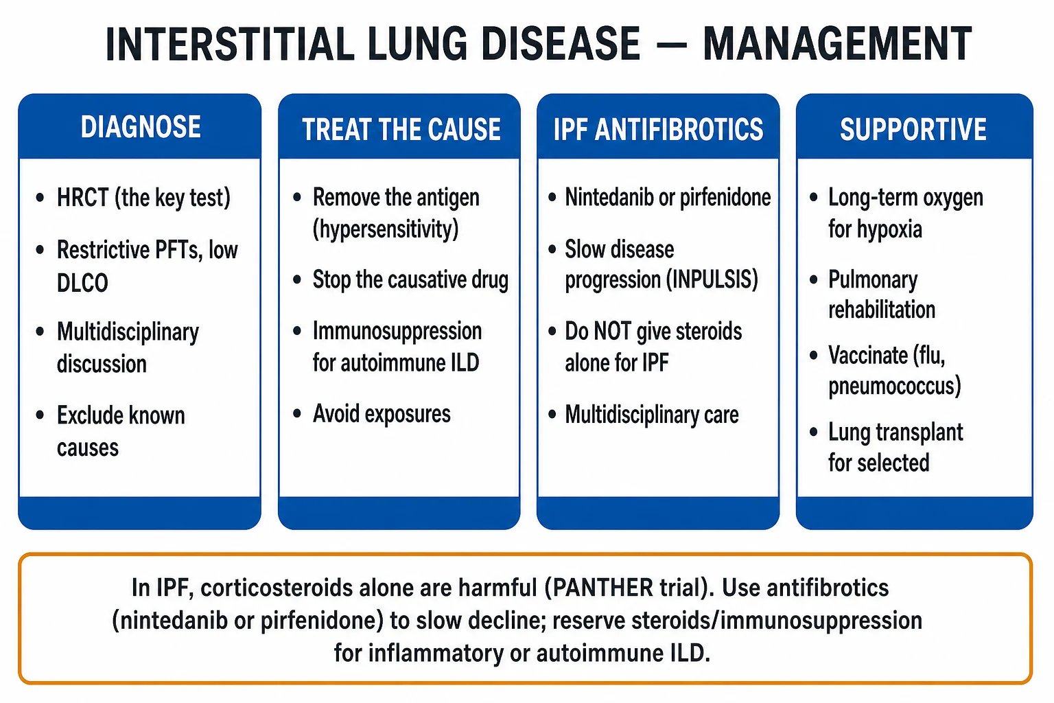
1. Treat the underlying cause (when one is identified)
- Stop the offending drug — amiodarone, methotrexate, nitrofurantoin, checkpoint inhibitor; switch to an alternative if essential.
- Antigen avoidance in hypersensitivity pneumonitis — remove birds, remediate mould, replace humidifiers.
- Treat the CTD with immunosuppression under rheumatology guidance (corticosteroids, mycophenolate, azathioprine, rituximab, tocilizumab, JAK inhibitors).
- Pneumoconiosis — remove from exposure; document for compensation. [1]
2. Antifibrotic therapy for IPF and progressive fibrosing ILD
Two antifibrotics are licensed; both slow the rate of FVC decline by approximately 100 mL per year without reversing fibrosis. [1]
[1]The INPULSIS trial demonstrated that nintedanib reduced the annual rate of FVC decline from 239 mL/year (placebo) to 113 mL/year in IPF.[2] The ASCEND trial showed pirfenidone reduced FVC decline and improved progression-free survival.[3] The INBUILD trial extended nintedanib's benefit to progressive fibrosing ILDs beyond IPF — fibrotic NSIP, HP, CTD-ILD, asbestos-related fibrosis.[4]
3. Drugs NOT to use in IPF
- Azathioprine + prednisolone + N-acetylcysteine (NAC) combination — the PANTHER-IPF trial (2012) was stopped early because of increased mortality, hospitalisation and adverse events; combination immunosuppression is contraindicated in IPF.
- Anticoagulation (warfarin) — no benefit; bleeding risk.
- Corticosteroid monotherapy — no benefit in IPF; may cause harm.
- Stem cell therapy, imatinib, endothelin antagonists (bosentan, macitentan), TNF-α inhibitors — no demonstrated benefit; possible harm. [1]
4. Long-term oxygen therapy (LTOT)
Prescribe if PaO₂ at or below 55 mmHg (7.3 kPa) at rest (room air, stable state), SpO₂ at or below 88%, or PaO₂ at or below 60 mmHg with evidence of pulmonary hypertension, cor pulmonale, or secondary polycythaemia. Use at least 15 hours per day including sleep. [1]
5. Pulmonary rehabilitation
Structured 6–8 week exercise, education, breathlessness-management programme improves 6MWT distance, dyspnoea score, and quality of life in ILD (evidence extrapolated from COPD and corroborated by ILD-specific trials). [1]
6. Lung transplantation — the only cure
Refer early to a transplant centre — ideally within three months of IPF diagnosis — because waiting-list mortality in IPF is the highest of any transplant indication. Bilateral sequential lung transplant is preferred in IPF. The lung allocation score (LAS) is weighted towards severity and wait-list urgency, favouring IPF. Five-year post-transplant survival is approximately 50–60%. Upper age limits (commonly 65–70 years) and frailty constrain eligibility. [1]
7. Pulmonary hypertension therapy
Group 3 PH — manage the underlying lung disease aggressively first. Pulmonary vasodilators (sildenafil, bosentan, riociguat, macitentan) may be considered in severe PH, ideally within a specialist PH-MDT. Trials in IPF-PH have been largely disappointing (RISE-IIP, INSTAGE). [1]
8. Supportive and palliative measures
- Antacid therapy — NICE recommends for IPF with confirmed reflux; consider laparoscopic anti-reflux surgery in selected patients.
- Cough suppression — thalidomide (limited), low-dose morphine for refractory cough in palliative setting; speech and language therapy techniques.
- Vaccinations — annual influenza, pneumococcal (PCV13 + PPSV23), COVID-19, RSV (older adults), herpes zoster.
- Palliative care — early integration; low-dose opioids (oral morphine 2.5 mg every 4–6 hours) for refractory dyspnoea; oxygen even with normal SpO₂ if symptomatically helpful; advance care planning. [1]
Specific Subtypes & Scenarios
Idiopathic pulmonary fibrosis (IPF)
- The clinical archetype. Older smoker, basal Velcro crackles, clubbing, restrictive defect, UIP on HRCT. Diagnosis by MDT without biopsy when HRCT is diagnostic.
- Genetic/familial form — MUC5B rs35705950, telomerase (TERT/TERC) mutations. Screen first-degree relatives if onset under 50 or strong family history. [1]
Non-specific interstitial pneumonia (NSIP)
- Common in CTD (especially systemic sclerosis), occasionally idiopathic.
- Cellular subtype — inflammation, ground-glass, responds well to corticosteroids (prednisolone 0.5–1 mg/kg/day tapered) ± immunosuppression (mycophenolate, azathioprine).
- Fibrotic subtype — poorer prognosis, behaves like IPF; consider antifibrotics.
- HRCT — symmetric basal ground-glass, fine reticulation, sparing of the immediate subpleural stripe at the bases, traction bronchiectasis, no honeycombing. [1]
Cryptogenic organising pneumonia (COP / idiopathic BOOP)
- Subacute flu-like illness with dry cough, dyspnoea, fever, weight loss.
- HRCT — patchy, often peripheral or peribronchovascular consolidation and ground-glass, sometimes nodules, migratory on serial imaging.
- Dramatic response to corticosteroids — prednisolone 0.75–1 mg/kg/day, taper over 6–12 months; relapse common on taper. Macrolides, cyclophosphamide, rituximab in resistant cases.
- Secondary organising pneumonia — infection, drugs (amiodarone, methotrexate, checkpoint inhibitors), radiation, CTD, post-transplant; treat the cause. [1]
Hypersensitivity pneumonitis (HP)
- Acute HP — 4–12 hours after heavy antigen exposure; fever, chills, cough, dyspnoea; resolves in hours to days away from antigen.
- Subacute/chronic HP — insidious cough, exertional dyspnoea, weight loss, basal crackles.
- Classic antigens — bird fancier's (pigeon, budgerigar, droppings and bloom), farmer's lung (mouldy hay, Saccharopolyspora rectivirgula, Thermoactinomyces vulgaris), humidifier lung, hot-tub lung (Mycobacterium avium complex), mushroom worker, malt worker (Aspergillus clavatus).
- HRCT — mid/upper-zone, centrilobular ground-glass nodules, mosaic attenuation, air trapping on expiratory images; chronic stage shows upper-zone fibrosis.
- BAL — lymphocytosis with CD4/CD8 below 1.
- Management — antigen avoidance (essential and most effective); corticosteroids (prednisolone 0.5 mg/kg/day, taper) for subacute/chronic or severe acute; mycophenolate/azathioprine as steroid-sparing. [1]
Smoking-related IIPs — RB-ILD and DIP
- Respiratory bronchiolitis-associated ILD (RB-ILD) — smokers, mild symptoms, diffuse centrilobular micronodules and ground-glass, bronchial wall thickening. Treat with smoking cessation; steroids rarely needed.
- Desquamative interstitial pneumonia (DIP) — heavy smokers, diffuse ground-glass; smoking cessation + corticosteroids; better prognosis than IPF. [1]
Acute interstitial pneumonia (AIP / Hamman-Rich syndrome)
- Rapidly progressive (days to weeks) respiratory failure with diffuse alveolar damage histologically — indistinguishable from ARDS.
- Very high mortality (above 50%); treat supportively (lung-protective ventilation, oxygen, antibiotics, cautious steroids). [1]
Lymphoid interstitial pneumonia (LIP)
- Lymphocytic interstitial infiltrate; Sjögren syndrome, HIV (paediatric), common variable immunodeficiency.
- May progress to lymphoma; treat the underlying condition; corticosteroids ± rituximab. [1]
Connective tissue disease-associated ILD (CTD-ILD)
- Systemic sclerosis — NSIP most common; screen with HRCT and PFTs at diagnosis and annually; early mycophenolate mofetil 2–3 g/day or tocilizumab for progressive disease; nintedanib for progressive SSc-ILD (SENSCIS trial).
- Rheumatoid arthritis — often UIP pattern, worse prognosis than IPF; treat with mycophenolate or rituximab (avoid methotrexate if methotrexate lung suspected).
- Polymyositis/dermatomyositis / antisynthetase syndrome — anti-Jo-1, anti-PL-7, anti-PL-12, anti-EJ, anti-OJ; anti-MDA5 portends rapidly progressive ILD. Combination immunosuppression (high-dose steroids, tacrolimus/cyclophosphamide, rituximab, IVIG).
- Sjögren, SLE, MCTD — variable patterns; treat the CTD. [1]
Drug-induced ILD
- Amiodarone — phospholipidosis; chronic interstitial pneumonitis, organising pneumonia, rarely ARDS; HRCT may show high attenuation. Risk is dose-related (above 400 mg/day, longer than 2 years). Stop the drug, give corticosteroids.
- Bleomycin — dose-related (above 450 units cumulative); oxygen augments toxicity (use lowest FiO₂); pneumonitis can progress to fibrosis; treat with steroids.
- Methotrexate — granulomatous pneumonitis; eosinophilia; stop, give steroids.
- Nitrofurantoin — acute or chronic pneumonitis; chronic form can mimic IPF.
- Checkpoint inhibitors (nivolumab, pembrolizumab, ipilimumab) — immune-related pneumonitis any time; hold drug, grade-based steroids (1–2 mg/kg/day prednisolone), infliximab for refractory. [1]
Pneumoconioses
- Asbestosis — amphibole/crocidolite fibres; lower-lobe interstitial fibrosis, pleural plaques (radiological hallmark of exposure), benign pleural effusion, diffuse pleural thickening, rounded atelectasis, mesothelioma, and increased risk of lung cancer (synergistic with smoking).
- Silicosis — crystalline silica; upper-zone nodular opacities, progressive massive fibrosis, eggshell-calcified hilar lymph nodes; greatly increased risk of TB (silico-tuberculosis). Caplan syndrome (rheumatoid pneumoconiosis) with rheumatoid nodules.
- Coal worker's pneumoconiosis — upper-zone small rounded opacities; Caplan syndrome.
- Berylliosis — non-caseating granulomas indistinguishable from sarcoidosis; beryllium lymphocyte proliferation test (BeLPT) is diagnostic.
- Hard metal disease — cobalt/tungsten carbide exposure; giant cell interstitial pneumonitis. [1]
Radiation-induced lung disease
- Acute radiation pneumonitis (within 6–12 weeks) — dry cough, dyspnoea, geographic ground-glass confined to the radiation field; treat with prednisolone 1 mg/kg/day tapering over weeks.
- Chronic radiation fibrosis (6–12 months) — traction bronchiectasis and volume loss in the field; usually irreversible. [1]
Rare diffuse lung diseases
- Lymphangioleiomyomatosis (LAM) — women of reproductive age; diffuse thin-walled cysts throughout both lungs; chylous pleural effusion, pneumothorax, abdominal angiomyolipomas; sirolimus (everolimus) stabilises lung function (mTOR inhibitors).
- Pulmonary Langerhans cell histiocytosis (LCH) — smokers; upper-zone cysts and nodules, pneumothorax, pituitary and bone lesions; CD1a+, S100+, CD207/Langerin+ cells; smoking cessation is the cornerstone.
- Pulmonary alveolar proteinosis — surfactant accumulation; crazy-paving pattern; anti-GM-CSF antibodies (autoimmune); whole-lung lavage; inhaled or subcutaneous GM-CSF.
- Eosinophilic pneumonia — acute (Löffler-like) and chronic; peripheral eosinophilia; upper-lobe predominance of chronic form ("photographic negative of pulmonary oedema"); steroid responsive. [1]
Complications & Pitfalls
- Respiratory failure — type 1, then mixed type 2 in end-stage disease.
- Pulmonary hypertension (group 3) and cor pulmonale — independent marker of poor prognosis.
- Acute exacerbation of IPF — in-hospital mortality around 50%; median survival post-AE approximately three to four months.[1]
- Lung cancer — IPF carries a 3–7-fold increased risk; consider screening.
- Spontaneous pneumothorax — particularly in LAM, LCH, lymphoid interstitial pneumonia.
- Pleural disease — asbestosis (plaques, effusion, mesothelioma); rheumatoid effusion.
- Opportunistic infection — PJP, CMV, mycobacterial — particularly in immunosuppressed CTD-ILD.
- Drug toxicity — amiodarone pulmonary toxicity mortality up to 33%; bleomycin lung 3–25% mortality.
- Diagnostic pitfalls — overcalling UIP on HRCT that lacks honeycombing (favour NSIP); missing chronic HP behind a "fibrotic ILD" label; missing CTD behind "lung-dominant" disease; sending frail elderly patients for VATS biopsy when HRCT is diagnostic.
Prognosis & Disposition
Prognosis varies enormously across the ILDs — from excellent to rapidly fatal. [1]
- IPF median survival 3–5 years from diagnosis; some patients survive over 10 years.
- Acute exacerbation in-hospital mortality around 50%; median post-AE survival 3–4 months.
- AIP mortality above 50% within weeks.
- COP — steroid-responsive, excellent prognosis; relapses on taper common.
- Cellular NSIP — good prognosis; fibrotic NSIP behaves like IPF.
- RB-ILD / DIP — slow course with smoking cessation.
- Antisynthetase with anti-MDA5 — rapidly progressive ILD, 6-month mortality 20–60% if untreated. [1]
The GAP index — Gender, Age, Physiology staging system
The GAP index (Ley 2012) is a simple validated four-variable mortality predictor for IPF, reproduced verbatim.[5]
| Variable | Score 0 | Score 1 | Score 2 |
|---|---|---|---|
| Gender | Female (0) | Male (1) | — |
| Age (years) | under 60 (0) | 60–65 (1) | over 65 (2) |
| FVC (% predicted) | above 75 (0) | 50–75 (1) | below 50 (2) |
| DLCO (% predicted) | above 55 (0) | 36–55 (1) | below 36 (2) |
Total 0–8 → Stages I (0–3), II (4–5), III (6–8). Stage I: 1-year mortality 6%, 3-year 16%; Stage II: 1-year 17%, 3-year 48%; Stage III: 1-year 39%, 3-year 62%. [1]
The ILD-GAP extends the score to other ILDs (additional variables: underlying diagnosis, presence of pulmonary hypertension). The Composite Physiologic Index (CPI) uses FVC, FEV1 and DLCO and predicts mortality across fibrotic ILD. [1]
Disposition
- All patients with new, suspected, or progressive ILD are managed under a specialist ILD service / MDT.
- Antifibrotics initiated and monitored by the ILD team.
- Transplant referral early.
- Palliative care integrated as fibrosis progresses. [1]
Special Populations
- Paediatric ILD (chILD syndrome) — very different differential: neuroendocrine cell hyperplasia of infancy (NEHI), pulmonary interstitial glycogenosis (PIG), surfactant dysfunction disorders (SP-B, SP-C, ABCA3 mutations), developmental disorders. Requires specialist paediatric ILD centre.
- Pregnancy — most ILDs tolerate pregnancy if function preserved; pirfenidone and nintedanib are NOT recommended (insufficient teratogenicity data; nintedanib embryo-foetal toxicity in animals); plan pregnancy with the ILD team.
- Elderly — frailty and comorbidity dominate; HRCT-first strategy to avoid biopsy; antifibrotics started earlier; transplant rarely an option beyond 70.
- Immunocompromised / transplant / HIV — broad differential (opportunistic infection, drug pneumonitis, GVHD, organising pneumonia); BAL critical.
- Anticoagulated patients — balance bleeding risk against biopsy; HRCT-first diagnostic pathway.
- Occupational medicolegal — asbestosis, silicosis, berylliosis are compensable; document exposure meticulously; engage occupational health. [1]
Evidence, Guidelines & Regional Differences
- ATS/ERS/JRS/ALAT 2022 Clinical Practice Guideline (Raghu 2022) — current standard; conditional recommendation for pirfenidone and nintedanib; conditional recommendation against anticoagulation, immunosuppression, and stem cell therapy in IPF.[1]
- NICE CG163 (2013) Idiopathic Pulmonary Fibrosis in Adults — UK service specification including specialist ILD MDT, early access to antifibrotics, oxygen, pulmonary rehab, and palliative care.
- NICE TA379 Pirfenidone for IPF — restricted to FVC between 50% and 80% predicted; treatment stopped if disease progresses by 10% FVC in any 12-month period.
- INPULSIS (Richeldi 2014, NEJM) — nintedanib reduced FVC decline from 239 to 113 mL/year in IPF.[2]
- ASCEND (King 2014, NEJM) — pirfenidone reduced FVC decline by 132 mL at 52 weeks and improved progression-free survival.[3]
- INBUILD (Flaherty 2019, NEJM) — nintedanib reduced FVC decline by 107 mL/year across progressive fibrosing ILDs other than IPF.[4]
- PANTHER-IPF (NEJM 2012, 2014) — azathioprine + prednisone + NAC increased mortality and hospitalisation; combination immunosuppression abandoned.
- SENSCIS — nintedanib slowed FVC decline in SSc-ILD.
- BTS / BTS-ILD registry — UK epidemiology and outcomes.
- Regional deltas:
- UK — NICE CG163 service standard; antifibrotics restricted to FVC 50–80%.
- USA — ATS/ERS/JRS/ALAT guideline with broader antifibrotic use; LAS-based transplant allocation.
- India — high burden of silicosis (agate, slate, stone-cutting), farmer's lung and post-TB fibrotic ILD; antifibrotic access limited; emphasise occupational hygiene and LTOT.
- Japan — high IPF prevalence (MUC5B allele not the main driver; epistatic loci); pirfenidone developed and first licensed here.
Exam Pearls
Idiopathic interstitial pneumonias (ATS/ERS 2013) — 'I Never Dislike Cheese On Biscuits'
INDICOB
UIP pattern; commonest IIP; worst prognosis (3–5 yr)
Cellular (steroid responsive) vs fibrotic; common in CTD
Smoking-related; ground-glass; steroid responsive
Respiratory bronchiolitis-ILD; smoking-related
Patchy migratory consolidation; dramatically steroid responsive
Acute interstitial pneumonia; DAD pattern; very high mortality
Lymphoid interstitial pneumonia; Sjögren, HIV
Drug causes of ILD — 'ABCD-MIN'
ABCDMIN
Phospholipidosis; chronic pneumonitis; high HRCT attenuation
Dose-related; O₂ augments toxicity; can progress to fibrosis
Busulfan, BCNU; nivolumab/pembrolizumab pneumonitis
Granulomatous; eosinophilia
Acute or chronic pneumonitis; mimics IPF
Variable patterns
Less common; usually reversible on withdrawal
- Restrictive pattern — low TLC, low FVC, low DLCO, normal or raised FEV1/FVC (mirror image of obstructive).
- IPF = commonest IIP and worst prognosis — median survival 3–5 yr.
- Velcro crackles — fine, late-inspiratory, basal, non-clearing; the bedside signature of UIP.
- HRCT UIP — basal, subpleural, honeycombing + traction bronchiectasis, little ground-glass; diagnostic without biopsy when all four criteria met.
- Two antifibrotics — pirfenidone (titrate to 600 mg TID; LFT monitoring) and nintedanib (150 mg BID; diarrhoea, LFTs). Both slow decline by ~100 mL FVC/year; neither reverses fibrosis.
- PANTHER-IPF — azathioprine + pred + NAC harms; do NOT use combination immunosuppression in IPF.
- 6MWT desaturation — independent predictor of mortality.
- Lung transplant is the only cure — refer early; bilateral sequential; LAS favours IPF.
- COP/BOOP — the steroid-responsive ILD with patchy migratory consolidation.
- NSIP — symmetric basal ground-glass ± reticulation; cellular subtype responds to steroids; spares extreme subpleural base.
- Hypersensitivity pneumonitis — upper-zone, mosaic attenuation, centrilobular nodules; CD8+ BAL lymphocytosis.
- Asbestosis — lower-lobe fibrosis + pleural plaques; risk of mesothelioma and lung cancer.
- Silicosis — upper-zone, eggshell calcification of hilar nodes; high TB risk.
- Berylliosis — non-caseating granulomas; mimics sarcoid; BeLPT diagnostic.
- Amiodarone lung — phospholipidosis; high-attenuation lung on HRCT; mortality up to 33%.
- LAM — cystic, women, chylous effusion; sirolimus.
- LCH — smokers, upper-zone cysts + nodules; smoking cessation.
- Alveolar proteinosis — crazy-paving, anti-GM-CSF; whole-lung lavage.
- Telomerase (TERT/TERC) mutations — familial pulmonary fibrosis; worse prognosis.
- GAP index = Gender, Age, FVC, DLCO — stages I–III predict mortality. [1]
Exam application bank (NEET-PG / INICET)
One-line answer
Interstitial lung disease (ILD), also called diffuse parenchymal lung disease (DPLD), is a heterogeneous group of over 200 disorders that share inflammation and/or fibrosis of the lung interstitium (alveolar walls, septa, peribronchovascular and perilymphatic spaces), producing a restrictive ventilatory defect (low total lung capacity, low FVC, low DLCO with preserved or raised FEV1/FVC) and a diffuse abnormality on imaging. The clinical archetype is idiopathic pulmonary fibrosis (IPF) — chronic, progressive fibrosis of older adults with a usual interstitial pneumonia (UIP) pattern on HRCT (basal, subpleural reticulation, honeycombing, traction bronchiectasis, with little ground-glass) and a median survival of three to five years. Diagnosis is multidisciplinary, requires exclusion of known causes (connective tissue disease, drugs, occupation, hypersensitivity), and is anchored by thin-se
Worked stems (answer without another resource)
Stem 1 — Classic presentation. Map symptoms to mechanism; name the first investigation and first treatment step with dose/route if drug therapy is standard. [1]
Stem 2 — Unstable / complicated. List red flags that force immediate resuscitation, theatre, ICU, antidote, or reperfusion — and what you do in the first 15 minutes. [1]
Stem 3 — Atypical group. Elderly, pregnancy, child, or immunocompromised: how presentation and thresholds change. [1]
Stem 4 — Differential trap. Name the three closest mimics and one discriminator for each. [1]
Stem 5 — Disposition. Who goes home with safety-netting, who is admitted, who needs HDU/ICU/theatre, and what follow-up is mandatory. [1]
Rapid viva checklist
- Definition + classification
- Pathophysiology chain
- Bedside signs / criteria
- Score with exact components (if any)
- Emergency bundle
- Definitive therapy with doses
- Complications of disease and of treatment
- Special populations
- Guideline/trial name if classic
- Three exam traps
Coverage self-check
If you cannot answer any stem above from this page alone, re-read the matching section — the page is intended to be self-sufficient for final-prof and NEET-PG/INICET questions on Interstitial Lung Disease.
References
- [1]Raghu G, Remy-Jardin M, Richeldi L, et al. Idiopathic Pulmonary Fibrosis (an Update) and Progressive Pulmonary Fibrosis in Adults: An Official ATS/ERS/JRS/ALAT Clinical Practice Guideline Am J Respir Crit Care Med, 2022.PMID 35486072
- [2]Richeldi L, du Bois RM, Raghu G, et al. Efficacy and safety of nintedanib in idiopathic pulmonary fibrosis N Engl J Med, 2014.PMID 24836310
- [3]King TE Jr, Bradford WZ, Castro-Bernardini S, et al. A phase 3 trial of pirfenidone in patients with idiopathic pulmonary fibrosis N Engl J Med, 2014.PMID 24836312
- [4]Flaherty KR, Wells AU, Cottin V, et al. Treatment of refractory ventricular tachycardia with combination of alcohol ablation and radiofrequency ablation J Geriatr Cardiol, 2019.PMID 31555335
- [5]Ley B, Ryerson CJ, Vittinghoff E, et al. The evolution and treatment of Korsakoff's syndrome: out of sight, out of mind? Neuropsychol Rev, 2012.PMID 22569770
- [6]Travis WD, Costabel U, Hansell DM, et al. Reach for the sky - tissue engineering in urology BJU Int, 2013.PMID 24028758