Respiratory · General Medicine
Lung Abscess
Also known as Lung abscess · Pulmonary abscess · Pyogenic lung abscess · Cavitating lung lesion · Necrotising pneumonia with cavitation
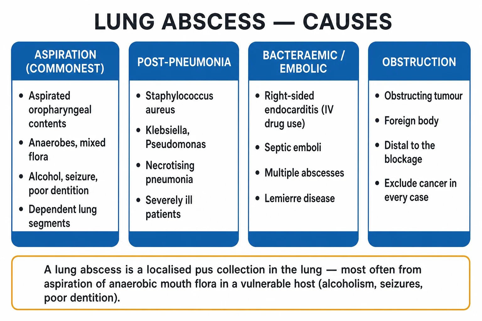
A lung abscess is a localised collection of pus within a cavitating area of lung parenchyma, classically produced by aspiration of oropharyngeal contents (a mixed anaerobic inoculum) in a host with impaired consciousness or swallowing — alcoholism, seizures, poor dentition, dysphagia, stroke. Other mechanisms are necrotising pneumonia (Staphylococcus aureus, Klebsiella, Pseudomonas), septic emboli (right-sided endocarditis in intravenous drug use, Lemierre syndrome), and abscess distal to an obstructing tumour or foreign body. The presentation is insidious over weeks with fever, foul-smelling (fetid) sputum, cough, weight loss and night sweats, and imaging shows a thick-walled cavity with an air-fluid level in a dependent segment. Treatment is prolonged antibiotics covering anaerobes — clindamycin or amoxicillin-clavulanate for 4 to 6 weeks, percutaneous or surgical drainage for large or failing abscesses, treatment of the predisposing cause, and bronchoscopy to exclude an obstructing tumour in any non-resolving case.
On this page & tools
Your progress
Saved locally on this device.
Practise this topic
Exam tags
Red flags
Overview & Definition
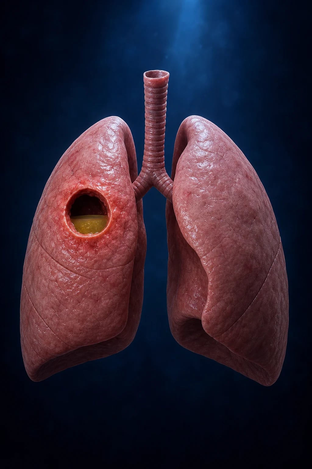
A lung abscess is a localised suppurative process that destroys lung parenchyma, leaving a cavity — usually greater than 2 cm in diameter, with a thick, irregular, organising wall and a central collection of pus and necrotic debris that frequently communicates with a bronchus, producing the characteristic air-fluid level on imaging.[1][5] The distinction from a pneumonic infiltrate is the cavity; the distinction from a simple pneumatocele (a thin-walled, air-filled, post-infectious cavity seen most often in children after Staphylococcal or Streptococcal pneumonia) is the thick wall, surrounding consolidation and purulent contents.[4]
The defining clinical idea — and the one every examiner returns to — is that a lung abscess is usually the late, organised endpoint of an inadequately treated or unusually virulent lung infection, evolving over one to several weeks rather than hours. The classical mechanism is aspiration of infected oropharyngeal contents into a dependent bronchopulmonary segment in a host with impaired consciousness or swallowing, producing a mixed anaerobic infection; but a substantial minority arise by necrotising pneumonia, haematogenous seeding (septic emboli) or obstruction.[1][5]
Lung abscess — the headline numbers
Classification
Lung abscess is classified along two axes that together predict the microbiology, the management and the prognosis: the mechanism by which infection reached the lung, and whether the abscess is primary (in an otherwise normal host, aspiration-driven) or secondary (distal to an obstruction, in immunosuppression, or hospital-acquired).[1][5]
Primary lung abscess
Aspiration, in a vulnerable host
- **Commonest form** — aspiration of oropharyngeal secretions in a host with impaired consciousness or swallowing
- Risk profile: **alcoholism, seizures, poor dentition, dysphagia, stroke, anaesthesia, drug overdose**
- **Mixed anaerobic** flora (Bacteroides, Prevotella, Fusobacterium, Peptostreptococcus, Actinomyces) plus oral streptococci
- **Insidious** over weeks; **dependent segments**; responds to **clindamycin or amoxicillin-clavulanate for 4 to 6 weeks**
Necrotising pneumonia abscess
Virulent aerobes, rapid cavitation
- **Staphylococcus aureus** (incl. PVL/CA-MRSA), **Klebsiella pneumoniae** (K1/K2 hypervirulent), **Pseudomonas**, type-3 *S. pneumoniae*
- Cavitation evolves **within days** of an acute pneumonia; often **multilobar** and bilateral
- **Severely ill or hospital-acquired** patients; broad empiric cover including MRSA and antipseudomonal agents
Bacteraemic / septic-embolic abscess
Multiple peripheral cavities
- **Right-sided (tricuspid) endocarditis in intravenous drug use** — *Staph aureus* septic emboli
- **Lemierre syndrome** — *Fusobacterium necrophorum* septicaemia from internal-jugular thrombophlebitis after pharyngitis
- **Multiple, peripheral, wedge-shaped or cavitating nodules**; treat the source (endocarditis, pharyngeal focus)
Post-obstructive abscess
Distal to a tumour or foreign body
- Abscess forms **distal to an obstructing bronchogenic carcinoma** (especially squamous-cell) or an **inhaled foreign body**
- **Impaired drainage and retained secretions** seed infection; antibiotic cure is **unlikely** until the obstruction is relieved
- **Bronchoscopy is mandatory** in any non-resolving abscess — cancer changes everything
By tempo, abscesses are acute (under 4 to 6 weeks, predominantly pyogenic) or chronic (over 6 weeks), the latter mandating a search for tuberculosis, fungal infection (Aspergillus, histoplasmosis), or cavitating malignancy. By number, a single abscess in a dependent segment suggests aspiration, while multiple, peripheral cavitating nodules point to septic emboli or necrotising pneumonia.[1][5]
Epidemiology & Risk Factors
The incidence of lung abscess has fallen sharply since the antibiotic era and with modern oral hygiene and swallowing care; it is now a relatively uncommon but not rare inpatient diagnosis. The risk profile is dominated by any condition that increases the bacterial load of aspirated material or impairs the host's ability to clear it.[1][5]
The unifying mechanism of aspiration-driven abscess is failure of one or more of the three protective layers of the airway: (1) glottic closure and the cough reflex (dependent on conscious level), (2) coordinated swallowing, and (3) mucociliary and alveolar-macrophage clearance. The classical aspiration-risk profile clusters these failures: alcoholism (depressed consciousness, vomiting, poor dentition), seizure disorders and post-ictal states, general anaesthesia, drug overdose, cerebrovascular disease and dysphagia, neuromuscular disease, oesophageal disorders (achalasia, stricture, Zenker diverticulum, reflux), poor dentition and gingival disease (which raise the bacterial load of the aspirate rather than the inoculum volume), and prolonged nasogastric or endotracheal intubation.[1][5]
[1]The non-aspiration risk clusters point to specific aetiologies: intravenous drug use (right-sided endocarditis, Staph/MRSA septic emboli), recent pharyngitis in a young adult (Lemierre syndrome), immunocompromise (HIV, transplant, neutropenia, prolonged steroids — Nocardia, Aspergillus, Mucorales, atypical mycobacteria), known or occult lung cancer (post-obstructive abscess), and hospital or ventilator acquisition (broad-spectrum aerobes — Klebsiella, Pseudomonas, MRSA).[1]
Pathophysiology
The lung is normally sterile below the vocal cords, defended by the cough reflex, glottic closure, the mucociliary escalator, and alveolar macrophages. A lung abscess develops when these defences are bypassed or overwhelmed by a large, virulent or poorly drainable inoculum, and the host's inflammatory response — rather than clearing the infection — liquefies the surrounding parenchyma to form a cavity.[1][5]
The aspiration cascade
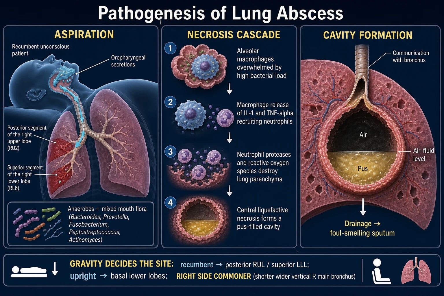
When oropharyngeal secretions carrying a heavy bacterial load — particularly the anaerobes that colonise gingival crevices and dental plaque — reach the alveoli, the inoculum is initially contained by alveolar macrophages. When the load exceeds macrophage capacity, macrophages release interleukin-1 and tumour necrosis factor-alpha, recruiting neutrophils that flood the alveolus and generate the purulent exudate we call consolidation. These same neutrophils, by releasing elastase, metalloproteases and reactive oxygen species, drive the liquefactive necrosis of the lung parenchyma that converts a pneumonia into an abscess. The necrotic core, surrounded by a rim of fibrous granulation tissue, slowly enlarges; once it erodes into a neighbouring bronchus, the pus partially drains (the foul-smelling expectoration that is the bedside hallmark), and air enters the cavity, creating the air-fluid level on imaging.[1][5]
The microbiology mirrors the oropharynx: in a community-acquired aspiration abscess the flora is mixed anaerobic — Bacteroides, Prevotella, Fusobacterium, Peptostreptococcus and Actinomyces — together with microaerophilic streptococci (the Streptococcus anginosus / milleri group) and aerobes such as Streptococcus pneumoniae, Haemophilus influenzae and Staphylococcus aureus. Anaerobes are notoriously missed on routine expectorated-sputum culture because they die in the oxygenated specimen cup and during transport; a "sterile" culture in a typical clinical picture therefore does not exclude an anaerobic abscess, and the fetid sputum and dependent-segment distribution are the real diagnostic clues. In hospital or ventilator-acquired abscess the flora shifts to Gram-negative bacilli (Klebsiella, Pseudomonas) and MRSA.[1]
Why does the sputum smell foul? Anaerobic metabolism produces volatile short-chain fatty acids, amines (putrescine, cadaverine) and sulphur compounds (hydrogen sulphide, methyl mercaptan) — the same molecules responsible for the smell of putrefaction. Fetid sputum is, in effect, the chemical fingerprint of an anaerobic infection. [1]
Why the dependent segments? — gravity decides the X-ray
Aspirated material is liquid or semi-liquid, so it flows to the lowest point the patient's posture offers. In a recumbent patient (bed-bound, unconscious, post-ictal) the dependent bronchopulmonary segments are the posterior segments of the upper lobes and the superior segments of the lower lobes — the classical X-ray location of an aspiration abscess. In a patient who aspirates upright, the material settles in the basal segments of the lower lobes, more often on the right because the right main bronchus is shorter, wider and more vertical — the single most tested anatomical fact in this topic.[1][5]
Other mechanisms
Necrotising pneumonia is produced by particularly virulent organisms whose toxins or tissue-invasive behaviour directly destroy parenchyma: Panton-Valentine leukocidin (PVL)-producing Staphylococcus aureus, hypervirulent K1/K2 capsular Klebsiella pneumoniae, Pseudomonas aeruginosa, and the type-3 capsular Streptococcus pneumoniae. These cavitate within days of an acute pneumonia, may be multilobar, and affect severely ill or immunocompromised hosts.[1]
Septic emboli arise when infected vegetations fragment from a right-sided (tricuspid) heart valve in an intravenous drug user (Staph/MRSA) or from a septic thrombophlebitis of the internal jugular vein after pharyngitis — Lemierre syndrome (Fusobacterium necrophorum). The emboli lodge in the pulmonary arterial tree, producing multiple, peripheral, often wedge-shaped or cavitating nodules that seed lung abscesses.[6]
Post-obstructive abscess develops distal to a bronchial obstruction — most often a squamous-cell bronchogenic carcinoma, less often an inhaled foreign body, bronchial stricture or an enlarged node. The obstruction impairs drainage and mucociliary clearance; retained secretions behind the block become infected and cavitate. Antibiotic cure is unlikely until the obstruction is relieved — the reason bronchoscopy is mandatory in any non-resolving abscess.[1]
Clinical Presentation
The tempo is the single most discriminating clinical feature: a lung abscess is subacute to chronic, unfolding over weeks, in contrast to the hours-to-days course of acute bacterial pneumonia or the minutes-to-hours chemical injury of aspiration pneumonitis.[1][5]
The classical presentation is an insidious illness of one to several weeks: low-grade fever (often with afternoon spikes), malaise, anorexia and weight loss (sometimes dramatic — patients are often misdiagnosed as having malignancy or tuberculosis), a productive cough with purulent, frequently foul-smelling (fetid) sputum, pleuritic chest pain, and haemoptysis (usually minor but occasionally massive). Night sweats and digital clubbing may appear in chronic cases — the latter a classic exam sign that overlaps with tuberculosis, bronchiectasis and lung cancer.[1]
Symptoms
The classical subacute picture
- **Insidious fever** (often low-grade), **malaise, anorexia, weight loss** over 1 to several weeks
- **Productive cough** with **purulent, foul-smelling (fetid) sputum** — the highest-yield clue to anaerobic infection
- **Pleuritic chest pain**, **dyspnoea**, **night sweats**, **haemoptysis** (usually minor)
- May mimic **tuberculosis, lung cancer or bronchiectasis** — weight loss and clubbing are common to all four
Signs
Consolidation plus cavity clues
- Signs of **consolidation** over the affected segment — **reduced expansion, dullness, bronchial breath sounds, crackles, increased vocal resonance**
- Over a **large, peripheral cavity** — **amphoric breath sounds** (low-pitched, cavernous, like blowing over a bottle) and **cavernous rhonchi**
- A **pleural rub** or signs of an **associated effusion/empyema** (stony dullness, absent breath sounds) if the abscess approaches the pleura
- **Digital clubbing** in chronic cases; signs of **sepsis** (tachycardia, hypotension, tachypnoea) if large or ruptured
Atypical / special hosts
The corners examiners live in
- **Elderly / dementia**: confusion, falls, anorexia, functional decline; fever and cough may be **absent**
- **Immunocompromised**: afebrile with neutropenia; consider **Nocardia, Aspergillus, Mucor, atypical mycobacteria**
- **Children**: often Staph or Strep pyogenes; may present with **high fever, dyspnoea, rapid cavity (pneumatocele) formation**
- **IVDU**: cough, fever and **multiple** cavitating lesions — look for tricuspid endocarditis and a murmur
A lung abscess may present acutely when a complication supervenes: rupture into the pleural space produces an empyema with a bronchopleural fistula (sudden expectoration of large volumes of foul sputum) and septic shock; erosion into a bronchial artery produces massive haemoptysis that can be life-threatening.[1]
Differential Diagnosis
A cavity on imaging is not a diagnosis — it is a radiological sign with a wide differential that the exam deliberately tests. The high-yield causes of a cavitating lung lesion, with the features that distinguish each:[1][5]
Pyogenic lung abscess
The diagnosis in question
- **Thick, irregular but relatively uniform wall**, **surrounding consolidation**, **air-fluid level**, **dependent location**
- **Fetid sputum**, aspiration-risk profile, **insidious** course over weeks; **fever and leucocytosis**
- Improves on **anaerobe-covering antibiotics** (clindamycin / amox-clav)
Pulmonary tuberculosis
Upper lobe, thin-walled, satellite lesions
- **Upper-lobe predominance (apical/posterior)**, often **thin-walled**, **satellite lesions**, **tree-in-bud** bronchogenic spread
- **Night sweats, chronic cough, haemoptysis, weight loss**; **sputum AFB positive**, GeneXpert/NAAT positive; **HIV risk**
- **Cavity wall often smoother** than a cancer cavity; epidemiological risk factors
Cavitating squamous-cell carcinoma
Thick eccentric wall, intracavitary mass
- **Thick, irregular, eccentric wall**; an **intracavitary mass** (the tumour); **little or no surrounding consolidation**; **no response to antibiotics**
- **Smoker, older adult**; may have **haemoptysis, weight loss**; biopsy diagnostic — **a post-obstructive abscess behind the tumour complicates the picture**
- **Bronchoscopy** is diagnostic and is mandatory in every non-resolving abscess
Infected bulla / pneumatocoele
Thin-walled, no surrounding consolidation
- **Thin wall**, **no or minimal air-fluid level**, **no surrounding consolidation**; a **pneumatocoele** follows Staph/Strep pneumonia in children and resolves
- **Bulla** in emphysema can become secondarily infected — but the wall is thin and the patient has underlying COPD
Septic emboli
Multiple peripheral cavitating nodules
- **Multiple, peripheral, wedge-shaped or cavitating nodules**; **right-sided endocarditis (IVDU)** or **Lemierre syndrome**
- Blood cultures positive; **tricuspid vegetation on echocardiography**; **internal jugular thrombophlebitis** in Lemierre
Fungal cavity
Aspergilloma, endemic mycoses
- **Aspergilloma**: a **fungal ball** moving within a pre-existing cavity (air-crescent sign); **haemoptysis** is common; immunocompromised or structural lung disease
- **Histoplasmosis / coccidioidomycosis**: endemic-region exposure; nodules that cavitate; serology and antigen tests
Non-infective cavitating lesions
Vasculitis, rheumatoid, infarct
- **Granulomatosis with polyangiitis (Wegener)**: cavitating nodules, sinusitis, glomerulonephritis, c-ANCA positive
- **Rheumatoid nodule**: cavitating peripheral nodule in seropositive rheumatoid arthritis; **cavitating infarct** (rare) after PE
Empyema with bronchopleural fistula
Pleural, not parenchymal
- **Loculated pleural collection with an air-fluid level**, **split-pleura sign** on CT (thickened visceral and parietal pleura); the cavity is **outside the lung**
- Distinguishing this matters — the **drain is a chest tube into the pleural space**, not a percutaneous lung drain
Always consider tuberculosis (sputum AFB and GeneXpert) and an underlying malignancy (bronchoscopy) when an "abscess" is non-resolving, recurrent, or in a smoker over 50.[1]
Clinical & Bedside Assessment
The focused examination has two jobs: characterise the respiratory lesion (a cavity with consolidation versus a pleural collection versus a complication), and assess the aspiration risk and predisposing cause that produced it.[1]
Respiratory examination in a typical abscess shows the classic signs of consolidation over the affected segment — reduced chest expansion, dullness to percussion, bronchial breath sounds, crackles and increased vocal resonance. Over a large, peripheral, subpleural cavity, the special auscultatory pearls appear: amphoric breath sounds (low-pitched, cavernous, like blowing across the mouth of an empty bottle) and cavernous rhonchi, both produced by resonance within the air-containing cavity. A silent hemithorax with stony dullness suggests an associated empyema; a sudden new finding of copious purulent sputum signals a bronchopleural fistula.[5]
Aspiration-risk assessment at the bedside — the part of the examination that finds the cause — should probe conscious level (GCS), a bedside swallow screen (a wet voice or cough on a sip of water is an unsafe swallow), dentition and oral hygiene, an alcohol and substance history, a seizure history and medication review (sedatives), and any neurological deficit suggesting stroke or neuromuscular disease. In an intravenous drug user, examine the jugular veins (a tender internal jugular vein = Lemierre) and listen for a tricuspid murmur.[1][6]
Severity is judged by the sepsis criteria, CURB-65 and the need for organ support: a tachypnoea over 30, hypotension, confusion, hypoxia or acute kidney injury mandates inpatient (often ICU) management.[1]
Investigations
Investigations confirm the cavity, characterise the lesion and its complications, identify the organism where possible, and — most distinctively — evaluate the underlying cause (aspiration, obstruction, immunosuppression, endocarditis).[1][5]
Imaging: [1]
- Chest X-ray — the first test. Shows a thick-walled cavity with an air-fluid level and surrounding consolidation, typically in a dependent segment (posterior upper lobe / superior lower lobe). A lateral or cross-table view may be needed to confirm the air-fluid level. Look for multiple lesions (septic emboli), an associated effusion (empyema risk), and mediastinal or hilar nodes (malignancy).[1]
- Contrast CT chest — the definitive imaging. Defines the wall thickness and character, the air-fluid level, the relationship to the pleura (imminent rupture), multiloculation, satellite lesions, mediastinal nodes, and crucially excludes an obstructing endobronchial mass. It also guides percutaneous drainage.[1]
Laboratory: [1]
- Full blood count — neutrophilic leucocytosis; raised CRP and ESR; anaemia of chronic disease in long-standing cases.
- Blood cultures (two sets, before antibiotics) — positive in bacteraemic/septic-embolic abscess; the single most useful microbiological specimen when positive.
- Sputum for Gram stain, culture and sensitivity — useful for aerobes but unreliable for anaerobes (which die in transit). Sputum AFB and GeneXpert/NAAT for tuberculosis in every cavity, especially upper-lobe, chronic, or with risk factors.
- Pleural fluid (if an effusion is present) — Light's criteria; send for pH, Gram stain, culture and cytology; a pH under 7.2 or frank pus demands chest-tube drainage (empyema).[1]
Microbiological specimens — the key teaching is that expectorated sputum under-represents anaerobes. When definitive microbiology matters (severe, immunocompromised, failing therapy), obtain specimens from less-contaminated sites: bronchoalveolar lavage (BAL) or a protected specimen brush at bronchoscopy, a percutaneous transthoracic aspirate, or culture of the drain fluid from a percutaneous catheter.[1]
Bronchoscopy is performed when the diagnosis is in doubt, when an obstructing tumour or foreign body must be excluded (every non-resolving abscess), to obtain BAL in the immunocompromised, or to remove a foreign body (therapeutic).[1]
Tests for the underlying cause — every abscess deserves a hunt for the predisposing condition: aspiration-risk review (alcohol, swallow, dentition), CT or bronchoscopy for obstruction/malignancy, echocardiography for right-sided endocarditis when septic emboli are suspected, HIV test in the appropriate host, and serology for endemic mycoses where relevant.[1][6]
Management — Resuscitation
ABCDE first. Most lung abscesses are subacute and do not require emergency resuscitation, but a septic, hypoxic or ruptured abscess is a time-critical emergency.[1]
- Airway and breathing — supplemental oxygen to maintain saturations 94 to 98 percent (or 88 to 92 percent in COPD); intubation and mechanical ventilation for respiratory failure, a deeply comatose patient at risk of further aspiration, or after massive haemoptysis to protect the airway.
- Circulation — IV fluid resuscitation for sepsis-induced hypotension; follow a sepsis bundle: two large-bore cannulae, 30 mL/kg crystalloid bolus, lactate, and blood cultures before antibiotics.[1]
- Antibiotics early — after cultures, give broad empiric cover including anaerobes within the first hour of sepsis; narrow once cultures return.
- Complication-specific emergencies — see the RedFlag below.
Management — Definitive & Stepwise
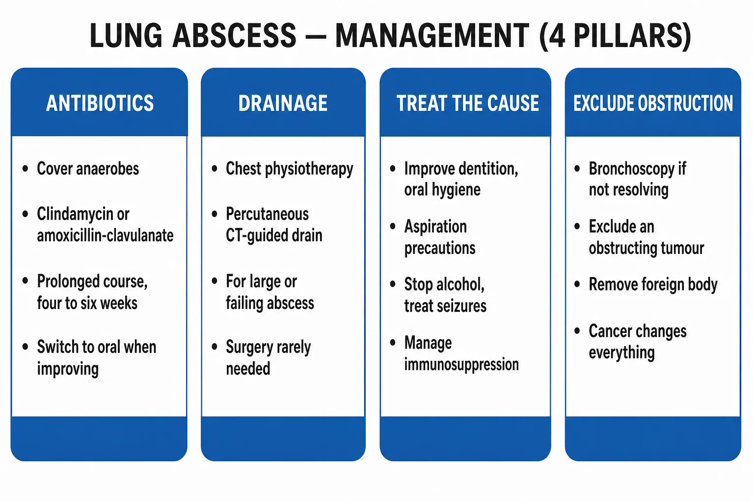
Definitive management rests on four pillars: antibiotics, drainage where indicated, treatment of the predisposing cause, and exclusion of obstruction. The principles — cover anaerobes, treat long enough, drain the large or failing abscess, exclude cancer — are the spine of every exam answer.[1][2]
Empiric antibiotics — drug, dose, route, duration, rationale
Community aspiration abscess
First line — anaerobe cover
- **Clindamycin 600 mg IV TDS** (then 300 to 450 mg PO QDS) **OR amoxicillin-clavulanate 1.2 g IV TDS** (then 625 to 875 mg PO TDS)
- Rationale: cover **anaerobes (Bacteroides, Prevotella, Fusobacterium, Actinomyces) AND microaerophilic streptococci (milleri group)**
- **Duration 4 to 6 weeks** — until clinical and radiographic resolution or a stable small residual scar
Necrotising / hospital-acquired / severe
Broad cover
- **Piperacillin-tazobactam 4.5 g IV TDS** OR a **carbapenem (meropenem 1 g IV TDS)**; **add vancomycin 15 to 20 mg/kg IV BD or linezolid 600 mg IV BD for MRSA**
- Cover **Klebsiella, Pseudomonas, Staph aureus (incl MRSA)**; narrow to culture results
- Duration guided by clinical and radiographic response — often **4 to 8 weeks**
Septic-embolic abscess (IVDU endocarditis / Lemierre)
Source-directed
- **IVDU endocarditis**: **flucloxacillin 2 g IV QDS** (add vancomycin/linezolid if MRSA likely), plus **gentamicin** per endocarditis guidelines
- **Lemierre**: **clindamycin OR piperacillin-tazobactam OR carbapenem** for *Fusobacterium necrophorum*; **anticoagulation controversial** — treat the source
Metronidazole — when NOT to use alone
Common exam trap
- **Metronidazole alone is inadequate** for an aspiration abscess — it has poor activity against **microaerophilic streptococci and Actinomyces**
- Use **only in combination** with a beta-lactam (e.g. **benzylpenicillin + metronidazole**, or **ceftriaxone + metronidazole**)
Why clindamycin historically leads for anaerobic abscess: clindamycin has excellent anaerobe and oral-flora activity, high lung-tissue penetration, and a convenient oral formulation. The landmark Levison 1983 randomised trial showed clindamycin superior to penicillin for anaerobic lung abscess (shorter fever, fewer failures), which is why it remains the textbook first-line agent. Its limitations — emerging resistance in Bacteroides fragilis and a substantial C. difficile risk — increasingly favour amoxicillin-clavulanate (or piperacillin-tazobactam in severe cases), and have driven modern guidelines away from clindamycin as the sole choice.[1]
Duration — typically 4 to 6 weeks, continuing until the patient is clinically well and the cavity has resolved or a stable small residual scar remains. IV-to-oral switch is appropriate once the patient is afebrile, haemodynamically stable, improving clinically, with a falling CRP and able to tolerate oral medication. Radiographic resolution lags behind clinical recovery by weeks to months, so a persistent thin-walled cavity is not, by itself, a reason to prolong antibiotics.[1]
Drainage — when and how
Most aspiration abscesses drain spontaneously through the bronchial tree and resolve with antibiotics alone. Interventional drainage is reserved for specific indications:[2][3]
Indications for drainage of a lung abscess — DRAIN
DRAIN
Large abscesses fail medical therapy more often; drainage improves cure
Failure to improve after 1 to 2 weeks of appropriate antibiotics; persistent fever, rising CRP
Risk of rupture into the pleura (empyema) — drain before it does
Patients less likely to cure on antibiotics alone; lower threshold to drain
Severe sepsis or a hospital-acquired organism; source control urgent
Percutaneous CT-guided catheter drainage is the procedure of choice — inserted under imaging into a mature, peripheral cavity with a safe window avoiding vessels and the pleural fissure; left on free drainage with regular flushes until output stops and the cavity collapses. It cures the majority, with low complication rates (pneumothorax, bleeding, bronchopleural fistula).[2][3]
Chest physiotherapy and postural drainage are adjuncts, used cautiously — they are contraindicated in patients at risk of flooding the contralateral lung (a large abscess communicating with a bronchus) or of massive haemoptysis.[1]
Surgical resection (lobectomy or, rarely, pneumonectomy) is reserved for failure of antibiotics and drainage, massive or uncontrolled haemoptysis, a suspected underlying malignancy, a giant abscess, or a bronchopleural fistula that will not close. It carries significant morbidity and is now uncommon.[1][3]
Escalation triggers
Escalate to drainage or surgery when any of the following occurs despite 1 to 2 weeks of appropriate antibiotics: persistent or spiking fever, an enlarging cavity, rising inflammatory markers, new pleural collection (empyema), haemodynamic instability or worsening sepsis, or haemoptysis.[1]
Specific Subtypes & Scenarios
Each mechanism reframes both the empiric cover and the source-control priority. [1]
- Aspiration lung abscess (commonest) — insidious, dependent-segment, mixed anaerobic; clindamycin or amoxicillin-clavulanate for 4 to 6 weeks; treat the aspiration risk (dentition, swallow, alcohol, seizures).[1]
- Necrotising pneumonia abscess — Staph aureus (PVL/CA-MRSA), Klebsiella, Pseudomonas; rapid multilobar cavitation in a severely ill patient; broad empiric cover including MRSA and antipseudomonal agents; drainage more often needed.[1]
- Bacteraemic / septic-embolic abscesses — tricuspid endocarditis in IVDU (Staph/MRSA) or Lemierre syndrome (Fusobacterium necrophorum after pharyngitis, internal-jugular thrombophlebitis). Multiple peripheral cavitating nodules; treat the source (endocarditis regimen, ENT source control for Lemierre) plus lung-directed antibiotics.[6]
- Post-obstructive abscess — distal to a tumour or foreign body; bronchoscopy is mandatory; antibiotic cure unlikely until the obstruction is relieved (tumour resection, stent, foreign-body removal).[1]
- Paediatric lung abscess — Staph aureus and anaerobes commonest; predominantly medical cure; drainage reserved for failure, large size or empyema; look for underlying aspiration, immunodeficiency or foreign body.[4][7]
- Immunocompromised lung abscess / cavity — Nocardia, Aspergillus, Mucorales, atypical mycobacteria; broad diagnostic sampling (BAL, biopsy); outcome worse.
Complications & Pitfalls
Local complications of inadequately treated or ruptured abscess include empyema (rupture into the pleural space), bronchopleural fistula, massive haemoptysis (erosion of a bronchial artery), lung gangrene (extensive necrosis), ARDS from overwhelming sepsis, and spread to the contralateral lung from spillage. Systemic complications include sepsis and septic shock, bacteraemia with metastatic infection (brain abscess, endocarditis, septic arthritis), acute kidney injury, and — in chronic cases — secondary amyloidosis.[1][5]
Treating an obstruction blindly
Doomed antibiotic cure
- Antibiotics **cannot cure** a post-obstructive abscess while the tumour or foreign body remains; **bronchoscopy first**
Mistaking a cavitating cancer for an abscess
Delayed cancer diagnosis
- A **squamous-cell carcinoma** may cavitate and be treated as an abscess for weeks; **bronchoscopy and biopsy** in every non-resolving case, especially a smoker over 50
Too short a course
Relapse and chronicity
- Stopping antibiotics early (before radiographic resolution or a stable scar) **relapses**; the 4 to 6 week duration is non-negotiable for an aspiration abscess
Over-long broad-spectrum therapy
Collateral harm
- Unnecessary broad cover drives **C. difficile, fungal superinfection, resistance and cost**; narrow as soon as cultures allow
Dangerous postural drainage
Spillage and bleeding
- Postural drainage can **flood the contralateral lung** (large abscess with bronchial communication) or **provoke massive haemoptysis** — avoid in these patients
Prognosis & Disposition
With appropriate prolonged antibiotic therapy and judicious drainage, most lung abscesses resolve; overall mortality with modern treatment is around 5 to 10 percent. Outcome is worse with large cavity (over 6 cm), age over 60, immunosuppression, Staph/Klebsiella/Pseudomonas aetiology, bronchopleural fistula, nosocomial acquisition, or an obstructing tumour — any of which can push mortality toward 20 to 30 percent. A non-resolving or recurrent abscess should prompt the search for cancer, tuberculosis, foreign body, or immunodeficiency.[1][2]
Discharge is safe when the patient is clinically stable, afebrile, with a falling CRP, tolerating oral antibiotics, and with a plan for outpatient follow-up. Radiographic resolution lags behind clinical recovery by weeks to months — a residual thin-walled cavity or a small linear scar may persist and is not, by itself, a treatment failure. A follow-up chest X-ray (and CT where needed) at 6 to 8 weeks is mandatory to confirm resolution and to exclude an underlying malignancy (post-obstructive pneumonia behind a tumour), particularly in smokers over 50.[1]
Special Populations
- Paediatric lung abscess — Staph aureus and anaerobes commonest; a pneumatocele may form after Staph/Strep pneumonia (thin-walled, resolves); management is predominantly medical, with drainage reserved for failure, large size or empyema; look for an underlying cause (aspiration, immunodeficiency, foreign body, congenital lung lesion).[4][7]
- Pregnancy — aspiration risk in obstetric anaesthesia (delayed gastric emptying, raised intra-gastric pressure, reduced lower-oesophageal sphincter tone); prefer beta-lactam antibiotics (avoid clindamycin in late pregnancy where possible); imaging with shielded CXR first, CT only if essential and after informed consent.[1]
- Elderly — atypical presentation (confusion, falls, anorexia), higher comorbidity, aspiration risk from stroke and dysphagia, higher mortality, and a heightened need to exclude lung cancer.[1]
- Immunocompromised (HIV, neutropenia, transplant, steroids) — broader differential (Nocardia, Aspergillus, Mucorales, atypical mycobacteria), need for invasive diagnostic sampling (BAL, biopsy), and worse prognosis.[1]
- Intravenous drug user — septic emboli, right-sided endocarditis, Staph/MRSA, Hepatitis C/HIV co-infection; tricuspid vegetation on echo, multiple peripheral cavitating nodules.[6]
Evidence, Guidelines & Regional Differences
The British Thoracic Society (BTS) 2009 local antimicrobial prescribing guidance and the ATS/IDSA 2019 CAP guideline endorse empiric anaerobic coverage for aspiration-related lung infection and recognise lung abscess as a prolonged-therapy entity (4 to 6 weeks). Both favour a beta-lactam/beta-lactamase inhibitor (amoxicillin-clavulanate) or clindamycin as first-line, with broader cover (piperacillin-tazobactam, carbapenem) plus MRSA cover for hospital-acquired or severe disease.[1]
The Levison 1983 randomised trial remains the pivotal evidence on antibiotic choice: clindamycin was superior to penicillin for anaerobic lung abscess (shorter time to defervescence, fewer failures), which established clindamycin as the historical first-line — now increasingly supplanted by amoxicillin-clavulanate because of clindamycin's C. difficile risk and Bacteroides resistance.[1]
The Zhang 2025 Bayesian network meta-analysis of drainage methods concluded that percutaneous catheter drainage is effective and safe for lung abscess, with lower morbidity than open surgery and comparable success to percutaneous needle aspiration for the appropriate cavity — supporting percutaneous drainage as the intervention of choice when drainage is indicated.[3]
Regional antibiotic deltas: [1]
| Region / guideline | First-line empiric for an aspiration lung abscess |
|---|---|
| UK / NICE | Amoxicillin-clavulanate 1.2 g IV TDS (or clindamycin if penicillin-allergic); metronidazole added only with a second agent |
| US / IDSA | Clindamycin 600 mg IV TDS or amoxicillin-clavulanate; broaden to piperacillin-tazobactam for hospital-acquired/severe |
| India / ICMR | Amoxicillin-clavulanate or clindamycin, guided by local antibiogram; consider carbapenem where ESBL/Klebsiella prevalent; AMR stewardship limits clindamycin |
Where the evidence is weak — there are no large modern randomised trials on optimal antibiotic duration or drainage timing; most data are observational or retrospective. The paediatric literature is largely single-centre retrospective series (e.g. Madhani 2016; Unal 2026).[4][7]
Exam Pearls
Lung abscess — the high-yield one-liners
CAVITY
Thick-walled, in a dependent segment — the radiological signature
Mixed anaerobic aspiration in alcoholism, seizures, poor dentition — foul-smelling sputum
Impaired consciousness or swallowing (alcohol, stroke, dysphagia, anaesthesia)
Exclude obstruction (tumour/foreign body) with bronchoscopy in any non-resolving case
Plus drainage for large/failing abscess; treat the underlying risk
Posterior upper lobe / superior lower lobe in a recumbent patient; right side commoner
Why is metronidazole alone inadequate for an aspiration lung abscess?
Metronidazole has excellent activity against strict anaerobes but poor activity against the microaerophilic streptococci (the Streptococcus anginosus / milleri group) and Actinomyces that co-infect aspirated lung, and it lacks aerobic cover. It must therefore be combined with a beta-lactam (e.g. amoxicillin or ceftriaxone plus metronidazole) rather than used alone.
- The one-liner: a lung abscess is a localised pus cavity in the lung; air-fluid level on imaging; aspiration of anaerobes commonest; clindamycin or amoxicillin-clavulanate for 4 to 6 weeks.[1]
- Four causes: aspiration (alcohol) → necrotising pneumonia (Staph/Klebsiella) → septic emboli (IVDU endocarditis, Lemierre) → obstruction (cancer).[1][6]
- Dependent segments: recumbent → posterior segment of the upper lobe / superior segment of the lower lobe; upright → basal lower lobe; right side commoner (shorter, wider, more vertical right main bronchus).[5]
- Anaerobes — BPPFPA: Bacteroides, Prevotella, Peptostreptococcus, Fusobacterium, Actinomyces — missed on routine culture.[1]
- Foul-smelling sputum = anaerobes — the single highest-yield bedside clue.[5]
- Non-resolving abscess → bronchoscopy to exclude an obstructing cancer.[1]
- Metronidazole must NOT be used alone for an aspiration abscess.[1]
- Digital clubbing may appear in a chronic lung abscess (overlap with TB, bronchiectasis, cancer).[5]
- Amphoric breath sounds (low-pitched, cavernous, like blowing over a bottle) over a large peripheral cavity — the classic auscultatory pearl.[5]
Exam application bank (NEET-PG / INICET)
One-line answer
A lung abscess is a localised collection of pus within a cavitating area of lung parenchyma, classically produced by aspiration of oropharyngeal contents (a mixed anaerobic inoculum) in a host with impaired consciousness or swallowing — alcoholism, seizures, poor dentition, dysphagia, stroke. Other mechanisms are necrotising pneumonia (Staphylococcus aureus, Klebsiella, Pseudomonas), septic emboli (right-sided endocarditis in intravenous drug use, Lemierre syndrome), and abscess distal to an obstructing tumour or foreign body. The presentation is insidious over weeks with fever, foul-smelling (fetid) sputum, cough, weight loss and night sweats, and imaging shows a thick-walled cavity with an air-fluid level in a dependent segment. Treatment is prolonged antibiotics covering anaerobes — clindamycin or amoxicillin-clavulanate for 4 to 6 weeks, percutaneous or surgical drainage for large or
Worked stems (answer without another resource)
Stem 1 — Classic presentation. Map symptoms to mechanism; name the first investigation and first treatment step with dose/route if drug therapy is standard. [1]
Stem 2 — Unstable / complicated. List red flags that force immediate resuscitation, theatre, ICU, antidote, or reperfusion — and what you do in the first 15 minutes. [1]
Stem 3 — Atypical group. Elderly, pregnancy, child, or immunocompromised: how presentation and thresholds change. [1]
Stem 4 — Differential trap. Name the three closest mimics and one discriminator for each. [1]
Stem 5 — Disposition. Who goes home with safety-netting, who is admitted, who needs HDU/ICU/theatre, and what follow-up is mandatory. [1]
Rapid viva checklist
- Definition + classification
- Pathophysiology chain
- Bedside signs / criteria
- Score with exact components (if any)
- Emergency bundle
- Definitive therapy with doses
- Complications of disease and of treatment
- Special populations
- Guideline/trial name if classic
- Three exam traps
Coverage self-check
If you cannot answer any stem above from this page alone, re-read the matching section — the page is intended to be self-sufficient for final-prof and NEET-PG/INICET questions on Lung Abscess.
References
- [1]Hadid W, Stella GM, Maskey AP, et al. Lung abscess: the non-conservative management: a narrative review J Thorac Dis, 2024.PMID 38883669
- [2]Kelogrigoris M, Tsagouli P, Stathopoulos K, et al. CT-guided percutaneous drainage of lung abscesses: review of 40 cases JBR-BTR, 2011.PMID 21980735
- [3]Zhang X, et al. Effectiveness and safety of four drainage methods for lung abscess: a Bayesian network meta-analysis and systematic review Front Med (Lausanne), 2025.PMID 41585276
- [4]Unal A, et al. Pediatric lung abscesses: a 13-year journey from diagnosis to treatment BMC Infect Dis, 2026.PMID 41808029
- [5]Pennza PT. Aspiration pneumonia, necrotizing pneumonia, and lung abscess Emerg Med Clin North Am, 1989.PMID 2653801
- [6]Escher R, Glauser MP, Meylan P, et al. Advanced Lemierre syndrome requiring surgery Infection, 2008.PMID 18791836
- [7]Madhani K, Akhilesh P, Phurailatpam J, et al. A 10-year retrospective review of pediatric lung abscesses from a single center Ann Thorac Med, 2016.PMID 27512508