Respiratory · General Medicine
Lung Cancer
Also known as Bronchogenic carcinoma · Lung carcinoma · Non-small cell lung cancer · NSCLC · Small cell lung cancer · SCLC · Pulmonary malignancy
Lung cancer is a malignant tumour arising from the bronchial or alveolar epithelium, the leading cause of cancer death worldwide. The fundamental divide is non-small cell lung cancer (NSCLC, ~85%) — adenocarcinoma (commonest, peripheral, never-smokers, EGFR/ALK-driven), squamous cell carcinoma (central, cavitates, PTHrP hypercalcaemia), large cell — versus small cell lung cancer (SCLC, ~15%) (central, smoker, neuroendocrine, paraneoplastic SIADH and Lambert-Eaton, early metastasis, chemo- and radio-sensitive). Risk is dominated by tobacco smoking (80 to 90%); also asbestos, radon, second-hand smoke. Presentation: persistent cough, haemoptysis, dyspnoea, weight loss, chest pain, plus paraneoplastic syndromes and metastatic manifestations (bone, brain, liver, adrenal). Diagnosis by CT chest ± PET-CT, confirmed on histology/cytology (bronchoscopy, EBUS-TBNA, CT-guided biopsy); molecular testing (EGFR, ALK, ROS1, BRAF, PD-L1) drives targeted therapy. Stage by TNM 8th edition (NSCLC) or limited vs extensive (SCLC). Management: NSCLC early stage (I to II) — surgical resection (lobectomy) ± adjuvant chemo; locally advanced (III) — chemoradiotherapy ± durvalumab; advanced (IV) — platinum doublet plus pembrolizumab/atezolizumab, with EGFR/ALK-targeted TKIs for driver-mutant disease. SCLC — etoposide plus platinum ± radiotherapy; prophylactic cranial irradiation. Prognosis overall 5-year survival ~20%; stage I ~60%, stage IV under 10%. Low-dose CT screening reduces mortality in high-risk smokers.
On this page & tools
Your progress
Saved locally on this device.
Practise this topic
Exam tags
Red flags
Overview & Definition
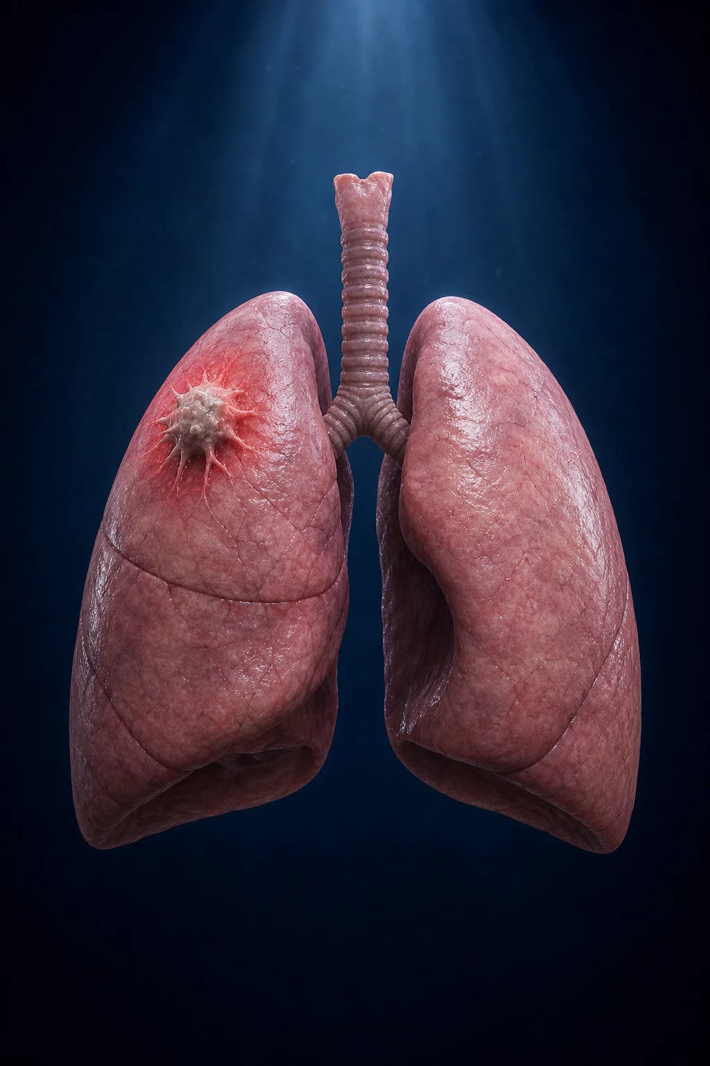
Lung cancer is a malignant tumour arising from the epithelial cells of the bronchi, bronchioles, or alveoli (bronchogenic carcinoma), and it is the single leading cause of cancer death worldwide in both men and women — killing more people each year than breast, prostate and colorectal cancer combined.[3]
Two histological families with radically different biology, treatment and prognosis dominate: [1]
- Non-small cell lung cancer (NSCLC) — about 85% of cases. The behaviour, molecular drivers and treatment algorithms differ by subtype, which is why modern practice demands not just "NSCLC vs SCLC" but the specific NSCLC subtype plus molecular profile (EGFR, ALK, ROS1, BRAF, PD-L1) before initiating therapy.[3]
- Small cell lung cancer (SCLC) — about 15%. A high-grade neuroendocrine tumour of smoking, central in location, with early and widespread metastasis and a striking responsiveness (often dramatic but transient) to chemotherapy and radiotherapy.
The clinical skill in lung cancer is four-fold: (1) recognise the presentation and don't dismiss a smoker's changing cough; (2) obtain adequate tissue for both histology AND molecular profiling; (3) stage accurately (TNM for NSCLC; limited/extensive for SCLC) because stage dictates treatment intent; and (4) treat within a multidisciplinary team (MDT) that can deliver surgery, radiotherapy, systemic therapy and palliation in the right sequence. The single biggest public-health lever is tobacco control and low-dose CT screening of high-risk smokers.[1]
Classification
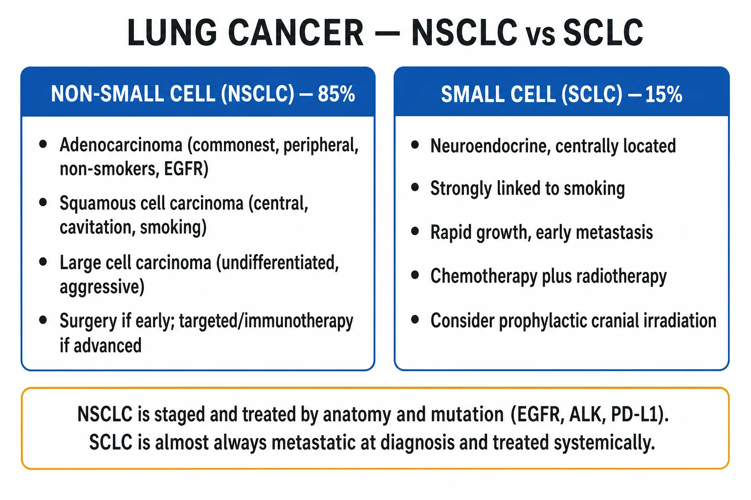
By histology (WHO 2015)[3] — the classification that drives treatment:
- NSCLC (~85%):
- Adenocarcinoma (~40%, the commonest subtype overall) — typically peripheral, arises from glandular/bronchiolar epithelium, may produce mucin, histology shows gland formation and/or papillary/acinar/lepidic patterns; the lepidic (bronchioloalveolar) pattern grows along intact alveolar walls. Most common lung cancer in never-smokers and in women. Molecular drivers: EGFR mutation (especially Asian never-smokers, women, adenocarcinoma), ALK and ROS1 rearrangements, KRAS mutation (smokers), BRAF, MET, RET, HER2, NTRK.
- Squamous cell carcinoma (~25 to 30%) — typically central (arising in major bronchi), histology shows keratin pearls and intercellular bridges; prone to cavitation and to PTHrP-mediated hypercalcaemia. More strongly smoking-associated than adenocarcinoma.
- Large cell carcinoma (~2 to 5%) — undifferentiated, lacks glandular or squamous features; peripheral, aggressive, poor prognosis.
- Adenosquamous, sarcomatoid — uncommon variants.
- SCLC (~15%) — high-grade neuroendocrine carcinoma; small dark-blue cells with scant cytoplasm, granular chromatin, nuclear moulding and frequent mitoses and necrosis; positive for synaptophysin, chromogranin, CD56.
- Carcinoid tumours (~1 to 2%) — low-grade/intermediate neuroendocrine tumours; young, non-smokers, central, carcinoid syndrome uncommon; treated surgically, indolent prognosis.
- Mesothelioma — distinct entity (pleural, asbestos, poor prognosis) — see Special Subtypes. [1]
By stage — NSCLC uses TNM 8th edition[2] (see Investigations); SCLC uses the simpler Valstgas/limited vs extensive scheme.
[1]Epidemiology & Risk Factors
Lung cancer is the most commonly diagnosed cancer in men and the leading cause of cancer death in both sexes worldwide. Globally there are roughly 2.2 to 2.5 million new cases and ~1.8 million deaths annually. Peak incidence is in the sixth and seventh decades; uncommon under 40. [1]
Tobacco smoking is by far the dominant risk factor — accounting for ~80 to 90% of all lung cancer deaths. The risk is dose- and duration-dependent (pack-years); smoking multiplies lifetime risk roughly 20- to 30-fold in heavy smokers. Cessation progressively reduces risk but never back to never-smoker baseline. All forms of smoking (cigarettes, bidi, cigars, pipes) carry risk; second-hand (passive) smoke increases risk by ~20 to 30%. [1]
Other risk factors and the lesions they favour (high-yield): [1]
| Risk factor / exposure | Lung cancer subtype / association |
|---|---|
| Tobacco smoking (80 to 90%) | All types; esp. squamous, SCLC |
| Asbestos (crocidolite worst) | Mesothelioma (pleural); also bronchogenic carcinoma; synergistic with smoking (multiplicative) |
| Radon gas (residential, uranium mining) | Adenocarcinoma; leading environmental cause in never-smokers |
| Ionising radiation (uranium, radium; therapeutic radiotherapy; prior Hodgkin lymphoma radiotherapy) | All types |
| Second-hand smoke | ~20 to 30% increased risk |
| Occupational: arsenic, chromium, nickel, cadmium, beryllium, silica, diesel exhaust, soot/PAHs | All types |
| Pulmonary fibrosis / interstitial lung disease | Increased risk (esp. adenocarcinoma, lower lobes) |
| COPD / emphysema | Independent risk (over and above smoking) |
| Prior cancer (head & neck, bladder) | Field cancerisation |
| HIV / immunosuppression | Modestly increased |
| Genetic: family history, Li-Fraumeni (TP53), germline EGFR T790M, retinoblastoma | Hereditary susceptibility |
Pathophysiology
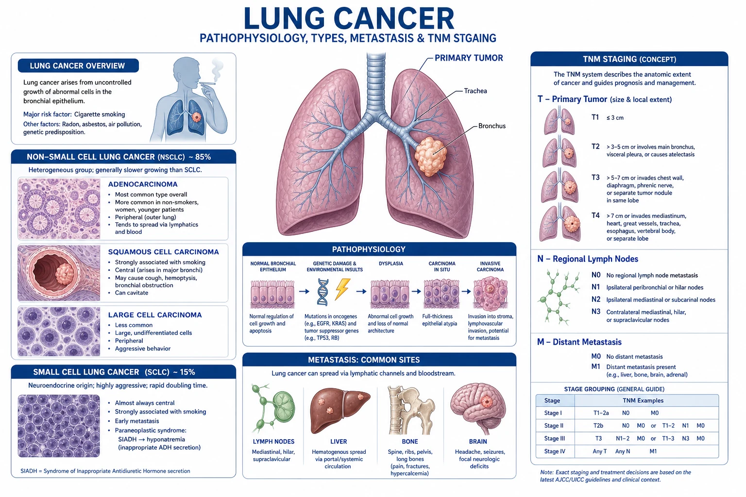
Step 1 — carcinogen exposure. Tobacco smoke contains more than 60 known carcinogens, of which the polycyclic aromatic hydrocarbons (benzo[a]pyrene) and tobacco-specific N-nitrosamines (NNK) are best characterised. Asbestos fibres and radon alpha-emissions act by analogous DNA-damaging mechanisms. These agents are inhaled and concentrate at airway bifurcations, where the highest tumour yield occurs. [1]
Step 2 — DNA adducts, mutations and field cancerisation. Carcinogens form covalent DNA adducts; if nucleotide-excision repair fails, the resulting G:C to T:A transversions become fixed mutations. Smoking produces a characteristic mutational signature (typically KRAS, TP53) and a strikingly high overall tumour mutational burden (~10 mutations/Mb). Because the entire bronchial epithelium is bathed in the same carcinogen, multiple foci of premalignant change coexist — field cancerisation, explaining second primary tumours. [1]
Step 3 — driver mutations and clonal expansion. Critical "driver" events activate oncogenes and disable tumour-suppressor genes:[3]
- Smoking-associated: KRAS (adenocarcinoma, ~25 to 30%), TP53 (very common), STK11, KEAP1, BRAF (non-V600E).
- Never-smoker adenocarcinoma: EGFR mutation (~10 to 15% of Western, 40 to 50% of East-Asian adenocarcinomas), ALK rearrangement (~5%), ROS1 (~1 to 2%), RET, NTRK, MET exon-14 skipping, HER2, and BRAF V600E. These driver-mutant tumours are typically sensitive to specific targeted tyrosine-kinase inhibitors (TKIs) — the foundation of precision oncology in NSCLC.[4][5][8]
Step 4 — invasion and metastasis. Mutated cells evade apoptosis (e.g., via p53 loss), drive angiogenesis (VEGF, the target of bevacizumab), and acquire the capacity to degrade the basement membrane (matrix metalloproteinases) and intravasate. Spread is by lymphatics (hilar → mediastinal → supraclavicular nodes — the classic Virchow's node, a Troisier's sign in left supraclavicular fossa) and haematogenous (the "four typical" metastatic sites — brain, bone, liver, adrenal). [1]
Step 5 — why SCLC behaves so differently. SCLC derives from neuroendocrine Kulchitsky cells of the bronchial mucosa; nearly all show biallelic inactivation of RB1 and TP53. Their neuroendocrine origin explains the paraneoplastic endocrine secretion (ectopic ADH → SIADH; ectopic ACTH → Cushing syndrome) and the immune-mediated paraneoplastic neurological syndromes (Lambert-Eaton myasthenic syndrome, anti-Hu paraneoplastic encephalomyelitis) caused by antibodies directed against neuronal voltage-gated calcium channels and Hu antigens. [1]
Why paraneoplastic syndromes are "remote" effects of the tumour: the tumour secretes peptide hormones (PTHrP, ADH, ACTH, GHRH) or triggers immune cross-reactivity against neural antigens, producing systemic effects disproportionate to tumour bulk and sometimes before the lung lesion is symptomatic. This is the mechanism behind "the lung mass that presents with confusion, weakness or hypercalcaemia." [1]
Clinical Presentation
Lung cancer is clinically silent for much of its course; symptoms often appear only with endobronchial obstruction, local invasion, metastasis, or paraneoplastic activity. Presentation falls into four groups: [1]
1. Local (respiratory) symptoms — from the primary tumour: [1]
- Persistent or changing cough — the commonest symptom; a smoker's "new or worsening" cough is the cardinal red flag.
- Haemoptysis — streaking of blood to frank bleeding; any unexplained haemoptysis in a smoker over 40 is cancer until proven otherwise.
- Dyspnoea — from bronchial obstruction, collapse, pleural effusion, phrenic nerve palsy (diaphragm), or pericardial involvement.
- Chest pain — pleuritic (pleural/pleural involvement) or dull aching (chest-wall invasion, Pancoast).
- Wheeze / stridor — fixed localized wheeze suggests partial bronchial obstruction; stridor = emergency.
- Recurrent or slowly resolving pneumonia in the same lobe — post-obstructive infection behind a tumour.
- Hoarseness — left recurrent laryngeal nerve palsy (loops under the aortic arch; a left hilar/mediastinal tumour catches it).
- New unilateral wheeze and collapse on CXR behind an obstructing lesion. [1]
2. Constitutional symptoms: weight loss, anorexia, fatigue, cachexia, fever (tumour or obstructive pneumonia). Cachexia is driven by tumour cytokines (TNF-alpha, IL-6, PIF) and is a poor prognostic sign. [1]
3. Metastatic symptoms (brain, bone, liver, adrenal): [1]
- Brain — headache, nausea, focal deficit, seizures, confusion, ataxia; brain metastases are common at presentation in SCLC and adenocarcinoma.
- Bone — severe, progressive, night pain, back pain (worry for spinal cord compression); pathological fracture; hypercalcaemia.
- Liver — hepatomegaly, jaundice, right upper quadrant pain.
- Adrenal — usually silent; occasionally adrenal insufficiency if bilateral destruction.
- Lymph nodes — supraclavicular (Virchow's / Troisier's), axillary. [1]
4. Paraneoplastic syndromes — an examiner favourite (grouped by mechanism):[3]
Endocrine (tumour secretes peptide)
- SIADH (ADH) — SCLC; hyponatraemia, euvolaemic, inappropriately concentrated urine
- Ectopic ACTH / Cushing syndrome — SCLC; hypokalaemia, hyperglycaemia, proximal myopathy, moon face
- PTHrP hypercalcaemia — squamous cell carcinoma; confusion, constipation, polyuria, bone pain
- Ectopic FSHH/LH/GHRH — rarer, gynaecomastia, acromegaly
Neurological (immune cross-reactivity)
- Lambert-Eaton myasthenic syndrome (anti-VGCC, P/Q-type) — SCLC; proximal weakness that IMPROVES with repeated activity
- Paraneoplastic cerebellar degeneration (anti-Yo / anti-Hu) — ataxia
- Paraneoplastic encephalomyelitis (anti-Hu) — SCLC
- Sensory neuropathy, limbic encephalitis
Musculoskeletal / dermatological
- Hypertrophic pulmonary osteoarthropathy (HPOA) — NSCLC; clubbing + periostitis of long bones + arthralgia
- Digital clubbing (frequently NSCLC, esp. squamous and large cell)
- Trousseau syndrome (migratory thrombophlebitis) — hypercoagulability
- Dermatomyositis — adult-onset, a known malignancy-associated myopathy
- Acanthosis nigricans (malignant, gastric > lung)
Haematological / metabolic
- Non-bacterial thrombotic (marantic) endocarditis
- Anaemia of chronic disease, microangiopathic haemolysis
- Disseminated intravascular coagulation
- Leukocytosis from tumour G-CSF secretion
Atypical and special presentations to recognise: [1]
- Never-smoker with adenocarcinoma — cough, dyspnoea, or an incidental nodule; test EGFR/ALK/ROS1 — they are often TKI-sensitive. Asian women, younger age.
- Elderly "failure to thrive" / weight loss — lung cancer may present as cachexia and falls without prominent respiratory symptoms.
- Young adult carcinoid — recurrent pneumonia, haemoptysis, or carcinoid syndrome; central lesion.
- Asbestos-exposed worker with dyspnoea and pleural effusion — mesothelioma (see Special Subtypes).
- Pancoast (superior sulcus) tumour — apical NSCLC invading the brachial plexus (C8-T1) and stellate ganglion → severe shoulder/arm pain, Horner syndrome (ptosis, miosis, anhidrosis), and hand muscle wasting (thenar hypoplasia).
- Superior vena cava obstruction (SVCO) — usually a right-sided central tumour or mediastinal nodes compressing the SVC; facial plethora, distended neck veins, arm swelling, headache, JVP raised — an oncological emergency. [1]
Differential Diagnosis
A lung mass, nodule, or cavitating lesion is not always cancer — and the trap is subjecting a patient with a benign lesion to invasive staging, or missing an infective mimic. Distinguish:[3]
- Pulmonary tuberculosis — young/high-risk, night sweats, upper-lobe cavity, sputum AFB positive, caseating granulomas on biopsy; a cavity can mimic squamous cancer.
- Lung abscess — fever, productive foul-smelling sputum, air-fluid level on CXR; usually follows aspiration or pneumonia.
- Pneumonia / organising pneumonia — acute febrile illness that resolves (or fails to resolve — the worry sign). A non-resolving pneumonia in a smoker over 40 mandates CT and bronchoscopy.
- Benign tumour — hamartoma (young, peripheral "popcorn" calcification), chondroma, fibroma.
- Metastasis from elsewhere — breast, colorectal, renal, melanoma, sarcoma; usually multiple, well-defined, round ("cannonball") lesions.
- Mesothelioma — asbestos, pleural-based, diffuse thickening, pleural effusion; calretinin-positive.
- Sarcoidosis — bilateral hilar lymphadenopathy and interstitial infiltrates; non-caseating granulomas; usually younger, non-smoker.
- Fungal infection — histoplasmosis, coccidioidomycosis, aspergilloma (fungus ball in a pre-existing cavity); endemic exposure, positive serology.
- Arteriovenous malformation / pulmonary sequestration — vascular, contrast-filling on CT.
- Rounded atelectasis — peripheral wedge with "comet-tail" vessels, asbestos exposure. [1]
A can't-miss rule: in a smoker over 40 with a new lung lesion, assume lung cancer until proven otherwise; obtain histology. In a never-smoker with a solitary pulmonary nodule, calculate malignancy risk (size, spiculation, growth) and investigate proportionately. [1]
Clinical & Bedside Assessment
Vital signs (respiratory rate, oxygen saturation, blood pressure, heart rate, temperature) and performance status (ECOG, 0 to 4) — performance status is one of the strongest prognostic factors and decides fitness for treatment: [1]
- ECOG 0 — fully active; ECOG 1 — light work; ECOG 2 — self-care but no work; ECOG 3 — limited self-care; ECOG 4 — bedbound. Most surgery and curative-intent chemo/radiotherapy require ECOG 0 to 2. [1]
General examination — the four systems an examiner wants: [1]
- Hands: digital clubbing (a clue to NSCLC; if with bone pain and arthralgia = HPOA), nicotine staining, Pancoast hand wasting.
- Nodes: cervical, supraclavicular (Virchow's) — palpate carefully; a hard supraclavicular node is a biopsy target that can secure diagnosis without invasive thoracic sampling.
- Chest: signs of collapse (deviation towards), effusion (deviation away, dullness), consolidation, tracheal position, apex beat, unilateral reduced breath sounds, wheeze/stridor.
- Neurological: Horner syndrome, proximal weakness that improves on sustained contraction (Lambert-Eaton), cerebellar signs (paraneoplastic), spinal tenderness, leg weakness (cord compression). [1]
Signs of oncological emergencies to seek at the bedside: [1]
- SVCO — facial plethora, distended neck and chest-wall veins, arm oedema, raised JVP.
- Spinal cord compression — back pain, paraparesis, sensory level, sphincter disturbance; urgent MRI whole spine.
- Hypercalcaemic crisis — confusion, dehydration, abdominal pain, ECG short QT.
- Stridor — partial upper-airway obstruction. [1]
Investigations
Diagnostic principle: (1) image (CT); (2) obtain tissue for histology/cytology AND molecular profiling; (3) stage accurately; (4) assess fitness for treatment.[2]
First-line imaging: [1]
- Chest X-ray — often the first test; may show a mass, nodule, collapse, consolidation, pleural effusion, mediastinal widening, hilar enlargement, raised hemidiaphragm (phrenic palsy), rib erosion, or cavitating lesion. A normal CXR does not exclude cancer — if clinical suspicion is high, proceed to CT.
- Contrast-enhanced CT chest (and abdomen/pelvis) — confirms the lesion, characterises it (size, margins, spiculation, cavitation, fat/calcification for hamartoma), and assesses hilar/mediastinal nodes, liver, adrenal and bone involvement for staging. [1]
Obtaining tissue — choose by lesion location: [1]
- Central / endobronchial lesion — bronchoscopy with biopsy (forceps), brushings and washings; sputum cytology may suffice for central exfoliating tumours.
- Hilar/mediastinal nodes — endobronchial ultrasound with transbronchial needle aspiration (EBUS-TBNA), or endoscopic ultrasound (EUS) — minimally invasive mediastinal staging; if negative but suspicion remains, mediastinoscopy.
- Peripheral lesion — CT-guided transthoracic needle biopsy (risk of pneumothorax, bleeding).
- Pleural effusion — thoracentesis for cytology; if negative, local-anaesthetic thoracoscopy/pleural biopsy.
- Palpable supraclavicular node — fine-needle aspiration or excision biopsy (least invasive route to a tissue diagnosis). [1]
Staging — NSCLC (TNM 8th edition, IASLC)[2]:
- T — primary tumour size and invasion:
- T1 — tumour under 3 cm (T1a under 1 cm; T1b 1 to under 2 cm; T1c 2 to under 3 cm), surrounded by lung/visceral pleura.
- T2 — 3 to under 5 cm, OR involves main bronchus (over 2 cm from carina), visceral pleura, or atelectasis/pneumonitis; T2a 3 to under 4 cm, T2b 4 to under 5 cm.
- T3 — 5 to under 7 cm, OR separate tumour nodules in the same lobe, OR invades chest wall, phrenic nerve, parietal pericardium.
- T4 — over 7 cm, OR separate tumour nodule(s) in a different ipsilateral lobe, OR invades mediastinum, heart, great vessels, trachea, recurrent laryngeal nerve, oesophagus, vertebra, carina.
- N — nodes: N0 none; N1 ipsilateral peribronchial/hilar/intrapulmonary; N2 ipsilateral mediastinal/subcarinal; N3 contralateral mediastinal/hilar, or any scalene/supraclavicular.
- M — metastasis: M0 none; M1a contralateral lung nodules, pleural/pericardial nodules or effusion (malignant); M1b single extrathoracic metastasis; M1c multiple extrathoracic metastases.
- Stage groups — broadly: stage I (T1-2 N0); stage II (T2b-3 N0, or T1-2 N1); stage III (N2/N3 disease, or T3/T4 with node involvement — locally advanced); stage IV (any M1). [1]
Staging — SCLC (simpler scheme): [1]
- Limited disease — tumour confined to one hemithorax, within a single radiotherapy port (ipsilateral hilar/mediastinal nodes, contralateral mediastinal/supraclavicular nodes allowed).
- Extensive disease — spread beyond one radiotherapy port (contralateral lung, malignant effusion, extrathoracic metastasis). [1]
Further staging and fitness tests: [1]
- PET-CT (18F-FDG) — for NSCLC staging (detects nodal and distant metastases, avoids futile surgery) and to characterise an indeterminate nodule.
- MRI brain — mandatory in SCLC (high rate of brain metastasis) and in NSCLC stage III/IV or symptomatic; CT brain if MRI contraindicated.
- Bone scan — largely supplanted by PET-CT; used when PET unavailable.
- Pulmonary function tests (spirometry, DLCO) — before curative-intent surgery or radiotherapy; resectability thresholds guide operability (e.g. predicted postoperative FEV1 and DLCO over 40% predicted reduces peri-operative risk).
- Cardiopulmonary exercise testing for borderline fitness.
- Baseline bloods — FBC, U&E, LFTs, calcium, renal function; LDH (SCLC prognostic). [1]
Molecular / biomarker testing — the modern essential in advanced NSCLC:[4][5][6][8]
- EGFR mutation (exon 19 deletion, L858R; resistance T790M) — IHC/PCR on biopsy or circulating tumour DNA; selects for EGFR-TKIs (gefitinib, erlotinib, afatinib, osimertinib).
- ALK and ROS1 rearrangements (FISH/IHC) — select for crizotinib, alectinib, lorlatinib.
- BRAF V600E, MET exon-14, RET, NTRK, HER2, KRAS G12C — emerging targeted therapies.
- PD-L1 (programmed death-ligand 1) immunohistochemistry — expressed as a tumour proportion score (TPS); TPS at least 50% selects for first-line pembrolizumab monotherapy; TPS at least 1% for combination or second-line checkpoint inhibition.[6][7]
- SCLC is NOT routinely tested for these drivers — targeted therapies are not standard.
Lung cancer — the numbers that decide an answer
Management — Resuscitation
Lung cancer is rarely a time-critical-resuscitation diagnosis, but several oncological emergencies demand immediate recognition and treatment — and these are exam favourites: [1]
- Superior vena cava obstruction (SVCO): sit the patient upright, give high-flow oxygen, dexamethasone (e.g., 8 to 16 mg PO/IV daily) to reduce tumour/peri-tumour oedema; urgent histological diagnosis if not already known; treat the underlying tumour with chemotherapy (SCLC, chemosensitive) or radiotherapy; endovascular stenting for severe/rapidly progressive symptoms or resistant tumour.
- Spinal cord compression: urgent MRI whole spine, dexamethasone 16 mg IV stat then 16 mg/day, urgent clinical oncology / neurosurgery review for radiotherapy ± decompression. Permanent paraplegia develops within hours of complete cord transection.
- Hypercalcaemia of malignancy (PTHrP-mediated, usually squamous): aggressive IV normal saline (3 to 6 L/day), IV bisphosphonate (e.g., zoledronic acid 4 mg IV or pamidronate 60 to 90 mg IV), or denosumab; correct the underlying tumour. Avoid thiazides; calcitonin for rapid transient effect.
- Massive haemoptysis: protect the uninvolved lung (lateral decubitus, bleeding side down), secure airway, bronchoscopic tamponade/ice-saline, bronchial artery embolisation, or surgery in selected operable cases.
- Stridor / impending airway obstruction: urgent anaesthetics/ENT, heliox, dexamethasone, definitive airway (intubation or tracheostomy) and oncology treatment.
- Symptomatic brain metastases with raised intracranial pressure: dexamethasone, mannitol if acute deterioration, urgent radiotherapy and surgery for a single accessible lesion with mass effect. [1]
Management — Definitive & Stepwise
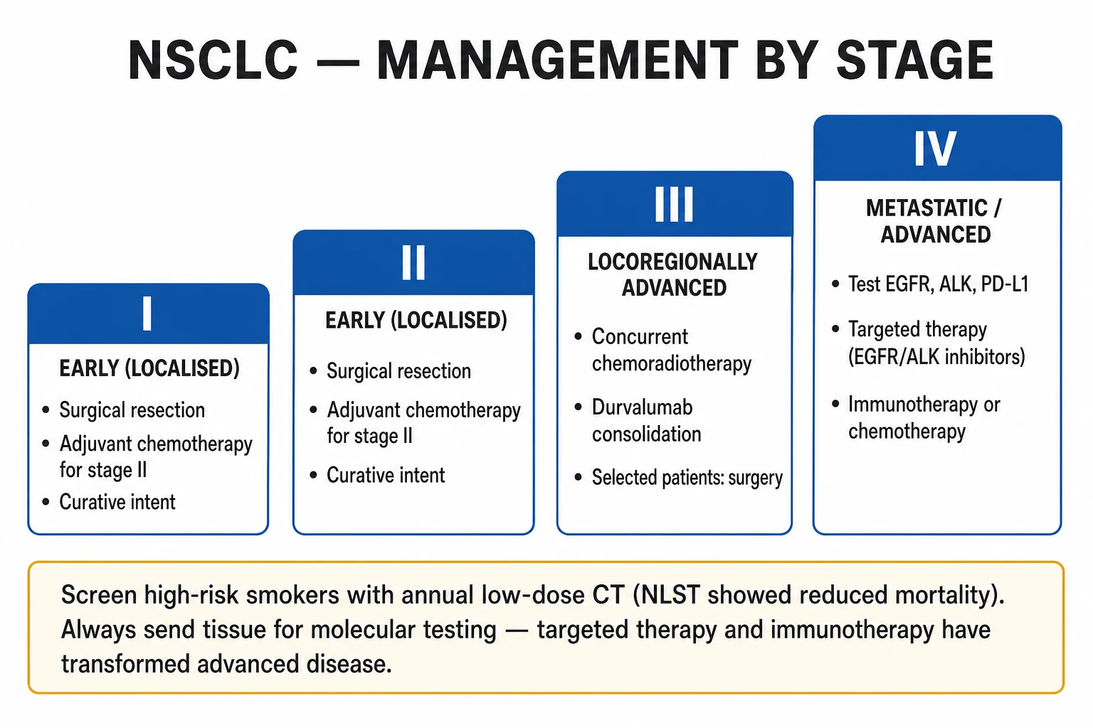
Treatment is stage- and subtype-driven, decided by a multidisciplinary team (MDT) including respiratory physician, medical and clinical oncologist, thoracic surgeon, radiologist, pathologist and specialist nurse.[2]
NSCLC — by stage
Stage I and II (early, operable): [1]
- Surgical resection is the treatment of choice with curative intent. Standard operation is a lobectomy with mediastinal lymph node sampling/dissection via VATS (video-assisted thoracoscopic surgery) or thoracotomy. Sublobar (wedge/segmentectomy) is acceptable for small (T1a) peripheral tumours or poor lung reserve.
- Adjuvant chemotherapy (e.g., cisplatin + vinorelbine, or cisplatin + pemetrexed for non-squamous) for stage II to resectable IIIA (improves survival). Adjuvant immunotherapy (atezolizumab or pembrolizumab) for select PD-L1-positive resected disease.
- Stereotactic ablative radiotherapy (SABR/SBRT) — for patients unfit for surgery or who decline it; curative in medically inoperable stage I NSCLC. [1]
Stage III (locally advanced): [1]
- Resectable IIIA (T3N1, selected T1-2N2): surgery + adjuvant chemo ± radiotherapy.
- Unresectable III (N2 bulky, IIIA/B): concurrent platinum-based chemoradiotherapy followed by consolidation durvalumab (the PACIFIC regimen) — improves progression-free and overall survival. [1]
Stage IV (advanced/metastatic):[4][5][6][9][10]
- Driver-mutant tumours — targeted TKI first-line:
- EGFR mutation → osimertinib (third-generation, FLAURA — superior to first-gen TKIs and active against brain metastases); alternatives gefitinib, erlotinib, afatinib.[4][5]
- ALK rearrangement → alectinib (preferred first-line), crizotinib, lorlatinib.[8]
- ROS1 → crizotinib; BRAF V600E → dabrafenib + trametinib; MET exon-14 → capmatinib/tepotinib; NTRK → larotrectinib/entrectinib.
- PD-L1-high (TPS at least 50%), EGFR/ALK-negative → pembrolizumab monotherapy (KEYNOTE-024 — improved overall survival vs platinum doublet).[6][7]
- PD-L1-low/negative, no driver → platinum doublet + pembrolizumab or atezolizumab (e.g., carboplatin + paclitaxel + atezolizumab ± bevacizumab, the IMpower150 regimen).[9][10]
- Squamous histology — avoid pemetrexed and bevacizumab; use carboplatin + paclitaxel or nab-paclitaxel as the backbone.
- Palliative radiotherapy to painful bone metastases, brain metastases, or symptomatic chest disease; stereotactic radiosurgery for limited brain metastases.
Stage I NSCLC
- Surgical lobectomy + lymph node dissection (VATS preferred)
- Sublobar resection if T1a/peripheral or poor reserve
- SABR/SBRT if medically inoperable — curative
- 5-year survival ~60 to 80%
Stage II NSCLC
- Surgery + adjuvant cisplatin-based chemotherapy
- Consider adjuvant immunotherapy if PD-L1-positive
- 5-year survival ~35 to 55%
Stage III NSCLC (unresectable)
- Concurrent chemoradiotherapy (platinum doublet + thoracic RT)
- Consolidation durvalumab (PACIFIC regimen)
- Selected IIIA: surgery + adjuvant therapy
- 5-year survival ~15 to 30%
Stage IV NSCLC — driver-mutant
- EGFR → osimertinib (FLAURA)
- ALK → alectinib; ROS1 → crizotinib
- BRAF V600E → dabrafenib + trametinib
- Median overall survival now 2 to 4 years with targeted therapy
Stage IV NSCLC — no driver
- PD-L1 TPS ≥50% → pembrolizumab monotherapy (KEYNOTE-024)
- PD-L1 low/neg → platinum doublet + pembrolizumab/atezolizumab (IMpower150)
- Squamous: carboplatin + paclitaxel (avoid pemetrexed/bevacizumab)
- Palliative radiotherapy; 5-year survival under 10%
SCLC — limited
- Etoposide + cisplatin/carboplatin + concurrent thoracic radiotherapy
- Prophylactic cranial irradiation (PCI) if response
- Median survival 16 to 24 months; 5-year survival ~20 to 25%
SCLC — extensive
- Etoposide + carboplatin ± atezolizumab/durvalumab
- PCI if response (reduces brain metastases, may improve survival)
- Palliative radiotherapy for symptoms
- Median survival 7 to 12 months; 5-year survival under 5%
SCLC — by stage
SCLC is almost always inoperable at presentation (early metastasis); treatment is systemic. [1]
- Limited disease — etoposide + cisplatin (or carboplatin) for 4 to 6 cycles, with concurrent thoracic radiotherapy (started early, around cycle 2). Responders receive prophylactic cranial irradiation (PCI) — reduces brain metastases and improves survival.
- Extensive disease — etoposide + carboplatin, with atezolizumab or durvalumab added (IMpower133, CASPIAN — modest survival benefit). PCI in responders. Palliative radiotherapy for symptomatic sites. [1]
Supportive and palliative care
- Pain — WHO analgesic ladder; opioids (morphine, oxycodone) for moderate/severe; treat bone pain with NSAIDs, bisphosphonates/denosumab, radiotherapy.
- Dyspnoea — treat reversible causes (effusion, anaemia, infection); opioids for refractory dyspnoea; oxygen only if hypoxaemic.
- Cough — treat cause; codeine, morphine suppress; steroids for radiation pneumonitis.
- Pleural effusion — therapeutic thoracentesis, indwelling pleural catheter, or talc pleurodesis.
- Nutrition, psychological support, advance care planning, hospice — early integration of palliative care improves quality of life and (in some studies) survival. [1]
Specific Subtypes & Scenarios
- Solitary pulmonary nodule (SPN) — approach by malignancy risk (size, age, smoking, spiculation, growth). Under 6 mm: low risk, surveil in selected smokers. 6 to 8 mm: CT surveillance at 6 to 12 months. Over 8 mm: PET-CT, then biopsy or surgical excision. Use validated calculators (e.g., Brock / Mayo / Herder models).
- Pancoast (superior sulcus) tumour — apical NSCLC invading the brachial plexus and stellate ganglion. Triad: severe shoulder/arm pain (C8-T1 distribution), Horner syndrome (ptosis, miosis, anhidrosis), and hand muscle wasting. Treatment: concurrent chemoradiotherapy then surgery if resectable.
- Carcinoid tumour — low-grade neuroendocrine; young non-smokers; central; treat with surgical resection (sleeve or lobectomy); somatostatin-receptor scintigraphy (octreoscan) for staging; carcinoid syndrome rare.
- Mesothelioma — distinct tumour of the pleura (rarely peritoneum); strongly linked to asbestos (latency 20 to 50 years). Presents with progressive dyspnoea, chest pain, and a unilateral pleural effusion; CT shows pleural thickening and nodularity; histology calretinin, WT-1, D2-40 positive. Treatment options: pleurectomy/decortication or extrapleural pneumonectomy (select patients), chemotherapy (pemetrexed + cisplatin), radiotherapy. Prognosis is poor (median survival 8 to 12 months); incurable in most.
- SVC obstruction — usually a right-sided tumour (often SCLC or NSCLC) compressing the SVC; treat underlying tumour (chemo for SCLC, RT for NSCLC), stenting for refractory symptoms.
- Brain metastases — common; treat with corticosteroids, whole-brain radiotherapy or stereotactic radiosurgery (for 1 to 4 lesions), and occasionally surgical resection; EGFR/ALK-targeted TKIs (osimertinib, alectinib) cross the blood-brain barrier.
- Never-smoker adenocarcinoma — always test for driver mutations (EGFR, ALK, ROS1); these patients often respond dramatically to targeted TKIs. [1]
Complications & Pitfalls
Disease-related: metastases (brain, bone, liver, adrenal), paraneoplastic syndromes (SIADH, Cushing, hypercalcaemia, Lambert-Eaton, HPOA), pleural effusion and malignant pleural disease, post-obstructive pneumonia/lung abscess, SVCO, recurrent laryngeal nerve palsy (hoarseness), phrenic nerve palsy (raised hemidiaphragm), spinal cord compression, pathological fracture, cachexia and thromboembolism (Trousseau). [1]
Treatment-related: surgical (pneumothorax, bleeding, bronchopleural fistula, post-lungectomy complications, arrhythmia); radiotherapy (radiation pneumonitis, oesophagitis, fatigue, fibrosis); chemotherapy (myelosuppression, nausea, neuropathy from cisplatin, renal toxicity); targeted TKIs (EGFR-TKI acneiform rash, diarrhoea, interstitial lung disease; crizotinib hepatotoxicity/visual disturbance); immunotherapy — immune-related adverse effects (pneumonitis, colitis, hepatitis, endocrinopathies — thyroiditis, hypophysitis, type 1 diabetes, adrenal insufficiency), which demand prompt corticosteroids and immunosuppression. [1]
Classic pitfalls: [1]
- Dismissing a smoker's changing cough as "just a cold" or "COPD exacerbation" — the cardinal missed diagnosis.
- Failing to obtain tissue for molecular testing — sending insufficient biopsy; always request EGFR/ALK/ROS1/PD-L1 in advanced NSCLC.
- Over-staging or under-staging — omitting PET-CT or brain MRI before curative-intent treatment.
- Treating SCLC like NSCLC (or vice-versa) — radically different chemo, no surgery in SCLC.
- Missing a paraneoplastic syndrome as the first clue — e.g., SIADH or Lambert-Eaton with weight loss in a smoker.
- Forgetting that a "non-resolving pneumonia" in a smoker over 40 is cancer until proven otherwise.
- Confusing a bulla or rounded atelectasis with a tumour — get histology before resecting. [1]
Prognosis & Disposition
Lung cancer prognosis is driven by stage at diagnosis, histology, performance status, weight loss, and molecular profile: [1]
- Overall 5-year survival ~20% (all comers) — most patients present late.
- NSCLC: stage I ~60 to 80%; stage II ~35 to 55%; stage III ~15 to 30%; stage IV ~under 10% (median survival 8 to 12 months with best supportive care, extended to 2 to 4 years with immunotherapy/targeted therapy in selected patients).
- SCLC: limited disease — median survival 16 to 24 months, 5-year survival ~20 to 25%; extensive disease — median 7 to 12 months, 5-year survival under 5%.
- Mesothelioma — median 8 to 12 months; incurable in most.
- Poor prognostic factors: advanced stage, poor performance status (ECOG 3 to 4), weight loss (over 5 to 10% in 6 months), elevated LDH, hypercalcaemia, brain/liver metastases, squamous in advanced stage, untreated disease. [1]
Disposition: curative-intent treatment (surgery, chemoradiotherapy, definitive SBRT) for stage I to III NSCLC and limited SCLC; systemic therapy and palliation for stage IV NSCLC and extensive SCLC; early palliative care integration alongside active treatment. Follow-up CT surveillance for recurrence and second primaries. [1]
Special Populations
- Never-smokers with adenocarcinoma — typically younger, female, Asian; high rate of driver mutations (EGFR, ALK, ROS1); excellent response to targeted TKIs; always test molecular profile.
- Elderly and frail — comorbidity and performance status dominate treatment choice; SBRT for inoperable stage I; single-agent chemotherapy or best supportive care for advanced disease in ECOG 3 to 4.
- Pregnancy — imaging favours MRI brain and limited CT; treatment delayed where possible; if treatment needed, weigh maternal prognosis against fetal risk; chemotherapy in second/third trimester if essential.
- Immunocompromised (HIV, transplant) — modestly higher risk; treat along standard lines but watch for opportunistic infections during chemo.
- Severe COPD / pulmonary fibrosis — borderline respiratory reserve; SBRT preferred to surgery; PFTs and cardiopulmonary exercise testing to assess fitness.
- Patients with chronic kidney disease or neuropathy — avoid cisplatin (use carboplatin); dose-adjust for renal function.
- Mesothelioma from occupational asbestos — recognised industrial disease; compensation may apply. [1]
Evidence, Guidelines & Regional Differences
Screening — the NLST and beyond: the National Lung Screening Trial (NLST, 2011)[1] randomised high-risk smokers (age 55 to 74, at least 30 pack-years, current or quit under 15 years ago) to three annual low-dose CT scans vs chest X-ray and showed a 20% reduction in lung-cancer mortality (and 7% all-cause mortality). This established low-dose CT screening as the standard for high-risk smokers, adopted by USPSTF, NICE (Targeted Lung Health Checks) and ICMR-aligned programmes. The European NELSON trial subsequently confirmed a mortality benefit in men (with a smaller effect in women). Balancing harms: false positives, incidental findings, radiation, overdiagnosis.
IASLC 8th edition TNM staging (Goldstraw 2016)[2] — refined T categories by tumour size cut-offs (1, 2, 3, 5, 7 cm), reclassified separate tumour nodules and pleural/pericardial deposits, and split M1 into M1a/b/c — improving stage-prognostic discrimination globally.
WHO 2015 classification (Travis)[3] — reclassified lung tumours with full integration of immunohistochemistry and molecular markers; redefined adenocarcinoma subtypes and dropped the term "bronchioloalveolar carcinoma" in favour of adenocarcinoma in situ / lepidic patterns.
Targeted therapy — landmark trials: IPASS (Mok 2009)[4] — gefitinib superior to carboplatin-paclitaxel in EGFR-mutant adenocarcinoma (the proof that genotype selects therapy). FLAURA (Soria 2018)[5] — osimertinib superior to first-generation EGFR-TKIs (and active in the CNS). PROFILE 1014 (Solomon 2014)[8] — first-line crizotinib superior to chemotherapy in ALK-positive NSCLC. ECOG 4599 (Sandler 2006)[9] — adding bevacizumab to carboplatin-paclitaxel improved survival in advanced non-squamous NSCLC.
Immunotherapy — landmark trials: KEYNOTE-001 (Garon 2015)[7] established PD-L1 TPS as a predictive biomarker for pembrolizumab. KEYNOTE-024 (Reck 2016)[6] — pembrolizumab superior to platinum doublet in PD-L1 TPS at least 50% treatment-naïve NSCLC, with fewer adverse events — a paradigm shift. IMpower150 (Socinski 2018)[10] — atezolizumab + bevacizumab + carboplatin + paclitaxel improved survival in metastatic non-squamous NSCLC, even in some EGFR/ALK-positive patients.
Regional deltas: [1]
- UK (NICE NG122) — emphasises urgent 2-week-wait referral for haemoptysis or red-flag symptoms in over-40s; Targeted Lung Health Check LDCT screening; treatment via regional cancer MDTs; NICE-approved drug pathways.
- USA (NCCN / ACCP) — widespread LDCT screening per USPSTF; comprehensive molecular testing and immunotherapy in advanced disease; aggressive SBRT for medically inoperable stage I.
- India (ICMR / NMC) — lung cancer rising; high prevalence of smoking, biomass-fuel exposure and TB makes differential diagnosis (especially TB vs cancer) critical; adenocarcinoma now the commonest subtype; EGFR mutation rate is high (10 to 40%) reflecting ethnicity and the large never-smoker fraction; access to molecular testing and immunotherapy is expanding but uneven.
- East Asia — exceptionally high EGFR mutation prevalence in adenocarcinoma (40 to 50%), driving first-line EGFR-TKI use as standard. [1]
Exam Pearls
Paraneoplastic syndromes of lung cancer
LUNG-PN
anti-VGCC; proximal weakness that IMPROVES with repeated activity; SCLC
hyponatraemia, euvolaemic, inappropriately concentrated urine; SCLC
paraneoplastic encephalomyelitis / sensory neuropathy; SCLC
clubbing + periostitis of long bones; NSCLC (esp. squamous)
confusion, polyuria, constipation; squamous cell carcinoma
hypokalaemia, hyperglycaemia, proximal myopathy; SCLC
The four typical metastatic sites of lung cancer
BB-LA
headache, seizures, focal deficit — common at presentation
night pain, pathological fracture, hypercalcaemia, cord compression
hepatomegaly, jaundice, deranged LFTs
usually silent on imaging; rarely Addisonian if bilateral
- Clubbing + a lung mass in a smoker = HPOA (NSCLC, esp. squamous/large cell).
- Squamous = central + cavitate + hypercalcaemia (PTHrP).
- Adenocarcinoma = peripheral + never-smoker + EGFR.
- SCLC = central + smoker + SIADH / Cushing / Lambert-Eaton + early brain mets.
- Horner syndrome + arm pain = Pancoast (superior sulcus) tumour.
- Hoarseness = left recurrent laryngeal nerve palsy (left hilar tumour).
- Raised hemidiaphragm = phrenic nerve palsy.
- Virchow's node (left supraclavicular) = metastatic gastric or lung cancer.
- Mesothelioma = asbestos + pleural effusion + calretinin positive + poor prognosis.
- Hamartoma = young + popcorn calcification.
- Carcinoid = young non-smoker + central + surgical + indolent.
- EGFR-TKI side effects: acneiform rash, diarrhoea, ILD.
- Immunotherapy pneumonitis/colitis/endocrinopathy — give corticosteroids.
- Hypercalcaemia of malignancy: saline + IV bisphosphonate (zoledronate). [1]
Exam application bank (NEET-PG / INICET)
One-line answer
Lung cancer is a malignant tumour arising from the bronchial or alveolar epithelium, the leading cause of cancer death worldwide. The fundamental divide is non-small cell lung cancer (NSCLC, ~85%) — adenocarcinoma (commonest, peripheral, never-smokers, EGFR/ALK-driven), squamous cell carcinoma (central, cavitates, PTHrP hypercalcaemia), large cell — versus small cell lung cancer (SCLC, ~15%) (central, smoker, neuroendocrine, paraneoplastic SIADH and Lambert-Eaton, early metastasis, chemo- and radio-sensitive). Risk is dominated by tobacco smoking (80 to 90%); also asbestos, radon, second-hand smoke. Presentation: persistent cough, haemoptysis, dyspnoea, weight loss, chest pain, plus paraneoplastic syndromes and metastatic manifestations (bone, brain, liver, adrenal). Diagnosis by CT chest ± PET-CT, confirmed on histology/cytology (bronchoscopy, EBUS-TBNA, CT-guided biopsy); molecular tes
Worked stems (answer without another resource)
Stem 1 — Classic presentation. Map symptoms to mechanism; name the first investigation and first treatment step with dose/route if drug therapy is standard. [1]
Stem 2 — Unstable / complicated. List red flags that force immediate resuscitation, theatre, ICU, antidote, or reperfusion — and what you do in the first 15 minutes. [1]
Stem 3 — Atypical group. Elderly, pregnancy, child, or immunocompromised: how presentation and thresholds change. [1]
Stem 4 — Differential trap. Name the three closest mimics and one discriminator for each. [1]
Stem 5 — Disposition. Who goes home with safety-netting, who is admitted, who needs HDU/ICU/theatre, and what follow-up is mandatory. [1]
Rapid viva checklist
- Definition + classification
- Pathophysiology chain
- Bedside signs / criteria
- Score with exact components (if any)
- Emergency bundle
- Definitive therapy with doses
- Complications of disease and of treatment
- Special populations
- Guideline/trial name if classic
- Three exam traps
Coverage self-check
If you cannot answer any stem above from this page alone, re-read the matching section — the page is intended to be self-sufficient for final-prof and NEET-PG/INICET questions on Lung Cancer.
[1]References
- [1]National Lung Screening Trial Research Team; Aberle DR, Adams AM, Berg CD, et al. Reduced lung-cancer mortality with low-dose computed tomographic screening N Engl J Med, 2011.PMID 21714641
- [2]Goldstraw P, Chansky K, Crowley J, et al. The IASLC Lung Cancer Staging Project: Proposals for Revision of the TNM Stage Groupings in the Forthcoming (Eighth) Edition of the TNM Classification for Lung Cancer J Thorac Oncol, 2016.PMID 26762738
- [3]Travis WD, Brambilla E, Nicholson AG, et al. The 2015 World Health Organization Classification of Lung Tumors: Impact of Genetic, Clinical and Radiologic Advances Since the 2004 Classification J Thorac Oncol, 2015.PMID 26291008
- [4]Mok TS, Wu YL, Thongprasert S, et al. Gefitinib or carboplatin-paclitaxel in pulmonary adenocarcinoma N Engl J Med, 2009.PMID 19692680
- [5]Soria JC, Ohe Y, Vansteenkiste J, et al. (FLAURA). Osimertinib in Untreated EGFR-Mutated Advanced Non-Small-Cell Lung Cancer N Engl J Med, 2018.PMID 29151359
- [6]Reck M, Rodriguez-Abreu D, Robinson AG, et al. (KEYNOTE-024). Pembrolizumab versus Chemotherapy for PD-L1-Positive Non-Small-Cell Lung Cancer N Engl J Med, 2016.PMID 27718847
- [7]Garon EB, Rizvi NA, Hui R, et al. (KEYNOTE-001). Pembrolizumab for the treatment of non-small-cell lung cancer N Engl J Med, 2015.PMID 25891174
- [8]Solomon BJ, Mok T, Kim DW, et al. (PROFILE 1014). First-line crizotinib versus chemotherapy in ALK-positive lung cancer N Engl J Med, 2014.PMID 25470694
- [9]Sandler A, Gray R, Perry MC, et al. (ECOG 4599). Paclitaxel-carboplatin alone or with bevacizumab for non-small-cell lung cancer N Engl J Med, 2006.PMID 17167137
- [10]Socinski MA, Jotte RM, Cappuzzo F, et al. (IMpower150). Atezolizumab for First-Line Treatment of Metastatic Nonsquamous NSCLC N Engl J Med, 2018.PMID 29863955