Respiratory · General Medicine
Obstructive Sleep Apnoea
Also known as Obstructive sleep apnoea · OSA · OSAHS · Sleep apnoea · Sleep-disordered breathing · Apnoea-hypopnoea index
Obstructive sleep apnoea (OSA) is recurrent collapse of the upper airway during sleep, causing apnoeas and hypopnoeas with oxygen desaturation and arousal. It presents with loud snoring, witnessed apnoeas (reported by a partner), excessive daytime sleepiness and unrefreshing sleep. Risk factors are obesity (large neck), male sex, middle age, alcohol/sedatives and craniofacial narrowing. Untreated, it drives systemic hypertension, atrial fibrillation, myocardial infarction, stroke, type 2 diabetes and a high risk of road-traffic accidents. Polysomnography is diagnostic, quantifying the apnoea-hypopnoea index (AHI) — mild 5-15, moderate 15-30, severe over 30 — and STOP-BANG screens for risk. Management is weight loss and lifestyle change, CPAP (the gold standard for moderate-severe disease), a mandibular advancement device for mild/CPAP-intolerant cases, and surgery only in selected patients.
On this page & tools
Your progress
Saved locally on this device.
Practise this topic
Exam tags
Red flags
Overview & Definition
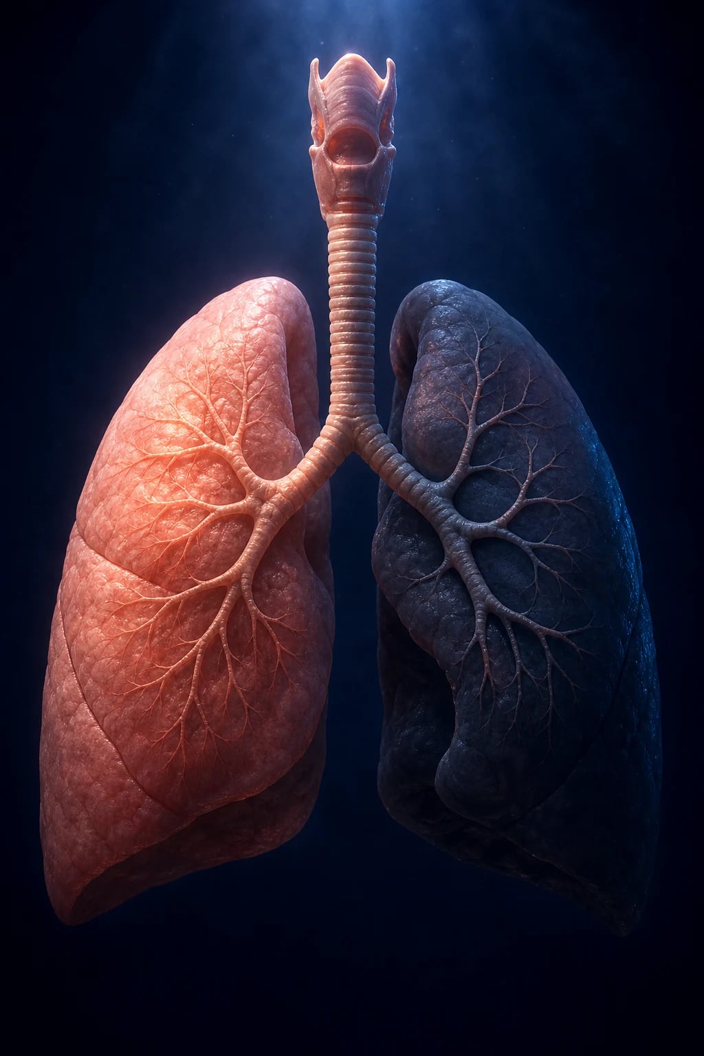
Obstructive sleep apnoea (OSA) is the commonest sleep-related breathing disorder. During sleep the loss of pharyngeal muscle tone lets the upper airway collapse repeatedly, producing apnoeas (complete cessation of airflow for 10 seconds or more despite ongoing respiratory effort) and hypopnoeas (a fall in airflow of at least 30% for 10 seconds or more with an oxygen desaturation of at least 3% or an arousal). Each event is terminated by a brief cortical arousal that restores airway tone and ventilation, but at the cost of sleep fragmentation, and the accompanying cyclic intermittent hypoxia drives systemic cardiovascular and metabolic harm.[1]
OSA lies on a spectrum of sleep-disordered breathing that runs from simple (primary) snoring (no apnoeas, hypopnoeas, desaturation or sleepiness) through to the obstructive sleep apnoea–hypopnoea syndrome (OSAHS) — the combination of OSA with daytime symptoms (sleepiness) or cardiovascular sequelae. The Wisconsin Sleep Cohort established that sleep-disordered breathing is remarkably common in middle-aged adults, refuting the older idea that OSA is a rare curio.[1]
The clinical importance of OSA is twofold. First, the fragmented sleep causes excessive daytime sleepiness, cognitive impairment and a markedly raised risk of road-traffic and occupational accidents. Second, the repetitive nocturnal hypoxia is an independent driver of systemic hypertension, atrial fibrillation, coronary artery disease, heart failure, stroke, type 2 diabetes and metabolic syndrome — the Sleep Heart Health Study demonstrated a robust, independent association between OSA and cardiovascular disease.[6]
Classification
Sleep-related breathing disorders are classified by pathophysiology and, for OSA, by severity measured on polysomnography. [1]
OSA versus central sleep apnoea (CSA)
The fundamental distinction is whether respiratory effort is present during the event: [1]
- Obstructive apnoea/hypopnoea — airflow is reduced or absent but respiratory effort continues (chest and abdominal belts still move). The airway is physically obstructed. This is OSA.
- Central apnoea — airflow is absent and respiratory effort is also absent (no chest/abdominal movement). The brain is not sending the signal to breathe. Seen in heart failure (Cheyne-Stokes), stroke, opioids, high-altitude periodic breathing, and idiopathic central sleep apnoea.
- Mixed apnoea — a central component followed by an obstructive component in the same event. [1]
The bedside discriminator is the respiratory effort channel on polysomnography: in OSA the patient strains against a closed airway; in CSA there is no strain. "OSA = effort, no flow; CSA = no effort, no flow." [1]
Severity by the apnoea-hypopnoea index (AHI)
The AHI is the number of apnoeas plus hypopnoeas per hour of sleep. The AASM/AASM–ICSD-3 severity grading: [1]
- Mild — AHI 5 to 15 events per hour
- Moderate — AHI 15 to 30 events per hour
- Severe — AHI over 30 events per hour [1]
Symptoms and cardiovascular risk rise with AHI, but even mild symptomatic OSA warrants treatment. A related index, the respiratory disturbance index (RDI), additionally counts respiratory-effort-related arousals (RERAs) and is therefore higher than the AHI; it is used in some scoring systems but the AHI is the standard metric. [1]

Related syndromes (must know)
- Obstructive sleep apnoea–hypopnoea syndrome (OSAHS / OSAS) — OSA plus symptoms of daytime sleepiness or cardiovascular sequelae. The term distinguishes the symptomatic clinical syndrome from asymptomatic sleep-disordered breathing.
- Obesity hypoventilation syndrome (OHS) — defined by all three of BMI over 30, daytime hypercapnia (PaCO2 over 45 mmHg / 6 kPa), and sleep-disordered breathing, with no alternative cause for hypoventilation. OSA coexists in the majority but OHS is a distinct entity that needs non-invasive ventilation (BiPAP/NIV), not CPAP alone, plus weight loss.
- Overlap syndrome (OSA + COPD) — the combination carries a worse prognosis than either alone, with a high risk of pulmonary hypertension, chronic hypercapnia and cor pulmonale.
- Upper-airway resistance syndrome (UARS) — recurrent arousals caused by increased upper-airway resistance (flow limitation) with a normal AHI; more common in young, thin, female patients. Treated with mandibular advancement or CPAP. [1]
Epidemiology & Risk Factors
Prevalence
The Wisconsin Sleep Cohort found that among middle-aged adults, roughly 9% of women and 24% of men have an AHI over 5, while 2–4% have the OSAHS (sleep-disordered breathing plus daytime sleepiness).[1] Prevalence rises steeply with age and with the obesity epidemic; modern estimates suggest that a large majority of people with moderate-severe OSA remain undiagnosed. The burden is increasing in India and globally in parallel with obesity.
Risk factors
Modifiable
- Obesity — especially central (waist-to-hip); BMI over 30; the strongest reversible risk factor
- Large neck circumference — over 43 cm (17 in) in men, over 40 cm (16 in) in women; a marker of peripharyngeal fat
- Alcohol and sedatives — relax pharyngeal dilator muscles, worsen OSA the same night
- Smoking — causes airway inflammation and reduces nicotine-related respiratory drive overnight
- Nasal obstruction — allergic rhinitis, polyps, septal deviation increase inspiratory resistance
- Supine sleep position — gravity draws the tongue and soft palate backward
Fixed / constitutional
- Male sex — 2 to 3 times the risk of women (until menopause, when the gap narrows)
- Middle and older age — peak 40–60 years; airway tone falls with age
- Post-menopausal status — loss of the protective effect of progesterone/oestrogen
- Craniofacial variants — retrognathia, micrognathia, midface hypoplasia, macroglossia, high arched palate
- Enlarged tonsils/adenoids — the dominant cause in children
- Family history / genetics — heritability of AHI is about 40%
- Endocrine — hypothyroidism, acromegaly (macroglossia, tissue oedema), polycystic ovarian syndrome
- Race/ethnicity — Asians have higher OSA risk at lower BMI due to craniofacial brachyfacial build
Paediatric epidemiology
OSA affects 1–5% of children, with a peak at age 2–8 years, most commonly due to adenotonsillar hypertrophy. Children with Down syndrome, craniofacial syndromes (Pierre Robin sequence, Treacher Collins, Crouzon) and cerebral palsy are at much higher risk. [1]
Pathophysiology
Why the pharynx collapses
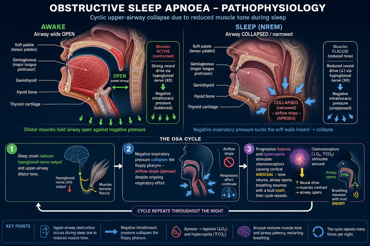
The pharyngeal airway is a collapsible muscular tube with no bony or cartilaginous support along most of its length. While awake it is held open against negative inspiratory pressure by the tonic and phasic activity of the pharyngeal dilator muscles — principally the genioglossus (CN XII, hypoglossal nerve), tensor veli palatini, geniohyoid and stylopharyngeus. During NREM sleep the wakefulness-related excitatory drive (noradrenergic, serotonergic, orexinergic) to these muscles is withdrawn, so hypoglossal output to genioglossus falls and the dilator muscles relax. The airway becomes narrower and, in a person whose airway is already anatomically small or whose neuromuscular compensation is inadequate, collapses. [1]
The Starling resistor / Pcrit model
The airway behaves like a Starling resistor: flow ceases when the pressure outside the tube (the tissue pressure surrounding the pharynx) exceeds the pressure inside it (the airway luminal pressure). Each individual has a critical closing pressure (Pcrit). Patients with OSA have a higher (less negative) Pcrit than unaffected people — their airways collapse at lower negative pressures. Pcrit is determined by anatomy (peripharyngeal fat, airway calibre, craniofacial structure), neuromuscular tone, and lung volume (a lower lung volume pulls less caudal traction on the airway). [1]
The four phenotypic traits (AASM / Wellman model)
OSA arises from the interaction of four physiological traits: [1]
- Anatomical narrowing / high Pcrit — small or crowded airway (the dominant trait in most patients).
- Low neuromuscular tone — inadequate dilator muscle response to the falling airway pressure.
- High loop gain (unstable ventilatory control) — an oversensitive chemoreflex feedback loop: a small rise in CO2 triggers excessive ventilation, which over-corrects and drives CO2 below the apnoeic threshold, causing a central pause that unloads the obstruction.
- Low arousal threshold — waking up too easily from mild respiratory stimuli prevents the deeper sleep needed for stable breathing, fragmenting sleep without fully relieving obstruction. [1]
Understanding the phenotype matters because it predicts response to therapy (e.g. a high-loop-gain or low-arousal-threshold patient may respond to oxygen or hypnotics respectively, while anatomical OSA responds to CPAP). [1]
The apnoea cycle (step by step)
- Sleep onset — loss of wakefulness drive, pharyngeal dilators relax.
- Airway narrowing then collapse — airflow ceases while effort continues.
- Progressive hypoxia and hypercapnia — oxygen falls, CO2 rises, pH falls.
- Chemoreceptor stimulation — carotid body and central chemoreceptors escalate ventilatory drive against the closed airway; intrathoracic pressure swings become hugely negative (straining).
- Cortical arousal — when the physiological stress exceeds threshold, the brain wakes briefly; muscle tone returns and the airway reopens, usually with a loud snort/gasp.
- Ventilation resumes, CO2 falls, oxygen recovers.
- Sleep resumes — tone falls again, and the cycle repeats, often dozens to hundreds of times a night. [1]
How cyclic hypoxia causes systemic disease
Each apnoea produces a swing from hypoxia to reoxygenation — intermittent hypoxia — which is far more injurious than sustained steady hypoxia because of the repeated ischaemia-reperfusion pattern: [1]
- Oxidative stress — bursts of reactive oxygen species (ROS) on reoxygenation.
- Sympathetic nervous system activation — a surge of catecholamines with every arousal and every desaturation, sustaining daytime and nocturnal sympathetic overactivity and driving systemic hypertension.[6]
- Systemic inflammation — raised CRP, IL-6, TNF-alpha, and adhesion molecules promoting atherogenesis.
- Endothelial dysfunction — reduced nitric oxide (NO) bioavailability and impaired flow-mediated dilation.
- Metabolic dysregulation — oxidative stress activates the HIF-1 pathway, increasing insulin resistance and the risk of type 2 diabetes; raised leptin with central leptin resistance (leptin normally stimulates respiration; resistance contributes to obesity hypoventilation).
These mechanisms explain why OSA is independently associated with hypertension, atrial fibrillation, coronary disease, heart failure, stroke and diabetes.[5][6]
Why arousals are a double-edged sword
Arousals are protective (they reopen the airway and restore oxygenation) but harmful (they fragment sleep, prevent restorative slow-wave sleep, and themselves trigger sympathetic activation). This is the core reason for the daytime sleepiness and cardiovascular harm of OSA: the patient is being woken — often without remembering — many times an hour, all night. [1]
Clinical Presentation
Nocturnal features
- Loud, habitual, disruptive snoring — the cardinal symptom, reported by the bed partner.
- Witnessed apnoeas — the partner observes that breathing stops, then resumes with a gasp; the single most specific symptom.
- Gasping, choking or snorting arousals — the patient may sit up or feel they are suffocating.
- Nocturia — from atrial stretch (negative intrathoracic pressure) releasing atrial natriuretic peptide; often 3 to 5 times a night.
- Restless, agitated sleep; tossing and turning.
- Nocturnal sweating, acid reflux (negative intrathoracic pressure draws gastric contents up), dry mouth/sore throat on waking (mouth breathing).
- Reduced libido / erectile dysfunction (more common and reversible with treatment). [1]
Daytime features
- Excessive daytime sleepiness (EDS) — falling asleep while driving, at work, in conversation, in front of the TV. Quantify with the Epworth Sleepiness Scale.
- Unrefreshing sleep despite adequate hours in bed; morning grogginess (sleep inertia).
- Fatigue, lethargy, poor concentration, impaired short-term memory and executive function.
- Morning headache (CO2 retention, typically frontal, resolves within an hour).
- Irritability, mood change, depression and anxiety. [1]
Atypical presentations (high-yield)
OSA — atypical & under-recognised presentations
- Women — more often report fatigue, insomnia, depression than witnessed apnoea; AHI thresholds for equivalent symptoms are lower; under-diagnosed and under-treated.
- Elderly — frequently less obese; present with falls, cognitive decline, nocturia, insomnia rather than classic sleepiness.
- Pregnancy — new snoring and sleepiness; strongly linked to gestational hypertension, pre-eclampsia and gestational diabetes.
- Children — mouth-breathing, failure to thrive, behavioural problems, hyperactivity mimicking ADHD, enuresis, poor school performance; not sleepiness. [1]
The patient presenting with a complication
Recognise OSA as the hidden driver when a patient presents with: resistant hypertension, paroxysmal (often nocturnal) atrial fibrillation, nocturnal angina, unexplained pulmonary hypertension or cor pulmonale, cryptogenic stroke (especially on waking), or a road-traffic accident from falling asleep at the wheel. In each, the question to ask is: "Could this be OSA?" and to screen with STOP-BANG and confirm with polysomnography. [1]
Differential Diagnosis
Simple snoring
- Snoring WITHOUT apnoeas, hypopnoeas, desaturation or daytime sleepiness
- AHI under 5 on polysomnography
- No cardiovascular sequelae; reassurance and snoring measures only
Central sleep apnoea (CSA)
- Absent respiratory effort on PSG (no chest/abdominal movement)
- Seen in heart failure (Cheyne-Stokes), stroke, opioids, high altitude
- Treatment is the underlying cause + adaptive servo-ventilation (ASV), NOT CPAP first-line
Obesity hypoventilation (OHS)
- Daytime hypercapnia PaCO2 over 45 mmHg + BMI over 30 + sleep-disordered breathing
- Often with raised bicarbonate (compensated respiratory acidosis), daytime hypoxaemia
- Needs NIV/BiPAP (pressure support), not CPAP alone
Upper-airway resistance syndrome (UARS)
- Flow limitation arousals with NORMAL AHI
- Young, thin, often female; fatigue/insomnia
- Treat with mandibular advancement device or CPAP
Narcolepsy
- Excessive daytime sleepiness + cataplexy, sleep paralysis, hypnagogic hallucinations
- Normal AHI; MSLT shows mean sleep latency under 8 min with 2+ SOREMPs
- Treat with stimulants (modafinil) + sodium oxybate; cataplexy with antidepressants
Other causes of sleepiness
- Insufficient sleep syndrome, shift-work, depression, idiopathic hypersomnia
- Periodic limb movement disorder & restless legs syndrome (urge to move legs, worse evening/rest, relieved by movement)
- Medications (sedatives, antihistamines), chronic fatigue syndrome
Other nocturnal noises/symptoms
- Nocturnal asthma (wheeze, morning dip), laryngospasm
- Nocturnal seizures (post-ictal confusion, tongue-biting, incontinence)
- GERD-related choking, panic disorder / night terrors
The decisive test separating OSA from its mimics is polysomnography, which simultaneously documents (a) whether events are obstructive or central, (b) the AHI, and (c) the presence of flow limitation arousals. [1]
Clinical & Bedside Assessment
The focused history
Always interview the bed partner — the patient under-reports nocturnal symptoms. Ask specifically about: loud snoring; witnessed apnoeas (the most specific history item); gasping/choking; nocturia; restless sleep; morning headache; daytime sleepiness (Epworth); cardiovascular comorbidity; alcohol and sedative use (including timing); and occupation (driving, machinery, safety-critical work). [1]
Epworth Sleepiness Scale (ESS)
The ESS quantifies the habitual tendency to doze across 8 common situations (sitting reading, watching TV, sitting inactive in a public place, as a passenger in a car for an hour, lying down in the afternoon, sitting talking to someone, sitting quietly after lunch without alcohol, in a car stopped in traffic). Each is scored 0 (would never doze) / 1 (slight) / 2 (moderate) / 3 (high chance). The sum ranges 0–24.[7]
- 0–9 — normal daytime alertness
- 10–12 — mild excessive sleepiness
- 13–15 — moderate
- 16–24 — severe [1]
An ESS over 10 indicates excessive daytime sleepiness and supports a diagnosis of OSAHS when combined with an abnormal AHI. [1]
STOP-BANG questionnaire (screening)
An 8-item screening tool — fast, validated, and the commonest screen used in clinic and pre-operatively: [1]
- S — Snoring loudly?
- T — Tiredness / daytime sleepiness?
- O — Observed apnoeas (witnessed)?
- P — high blood Pressure (treated or over 140/90)?
- B — BMI over 35?
- A — Age over 50?
- N — Neck circumference over 40 cm?
- G — male Gender? [1]
Each item scores 1. 0–2 = low risk, 3–4 = intermediate risk, 5–8 = high risk of moderate-severe OSA. A high score warrants polysomnography. (The Berlin questionnaire is an alternative three-category screen with similar intent.) [1]
Anthropometric and upper-airway examination
- BMI and waist circumference (central obesity).
- Neck circumference measured at the cricothyroid membrane — over 43 cm (men) / 40 cm (women) is high-risk.
- Blood pressure — baseline hypertension is common.
- Oropharyngeal examination — Mallampati / Friedman tongue position (grade III–IV = crowded), tonsil size (0–4), uvula size, tongue volume (macroglossia), high arched palate.
- Jaw — retrognathia / micrognathia (overjet, receding chin).
- Nose — patency, septal deviation, polyps.
- Cardiorespiratory — signs of right-heart failure / cor pulmonale (raised JVP, loud P2, ankle oedema) in severe disease.
- Endocrine screen — features of acromegaly (coarse facies, prognathism, large hands) and hypothyroidism (bradycardia, slow-relaxing reflexes, goitre, dry skin). [1]
Red flags mandating urgent assessment
- Sleepiness with imminent driving/occupational risk — advise immediate driving cessation until assessed and treated.
- Severe cardiovascular comorbidity (heart failure, recent stroke/MI).
- Suspected obesity hypoventilation — daytime somnolence, morning headache, lower-limb oedema, SpO2 under 94% awake → check venous/arterial blood gas for hypercapnia. [1]
Investigations
Polysomnography (PSG) — the gold standard
In-lab, attended polysomnography (Level 1) is the diagnostic gold standard. It records: [1]
- EEG (for sleep staging and arousals), EOG (eye movements), chin EMG (muscle tone) — to stage sleep and detect arousals.
- Airflow — both nasal pressure (sensitive) and oronasal thermistor (detects apnoea).
- Respiratory effort — chest and abdominal belts (distinguishes obstructive from central events).
- Oximetry — SpO2 trend and oxygen desaturation index (ODI).
- ECG — arrhythmias.
- Leg EMG — periodic limb movements.
- Body position (supine vs lateral) and a snoring microphone. [1]
From these the AHI is calculated and the AASM hypopnoea rule applied: a hypopnoea is a drop in airflow of at least 30% for 10 seconds or more with at least a 3% oxygen desaturation or an arousal. [1]
Home sleep apnoea testing (HSAT / Level 3 portable monitor)
A simplified device (oximetry, airflow, respiratory effort, sometimes heart rate) used at home. It is cheaper and more accessible and is appropriate in patients with a high pre-test probability of OSA without significant comorbidity. It underestimates the AHI versus in-lab PSG (it cannot detect arousals or central events reliably) and is not suitable if comorbidity is present (heart failure, COPD overlap, neuromuscular disease) or if CSA is suspected. A negative HSAT in a symptomatic patient should be followed by in-lab PSG. [1]
Ancillary investigations
- Full blood count — secondary polycythaemia (raised haematocrit from chronic hypoxia).
- Fasting glucose / HbA1c — type 2 diabetes (OSA is an independent risk factor).
- Lipid profile — dyslipidaemia.
- Thyroid function — exclude hypothyroidism.
- IGF-1 — exclude acromegaly in a patient with the phenotype.
- Venous or arterial blood gas — detect daytime hypercapnia (PaCO2 over 45 mmHg / 6 kPa) defining OHS; look for a compensated respiratory acidosis (raised bicarbonate over 27 mmol/L).
- ECG — LVH, ischaemia, arrhythmia.
- Echocardiogram — left ventricular hypertrophy, pulmonary hypertension, right-heart dimensions and function.
- Chest X-ray and spirometry — cardiomegaly, COPD overlap, asthma. [1]
Confirming obesity hypoventilation syndrome
OHS is defined on blood gas: awake PaCO2 over 45 mmHg (6 kPa), BMI over 30, and sleep-disordered breathing, with no other cause of hypoventilation (no severe COPD, neuromuscular disease, chest wall deformity or drug cause). A raised serum bicarbonate (compensated respiratory acidosis) is a useful clue to check a blood gas. [1]
Multiple Sleep Latency Test (MSLT) — when narcolepsy is suspected
If the differential is narcolepsy rather than OSA, the MSLT objectively measures daytime sleepiness: mean sleep latency under 8 minutes with 2 or more sleep-onset REM periods (SOREMPs) supports narcolepsy (after excluding OSA, which must be treated first as it fragmentates sleep and confounds the MSLT). [1]
Management — Resuscitation
OSA is ordinarily a chronic outpatient condition; acute decompensation is uncommon but must be recognised. Escalate urgently in these settings: [1]
Acute hypercapnic respiratory failure
Seen in overlap syndrome (OSA + COPD), obesity hypoventilation syndrome, or post-operative respiratory depression (sedatives/opioids in an OSA patient): [1]
- Controlled oxygen — target SpO2 88–92% in the chronically hypercapnic patient; uncontrolled high-flow oxygen worsens CO2 retention.
- Arterial blood gas — confirm and quantify hypercapnia/acidosis.
- Non-invasive ventilation (BiPAP/NIV), not CPAP alone — the hypercapnic patient needs pressure support (IPAP/EPAP) to augment ventilation, not just a continuous pressure to splint the airway.
- Identify and treat the precipitant — sedative/opioid overdose (reversal agents where appropriate: naloxone/flumazenil), infection, cardiac decompensation.
- Escalate to ICU if severe acidosis (pH under 7.25), exhaustion, hypoxaemia despite NIV, or reduced consciousness — for intubation and mechanical ventilation. [1]
Peri-operative pathway
OSA patients are at high risk of airway difficulty and post-operative respiratory depression: [1]
- Anticipate a difficult airway — high Mallampati, large neck, obesity; have senior anaesthetic help and difficult-airway equipment ready.
- Use short-acting anaesthetic agents (propofol, desflurane/sevoflurane, remifentanil); avoid long-acting opioids and sedatives.
- Recover in a semi-upright position; monitor on a high-dependency unit (continuous oximetry).
- Ensure the patient's own CPAP/NIV is brought in and used immediately post-op; never interrupt CPAP peri-operatively. [1]
Immediate driving advice
A sleepy driver with suspected OSA must stop driving and notify the licensing authority (DVLA in the UK) until assessed and effectively treated. This is a medico-legal obligation — a sleepy driver with OSA is as impaired as one over the alcohol limit, with a 2–7 fold increased crash risk. [1]
Management — Definitive & Stepwise
Pillar 1 — Conservative measures (for ALL patients)
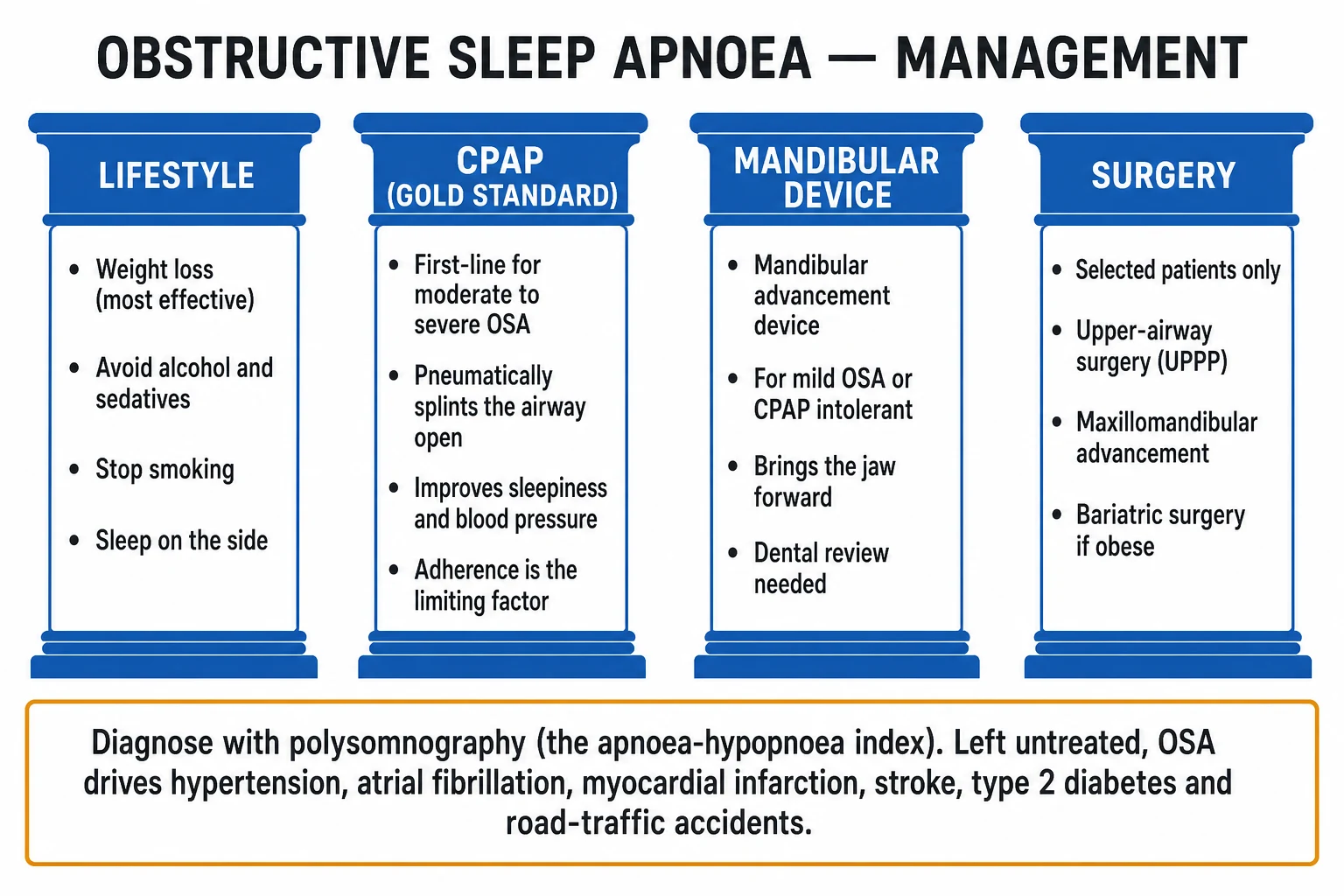
- Weight loss — the single most effective reversible intervention; a 10% weight loss reduces AHI by roughly 20–30%. Combine diet, exercise, behavioural change; consider pharmacological weight-loss agents and bariatric surgery (Roux-en-Y gastric bypass, sleeve gastrectomy) in morbid obesity (BMI over 35–40), which can produce OSA remission.
- Sleep hygiene and adequate sleep duration (7–9 hours).
- Positional therapy — for supine-dominant OSA, use a positional device (tennis-ball vest, wedge, lateral pillow) to encourage lateral sleep; effective in a subset.
- Avoid alcohol and sedatives for 4–6 hours before bed (they relax the dilator muscles and worsen OSA the same night).
- Stop smoking.
- Treat nasal obstruction — intranasal corticosteroid (e.g. fluticasone 50 micrograms, 2 sprays each nostril daily) for rhinitis, and surgical correction of structural obstruction if relevant. [1]
Pillar 2 — CPAP (the gold standard)
Continuous positive airway pressure (CPAP), typically 4–20 cmH2O delivered by auto-titrating (APAP) or a fixed pressure, is the gold-standard first-line therapy for moderate-to-severe OSA.[2] It works by providing a pneumatic splint that holds the upper airway open throughout the respiratory cycle, abolishing apnoeas, hypopnoeas, snoring and the associated desaturations and arousals.
Benefits of effective CPAP:
- Resolves snoring and witnessed apnoeas; restores bed-partner sleep.
- Reduces excessive daytime sleepiness and improves cognition and quality of life.
- Reduces blood pressure (a modest but real fall, ~2–3 mmHg systolic).[5]
- In observational data, reduces cardiovascular events and mortality; CPAP-treated patients have fewer cardiovascular events than untreated OSA.[4]
Adherence is the limiting factor (mean use often only 4–5 hours/night). Support adherence with:
- Expert mask fitting (nasal, full-face, or nasal-pillows; try several).
- Heated humidification to reduce nasal dryness/congestion.
- Ramp feature (pressure rises gradually as the patient falls asleep) and pressure relief on exhalation (C-Flex/EPR).
- Behavioural support, early and regular follow-up, and troubleshooting of mask leaks, claustrophobia, and side-effects. [1]
Effective use is conventionally defined as CPAP used for at least 4 hours per night on at least 70% of nights.[2]
Pillar 3 — Mandibular advancement device (MAD / MAS)
A custom-made dental splint that protrudes the mandible (and hence the tongue base) forward, widening the pharyngeal airway. Indicated for:
- Mild OSA (AHI 5–15), especially if symptomatic or with cardiovascular risk.
- CPAP-intolerant or CPAP-refusing patients with moderate-severe disease.
- Patients with retrognathia / a crowded oropharynx and adequate dentition. [1]
Requires dental assessment and adequate teeth; can cause temporomandibular joint discomfort and dental movement. Less effective than CPAP for severe OSA but better tolerated, so adherence is higher. [1]
Pillar 4 — Surgery
Surgery is not first-line for adult OSA. It is reserved for selected patients with a correctable anatomical cause or who have failed conservative therapy: [1]
- Uvulopalatopharyngoplasty (UPPP) — removes redundant uvula/soft palate and tonsillar tissue; benefits a subset with retropalatal collapse; variable long-term success.
- Maxillomandibular advancement (MMA) — moves the maxilla and mandible forward, enlarging the entire pharyngeal airway; the most anatomically effective upper-airway surgery.
- Genioglossus advancement / hyoid suspension — for tongue-base collapse.
- Multi-level / stepwise surgery — staged approach.
- Nasal surgery (septoplasty, turbinate reduction) — for nasal obstruction; may improve CPAP tolerance.
- Tonsillectomy — first-line in children with adenotonsillar hypertrophy.
- Bariatric surgery (Roux-en-Y gastric bypass, sleeve gastrectomy) — highly effective for OSA in morbid obesity, often producing durable remission by addressing the root cause.
- Hypoglossal nerve stimulation (e.g. Inspire) — an implantable device that stimulates the hypoglossal nerve in synchrony with respiration; for selected moderate-severe OSA with BMI under 32 and CPAP failure. [1]
Pharmacological therapy (limited role)
There is no effective drug for OSA itself. Drugs are reserved for:
- Residual daytime sleepiness despite compliant CPAP — modafinil or armodafinil (wakefulness-promoting agents), added on top of (not instead of) CPAP.
- Acetazolamide in selected cases (stimulates ventilation via metabolic acidosis; used in high-altitude periodic breathing and rare refractory cases). [1]
Treatment-emergent central sleep apnoea (complex sleep apnoea)
In some patients, CPAP unmasking central apnoeas produces treatment-emergent CSA (persistent central events on adequate CPAP). Manage by optimising CPAP pressure and switching to adaptive servo-ventilation (ASV) or BiPAP with a back-up respiratory rate, which stabilises ventilatory control. [1]
Specific Subtypes & Scenarios
Obesity hypoventilation syndrome (OHS)
Defined by BMI over 30 + daytime PaCO2 over 45 mmHg + sleep-disordered breathing, excluding other causes. Patients are somnolent, hypoxaemic, and at high risk of pulmonary hypertension and cor pulmonale. CPAP alone is often insufficient — prefer NIV (BiPAP) providing pressure support to augment ventilation, alongside aggressive weight loss and treatment of any OSA component. Mortality is high if untreated. [1]
Overlap syndrome (OSA + COPD)
The combination carries a worse prognosis than either alone: higher risk of pulmonary hypertension, chronic hypercapnia, cor pulmonale and mortality. Treat with CPAP (for the obstructive component) ± long-term oxygen therapy for chronic hypoxaemia, smoking cessation, and pulmonary rehabilitation. Avoid uncontrolled oxygen (CO2 retention). [1]
OSA in children
Most commonly due to adenotonsillar hypertrophy (peak 2–8 years). Also in Down syndrome, Pierre Robin sequence and other craniofacial syndromes, and cerebral palsy. Adenotonsillectomy is first-line. Use CPAP at weight-appropriate pressures if residual OSA after surgery or if surgery is contraindicated. [1]
OSA in pregnancy
Snoring and OSA are common and increase the risk of gestational hypertension, pre-eclampsia, gestational diabetes and low birth weight. CPAP is safe and effective in pregnancy; lateral sleep positioning and weight management within recommended ranges also help. [1]
OSA and resistant hypertension
Screen for OSA in any patient with resistant hypertension (BP above target despite three antihypertensives including a diuretic). CPAP produces a modest but meaningful fall in blood pressure and improves nocturnal dipping.[5]
OSA and atrial fibrillation
OSA is strongly associated with paroxysmal (often nocturnal) AF, and untreated OSA predicts recurrence after cardioversion or ablation. Treating OSA with CPAP improves the maintenance of sinus rhythm. [1]
Peri-operative OSA
High risk of difficult airway, post-operative respiratory depression, hypoxaemia, atrial fibrillation, and prolonged hospital stay. Identify pre-operatively (STOP-BANG), plan the airway, use short-acting agents, monitor in HDU, and continue CPAP throughout the peri-operative period. [1]
OSA secondary to endocrine disease
In acromegaly (macroglossia, tissue oedema), hypothyroidism (macroglossia, myopathy of dilators), and polycystic ovarian syndrome (obesity, hyperandrogenism), OSA is common. Treat the endocrine cause as well as the OSA — OSA may resolve with control of acromegaly or replacement of thyroid hormone. [1]
Treatment-emergent / complex sleep apnoea
Persistent central apnoeas on adequate CPAP. Switch from CPAP to adaptive servo-ventilation (ASV), which dynamically adjusts support to stabilise ventilation and abolish central events. [1]
Complications & Pitfalls
Cardiovascular complications
- Systemic hypertension — including drug-resistant hypertension; OSA is found in a large fraction of resistant hypertensives.[5][6]
- Atrial fibrillation — often paroxysmal and nocturnal; recurrence after cardioversion/ablation if untreated.
- Other arrhythmias — bradyarrhythmias, premature ventricular complexes, nocturnal atrioventricular block, sudden cardiac death.
- Coronary artery disease / myocardial infarction.
- Heart failure — both systolic and with preserved ejection fraction.
- Pulmonary hypertension and cor pulmonale in severe disease.
- Stroke — OSA is an independent risk factor, especially for cryptogenic stroke on waking.
Metabolic complications
- Insulin resistance and type 2 diabetes (independent of obesity).
- Dyslipidaemia.
- Non-alcoholic fatty liver disease (NAFLD). [1]
Neurocognitive and psychosocial
- Impaired vigilance, memory and executive function; depression and anxiety; reduced quality of life. [1]
Safety
- Road-traffic accidents — 2–7 fold increased risk; the leading acutely preventable harm.
- Occupational accidents. [1]
Pregnancy complications
- Gestational hypertension, pre-eclampsia, low birth weight. [1]
Classic pitfalls
- Not taking a partner's history — the patient under-reports nocturnal events.
- Under-using validated questionnaires (STOP-BANG, Epworth).
- Assuming a normal spot SpO2 excludes OSA — daytime oximetry is often normal; the desaturations occur only during sleep.
- Using CPAP alone in OHS — the hypercapnic patient needs NIV/BiPAP.
- Failing to advise driving cessation in a sleepy patient.
- Not screening patients with resistant hypertension, AF or cryptogenic stroke for OSA.
- Ignoring residual sleepiness on CPAP — first check adherence (download the device), mask fit and pressure adequacy before adding modafinil.
- Not addressing weight and alcohol — these are modifiable drivers of disease severity. [1]
Prognosis & Disposition
- Untreated severe OSA is associated with increased all-cause and cardiovascular mortality.[4]
- Effective CPAP (used for at least 4 hours per night on at least 70% of nights) reduces daytime sleepiness, improves blood pressure, quality of life and metabolic markers; cardiovascular and mortality benefit is greatest in moderate-severe, symptomatic, adherent patients.[2][4]
- Weight loss (lifestyle or bariatric surgery) can produce durable OSA remission.
- Adherence to CPAP is the key determinant of long-term outcome; long-term follow-up is essential.
- Drivers — once effectively treated and no longer sleepy, the patient may resume driving; some licensing authorities require objective confirmation of CPAP adherence before recertifying a commercial driver.
Special Populations
Children (1–5% prevalence)
Adenotonsillar hypertrophy is the usual cause; also Down syndrome, craniofacial syndromes (Pierre Robin, Treacher Collins, Crouzon), cerebral palsy. Behavioural and growth effects are prominent (hyperactivity mimicking ADHD, failure to thrive, enuresis). Adenotonsillectomy is first-line; CPAP at weight-appropriate pressures if residual or surgery contraindicated. [1]
Pregnancy
New snoring and OSA raise the risk of gestational hypertension, pre-eclampsia and gestational diabetes. CPAP is safe and effective; favour lateral sleep positioning. [1]
Elderly
Often less obese; present atypically (falls, cognitive decline, nocturia). Treatment benefit is similar but watch for polypharmacy (sedatives) and CPAP tolerance. [1]
Women / post-menopausal
Under-diagnosed; present with fatigue/insomnia rather than witnessed apnoeas. Lower AHI thresholds for equivalent symptoms. Risk rises after menopause. [1]
Obesity (OHS)
Daytime hypercapnia; NIV/BiPAP preferred over CPAP alone; weight loss essential; high mortality untreated. [1]
Patients with cardiovascular disease
OSA worsens hypertension, AF, heart failure; treat the OSA and the cardiovascular risk aggressively in tandem. [1]
Peri-operative
Anticipate difficult airway and post-op respiratory depression; continue CPAP through the peri-operative period; use short-acting anaesthetics. [1]
Commercial drivers
Fitness-to-drive regulations apply; OSA must be diagnosed, treated and objectively controlled before driving professionally. [1]
[1]Evidence, Guidelines & Regional Differences
Landmark evidence
- Wisconsin Sleep Cohort (Young 1993, NEJM) — established the high prevalence of sleep-disordered breathing in middle-aged adults (~24% of men, ~9% of women with AHI over 5; ~2–4% with the clinical syndrome). The defining epidemiology of OSA.[1]
- Sleep Heart Health Study (Nieto 2000, JAMA) — a large community-based cohort showing an independent, dose-response association between OSA and hypertension and cardiovascular disease, even after adjusting for obesity.[6]
- Marin 2005 (Lancet) and Marin 2012 (JAMA) — observational data showing that untreated severe OSA has a higher incidence of fatal and non-fatal cardiovascular events and new hypertension, while CPAP-treated patients approach the risk of simple snorers — strong observational evidence for the cardiovascular benefit of treatment.[4][5]
- SAVE trial (McEvoy 2016, NEJM) — the pivotal randomised trial of CPAP in patients with moderate-severe OSA and established cardiovascular disease. CPAP did not reduce the composite of recurrent cardiovascular events. Interpretation: CPAP must be given early, before end-organ damage; in patients with established CVD, treating OSA alone is too late. CPAP improved sleepiness and quality of life regardless.[3]
- Johns 1991 (Sleep) — derivation of the Epworth Sleepiness Scale, the standard subjective measure of daytime sleepiness.[7]
- Kushida 2006 (AASM practice parameters, Sleep) — the foundational guidance for CPAP and BiPAP in adult sleep-related breathing disorders.[2]
The central controversy
The SAVE trial's negative cardiovascular result in established disease sits alongside clear symptomatic benefit (sleepiness, quality of life, blood pressure) and strong observational signals of cardiovascular prevention. The synthesis: treat OSA early (the symptomatic and CV-prevention benefit is real in moderate-severe disease before irreversible end-organ damage), and do not expect CPAP alone to reverse established atherosclerotic disease. [1]
Regional guidance
- AASM (American Academy of Sleep Medicine) 2017 clinical practice guideline — recommends CPAP, mandibular advancement devices, expiratory positive airway pressure, and selected surgery, graded by evidence quality.
- ICSD-3 (International Classification of Sleep Disorders, 3rd edition) — defines OSA and OSAHS.
- NICE NG202 (UK, 2021) — sleep-disordered breathing: home respiratory polygraphy or PSG for diagnosis; CPAP first-line for moderate-severe OSA; MAD for mild or CPAP-intolerance; sleep clinic follow-up.
- Indian Sleep Disorders Association / AIIMS consensus — adapted to the Indian setting, where the obesity phenotype and craniofacial (brachyfacial) build produce OSA at lower BMI, and where access to PSG and CPAP is uneven. [1]
Exam Pearls
STOP-BANG — the OSA screen
STOPBANG
Loud, habitual, reported by partner
Excessive daytime sleepiness
Witnessed apnoeas (most specific symptom)
Blood pressure — treated or over 140/90
Over 35
Over 50
Circumference over 40 cm
Male
High-yield definitions
- Apnoea — cessation of airflow for 10 seconds or more despite ongoing respiratory effort.
- Hypopnoea — fall in airflow of at least 30% for 10 seconds or more with at least a 3% desaturation or an arousal.
- AHI = (apnoeas + hypopnoeas) per hour of sleep. Mild 5–15, moderate 15–30, severe over 30. [1]
The decisive distinctions
- OSA = effort, no flow; CSA = no effort, no flow.
- OHS = BMI over 30 + daytime PaCO2 over 45 mmHg + sleep-disordered breathing → use NIV/BiPAP, not CPAP alone.
- Epworth over 10/24 = excessive daytime sleepiness.
- CPAP is gold standard for moderate-severe OSA; MAD for mild or CPAP-intolerant; surgery in selected patients only. [1]
Always remember
- Always take a partner's history — the patient under-reports nocturnal symptoms.
- Screen for OSA in resistant hypertension, paroxysmal AF, nocturnal angina, unexplained pulmonary hypertension, cryptogenic stroke, and after a road-traffic accident.
- In the obese hypercapnic patient, think OHS — PaCO2 over 45 mmHg, BMI over 30 — and use NIV/BiPAP.
- Peri-operative OSA: difficult airway + post-op respiratory depression — continue CPAP.
- SAVE trial: in established CVD, CPAP did not reduce recurrent events — treat OSA early.
- First-line in children with OSA is adenotonsillectomy. [1]
Exam application bank (NEET-PG / INICET)
One-line answer
Obstructive sleep apnoea (OSA) is recurrent collapse of the upper airway during sleep, causing apnoeas and hypopnoeas with oxygen desaturation and arousal. It presents with loud snoring, witnessed apnoeas (reported by a partner), excessive daytime sleepiness and unrefreshing sleep. Risk factors are obesity (large neck), male sex, middle age, alcohol/sedatives and craniofacial narrowing. Untreated, it drives systemic hypertension, atrial fibrillation, myocardial infarction, stroke, type 2 diabetes and a high risk of road-traffic accidents. Polysomnography is diagnostic, quantifying the apnoea-hypopnoea index (AHI) — mild 5-15, moderate 15-30, severe over 30 — and STOP-BANG screens for risk. Management is weight loss and lifestyle change, CPAP (the gold standard for moderate-severe disease), a mandibular advancement device for mild/CPAP-intolerant cases, and surgery only in selected patien
Worked stems (answer without another resource)
Stem 1 — Classic presentation. Map symptoms to mechanism; name the first investigation and first treatment step with dose/route if drug therapy is standard. [1]
Stem 2 — Unstable / complicated. List red flags that force immediate resuscitation, theatre, ICU, antidote, or reperfusion — and what you do in the first 15 minutes. [1]
Stem 3 — Atypical group. Elderly, pregnancy, child, or immunocompromised: how presentation and thresholds change. [1]
Stem 4 — Differential trap. Name the three closest mimics and one discriminator for each. [1]
Stem 5 — Disposition. Who goes home with safety-netting, who is admitted, who needs HDU/ICU/theatre, and what follow-up is mandatory. [1]
Rapid viva checklist
- Definition + classification
- Pathophysiology chain
- Bedside signs / criteria
- Score with exact components (if any)
- Emergency bundle
- Definitive therapy with doses
- Complications of disease and of treatment
- Special populations
- Guideline/trial name if classic
- Three exam traps
Coverage self-check
If you cannot answer any stem above from this page alone, re-read the matching section — the page is intended to be self-sufficient for final-prof and NEET-PG/INICET questions on Obstructive Sleep Apnoea.
References
- [1]Young T, Palta M, Dempsey J, et al. The occurrence of sleep-disordered breathing among middle-aged adults N Engl J Med, 1993.PMID 8464434
- [2]Kushida CA, Littner MR, Hirshkowitz M, et al. Practice parameters for the use of continuous and bilevel positive airway pressure devices to treat adult patients with sleep-related breathing disorders Sleep, 2006.PMID 16553024
- [3]McEvoy RD, Antic NA, Heeley E, et al. (SAVE trial). CPAP for Prevention of Cardiovascular Events in Obstructive Sleep Apnea N Engl J Med, 2016.PMID 27571048
- [4]Marin JM, Carrizo SJ, Vicente E, Agusti AG. Long-term cardiovascular outcomes in men with obstructive sleep apnoea-hypopnoea with or without treatment with continuous positive airway pressure: an observational study Lancet, 2005.PMID 15781100
- [5]Marin JM, Agusti A, Villar I, et al. Association between treated and untreated obstructive sleep apnea and risk of hypertension JAMA, 2012.PMID 22618924
- [6]Nieto FJ, Young TB, Lind BK, et al. (Sleep Heart Health Study). Association of sleep-disordered breathing, sleep apnea, and hypertension in a large community-based study. Sleep Heart Health Study JAMA, 2000.PMID 10770144
- [7]Johns MW. A new method for measuring daytime sleepiness: the Epworth sleepiness scale Sleep, 1991.PMID 1798888