Respiratory · General Medicine
Pleural Effusion
Also known as Pleural effusion · Hydrothorax · Transudative effusion · Exudative effusion · Parapneumonic effusion · Empyema · Hepatic hydrothorax · Chylothorax
Pleural effusion is an accumulation of fluid in the normally near-dry pleural space. It is classified by Light's criteria (1972) as a transudate (systemic cause: heart failure, cirrhosis, nephrotic syndrome, peritoneal dialysis, myxoedema, pulmonary embolism) or an exudate (local pleural disease: parapneumonic, malignancy, tuberculosis, pulmonary embolism, autoimmune, pancreatitis, chylothorax, haemothorax). Diagnosis rests on chest X-ray (blunted costophrenic angle over 200 mL, meniscus sign, mediastinal shift), thoracic ultrasound (loculation, septation, guidance) and diagnostic thoracentesis with pleural fluid analysis (protein, LDH, glucose, pH, cell count, Gram stain and culture, cytology, ADA, amylase, triglycerides, NT-proBNP). Treatment is cause-specific; therapeutic thoracentesis, small-bore Seldinger chest drain, talc pleurodesis and indwelling pleural catheter are the procedural pillars.
On this page & tools
Your progress
Saved locally on this device.
Exam tags
Red flags
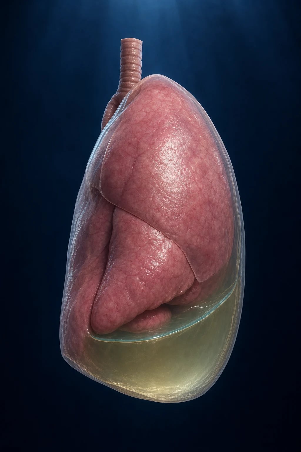
Overview & Definition
A pleural effusion is the pathological accumulation of excess fluid in the pleural space — the thin, normally near-dry potential space between the visceral pleura investing the lung and the parietal pleura lining the chest wall, mediastinum and diaphragm. The healthy pleural space contains only 0.1 to 0.3 mL/kg of a low-protein lubricating fluid (around 5 to 15 mL in an adult), continuously generated by the systemic capillaries of the parietal pleura and reabsorbed by the parietal lymphatics at a rate of 0.01 mL/kg/h, with a maximal lymphatic clearance of roughly 0.4 mL/kg/h — a generous reserve that must be overwhelmed before fluid accumulates. When formation exceeds absorption, fluid accumulates, the lung collapses outward, and the characteristic syndrome of dyspnoea, pleuritic chest pain, dullness to percussion and reduced breath sounds emerges.[2][10]
Pleural effusion is not a diagnosis — it is a sign of an underlying disease. The clinical task is twofold: first to confirm the effusion, then to identify its cause. The single most powerful diagnostic step, introduced by Richard Light in 1972 and unchanged in half a century, is to classify the fluid as a transudate or an exudate using Light's criteria. That single distinction collapses a differential of dozens of causes into two manageable lists, and it dictates which further pleural fluid tests are worth sending.[1][2]
Pleural effusion is common. The estimated annual incidence in the United States is over 1.5 million, making it one of the most frequently encountered pleural problems in hospital medicine. Heart failure alone accounts for roughly 40 per cent of all effusions, parapneumonic effusion about 25 per cent, malignancy 15 to 25 per cent, and pulmonary embolism, tuberculosis and autoimmune disease make up much of the rest. The epidemiology is regional: in tuberculosis-endemic countries tuberculous pleuritis is a leading cause of a unilateral exudate, whereas in western series malignancy dominates that slot.[2][10]
Classification
Pleural effusion is classified along three axes: fluid composition (transudate versus exudate, by Light's criteria), aetiology (the underlying disease), and macroscopic appearance (serous, bloody, turbid, milky, frankly purulent, bile-stained, food- contaminated). The composition axis is the master axis because it determines the cause list and the subsequent fluid tests.[1][4]
The transudate–exudate distinction is not merely biochemical. A transudate forms when systemic forces alter the balance of Starling forces across an intact, healthy pleura — increased hydrostatic pressure (heart failure), reduced oncotic pressure (cirrhosis, nephrotic syndrome), or a combination. The pleura itself is normal; the fluid "leaks" through it because the system is disturbed. An exudate forms when the pleura itself is diseased — by inflammation increasing capillary permeability (infection, autoimmune), by tumour blocking lymphatic reabsorption (malignancy), by direct violation of the lymphatics (chylothorax) or by a blood vessel rupture (haemothorax). The pleural fluid therefore reflects the local disease.[1][2]
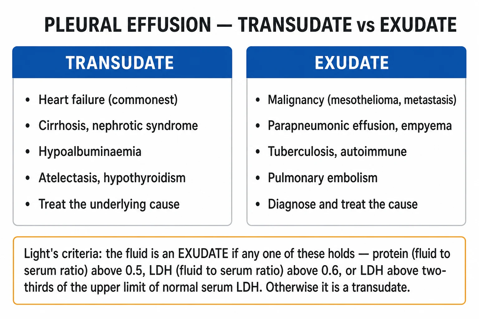
Transudate (Light negative)
- Light's criteria: all three negative
- Cause is systemic; pleura is normal
- Heart failure (about 40 per cent) — most common, often bilateral, right greater
- Hepatic hydrothorax (cirrhosis) — usually right-sided
- Nephrotic syndrome — bilateral, low oncotic pressure
- Peritoneal dialysis, myxoedema, urinothorax
- Constrictive pericarditis, mitral stenosis
- Rescue tests: serum-pleural albumin gradient over 1.2 g/dL; NT-proBNP over 1500 ng/L
Exudate (Light positive)
- Light's criteria: any one positive
- Cause is pleural or local disease
- Parapneumonic — uncomplicated, complicated or empyema (about 25 per cent)
- Malignant — lung, breast, mesothelioma, lymphoma, ovarian (15 to 25 per cent)
- Pulmonary embolism — often bloody; can be transudate or exudate
- Tuberculous — lymphocytic, ADA over 40 U/L
- Autoimmune — rheumatoid, lupus; pancreatitis (high amylase)
- Chylothorax (triglycerides over 1.24 mmol/L) and haemothorax
Pleural effusion — the numbers that decide a question
Epidemiology & Risk Factors
The epidemiology of pleural effusion is the epidemiology of its causes. Heart failure is the commonest cause worldwide: roughly two-thirds of cardiac effusions are bilateral, and when unilateral they are right-sided in roughly two-thirds of cases because the larger right lung and right pleural space, combined with the gravitational distribution of pulmonary venous congestion, favour the right side. The presence of a unilateral right-sided effusion in a patient with heart failure should not prompt a search for an alternative cause before the more common explanation is considered.[10]
Parapneumonic effusion complicates roughly 20 to 40 per cent of bacterial pneumonias that require hospitalisation, and a small minority progress to frank empyema. The microbiology mirrors community-acquired pneumonia (Streptococcus pneumoniae, Haemophilus influenzae) in early cases and shifts to anaerobes, staphylococci (including MRSA) and gram-negatives (Klebsiella, Pseudomonas) in established and hospital-acquired empyema. Streptococcus anginosus (milleri) group is particularly associated with empyema.[5]
Malignant pleural effusion is a marker of advanced cancer. The commonest primaries are lung (about one-third), breast (about one-fifth), lymphoma and ovarian, with mesothelioma a less common but important cause, especially in patients with asbestos exposure. Roughly 15 per cent of all lung cancer patients develop a malignant effusion during their illness, and it carries a poor prognosis with median survival of 3 to 12 months depending on primary tumour type and performance status. Mesothelioma-related effions do worse.[3][11]
Tuberculous pleuritis is a leading cause of a unilateral exudative effusion in tuberculosis-endemic regions (India, sub-Saharan Africa, South-East Asia). It represents a delayed hypersensitivity reaction to mycobacterial antigens in the pleural space rather than direct disseminated infection, which is why pleural fluid cultures are positive in only 20 to 30 per cent and biopsy cultures in roughly 50 to 70 per cent. The effusion is usually unilateral, small to moderate, lymphocyte-predominant, with a high adenosine deaminase (ADA over 40 U/L).[10]
The risk factors therefore depend on cause: cardiac disease (ischaemic, hypertensive, valvular), cirrhosis with portal hypertension, nephrotic-range proteinuria, recent pneumonia, known or occult malignancy, asbestos exposure, thromboembolic risk (immobility, malignancy, pregnancy, oestrogen), autoimmune disease (rheumatoid arthritis, systemic lupus erythematosus), pancreatitis and alcohol misuse, drug exposure (amiodarone, methotrexate, nitrofurantoin, bromocriptine, phenytoin), recent cardiac or thoracic surgery, and trauma.[2][4]
Pathophysiology
The pleural space is a dynamic, low-pressure lubricating compartment governed by Starling forces and lymphatic drainage. Fluid is filtered from the systemic capillaries of the parietal pleura (supplied by the intercostal arteries, systemic pressure) at a rate governed by the net hydrostatic minus oncotic pressure gradient. The visceral pleura, supplied by the low-pressure pulmonary circulation, contributes less. Fluid is reabsorbed mainly by the parietal pleural lymphatics (the stomata) into the intercostal lymph nodes, with a small contribution from pulmonary capillaries. The lymphatic pump can increase its clearance up to roughly twenty-eight times the basal rate before it is overwhelmed — a reserve that explains why early effusions are subtle and why, once they appear, the underlying disturbance is already substantial.[2]
Effusions arise by one or more of four pathophysiological mechanisms, and recognising the mechanism predicts the transudate–exudate split:[1][10]
- Increased hydrostatic pressure (transudate). Pulmonary venous and capillary pressure rises in left heart failure, mitral stenosis, fluid overload and constrictive pericarditis. Filtration exceeds lymphatic reabsorption; the fluid is protein-poor because the intact pleura still excludes large molecules.
- Decreased oncotic pressure (transudate). Hypoalbuminaemia from cirrhosis, nephrotic syndrome, protein-losing enteropathy or severe malnutrition lowers plasma oncotic pressure and widens the net filtration gradient. Hepatic hydrothorax is the paradigm: ascites tracks through small diaphragmatic defects (more common on the right where the diaphragmatic muscle fibres are thinner) into the negative-pressure pleural space.
- Increased capillary permeability (exudate). Inflammation from pneumonia, autoimmune disease (rheumatoid, lupus), pulmonary embolism with infarction, pancreatitis, uraemia, drugs or radiation opens intercellular junctions and allows protein-rich fluid — and often cells — to leak into the pleural space. Malignancy increases permeability directly through tumour-secreted cytokines (vascular endothelial growth factor) and by causing pleural inflammation.
- Impaired lymphatic drainage (exudate). The mediastinal and parietal lymphatics are the principal drainage route; obstruction by tumour (lymphangitic carcinomatosis, lymphoma, mesothelioma) or nodes raises the back-pressure and prevents reabsorption of even normal-constituency fluid, which then accumulates as an exudate because the protein is trapped and concentrated. [1]
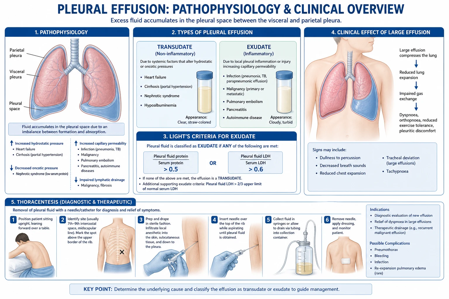
Two specific mechanisms deserve a sentence each because the examiner rewards them. Hepatic hydrothorax arises because cirrhotic portal hypertension generates ascites that tracks through tiny diaphragmatic defects (often in the tendinous right hemidiaphragm) into the negative-pressure pleural space; the effusion can dominate the picture even when the ascites is clinically small, and it is a transudate that nonetheless may have a protein a little higher than cardiac transudates because of chronic equilibration. Chylothorax is a true leak of chyle from the thoracic duct (post-traumatic, post-oesophagectomy or post-thoracic surgery, or from lymphoma that invades the duct); the fluid is milky white, alkaline, sterile, lymphocyte-predominant and rich in triglycerides (over 1.24 mmol/L or 110 mg/dL).[10][12]
Clinical Presentation
The clinical picture of a pleural effusion reflects three things — the size and rate of accumulation, the underlying cause, and the patient's cardiopulmonary reserve. A small effusion (under 300 to 500 mL) may be entirely asymptomatic and discovered on a chest X-ray taken for another reason. A moderate or large effusion produces the cardinal respiratory symptoms, and a massive effusion (filling the hemithorax) produces respiratory compromise that can be life-threatening.[2]
The classical symptom triad is progressive exertional dyspnoea, pleuritic chest pain and a dry cough. Dyspnoea is the commonest and most reliable symptom; it worsens as the effusion grows and the lung is compressed, and is relieved by sitting upright (which splints the diaphragm). Pleuritic chest pain — sharp, well-localised, worse on deep inspiration and coughing — arises from parietal pleural inflammation (the visceral pleura is insensate) and is therefore characteristic of exudative causes (pneumonia, pulmonary embolism, autoimmune) rather than transudates. The pain often eases as fluid accumulates and separates the inflamed pleural surfaces. A dry, non-productive cough results from reflex stimulation of the cough receptors in the distended pleura. Constitutional symptoms point to the cause: fever and rigors (infection, empyema), weight loss and night sweats (tuberculosis, lymphoma, malignancy), orthopnoea and paroxysmal nocturnal dyspnoea (heart failure), ankle and abdominal swelling (heart failure, cirrhosis, nephrotic).[2][4]
Examination follows the classical sequence and yields one of the most distinctive bedside findings in medicine. The affected hemithorax is expanded, moves less on respiration, and is dull to percussion. The percussion note over an effusion is described as "stony dull" — a dense, thud-like quality distinct from the dullness of consolidation, because fluid transmits vibration poorly. Breath sounds are reduced or absent over the effusion (sound transmission through fluid is attenuated), vocal resonance and tactile vocal fremitus are diminished, and a pleural rub may be audible at the upper margin where the two pleural surfaces still rub together. The trachea and apex beat are deviated away from the side in a massive effusion (a volume-displacement sign), which is the key distinction from collapse (where they deviate toward the affected side).[2]
Signs of the underlying cause must be sought deliberately: raised jugular venous pressure, a third heart sound, basal crackles and peripheral oedema (heart failure); jaundice, spider naevi, palmar erythema, ascites and caput medusae (cirrhosis); periorbital and dependent oedema, frothy urine (nephrotic); cachexia, lymphadenopathy, clubbing, hepatosplenomegaly (malignancy); fever, focal consolidation, sepsis (parapneumonic, empyema); calf swelling and tenderness, tachycardia (pulmonary embolism); malar rash, synovitis, Raynaud (autoimmune).[4]
Differential Diagnosis
The bedside finding of dullness with reduced breath sounds has a focused differential, and the examiner expects the candidate to distinguish each by the features that separate them. The four classical mimics are pleural effusion, consolidation (pneumonia), collapse (atelectasis) and a raised hemidiaphragm.[2][4]
Pleural effusion
- Stony dull percussion, reduced breath sounds and vocal resonance
- Trachea deviated AWAY in massive effusion
- CXR: meniscus sign, blunted costophrenic angle, mediastinal shift away
- Ultrasound confirms fluid; thoracentesis diagnostic
Consolidation (pneumonia)
- Dull but woody, not stony; bronchial breathing and increased vocal resonance
- Crackles and wheeze; fever, productive cough, sepsis
- Trachea central; CXR: alveolar opacification with air bronchograms
- Responds to antibiotics; small reactive effusion may coexist
Collapse / atelectasis
- Dull with reduced breath sounds — but trachea deviated TOWARD the lesion
- Volume loss: shifted mediastinum, narrowed intercostal spaces
- CXR: opacification with volume loss, no meniscus
- Causes: mucus plug, foreign body, tumour, pneumothorax (compressive)
Raised hemidiaphragm
- Dullness high in the axilla, normal breath sounds above
- Phrenic nerve palsy (lung cancer, mediastinal tumour, post-CABG)
- CXR: elevated dome; fluoroscopy sniff test shows paradoxical ascent
- No fluid meniscus; ultrasound confirms no effusion
Two less common but examinable mimics are a large pericardial effusion (which can dull the left base and mimic a pleural effusion) and a subpulmonic effusion (fluid trapped beneath the lung, which masquerades as an elevated hemidiaphragm with a flattened peak and an unusually lateral drop-off on the chest X-ray; lateral decubitus or ultrasound confirms it).[2]
The aetiological differential is enormous and is best organised by the transudate–exudate split: [1]
- Transudate: heart failure, hepatic hydrothorax, nephrotic syndrome, peritoneal dialysis, myxoedema, constrictive pericarditis, mitral stenosis, pulmonary embolism (occasionally), urinothorax, superior vena cava obstruction, atelectasis (trapped).
- Exudate: parapneumonic (uncomplicated, complicated, empyema), malignancy (lung, breast, lymphoma, mesothelioma, ovarian, gastric), pulmonary embolism (commonly bloody), tuberculosis, autoimmune (rheumatoid, systemic lupus erythematosus, granulomatosis with polyangiitis), pancreatitis (acute and chronic, pancreaticopleural fistula), post-cardiac injury (Dressler, post-CABG), drug-induced (amiodarone, methotrexate, nitrofurantoin, bromocriptine, phenytoin), chylothorax, haemothorax, oesophageal rupture (Boerhaave), benign asbestos pleural effusion, Meigs syndrome, yellow nail syndrome, trapped lung.[1][10]
Clinical & Bedside Assessment
A focused history and examination narrow the cause before any investigation. Take a dyspnoea history (onset, progression, orthopnoea, paroxysmal nocturnal dyspnoea, exercise tolerance), a pleuritic pain history (onset, character, relieving and exacerbating factors), and a cough and sputum history. Elicit constitutional symptoms (fever, night sweats, weight loss, anorexia) and symptoms of the common causes: ankle swelling and abdominal distension (heart failure, cirrhosis, nephrotic); palpitations, chest pain, syncope (cardiac); frothy urine and periorbital swelling (nephrotic); haemoptysis, weight loss, asbestos exposure (malignancy, mesothelioma); calf pain and recent immobility (pulmonary embolism); acute severe epigastric pain radiating to the back (pancreatitis); joint swelling and rash (autoimmune). A drug history (amiodarone, methotrexate, nitrofurantoin), occupational history (asbestos, shipyard, demolition), alcohol history (cirrhosis, pancreatitis), travel and tuberculosis contact, and recent surgery or trauma complete the picture.[4]
The bedside examination is conducted systematically. Begin with the vital signs — respiratory rate, oxygen saturation, heart rate, blood pressure, temperature — which flag the unwell patient. Inspect the chest for reduced movement on the affected side and intercostal indrawing. Palpate for chest expansion (reduced on the affected side), tracheal position (central, deviated away in massive effusion, toward in collapse), apex beat position, and tactile vocal fremitus (reduced over an effusion, increased over consolidation). Percuss for stony dullness in the dependent zone with a sharp upper level that slopes down laterally (Ellis's S-shaped line). Auscultate for reduced or absent breath sounds, reduced vocal resonance, an egophony at the upper border (the "e to a" change at the fluid-air interface), occasionally a pleural rub. Complete with a cardiovascular (raised JVP, third heart sound, murmurs of mitral stenosis), abdominal (hepatosplenomegaly, ascites, masses, caput medusae), peripheral (oedema, clubbing, lymphadenopathy, calf tenderness), and skin (malar rash, synovitis, spider naevi, gynaecomastia) examination.[2]
Bedside ultrasound is now an extension of the clinical examination and the BTS recommends it for every pleural procedure. It confirms the presence of fluid, estimates depth from skin to pleura, detects loculation and septation that a chest X-ray cannot, identifies the safe entry point and depth, and dramatically reduces the iatrogenic pneumothorax rate from blind thoracentesis. A skilled operator can characterise an anechoic effusion (likely simple), an echogenic or septated effusion (likely exudate or organising empyema) and a homogeneously echogenic effusion (likely haemorrhagic).[4]
Investigations
The investigation of a pleural effusion proceeds in two phases — confirm the effusion (imaging), then identify the cause (pleural fluid analysis, sometimes CT and biopsy). The cornerstone is the diagnostic thoracentesis with pleural fluid analysis interpreted through Light's criteria.[1][4]
Imaging
The chest X-ray is the entry test. A posterior-anterior film blunts the costophrenic angle when the effusion exceeds roughly 200 mL, producing the classical meniscus sign — a concave-up fluid line rising laterally because capillarity draws fluid up the chest wall (Ellis's S-shaped line). Larger effusions opacify the lower zone and eventually the whole hemithorax, with mediastinal shift away in a massive effusion. A lateral decubitus film detects as little as 10 mL of free-flowing fluid; a lateral chest film blunts the posterior costophrenic sulcus at roughly 50 to 75 mL. A subpulmonic effusion sits beneath the lung and mimics an elevated hemidiaphragm with a lateral peak and a steep medial drop-off. Complete opacification of a hemithorax with the trachea and mediastinum pulled toward the lesion suggests not effusion but atelectasis or post-pneumonectomy, whereas shift away confirms a massive effusion or tension.[2][4]
Thoracic ultrasound is the modern workhorse. It confirms fluid when the chest X-ray is opaque, detects loculation, septation and debris that change management, estimates volume, identifies the optimal puncture site and depth, and guides thoracentesis or drain insertion in real time. It is more sensitive than chest X-ray for small and loculated effusions and is recommended by the BTS before any pleural procedure.[4]
Contrast-enhanced computed tomography (CT) is the gold standard for characterising the underlying cause in an unexplained exudative effusion, in suspected malignancy, in a complex loculated collection, and to define pleural thickening, nodularity, masses and the pleural phase of enhancement that indicates pleural inflammation or malignancy. It is performed ideally with pleural fluid still present so that the contrast between fluid and enhancing pleura is maximal. It guides image-directed pleural biopsy when needed.[2][4]
Diagnostic thoracentesis and pleural fluid analysis
Diagnostic thoracentesis is indicated for every unexplained unilateral effusion, for any bilateral effusion with discordant appearances, for an effusion that does not resolve with heart failure treatment, and whenever infection or malignancy is suspected. It is performed under ultrasound guidance at the bedside, through the most dependent point of the fluid (posteriorly, one to two intercostal spaces below the upper level of the dullness, in the mid-axillary line for a moderate effusion), using a sterile technique and a 21 to 23 gauge needle with a syringe and three-way tap. The safe triangle (bordered by the anterior border of latissimus dorsi, the lateral border of pectoralis major, a line horizontal to the nipple, and the base just above the fifth intercostal space) is the preferred zone for any drain. About 20 to 50 mL is aspirated for analysis.[4]
Relative contraindications include a coagulopathy (INR over 1.5, platelets under 50), a small effusion (under 10 mm on ultrasound), and severe respiratory compromise that would not tolerate a pneumothorax. Anticoagulants and antiplatelets are not absolute contraindications for a diagnostic tap but warrant caution and ultrasound guidance.[4]
The first and decisive step is to apply Light's criteria to the fluid, comparing simultaneous serum protein and LDH:[1]
Light's criteria (1972)
- Pleural fluid protein / serum protein over 0.5
- Pleural fluid LDH / serum LDH over 0.6
- Pleural fluid LDH over two-thirds the upper limit of normal serum LDH
- EXUDATE if any ONE criterion is met; transudate only if ALL three are negative
- Sensitivity about 98 to 99 per cent; specificity about 70 to 80 per cent
- Overdiagnoses exudate in about 25 per cent of cardiac effusions on diuretics
Light's criteria are highly sensitive (about 99 per cent — they will not miss an exudate) but less specific (about 75 per cent), and they misclassify about 25 per cent of cardiac effusions in patients on diuretic therapy as exudates because diuresis concentrates the protein and LDH in the residual fluid. The two rescue tests for this diuretic-confounded situation are the serum-to-pleural fluid albumin gradient (over 1.2 g/dL supports a transudate) and pleural fluid NT-proBNP (over 1500 ng/L strongly supports a cardiac cause).[2][10]
Once the fluid is classified as a transudate, no further fluid tests are usually needed — the workup shifts to the systemic cause. Once it is an exudate, a panel of additional fluid tests refines the diagnosis. The macroscopic appearance gives the first clue: serous and straw-coloured (transudate or early exudate), bloody (malignancy, trauma, pulmonary embolism, iatrogenic), turbid or purulent (empyema), milky white (chylothorax or pseudochylothorax), greenish (rheumatoid pleuritis, bilious in biliary-pleural fistula), black (Aspergillus, rheumatoid), food particles (oesophageal rupture).[2][4]
Pleural fluid test
- Protein and LDH — Light's criteria
- Glucose — under 3.3 mmol/L (60 mg/dL): empyema, malignancy, RA, TB, oesophageal rupture
- pH — under 7.2: empyema, complicated parapneumonic, oesophageal rupture; under 7.3 borderline
- Cell count — neutrophils: bacterial/PE/pancreatitis; lymphocytes: TB, malignancy, chronic; eosinophils: drug, air, blood, parasitic
- Gram stain and culture, AFB — bacterial, mycobacterial
- Cytology — malignant cells; sensitivity 60 to 90 per cent, higher with repeat samples
- ADA — over 40 U/L in a lymphocytic effusion supports TB
- Amylase — high in pancreatitis, pancreaticopleural fistula, oesophageal rupture
- Triglycerides — over 1.24 mmol/L (110 mg/dL): chylothorax
- NT-proBNP — over 1500 ng/L: cardiac; RF, ANA: autoimmune; haematocrit: haemothorax if over 50% of serum
The decision to drain a parapneumonic effusion rests on three fluid values: a pH under 7.20, a glucose under 3.3 mmol/L (60 mg/dL) or frankly purulent fluid (or a positive Gram stain) — any one mandates a chest drain. The pH must be measured anaerobically in a heparinised blood gas syringe, on fluid free of lidocaine, and air must not be introduced. A pH between 7.20 and 7.30 is a grey zone; the BTS suggests drainage if there is also loculation or a large effusion.[5]
Further investigations
A pleural biopsy (CT-guided or thoracoscopic — medical thoracoscopy or VATS) is indicated for an unexplained exudative effusion, suspected malignancy with negative cytology, or suspected tuberculosis. Abrams' closed pleural biopsy (now rarely used) and thoracoscopic biopsy are options; the yield for malignancy rises to over 90 per cent with thoracoscopy, and for tuberculosis a combination of histology (caseating granuloma), AFB stain and culture is diagnostic in most cases.[2][11]
Bronchoscopy is indicated when there is an endobronchial lesion suspected — haemoptysis, a parenchymal mass, atelectasis, or a malignant effusion with a central lesion on CT — and is otherwise low yield in a cytology-negative undiagnosed exudate.[4]
PLEURAL
Management — Resuscitation
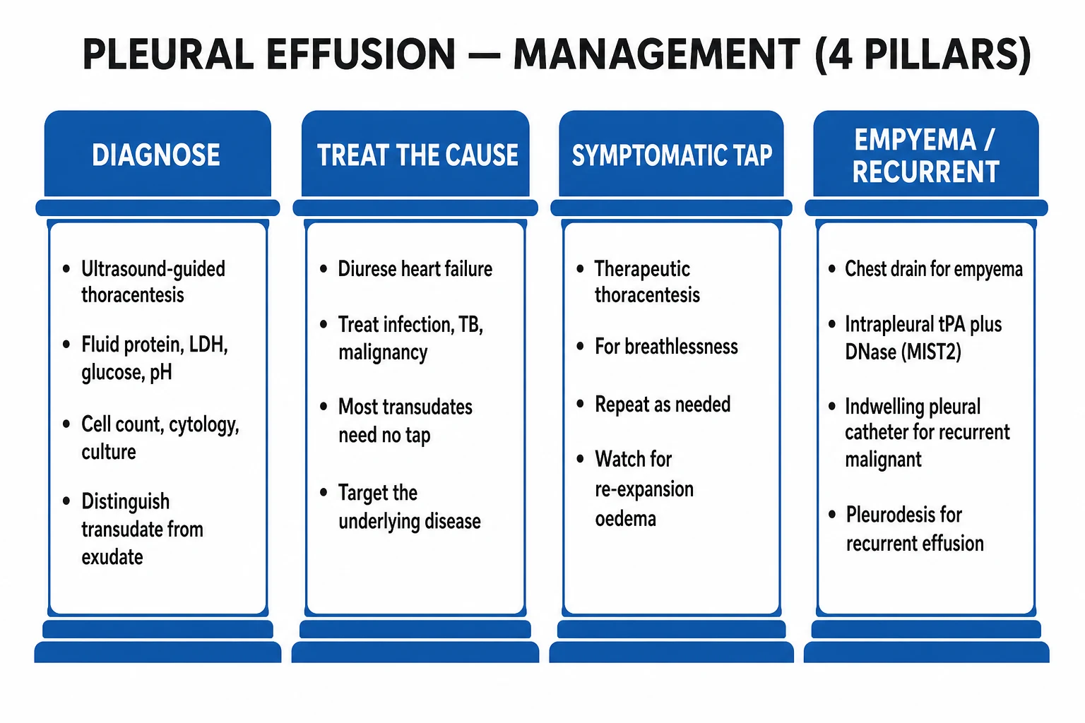
Most pleural effusions are managed electively, but a small number present as time-critical emergencies in which the resuscitation reflex precedes the diagnostic workup. The dangerous scenarios are tension pleural effusion, massive haemothorax, septic shock with empyema, and severe hypoxaemia from a large malignant effusion.[2][5]
Tension pleural effusion is rare but lethal — a large effusion under pressure that shifts the mediastinum away, compresses the great veins and produces hypotension, tachycardia, marked dyspnoea and hypoxaemia. The immediate management is emergency needle aspiration (a 14 to 16 gauge cannula in the second intercostal space mid-clavicular line or, more safely, the safe triangle in the mid-axillary line) followed promptly by an intercostal chest drain. Improvement in blood pressure and oxygenation should be rapid as the mediastinum returns to the midline.[2]
Massive haemothorax (over 1500 mL drained immediately, or over 200 mL per hour for two to four hours) is a surgical emergency. Insert a large-bore (28 to 36 French) intercostal drain, resuscitate with intravenous crystalloid and blood transfusion, correct coagulopathy, and refer immediately to thoracic surgery for thoracotomy, because ongoing bleeding indicates an intercostal or internal mammary artery laceration, a major lung laceration, or great vessel injury.[2]
Empyema with septic shock requires simultaneous resuscitation and source control: intravenous fluids, vasopressors (noradrenaline) for refractory hypotension, broad-spectrum antibiotics (a beta-lactam-beta-lactamase inhibitor or carbapenem plus cover for anaerobes and MRSA as appropriate), and urgent image-guided chest drain. Intrapleural tissue plasminogen activator (tPA) plus DNase is added for loculated collections (see below).[5][6]
A single thoracentesis must not drain more than 1.5 L (or be stopped if the patient develops chest discomfort, persistent cough, vasovagal symptoms or a drop in oxygen saturation) because of the risk of re-expansion pulmonary oedema — a rare but dangerous complication of too-rapid lung re-expansion in which the suddenly negative intrapleural pressure injures the pulmonary capillaries and floods the alveoli. It presents within 24 hours of drainage with cough, dyspnoea, hypoxaemia and frothy sputum; treatment is supportive (oxygen, diuretics, sometimes mechanical ventilation).[2]
Management — Definitive & Stepwise
Definitive management is cause-specific. The transudate–exudate split determines the strategy: a transudate is managed by treating the systemic cause, and an exudate is managed by treating the pleural disease plus drainage when indicated. The procedural toolkit — therapeutic thoracentesis, intercostal drain, pleurodesis, indwelling pleural catheter and surgery — is overlaid on the cause-specific treatment.[2][4]
Transudative effusions
Heart failure: optimise guideline-directed medical therapy (diuretics, ACE inhibitor or ARNI, beta-blocker, mineralocorticoid antagonist, SGLT2 inhibitor); the effusion usually resolves with diuresis. Therapeutic thoracentesis is reserved for refractory symptomatic effusions, and bilateral effusions that persist despite optimal therapy should prompt reconsideration of the cause (an exudative component from concomitant pneumonia, pulmonary embolism or malignancy is the classic pitfall).[10]
Hepatic hydrothorax: treat the ascites with sodium restriction, spironolactone (100 to 400 mg daily) plus furosemide (40 to 160 mg daily), large-volume paracentesis of ascites, and consider transjugular intrahepatic portosystemic shunt (TIPS) for refractory cases and liver transplantation as definitive therapy. Chest tube drainage should be avoided because it causes protein and fluid depletion, infection, and often a persistent bronchopleural fistula through the diaphragmatic defect. Symptomatic relief is achieved by serial therapeutic thoracentesis.[12]
Nephrotic syndrome, peritoneal dialysis, myxoedema and constrictive pericarditis: treat the underlying disease; therapeutic thoracentesis for symptoms.[10]
Parapneumonic effusion and empyema
Parapneumonic effusions are stratified into three stages that map directly onto management:[5]
Stage I — Uncomplicated
- Thin, free-flowing sterile fluid
- pH over 7.2, glucose over 3.3 mmol/L, LDH under 1000
- Gram stain and culture negative
- Antibiotics for the pneumonia alone — usually no drain
- Resolves with treatment of the pneumonia
Stage II — Complicated
- Neutrophilic, fibrinous, often loculated
- pH under 7.2, glucose under 3.3 mmol/L, LDH over 1000
- Gram stain may be positive; culture may be negative if antibiotics started
- Chest drain plus IV antibiotics; intrapleural tPA plus DNase if loculated
- Risk of progression to empyema if undrained
Stage III — Empyema
- Frank pus in the pleural space, single or multiloculated
- pH under 7.2, glucose low; bacterial culture often positive
- Chest drain plus IV antibiotics 2 to 6 weeks
- Intrapleural tPA plus DNase; VATS decortication if failed
- May organise into a fibrous peel (trapped lung) needing decortication
Antibiotics for pleural infection must cover the typical community-acquired pathogens plus anaerobes (a beta-lactam-beta-lactamase inhibitor such as amoxicillin-clavulanate 1.2 g IV every 8 hours, or ceftriaxone plus metronidazole); hospital-acquired empyema needs cover for MRSA (vancomycin or teicoplanin) and gram-negatives including Pseudomonas (piperacillin-tazobactam or meropenem). Duration is 2 to 6 weeks intravenously then orally, guided by clinical response and inflammatory markers.[5]
Intrapleural tPA plus DNase was established by the MIST-2 trial (Rahman 2011), which showed that alteplase 10 mg plus dornase alfa 5 mg, each daily for three days, significantly increased pleural fluid drainage and reduced the frequency of surgical referral and length of stay compared with either agent alone or placebo — tPA alone or DNase alone was no better than placebo, and indeed DNase alone was harmful. The combination is now first-line for a loculated, poorly draining empyema.[6]
Surgical decortication (VATS or open) is indicated for failed medical therapy (persistent sepsis, residual collection, trapped lung with a fibrous peel preventing re-expansion) and for multiloculated empyema at presentation in a fit patient. The RAPID score (Rahman 2014) — Renal (urea), Age, Purulence of fluid, Infection source (hospital-acquired scores worse), Dietary (albumin) — stratifies mortality risk at three months (low 1 to 5 per cent, medium 10 to 15 per cent, high 25 to 45 per cent) and can guide the threshold for early surgical referral.[5][9]
Malignant pleural effusion
A malignant effusion is a symptom-control problem layered on an oncology problem. The aims are relief of dyspnoea and prevention of reaccumulation. Options are therapeutic thoracentesis (for recurrence is impractical), talc pleurodesis (chemical, via chest drain or thoracoscopy), indwelling pleural catheter (IPC) (a tunnelled silicone catheter drained at home), VATS pleurodesis (mechanical abrasion plus talc poudrage), and treatment of the underlying tumour (chemotherapy for sensitive tumours such as small cell lung cancer, lymphoma, breast, ovarian).[3]
The TIME2 trial (Davies 2012) compared an indwelling pleural catheter with chest tube and talc slurry pleurodesis and found equivalent dyspnoea relief at 42 days, with the IPC group reporting better dyspnoea at six months and a shorter initial hospital stay. The IPC-Plus trial (Bhatnagar 2018) showed that 4 g of sterile talc slurry instilled through an existing IPC produced successful pleurodesis at 35 days in 43 per cent versus 23 per cent with placebo, without excess adverse events — combining the convenience of an IPC with the pleurodesis effect. The BTS 2010 guideline and current ATS/STS/STR guideline therefore present IPC and talc pleurodesis as equivalent first-line options, with the choice guided by lung re-expansion (a trapped lung mandates an IPC, since pleurodesis cannot succeed with a non-apposed pleura), expected prognosis (an IPC suits shorter prognosis), and patient preference.[3][7][8]
Tuberculous pleural effusion
Tuberculous pleuritis is treated with standard antituberculous therapy — two months of isoniazid, rifampicin, pyrazinamide and ethambutol, followed by four months of isoniazid and rifampicin (2HRZE plus 4HR), dosed according to weight bands. A short course of adjunctive prednisolone (0.5 to 1 mg/kg/day tapering over four to eight weeks) may speed symptom resolution and reduce residual pleural thickening, but is not routine. Drainage is rarely needed because the effusion is usually small and self-limiting; a large or highly symptomatic effusion is drained for symptomatic relief.[10]
Chylothorax and other causes
Chylothorax management begins with identifying and treating the cause. Conservative therapy — a low-fat, medium-chain triglyceride diet (medium-chain triglycerides are absorbed directly into the portal vein and bypass the thoracic duct) or total parenteral nutrition with bowel rest, plus octreotide (50 to 200 micrograms subcutaneously three times daily) — allows spontaneous closure in many post-surgical and non-traumatic cases. Persistent or high-output chylothorax (over 1 L per day for several days, or failure to resolve in one to two weeks) requires surgical thoracic duct ligation (usually VATS) or lymphatic embolisation by interventional radiology. The underlying cause must be addressed — a lymphoma-induced chylothorax resolves with chemotherapy.[2][10]
Haemothorax is managed with a large-bore (28 to 32 French) chest drain, resuscitation, correction of coagulopathy, and thoracic surgery referral for ongoing bleeding (over 200 mL per hour), a retained clotted haemothorax (risks empyema and trapped lung), or suspected great-vessel injury. Intrapleural tPA may lyse a retained clotted haemothorax and avoid surgery.[2]
Autoimmune pleuritis (rheumatoid, lupus) is treated with corticosteroids (prednisolone 0.5 to 1 mg/kg/day tapering) and immunosuppression; rheumatoid effusions are typically low-glucose (under 1.6 mmol/L), low-pH, high-LDH exudates. Pancreatic pleural effusion is treated by managing the pancreatitis or pancreaticopleural fistula (somatostatin, octreotide, endoscopic stenting, surgery). Drug-induced pleural effusion resolves on withdrawal of the offending drug (amiodarone, methotrexate, nitrofurantoin, bromocriptine). Post-cardiac injury (Dressler) and post-CABG effusions are usually self-limiting; NSAIDs, colchicine 500 micrograms twice daily, or a short course of corticosteroids help symptomatic cases.[2][10]
Chest tube selection and care
The modern principle is the smallest effective drain. A small-bore (8 to 14 French) Seldinger drain, inserted under ultrasound guidance, is preferred for most free-flowing effusions, complicated parapneumonic effusion and many empyemas, because it is more comfortable and equally effective when correctly placed. A large-bore (24 to 32 French) drain is reserved for frank pus, haemothorax and large-volume air leaks. The BTS recommends ultrasound guidance for every pleural drain. Drain management includes regular assessment of output and swing, flushing (20 mL normal saline every 6 to 12 hours to maintain patency), avoidance of clamping (a chest tube is never clamped in a ventilated patient or a pneumothorax), and removal when output falls below 200 mL per day and the lung is re-expanded on chest X-ray.[4][5]
Specific Subtypes & Scenarios
Hepatic hydrothorax deserves a paragraph because it is frequently mismanaged. It is a transudative pleural effusion in a patient with cirrhotic portal hypertension, typically right-sided (about 70 per cent), occasionally bilateral, rarely left-sided alone. The fluid tracks from the peritoneal cavity through tiny diaphragmatic defects that act as one-way valves; the effusion may be present without clinically obvious ascites. Diagnosis is by Light's criteria (transudate) plus cirrhosis, with the serum-to-pleural albumin gradient over 1.2 g/dL confirming a portal-hypertensive origin. The dangerous complications are spontaneous bacterial empyema (the pleural equivalent of spontaneous bacterial peritonitis, with a pleural fluid neutrophil count over 250 or a positive culture, treated with a third-generation cephalosporin), and hepatorenal syndrome triggered by over-diuresis or inappropriate chest tube drainage. The management ladder is sodium restriction, diuretics, therapeutic thoracentesis for symptoms, TIPS for refractory cases, and liver transplant as definitive treatment. Chest tubes should be avoided.[12]
Malignant pleural effusion is most often from lung cancer (one-third), breast cancer (one-fifth), lymphoma and ovarian cancer, with mesothelioma a less common but important asbestos-related cause. The fluid is usually an exudate (Light positive), often bloody, with malignant cells on cytology in 60 to 90 per cent depending on tumour type (adenocarcinomas yield better than mesothelioma or lymphoma). A trapped lung — lung encased by a tumour rind that cannot re-expand — precludes pleurodesis and mandates an indwelling pleural catheter. Mesothelioma-related effusions deserve thoracoscopy with biopsy for diagnosis and talc poudrage pleurodesis in the same sitting where possible.[3][11]
Chylothorax (true chyle) is milky, alkaline, sterile, lymphocyte-predominant, with triglycerides over 1.24 mmol/L (110 mg/dL). Causes are trauma or surgery (oesophagectomy, thoracic surgery, neck dissection, central line malposition), malignancy (lymphoma is the classic non-traumatic cause), congenital (Down syndrome, Noonan syndrome, lymphangiectasia in neonates), and idiopathic. Pseudochylothorax (cholesterol effusion) is a chronic, long-standing effusion (often from rheumatoid pleuritis or chronic tuberculosis) that looks milky but has high cholesterol and low triglycerides and cholesterol crystals — the distinction matters because pseudochylothorax is not managed with a low-fat diet.[2][10]
Post-cardiac injury and post-CABG effusions are common after cardiac surgery and myocardial infarction. They are usually left-sided, bloody or serosanguinous exudates, and most are self-limiting. The post-cardiotomy syndrome (Dressler-like) is treated with NSAIDs and colchicine 500 micrograms twice daily for one to three months; recurrent or large effusions may need therapeutic thoracentesis or, rarely, pericardial window if a pericardial component is symptomatic.[2]
Rheumatoid and lupus pleuritis are classic exudative autoimmune effusions. Rheumatoid pleuritis is distinctive — the fluid has a very low glucose (often under 1.6 mmol/L, sometimes under 0.3), very low pH (under 7.2), very high LDH (over 1000), high rheumatoid factor, low complement, and a lymphocytic predominance. It is a diagnosis of exclusion in a patient with established rheumatoid arthritis, and is managed with NSAIDs and corticosteroids. Lupus pleuritis is more often bilateral, painful, and responds briskly to corticosteroids; ANA positivity and low complement in the pleural fluid support the diagnosis.[2][10]
Complications & Pitfalls
The complications of pleural effusion divide into disease-related and procedure-related. Disease-related complications include progression to empyema and sepsis in an untreated parapneumonic effusion, trapped lung from an organising empyema or a malignant rind, respiratory failure from a massive effusion in a patient with limited reserve, fistula formation (bronchopleural in empyema), pleural thickening and fibrosis (chronic empyema, tuberculous pleuritis, haemothorax), and spontaneous bacterial empyema in hepatic hydrothorax.[5][10]
Procedure-related complications follow thoracentesis and chest tube insertion. The commonest is iatrogenic pneumothorax (roughly 3 to 6 per cent of blind thoracenteses, reduced to under 1 per cent with ultrasound guidance) — a small pneumothorax may be observed, a symptomatic or enlarging one needs a chest drain. Re-expansion pulmonary oedema follows too-rapid drainage of a large effusion (over 1.5 L); it is rare but carries a mortality up to 20 per cent. Bleeding and haemothorax from intercostal or internal mammary artery laceration, vasovagal reactions, infection of the pleural space or drain site, pain, subcutaneous emphysema, and splenic or hepatic injury from a low puncture are the others. The "safe triangle" (lateral border of pectoralis major, anterior border of latissimus dorsi, the line of the fifth intercostal space, and a horizontal line at the nipple) and ultrasound guidance minimise most of these.[2][4]
Exam application bank (NEET-PG / INICET)
One-line answer
Pleural effusion is an accumulation of fluid in the normally near-dry pleural space. It is classified by Light's criteria (1972) as a transudate (systemic cause: heart failure, cirrhosis, nephrotic syndrome, peritoneal dialysis, myxoedema, pulmonary embolism) or an exudate (local pleural disease: parapneumonic, malignancy, tuberculosis, pulmonary embolism, autoimmune, pancreatitis, chylothorax, haemothorax). Diagnosis rests on chest X-ray (blunted costophrenic angle over 200 mL, meniscus sign, mediastinal shift), thoracic ultrasound (loculation, septation, guidance) and diagnostic thoracentesis with pleural fluid analysis (protein, LDH, glucose, pH, cell count, Gram stain and culture, cytology, ADA, amylase, triglycerides, NT-proBNP). Treatment is cause-specific; therapeutic thoracentesis, small-bore Seldinger chest drain, talc pleurodesis and indwelling pleural catheter are the procedur [1]
Worked stems (answer without another resource)
Stem 1 — Classic presentation. Map symptoms to mechanism; name the first investigation and first treatment step with dose/route if drug therapy is standard. [1]
Stem 2 — Unstable / complicated. List red flags that force immediate resuscitation, theatre, ICU, antidote, or reperfusion — and what you do in the first 15 minutes. [1]
Stem 3 — Atypical group. Elderly, pregnancy, child, or immunocompromised: how presentation and thresholds change. [1]
Stem 4 — Differential trap. Name the three closest mimics and one discriminator for each. [1]
Stem 5 — Disposition. Who goes home with safety-netting, who is admitted, who needs HDU/ICU/theatre, and what follow-up is mandatory. [1]
Rapid viva checklist
- Definition + classification
- Pathophysiology chain
- Bedside signs / criteria
- Score with exact components (if any)
- Emergency bundle
- Definitive therapy with doses
- Complications of disease and of treatment
- Special populations
- Guideline/trial name if classic
- Three exam traps
Coverage self-check
If you cannot answer any stem above from this page alone, re-read the matching section — the page is intended to be self-sufficient for final-prof and NEET-PG/INICET questions on Pleural Effusion.
Prognosis & Disposition
The prognosis of a pleural effusion is the prognosis of its cause. Transudative effusions generally improve with treatment of the underlying disease — heart failure responds to guideline-directed therapy, hepatic hydrothorax to TIPS or transplant, nephrotic syndrome to immunosuppression. Parapneumonic effusion and empyema have an excellent prognosis when drained and treated early, but mortality rises steeply with delay, with hospital-acquired infection, in the elderly, and in the immunocompromised; the RAPID score quantifies this risk at three months (low 1 to 5 per cent, high 25 to 45 per cent) and guides the threshold for early surgery.[5][9]
Malignant pleural effusion is a marker of advanced disease with a median survival of 3 to 12 months (lung and mesothelioma do worse; breast, ovarian and lymphoma better, because they respond to systemic therapy). The LENT score (LDH, ECOG performance status, neutrophil-to-lymphocyte ratio, tumour type) refines the prognosis and informs the decision between pleurodesis and an indwelling catheter.[3]
Tuberculous pleuritis has an excellent prognosis with antituberculous therapy — relapse is rare, although residual pleural thickening occurs in a minority. Chylothorax prognosis depends on cause — surgical ligation is curative for traumatic leaks, lymphoma-induced chylothorax responds to chemotherapy, and neglected high-output chylothorax can be fatal from malnutrition and immunodeficiency (loss of T-cell-rich lymph).[2][10]
Disposition follows the cause and the patient's physiology. Outpatient management suits a stable transudate with a treatable cause (heart failure, cirrhosis, nephrotic) and a small exudate under investigation. Inpatient management is required for drainage (empyema, complicated parapneumonic, large symptomatic), sepsis, massive or tension effusion, haemothorax, diagnostic uncertainty needing bronchoscopy or thoracoscopy, and significant comorbidity. Every patient needs an explicit safety-net — return if worsening dyspnoea, fever, pleuritic chest pain or haemoptysis — and a follow-up chest X-ray to document resolution, because a non-resolving effusion mandates reinvestigation for malignancy.[4]
Special Populations
The elderly are more vulnerable to effusions and their complications — heart failure, pneumonia and malignancy all rise with age; reserve is lower; re-expansion oedema and iatrogenic pneumothorax are less well tolerated; and the threshold for inpatient management is lower. Atypical presentations are common: confusion rather than dyspnoea, a fall rather than pleuritic pain, or a incidental chest X-ray finding.[10]
In pregnancy, a small asymptomatic effusion may be physiological (especially in the third trimester or with pre-eclampsia), but a large or symptomatic effusion requires workup — peripartum cardiomyopathy, pre-eclampsia, pulmonary embolism, ovarian hyperstimulation syndrome (Meigs-like) and tuberculosis are the principal causes. The diagnostic thoracentesis is safe under ultrasound guidance; talc pleurodesis is avoided in pregnancy where possible.[10]
The immunocompromised (HIV, neutropenia, post-transplant, on biologics) are at risk for a broader spectrum of infection — bacterial, mycobacterial (including non-tuberculous), fungal (Aspergillus, Coccidioides), parasitic, and opportunistic viral — and a wider differential for exudative effusion (Kaposi sarcoma, lymphoma, drug reaction). Empirical coverage should be broader and early diagnostic biopsy (thoracoscopic) is more often needed.[10]
The anticoagulated patient needs the INR checked and corrected before thoracentesis if it is over 1.5; warfarin is held or reversed with vitamin K or prothrombin complex concentrate for an urgent tap, and the direct oral anticoagulants are held for 24 hours. A bloody effusion in an anticoagulated patient still needs investigation — iatrogenic haemothorax is excluded by a pleural fluid haematocrit under 50 per cent of the serum haematocrit (a higher ratio defines haemothorax).[4]
In TB-endemic regions (India, sub-Saharan Africa, South-East Asia), a unilateral lymphocytic exudate is tuberculosis until proven otherwise, and the diagnostic threshold for ADA over 40 U/L carries a high positive likelihood ratio; biopsy with histology and culture remains the gold standard but is not always accessible. In asbestos-exposed populations (shipyard, demolition, insulation workers), a benign asbestos pleural effusion and mesothelioma are higher on the differential, and a cytology-negative bloody effusion in a man over 60 with asbestos exposure is mesothelioma until excluded by thoracoscopic biopsy. In pregnancy, ovarian hyperstimulation and peripartum cardiomyopathy are the distinctive causes. In patients on immune checkpoint inhibitors, a serositis can mimic an exudative effusion.
Evidence, Guidelines & Regional Differences
The British Thoracic Society (BTS) 2010 Pleural Disease Guidelines remain the international reference standard, published as three companion documents: Hooper et al. on the investigation of a unilateral pleural effusion, Davies et al. on the management of pleural infection, and Roberts et al. on the management of a malignant pleural effusion. The 2010 BTS unilateral effusion guideline formalised the algorithm of chest X-ray, then ultrasound, then diagnostic thoracentesis with Light's criteria, the thresholds for drainage (pH under 7.2, glucose under 3.3 mmol/L, frank pus), and the role of CT, biopsy and thoracoscopy.[3][4][5]
The 2011 MIST-2 trial (Rahman et al.) transformed the management of loculated empyema by establishing intrapleural alteplase (tPA) 10 mg plus dornase alfa (DNase) 5 mg daily for three days as the regimen that increased drainage, reduced surgical referral and shortened stay; crucially, either agent alone was ineffective, and DNase alone was harmful. The 2014 RAPID score (Rahman et al.) then provided a validated three-month mortality risk score for pleural infection (renal-urea, age, purulence, infection source, dietary-albumin) that guides surgical thresholds.[6][9]
For malignant effusion, the TIME2 trial (Davies 2012) showed that an indwelling pleural catheter was non-inferior to chest tube and talc pleurodesis for dyspnoea at 42 days, with better six-month dyspnoea and shorter stay; the IPC-Plus trial (Bhatnagar 2018) showed that talc instilled through an IPC increased pleurodesis rates (43 per cent versus 23 per cent) without excess harm. The two trials together established the IPC and talc pleurodesis as equivalent first-line options, with the trapped lung and expected prognosis guiding the choice.[3][7][8]
The ERS 2024 statement on benign pleural effusions (Sundaralingam et al.) synthesises the contemporary approach to heart failure, hepatic hydrothorax, nephrotic and parapneumonic effusions, including the role of NT-proBNP and the serum-to-pleural albumin gradient in cardiac effusions mislabelled as exudate by diuretic-confounded Light's criteria.[10]
Regional guideline differences are modest because the BTS guidelines are widely adopted. The American College of Chest Physicians (ACCP) and the joint ATS/STS/STR guideline on malignant pleural effusion (2018) broadly align with the BTS, with slightly more permissive use of IPC first-line. The Indian RNTCP/NTEP framework governs the weight-banded daily antituberculous regimen (2HRZE plus 4HR) for tuberculous pleuritis. Controversies include the role of surgery versus intrapleural tPA-DNase in early multiloculated empyema (the MIST-2 data favour medical first-line in most), the optimal talc preparation (graded sterile talc, not surgical talc, to minimise systemic inflammation), and the timing of pleurodesis relative to oncology treatment.[3][6][10]
Exam Pearls
The examiner rewards precise one-liners and a handful of discriminating facts. The following are the high-yield minutiae that decide a pleural effusion question.[1][2][4]
The definition. A pleural effusion is fluid in the pleural space, classified by Light's criteria as a transudate (systemic cause, normal pleura) or an exudate (pleural disease). The normal pleural space holds 5 to 15 mL.[1]
Light's criteria (1972) — an effusion is an exudate if any ONE of: pleural/serum protein over 0.5; pleural/serum LDH over 0.6; pleural LDH over two-thirds the upper limit of normal serum LDH. Sensitivity about 99 per cent, specificity about 75 per cent; misclassifies about a quarter of cardiac effusions on diuretics — rescue with serum-pleural albumin gradient over 1.2 g/dL or pleural NT-proBNP over 1500 ng/L.[1][10]
The chest X-ray thresholds. Posterior-anterior film blunts the costophrenic angle at over 200 mL; lateral chest film at 50 to 75 mL; lateral decubitus detects as little as 10 mL. The meniscus sign is the classical appearance; mediastinal shift away in a massive effusion, toward in collapse.[2][4]
The drainage thresholds (parapneumonic). Drain if pH under 7.2, glucose under 3.3 mmol/L (60 mg/dL), or frank pus (or positive Gram stain). The pH is measured anaerobically in a heparinised blood gas syringe.[5]
The bedside signs. Stony dull percussion, reduced breath sounds, reduced vocal resonance, trachea deviated away (massive). Pleural rub early. Egophony at the upper border.[2]
The procedural limits. Drain under 1.5 L at a single thoracentesis to avoid re-expansion pulmonary oedema. Small-bore 8 to 14 French Seldinger for most free-flowing and parapneumonic; large-bore 28 to 32 French for frank pus and haemothorax.[4]
The specific-cause one-liners. Right-sided in heart failure (two-thirds of unilateral cardiac effusions); hepatic hydrothorax is right-sided through diaphragmatic defects, do not put a chest tube; chylothorax has triglycerides over 1.24 mmol/L (110 mg/dL); rheumatoid pleuritis has very low glucose (under 1.6 mmol/L) and low pH; tuberculous is lymphocytic with ADA over 40 U/L; malignant is often bloody, cytology 60 to 90 per cent sensitive.[1][2][10]
The landmark trials. MIST-2 (tPA 10 mg plus DNase 5 mg daily for three days for loculated empyema; neither alone works, DNase alone harmful). TIME2 (IPC non-inferior to talc pleurodesis for dyspnoea). IPC-Plus (talc through IPC increases pleurodesis). RAPID (three-month mortality risk in pleural infection).[6][7][8][9]
References
- [1]Light RW, Macgregor MI, Luchsinger PC, Ball WC Jr Pleural effusions: the diagnostic separation of transudates and exudates Ann Intern Med, 1972.PMID 4642731
- [2]Feller-Kopman D, Light R Pleural Disease N Engl J Med, 2018.PMID 29719174
- [3]Roberts ME, Neville E, Berrisford RG, Antunes G, Ali NJ Management of a malignant pleural effusion: British Thoracic Society Pleural Disease Guideline 2010 Thorax, 2010.PMID 20696691
- [4]Hooper C, Lee YCG, Maskell N Investigation of a unilateral pleural effusion in adults: British Thoracic Society Pleural Disease Guideline 2010 Thorax, 2010.PMID 20696692
- [5]Davies HE, Davies RJO, Davies CWH Management of pleural infection in adults: British Thoracic Society Pleural Disease Guideline 2010 Thorax, 2010.PMID 20696693
- [6]Rahman NM, Kahan BC, Miller RF, et al. Intrapleural use of tissue plasminogen activator and DNase in pleural infection N Engl J Med, 2011.PMID 21830966
- [7]Davies HE, Mishra EK, Kahan BC, et al. Effect of an indwelling pleural catheter vs chest tube and talc pleurodesis for relieving dyspnea in patients with malignant pleural effusion: the TIME2 randomized controlled trial JAMA, 2012.PMID 22610520
- [8]Bhatnagar R, Keenan EK, Morley AJ, et al. Outpatient Talc Administration by Indwelling Pleural Catheter for Malignant Effusion N Engl J Med, 2018.PMID 29617585
- [9]Rahman NM, Kahan BC, Miller RF, Gleeson FV, Nunn AJ, Maskell NA A clinical score (RAPID) to identify those at risk for poor outcome at presentation in patients with pleural infection Chest, 2014.PMID 24264558
- [10]Sundaralingam A, Eccleshall S, Sheth H, et al. ERS statement on benign pleural effusions in adults Eur Respir J, 2024.PMID 39060018
- [11]Porcel JM, Valencia L, Bielsa S Pleural mesothelioma Med Clin (Barc), 2022.PMID 35636988
- [12]Cilia BJ, Tabone S, Micallef K, et al. Hepatic hydrothorax as a manifestation of decompensated cirrhosis: An update on current management and future directions World J Hepatol, 2025.PMID 41179726