Respiratory · General Medicine
Pneumothorax
Also known as Pneumothorax · Spontaneous pneumothorax · Primary spontaneous pneumothorax · Secondary spontaneous pneumothorax · Tension pneumothorax · Collapsed lung
Pneumothorax is air in the pleural space. Loss of the normal negative intrapleural pressure uncouples the lung from the chest wall, and elastic recoil collapses the lung. It is classified as primary spontaneous (PSP — no underlying lung disease; tall, thin, young male smoker, ruptured apical subpleural bleb), secondary spontaneous (SSP — underlying disease: COPD, asthma, cystic fibrosis, Pneumocystis, TB, LAM), traumatic/iatrogenic, and the life-threatening tension pneumothorax (a one-way valve raising intrathoracic pressure until venous return and cardiac output collapse — hypotension, tracheal deviation away, distended neck veins). Diagnosis is clinical plus an erect chest X-ray (pleural line, absent lung markings), except tension which is a clinical diagnosis — decompress before imaging. Management is size- and type-driven per the BTS 2023 guideline: small/asymptomatic PSP — observe plus high-flow oxygen; large or symptomatic PSP — aspiration or small-bore drain; SSP — intercostal drain; recurrent/persistent air leak — surgical pleurodesis (VATS). Tension — immediate needle/finger decompression then a definitive chest drain.
On this page & tools
Your progress
Saved locally on this device.
Exam tags
Red flags
Overview & Definition
A pneumothorax is the presence of air in the pleural space — the normally potential, negative-pressure space between the visceral pleura (on the lung) and the parietal pleura (on the chest wall and diaphragm). That negative pressure (about -5 cmH₂O at end-expiration, -8 cmH₂O at end-inspiration) is what holds the lung against the chest wall: it couples the two mechanically so that, as the chest expands, the lung expands with it. When air enters and abolishes that pressure, the lung's intrinsic elastic recoil pulls it inward and it partially or completely collapses away from the chest wall. The cardinal clinical consequence is a sudden fall in compliance and a ventilation-perfusion mismatch producing breathlessness, and, if pressure rises enough to obstruct venous return, cardiovascular collapse.[4]
The great clinical divide — and the one an examiner will test — is between the spontaneous pneumothoraces (primary, with no underlying lung disease, and secondary, complicating established lung disease) and the acquired (traumatic and iatrogenic) — and the recognition of tension as an immediately life-threatening variant rather than a separate aetiology. Air can also coexist with fluid: a hydropneumothorax (air plus serous fluid), a haemopneumothorax (air plus blood, classic in trauma), or a pyopneumothorax (air plus pus, as in a bronchopleural fistula or ruptured empyema).[1]
The clinical skill in pneumothorax is not the X-ray diagnosis but three judgements: (1) is this tension — does the patient need decompression now, before imaging; (2) what is the type and size — which dictates aspiration versus drain versus surgery; and (3) what is the underlying cause — which determines recurrence risk and the threshold for definitive pleurodesis. The single most consequential process lever is recognising and decompressing tension pneumothorax without waiting for a film.[7][8]
Classification
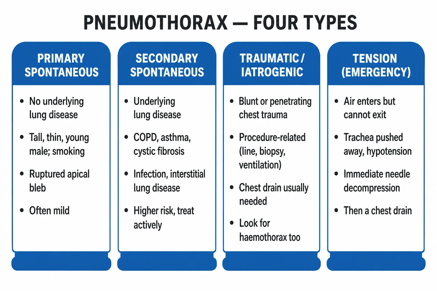
By aetiology — the classification that drives management:[1][2]
- Primary spontaneous pneumothorax (PSP) — occurs without preceding trauma and without clinically apparent underlying lung disease. Classic host: tall, thin, young male (late teens to early 30s), smoker, often with a family history. The rupture site is an apical subpleural bleb or bulla. Smoking multiplies risk several-fold (relative risk up to 20 in heavy smokers); Marfan-habitus, alpha-1-antitrypsin deficiency, and familial forms are recognised.[4]
- Secondary spontaneous pneumothorax (SSP) — occurs without trauma but in established lung disease. By far the commonest cause is COPD; others include asthma, cystic fibrosis, lung abscess, necrotising pneumonia (S. aureus, TB), Pneumocystis jirovecii (in HIV), idiopathic pulmonary fibrosis, Langerhans cell histiocytosis, lymphangioleiomyomatosis (LAM, young women), sarcoidosis, and malignancy. SSP carries higher morbidity because the underlying lung is already compromised and hypoxia is poorly tolerated.[4]
- Traumatic — blunt (rib fracture, deceleration shearing a bleb) or penetrating (stab, gunshot) chest trauma; frequently haemopneumothorax.
- Iatrogenic — a subset of traumatic: central venous catheterisation (subclavian greater than internal jugular), transthoracic needle biopsy, thoracocentesis, transbronchial biopsy, positive-pressure mechanical ventilation/barotrauma (high peak and plateau pressures, ARDS), cardiopulmonary resuscitation, and acupuncture.
- Catamenial pneumothorax — a recurrent SSP in women of reproductive age, right-sided in over 90%, occurring within 72 hours before to 72 hours after the onset of menses; the underlying mechanism is diaphragmatic defects and thoracic endometriosis. Classified as a cause of SSP with a specific hormonal/surgical management pathway.
- Tension pneumothorax — not an aetiology but a complication of any of the above; air accumulates under progressive positive pressure, obstructing venous return. It is the emergency that all pneumothoraces can become, especially in the ventilated patient.
By size — the BTS/UK definition that decides observation versus intervention:[1][9]
- Small — the visible rim of air between the lung edge and the chest wall at the level of the hilum is less than 2 cm (approximately less than 50% of the hemithorax).
- Large — the rim at the hilum is 2 cm or more (approximately 50% or more of the hemithorax). [1]
The American CHEST guideline uses a different and more sensitive cut — the apex-to-cupola distance of 3 cm or more defines a large pneumothorax. The two systems classify a different proportion of patients as large, which matters when comparing studies but not at the bedside: size is interpreted together with symptoms and underlying disease, never in isolation.[9]
Epidemiology & Risk Factors
Pneumothorax is common. PSP has an incidence of roughly 10 to 18 per 100 000 per year in men and 1 to 5 per 100 000 in women, peaking between the late teens and early 30s; SSP is more frequent still (around 20 to 60 per 100 000 per year in men) and increases with age because it tracks the prevalence of COPD. Iatrogenic pneumothorax is the commonest acquired form in hospital.[4]
Risk factors and the type they favour (high-yield): [1]
| Risk factor / host | Type / mechanism to consider |
|---|---|
| Tall, thin, young male | PSP (apical bleb rupture) |
| Smoking (any amount, dose-related) | PSP — multiplies risk up to 20-fold |
| Marfan syndrome, homocystinuria, alpha-1-antitrypsin deficiency | PSP (connective tissue / blebs) |
| COPD / emphysema | SSP — the commonest cause overall |
| Asthma exacerbation | SSP (air-trapping, alveolar rupture) |
| Cystic fibrosis | SSP — high recurrence, high morbidity |
| HIV / Pneumocystis jirovecii | SSP (pneumatocele rupture) — often bilateral |
| Tuberculosis, lung abscess, necrotising pneumonia | SSP |
| LAM (young women), Langerhans cell histiocytosis | SSP — recurrent, may be bilateral |
| Young woman, right-sided, with menses | Catamenial (diaphragmatic endometriosis) |
| Rib fracture / penetrating injury | Traumatic pneumothorax ± haemothorax |
| Central venous catheter, lung biopsy, thoracocentesis | Iatrogenic |
| Mechanical ventilation (high plateau pressure, ARDS) | Barotrauma — high risk of tension |
| Pregnancy / labour | Increased risk of tension in the ventilated or pushing patient |
| Previous ipsilateral pneumothorax | Recurrence — see Prognosis |
| Family history | Familial PSP (Birt-Hogg-Dubé syndrome: folliculin FLCN) |
The tallest, thinnest smokers are the canonical PSP stem in NEET-PG; the elderly COPD patient with sudden worsening dyspnoea is the canonical SSP stem. Remembering the non-COPD SSP causes — LAM, Pneumocystis, Langerhans — separates an above-average answer from an average one.[4]
Pathophysiology
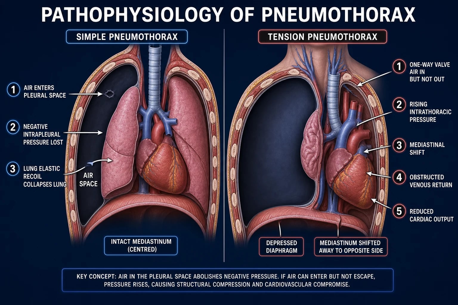
The pleural space normally holds only a few millilitres of fluid and is at negative pressure because the chest wall's tendency to spring outwards is exactly balanced by the lung's tendency to recoil inwards. When air enters — by rupture of a subpleural bleb, by a needle or bullet breaching the parietal pleura, or by alveolar over-distension rupturing into the interstitium and dissecting to the pleura (Macklin effect, the mechanism of barotrauma) — the pressure equalises with the atmosphere and the lung collapses to the extent of the air leak.[4]
Why the patient is hypoxic. Collapsed alveoli are perfused but not ventilated, producing a shunt and ventilation-perfusion mismatch; the larger the pneumothorax, the greater the shunt fraction. There is no primary diffusion defect — the gas-exchange surface of the inflated lung is intact — which is why hypoxaemia in a small PSP may be modest and why supplemental oxygen is so effective.[4]
Why high-flow oxygen accelerates re-expansion. Pleural gas is reabsorbed along a partial-pressure gradient: the sum of gas partial pressures in venous blood (about 660 mmHg total: water vapour 47, CO₂ 46, leaving ~570 for nitrogen plus oxygen) is less than atmospheric, so pleural air is slowly reabsorbed at roughly 1.25% of the volume per day on room air. Breathing high-flow supplemental oxygen lowers the alveolar and hence venous nitrogen partial pressure dramatically (nitrogen washout), widening the gradient and roughly quadrupling the reabsorption rate to about 4% per day — the rationale for oxygen as active treatment of a small pneumothorax. Caution: this is avoided or used with care in patients at risk of CO₂ retention (severe COPD), where controlled oxygen targets SpO₂ 88 to 92%.[1]
Tension pneumothorax — the haemodynamic mechanism. If the pleural breach behaves as a one-way valve (a flap of tissue, or, in the ventilated patient, the constant infusion of positive pressure), air enters on inspiration but cannot escape on expiration. Intrapleural pressure climbs above atmospheric, collapsing the lung entirely, pushing the mediastinum and trachea away from the affected side, depressing the diaphragm, and — critically — compressing the great veins and obstructing venous return, so preload and cardiac output fall. The result is hypotension, tachycardia, distended neck veins (raised systemic venous pressure) and hypoxaemia. Classic teaching emphasised tracheal deviation and hypoxia; the systematic review of Roberts and colleagues shows that in the ventilated patient the dominant features are hypotension and airway-pressure rise, and the textbook triad is frequently absent — so its absence does not exclude tension.[7][8]
Clinical Presentation
Typical spontaneous pneumothorax presents with the sudden onset (often at rest, sometimes during exertion) of ipsilateral pleuritic chest pain and dyspnoea. The pain is sharp, well-localised, and worse on deep inspiration or cough. Cough is usually absent or dry. The patient is often distressed but, in PSP, may tolerate the symptoms surprisingly well; in SSP the breathlessness is out of proportion because the underlying lung has no reserve.[4]
Examination of a moderate-to-large pneumothorax: [1]
- Reduced chest expansion on the affected side (the hemithorax moves less because it is splinted by trapped air).
- Hyper-resonance to percussion (the resonant note of air rather than the dullness of fluid or solid lung).
- Reduced or absent breath sounds and reduced vocal resonance / tactile fremitus on the affected side.
- Occasional subcutaneous emphysema (surgical emphysema) if air has tracked into the soft tissues.
- In tension: tachypnoea, tachycardia, hypotension, cyanosis, distended neck veins, trachea deviated AWAY from the side, and a hyper-resonant hemithorax. [1]
Tension pneumothorax — recognise and act. The bedside picture is of a rapidly deteriorating, hypoxic, hypotensive patient with reduced breath sounds and hyper-resonance on one side, distended neck veins from obstructed venous return, and tracheal deviation away from the affected side. The Roberts systematic review (2015) emphasises two corrections to the textbook picture: (1) the classic triad is often incomplete, particularly in the ventilated patient where rising airway pressures, hypotension and hypoxia may be the only clues; and (2) distended neck veins can be absent in hypovolaemic co-existing bleeding. The practical rule an examiner wants: a deteriorating ventilated patient with unilateral reduced air entry and rising airway pressures is a tension pneumothorax until proven otherwise — decompress.[7][8]
Atypical and special presentations: [1]
- Elderly / COPD patient (SSP) — the dyspnoea may be attributed to an exacerbation; a sudden deterioration out of keeping with the infection, or failure to improve with bronchodilators, should prompt auscultation and a CXR. Hyper-resonance is harder to detect in a hyperinflated emphysematous chest, so the asymmetry of breath sounds is the clue.
- Ventilated / ICU patient — presents as sudden hypotension, hypoxia and a rise in peak airway pressure with reduced chest-wall movement and breath sounds on one side. Tension develops fast and the textbook signs may be masked by sedation; a high index of suspicion saves the patient.[7]
- Catamenial pneumothorax — a woman (typically 30 to 40 years) with recurrent right-sided pneumothorax temporally linked to menses (within 72 hours before to after onset).[4]
- Neonate — sudden deterioration in an infant on continuous positive airway pressure or mechanical ventilation (meconium aspiration, respiratory distress syndrome) with asymmetrical chest movement — suspect pneumothorax.
Differential Diagnosis
Sudden pleuritic chest pain with dyspnoea is a broad differential — and the trap is treating a non-pneumothorax as one (or missing the more dangerous mimic). Distinguish:[4]
- Pulmonary embolism — sudden pleuritic pain, dyspnoea, risk factors (immobility, malignancy, post-operative, pregnancy); often low-grade fever; no hyper-resonance, normal or near-normal breath sounds, evidence of right-heart strain, and a wedge infarct or filling defect on CT pulmonary angiogram. The trap: PE and PSP can both cause sudden dyspnoea with a near-normal CXR.
- Acute coronary syndrome / myocardial infarction — central crushing chest pain (may radiate), diaphoresis, ECG changes, troponin rise; breath sounds are normal and the chest is resonant.
- Aortic dissection — tearing interscapular pain, pulse/BP asymmetry, widened mediastinum on CXR; a life-threatening mimic.
- Pericarditis — pleuritic, positional chest pain relieved by sitting forward, diffuse ST elevation, pericardial rub.
- Pleurisy / pleuritis (viral, autoimmune, pulmonary infarct) — pleuritic pain with a normal or near-normal CXR and normal breath sounds.
- Pneumonia with pleuritic pain — fever, productive cough, signs of consolidation (dullness, bronchial breath sounds, crackles), consolidation on CXR.
- Large emphysematous bulla — the classic imaging mimic of a pneumothorax: it also shows a thin wall with absent markings inside. Distinguishing features: the wall of a bulla is concave inward and parallel vessels may be seen; the pleural line of a pneumothorax is convex outward along the chest wall. Never drain a bulla as if it were a pneumothorax — it creates a bronchopleural fistula. A CT is definitive when there is doubt.
- Pleural effusion — dullness (not hyper-resonance), stony-dull note, meniscus on CXR; haemothorax in trauma.
- Diaphragmatic hernia / rupture — bowel sounds in the chest, loss of the diaphragmatic contour.
- Skin-fold artefact on CXR — the commonest false-positive "pneumothorax": a sharp edge that extends beyond the thorax or crosses ribs, with lung vessels visible beyond the line. Look for the line to extend outside the film and for the absence of a true pleural line. [1]
A can't-miss rule: in any sudden pleuritic chest pain with dyspnoea, consider PE, aortic dissection and tension pneumothorax first — all three are immediately dangerous — and only then move to the more benign mimics. [1]
Clinical & Bedside Assessment
Vital signs drive severity — respiratory rate, oxygen saturation, blood pressure, heart rate, and conscious level. Tachypnoea and tachycardia, hypoxia, and any hypotension move the patient towards intervention, not observation.[4]
Focused respiratory examination — the pneumothorax signs. Inspect for reduced expansion on the affected side and asymmetry of chest movement; palpate for tracheal position (central normally; deviated away in tension; deviated towards in collapse or fibrosis), apex beat, and subcutaneous emphysema (crepitus); percuss for hyper-resonance (compare side-to-side); auscultate for reduced breath sounds and reduced vocal resonance on the affected side. A pleural rub is occasionally heard. [1]
Side localisation — the high-yield bedside rule: the trachea and apex beat are pushed AWAY from the side in a tension pneumothorax (and in a large pleural effusion) and pulled TOWARDS the side in lung collapse (atelectasis, consolidation with collapse) or chronic fibrosis. Combining tracheal deviation with percussion note resolves most cases: hyper-resonance + deviation away = pneumothorax (tension if unwell); dullness + deviation away = effusion; dullness + deviation towards = collapse.[4]
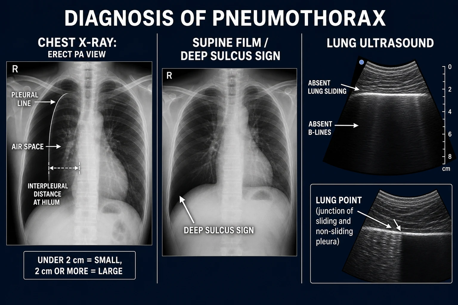
Point-of-care lung ultrasound. At the bedside (and in trauma as part of eFAST), a pneumothorax abolishes two normal artefacts: there is no lung sliding (the to-and-fro movement of the visceral against parietal pleura with respiration) and no B-lines (comet-tail artefacts arising from the pleural line). The lung point — the junction where sliding lung meets non-sliding pleura, seen as a rhythmic appearance and disappearance of sliding with respiration — is highly specific for pneumothorax and allows a size estimate. Absent sliding is sensitive but not specific (pleural adhesion, apneic patient); the lung point is specific but only seen when there is some residual pleural apposition. Ultrasound is most useful in the unstable or trauma patient where an erect CXR is impractical, and may be more sensitive than a supine CXR for an anterior pneumothorax.[4]
When the bedside exam IS the treatment. In suspected tension pneumothorax, the assessment ends with the diagnosis: a hypotensive, tachycardic, distressed patient with unilateral hyper-resonance, reduced breath sounds and (where present) tracheal deviation. Do not leave the bedside for imaging — decompress immediately. [1]
Investigations
First-line: an erect PA chest X-ray (inspiration). The diagnostic finding is a pleural line — the sharp outer edge of the collapsed visceral pleura — with absent lung markings beyond it (the radiolucent air space). Look for mediastinal shift (a clue to tension even on a film) and subcutaneous emphysema.[1]
Sizing the pneumothorax (BTS 2023 / 2010):[1]
- Measure the interpleural distance at the level of the hilum (the horizontal distance from the lung edge to the inner chest wall at the hilum).
- Small = under 2 cm; Large = 2 cm or more. The 2 cm cut approximates a 50% collapse.
- The American CHEST guideline instead measures the apex-to-cupola distance and defines large as 3 cm or more; this classifies more pneumothoraces as large and intervention-worthy. Use whichever your region uses, but always interpret size with symptoms and underlying disease.[9]
Adjuncts and when each test is right: [1]
- Expiratory film — a pneumothorax is easier to see on expiration (the lung is smaller so the air space is relatively larger); occasionally used when the inspiratory film is equivocal. No longer routine if the inspiratory film is clear.
- Supine film — air rises to the anteromedial and subpulmonic recesses in the supine trauma patient, so a supine film may show only a deep, lucent costophrenic sulcus (the deep sulcus sign) or increased lucency over the upper abdomen — a pitfall: a small traumatic pneumothorax is easily missed supine. Ultrasound or CT is more sensitive.
- CT chest — the gold standard; most sensitive for small, loculated or basal pneumothoraces and the definitive test to distinguish a pneumothorax from a bulla, to localise an air leak, to plan surgery, and in complex trauma. Not routine for straightforward PSP.
- Arterial blood gas — to quantify hypoxaemia and hypercapnia in the breathless or SSP patient; pulse oximetry is the bedside surrogate. Hypoxia drives intervention.
- Lung ultrasound — see bedside assessment; no lung sliding + absent B-lines, with the lung point as the specific sign. [1]
Scores and criteria reproduced: pneumothorax has no dedicated prognostic score (unlike CURB-65 for pneumonia). Severity is judged by symptoms, size, underlying disease and physiological derangement (hypoxia, hypotension) — the inputs to the BTS management algorithm. When a pleural effusion coexists and needs characterising, apply Light's criteria for exudate (pleural-fluid/serum protein ratio over 0.5, OR LDH ratio over 0.6, OR fluid LDH over two-thirds the upper limit of normal).[1]
Management — Resuscitation
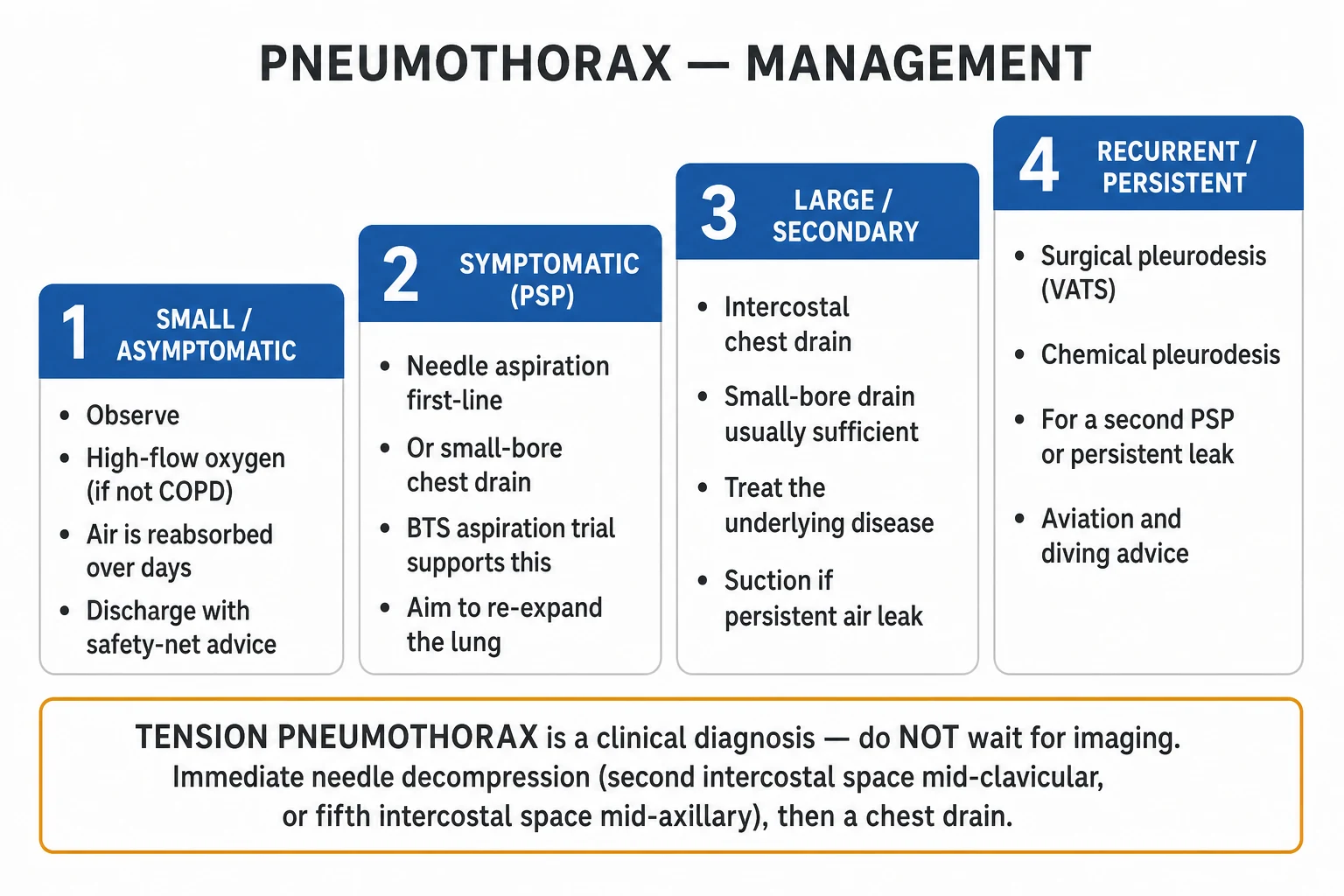
The single resuscitation rule: tension is a clinical diagnosis — decompress before imaging.[1][7]
Immediate needle decompression for suspected tension: [1]
- Site: the 2nd intercostal space, mid-clavicular line on the affected side (anterior approach) — the classical landmark. Current trauma guidance (and the BTS) increasingly favour the 5th intercostal space, anterior to the mid-axillary line (the same "safe triangle" used for a chest drain) because the chest wall is thinner there, the vessels and breast tissue are avoided, and shorter-standard needles reach the pleura; chest-wall thickness in the 2nd ICS often exceeds the length of a standard cannula.
- Device: a large-bore needle/cannula (classically 14 to 16G), but standard devices are frequently too short for the 2nd ICS in adults; modern pre-hospital practice uses a 3rd-or-4th-generation longer fenestrated catheter (e.g., 3.5 inch / 8 cm, 10G) to reach the pleura reliably and reduce kinking and blockage.
- Technique: aspirate air to confirm intrapleural placement; the rush of air confirms tension. Convert to a definitive intercostal chest drain as soon as possible — needle decompression is a bridge, not a treatment, and can kink, dislodge or block.
- In the ventilated patient, where the tension can reaccumulate instantly, proceed straight to a thoracostomy (an open intercostal incision left to air, or a formal tube) rather than relying on a needle. [1]
Airway and breathing in the unstable patient: high-flow oxygen (15 L/min via a non-rebreather mask, or target SpO₂ 94 to 98%, or 88 to 92% in COPD/CO₂-retainers), IV access, and treat the haemodynamic collapse with cautious fluids while the decompression is performed — the cause is mechanical and is corrected only by releasing the air. Reassess immediately after decompression: a patient who fails to improve has either misdiagnosis (e.g., a misplaced needle, or the wrong side) or a blocked/kinked device.[7]
For a non-tension pneumothorax in a deteriorating or ventilated patient: do not delay — a ventilated patient with a pneumothorax is one breath away from tension; insert a chest drain proactively.[1]
Management — Definitive & Stepwise
Management follows the BTS 2023 guideline (the 2010 guideline with 2023 refinement). Two axes decide treatment: type (PSP vs SSP vs traumatic/iatrogenic) and size and symptoms.[1][2]
Small PSP (under 2 cm, asymptomatic)
- Discharge with safety-net advice and review in 2 to 4 weeks
- High-flow supplemental oxygen if admitted and not CO₂-retainer, to speed reabsorption (nitrogen washout)
- Avoid air travel until full resolution; never dive until cleared
- Conservative (ambulatory) management now supported by the NAPPE/Wait-PT trials — many small PSPs resolve without intervention
Large or symptomatic PSP (≥2 cm or breathless)
- First-line: simple needle aspiration — 16G cannula in 2nd ICS MCL, aspirate up to 2.5 L
- Alternative / if aspiration fails: small-bore (8 to 14 Fr) Seldinger intercostal drain on underwater seal
- Admit for observation; assess for re-expansion
- Refer for surgery if recurrent, persistent air leak, or high-risk occupation
Secondary spontaneous (SSP, any size)
- Always admit — conservative management is unsafe in compromised lungs
- Intercostal chest drain (small-bore usually sufficient; large-bore if copious air or haemothorax)
- High-flow oxygen unless CO₂-retainer (then 88 to 92%)
- Lower threshold for surgical pleurodesis after first episode (high recurrence)
Tension (clinical diagnosis)
- Immediate needle decompression (2nd ICS MCL or 5th ICS mid-axillary line) — do NOT image first
- Convert at once to a definitive intercostal chest drain on underwater seal
- IV access, oxygen, treat the cardiovascular collapse
- Post-decompression CXR to confirm re-expansion and exclude haemothorax
Stepwise detail. [1]
1. Observation / conservative management. For a small (under 2 cm), asymptomatic PSP (and selected SSP that are tiny and stable), observe in the emergency department for a few hours; if the patient remains comfortable and not hypoxic, discharge with safety-net advice (return immediately if worsening breathlessness) and outpatient review in 2 to 4 weeks to confirm radiological resolution. Recent evidence (the NAPPE and Wait-PT randomised trials, summarised in the 2024 NEJM review) supports conservative management even of larger PSPs in stable patients — many resolve without intervention — though BTS still recommends intervention for the large or symptomatic PSP pending wider guideline adoption.[4]
2. High-flow oxygen (agent: supplemental oxygen; dose: 10 to 15 L/min via non-rebreather mask to target SpO₂ 94 to 98%; route: inhaled; timing: from presentation; rationale: nitrogen washout accelerates pleural air reabsorption roughly fourfold). Avoid or use with close monitoring in patients at risk of CO₂ retention (severe COPD — target SpO₂ 88 to 92%).[1]
3. Simple needle aspiration — first-line for a large or symptomatic PSP and acceptable for some SSP. Technique: insert a 16G cannula into the 2nd intercostal space, mid-clavicular line, on the affected side; aspirate using a three-way tap and 50 mL syringe or a one-way valve; aspirate up to 2.5 L (stop earlier if resistance or coughing — the lung has re-expanded). Success is defined as symptomatic relief plus radiological re-expansion; the Harvey and Prescott BTS RCT (1994) established that aspiration is at least as effective as intercostal tube drainage for initial PSP, with shorter stays. If aspiration fails to re-expand, proceed to a chest drain. Aspiration is less successful in SSP (underlying lung disease limits re-expansion).[3]
4. Intercostal chest drain — indicated for SSP of any size that is symptomatic or large, traumatic pneumothorax, a failed aspiration, a tension pneumothorax after decompression, and any pneumothorax in a ventilated patient. Insert through the safe triangle (bounded by the anterior border of latissimus dorsi, the lateral border of pectoralis major, a line above the 5th intercostal space, and the apex below the axilla) — i.e., the 4th to 5th intercostal space, anterior to the mid-axillary line. A small-bore (8 to 14 Fr) Seldinger drain is sufficient for most simple pneumothoraces and is more comfortable; a large-bore (20 to 32 Fr) drain is used for haemothorax, copious air leak, or when a small drain has failed. Connect to an underwater seal (the drain tip sits 2 cm below the water level, providing a one-way valve).[1]
5. Reading the underwater seal. Swinging (tidalling) — the water column moves with respiration (down with inspiration in spontaneous breathing), confirming the system is patent and connected to the pleural space. Bubbling — air is leaving the pleural space; a continuous bubbling indicates an ongoing air leak (bronchopleural fistula); intermittent bubbling with cough/respiration is normal as the lung re-expands. No bubbling with the lung re-expanded = the leak has sealed; clamp and assess (water-seal trial) before removal. [1]
6. Persistent air leak and suction. If the lung fails to re-expand or an air leak persists beyond 2 to 3 days, apply low-pressure, high-volume suction at -10 cmH₂O (e.g., a high-volume low-pressure system) to encourage apposition and sealing. A leak persisting beyond 3 to 5 days (some units 5 days) is an indication for surgical referral.[1]
7. Surgical and chemical pleurodesis — indicated for: second ipsilateral PSP (recurrence), first contralateral PSP, persistent air leak beyond 3 to 5 days, SSP at first presentation (high recurrence — lower threshold), bilateral pneumothorax, and patients in high-risk occupations (pilots, divers) after a first event. Surgical pleurodesis by video-assisted thoracoscopic surgery (VATS) — bullectomy of apical blebs plus mechanical/abrasive pleurodesis — is the gold standard, with recurrence under 5%. Chemical pleurodesis (intravenous talc slurry, or doxycycline/minocycline or povidone-iodine) through the drain obliterates the pleural space and is an option for patients unfit for surgery; the Chen (2013) Lancet RCT showed that minocycline pleurodesis after simple aspiration reduced PSP recurrence compared with aspiration alone.[5][6]
8. Drain removal. Once the lung is fully re-expanded and there is no bubbling for 24 hours (with the lung expanded on clamping and on subsequent CXR), remove the drain during a Valsalva manoeuvre (forced expiration against a closed glottis) to prevent air re-entry; close with a purse-string or occlusive dressing. Prophylactic antibiotics are not routinely required for elective drains in PSP but are used for traumatic drains.[1]
9. Discharge, follow-up, and lifestyle advice. Advise: no air travel until the pneumothorax has fully resolved on CXR (cabin pressure expands the gas — risk of tension); never scuba dive until definitive surgery (pleurodesis) and specialist clearance; stop smoking (halves recurrence). Follow-up CXR at 2 to 4 weeks to confirm resolution; recurrent or non-resolving cases to respiratory/thoracic surgery.[1]
Specific Subtypes & Scenarios
- Primary spontaneous pneumothorax (PSP) — managed as above; the aim is symptom relief and preventing recurrence. The apical subpleural bleb is the rupture site in most; CT is not routine before a first episode but is used for recurrent or complex disease.
- Secondary spontaneous pneumothorax (SSP) — because the underlying lung is compromised, admit all, drain (even small ones, if symptomatic or over 1 to 2 cm), give supplemental oxygen, and treat the underlying disease. Recurrence is higher and the threshold for pleurodesis at first presentation is lower.
- Tension pneumothorax — clinical diagnosis; immediate decompression then a definitive drain (see Resuscitation). Survivable if recognised, fatal if missed.
- Traumatic pneumothorax — exclude haemothorax (dullness, blood in the drain, dropping haemoglobin) and other thoracic injury (flail chest, cardiac tamponade, great-vessel injury); a large-bore chest drain is usual. A small, asymptomatic, occult traumatic pneumothorax found only on CT may be observed if the patient is not ventilated, but any ventilated patient with a traumatic pneumothorax must be drained because positive pressure converts it to tension.
- Iatrogenic pneumothorax — commonest from central venous catheterisation (use ultrasound, prefer the internal jugular route, and avoid the apex of the lung). Small, asymptomatic iatrogenic pneumothoraces can be observed with oxygen; symptomatic or enlarging ones need a small-bore drain.
- Barotrauma in the ventilated patient — high peak/plateau pressures and ARDS rupture alveoli via the Macklin effect; presents as sudden deterioration with rising airway pressures — decompress immediately and insert a chest drain; thereafter use lung-protective ventilation (low tidal volume 6 mL/kg ideal body weight, plateau pressure under 30 cmH₂O) to prevent recurrence.
- Catamenial pneumothorax — right-sided, perimenstrual; basis is diaphragmatic defects through which intra-abdominal gas and endometrial tissue pass. Management combines mechanical pleurodesis + diaphragmatic repair at VATS with hormonal suppression (GnRH analogue or combined oral contraceptive) to prevent recurrence.
- Pneumothorax in the ventilated / ICU patient — high risk of rapid progression to tension; insert a chest drain proactively rather than observing; ventilate with lung-protective settings.
- Hydropneumothorax / haemopneumothorax / pyopneumothorax — drain with a large-bore tube; in haemothorax, autotransfusion may be considered and a thoracic surgeon is involved if drainage exceeds 1500 mL acutely or over 200 mL/hour. [1]
Complications & Pitfalls
Disease-related: [1]
- Recurrence — the dominant long-term complication. PSP recurs in roughly 30% after a first episode (and again in 30% after a second); SSP recurs more often and more dangerously. Recurrence drives the indication for pleurodesis (after a second ipsilateral PSP, a first contralateral PSP, or an SSP at first presentation). Smoking cessation halves recurrence.[5]
- Persistent air leak / bronchopleural fistula — an ongoing communication between bronchus and pleura; managed with suction, then surgical repair/pleurodesis if not sealed by 3 to 5 days.
- Re-expansion pulmonary oedema — a rare but potentially fatal complication of rapid re-expansion of a large, chronically collapsed lung; presents as cough, frothy sputum and hypoxia within 24 hours of drainage. Prevention: do not drain large pneumothoraces too rapidly, avoid routine suction on the first drain, and observe after aspiration.[4]
Procedure-related (drain / decompression): [1]
- Pain, infection (cellulitis at the site), and empyema — sterile technique and (for traumatic drains) prophylactic antibiotics reduce this.
- Blockage, kinking, or dislodgement of the drain — the underwater seal stops swinging; never leave a non-swinging drain unassessed.
- Re-expansion pulmonary oedema (above) from over-rapid drainage.
- Injury to intercostal vessels or nerve (neurovascular bundle runs below each rib — always insert above a rib, never below), to the lung, or to solid organs; chronic intercostal neuralgia after surgery.
- Surgical emphysema — air in the soft tissues; usually benign but can be extensive if the drain is blocked or the tract is large. [1]
Classic pitfalls: [1]
- Misdiagnosing a bulla as a pneumothorax and draining it → iatrogenic bronchopleural fistula. Always check it is a true pleural line; CT if any doubt.[4]
- Waiting for a CXR in suspected tension — the classic fatal error. Tension is a clinical diagnosis; decompress first.[7]
- Inserting the drain below a rib (into the neurovascular bundle) — always go above the rib.
- Wrong-site decompression — confirm the side with the hyper-resonant, silent hemithorax; document the side before acting.
- Over-rapid re-expansion causing pulmonary oedema.
- Missing a small traumatic pneumothorax on a supine film (look for the deep sulcus sign; use ultrasound/CT).[4]
- Discharging a patient to fly or dive before full resolution / surgical clearance.
- Failing to drain a ventilated patient's pneumothorax → imminent tension.[7]
Prognosis & Disposition
Primary spontaneous pneumothorax usually resolves with aspiration or a small drain, but recurs in about 30% — after which the recurrence rate climbs further, justifying pleurodesis after a second ipsilateral event (and for high-risk occupations after a first). Smoking cessation roughly halves recurrence.[5]
Secondary spontaneous pneumothorax has higher morbidity and recurrence tied to the underlying disease; patients are older and less fit and tolerate hypoxia poorly. Disposition is admission for all but the very smallest, most stable cases.[4]
Tension pneumothorax is rapidly fatal if untreated (obstructive shock and cardiac arrest) but immediately and dramatically reversible with decompression — survival depends entirely on prompt recognition.[7]
Radiological resolution with conservative management proceeds at roughly 1 to 2% of the hemithorax per day on room air and faster with oxygen; follow-up CXR at 2 to 4 weeks confirms resolution. Disposition: PSP that is small and asymptomatic → discharge with safety-net; large/symptomatic PSP → admit for aspiration or drain; SSP, traumatic, ventilated, or tension → admit, drain, monitor. No air travel until the CXR is clear; no diving ever (after pneumothorax) without definitive surgery.[1]
Special Populations
- Pregnancy and labour — pneumothorax in pregnancy is rare but the risk of tension rises in labour (Valsalva, and positive pressure if requiring anaesthesia). Manage with aspiration or a small-bore drain, avoiding talc pleurodesis where possible; involve obstetric and thoracic teams. Avoid delaying decompression for any reason in suspected tension.
- Catamenial pneumothorax — women of reproductive age with recurrent right-sided perimenstrual pneumothorax; treat with VATS pleurodesis plus diaphragmatic repair and hormonal suppression.
- Paediatrics / neonates — neonatal pneumothorax complicates meconium aspiration, respiratory distress syndrome, and mechanical ventilation/CPAP; presents as sudden deterioration with asymmetrical chest movement. Needle decompression in a neonate uses a 22 to 24G cannula in the 2nd ICS MCL or 4th ICS mid-axillary line, followed by a small chest drain; size all equipment to weight. Spontaneous pneumothorax in older children follows the same algorithm as adults.
- The anticoagulated patient — drainage risks haemothorax; assess the bleeding risk versus urgency (tension overrides everything), correct or reverse anticoagulation (e.g., warfarin with vitamin K and prothrombin complex concentrate; DOACs according to agent and timing), and use a smaller drain where possible with close monitoring of drain output and haemoglobin.
- HIV / Pneumocystis and LAM — important non-COPD causes of SSP: Pneumocystis jirovecii pneumothorax (from pneumatocele rupture) is often bilateral, recurrent and difficult; lymphangioleiomyomatosis (young women, recurrent, may lead to lung transplant). Recognise the underlying cause to plan definitive management. [1]
Evidence, Guidelines & Regional Differences
BTS Pleural Disease Guideline (2010, MacDuff; 2023, Roberts) — the most influential UK guideline, defining the 2 cm size cut and the observe / aspirate / drain / surgery algorithm. The 2023 update refined indications, endorses small-bore Seldinger drains as first-line for most, and incorporates emerging evidence for conservative (ambulatory) management of larger PSPs.[1][2]
American CHEST guideline — uses the apex-to-cupola 3 cm cut for a large pneumothorax and is more interventionist for SSP; the Nikolić statistical comparison showed the two systems classify meaningfully different proportions as large, with consequent differences in management — so the guideline you quote matters in the exam.[9]
Landmark trials: [1]
- Harvey and Prescott (1994, BMJ) — BTS multicentre RCT: simple aspiration is at least as effective as intercostal tube drainage for initial PSP, with shorter hospital stay — the basis for aspiration as first-line for PSP.[3]
- Chen (2013, Lancet) — adding intrapleural minocycline pleurodesis after simple aspiration and drainage reduced PSP recurrence versus aspiration/drainage alone.[6]
- Sadikot (1997, Thorax) — defined the ~30% recurrence rate of PSP and its risk factors (smoking, taller stature), underpinning the threshold for pleurodesis.[5]
- Roberts DJ (2015, Annals of Surgery) — systematic review of tension pneumothorax presentation: the classic triad is often incomplete, especially in the ventilated patient; hypotension and airway-pressure rise dominate.[7]
- NAPPE (Boulay 2014) and Wait-PT (Brown 2020) trials — randomised evidence that conservative management of large PSPs in stable patients is non-inferior to interventional management and avoids procedures — summarised in the Smyth (2024) NEJM review, which represents the current state of the evidence.[4]
Regional deltas: [1]
- UK (BTS 2023) — 2 cm size cut; aspiration or small-bore drain first-line for PSP; conservative management increasingly accepted for stable larger PSPs.[2]
- US (CHEST / ACCP) — apex-to-cupola 3 cm cut; catheter aspiration and small-bore pigtail catheters widely used.[9]
- India (NMC / ICMR / ATG) — empirical practice follows BTS principles with limited access to VATS outside tertiary centres; chemical pleurodesis (talc, povidone-iodine) is used more widely where surgery is not accessible; smoking-related and TB-related SSP dominate, so always consider TB as a cause of SSP.[10]
- Tension decompression site — historically 2nd ICS MCL; trauma and military guidance now favour the 5th ICS mid-axillary line with a longer fenestrated catheter because chest-wall thickness defeats shorter needles — an evolving area.[7]
Exam Pearls
TENSION pneumothorax — the bedside six
TENSION
trachea and apex beat pushed away from the affected side
hyper-resonant, reduced movement on the affected side
reduced/absent breath sounds and vocal resonance
hypotension, tachycardia, cyanosis from obstructed venous return
resuscitate while you decompress; treat is mechanical
air in but not out — rising intrathoracic pressure; decompress BEFORE imaging
immediate needle decompression (2nd ICS MCL or 5th ICS mid-axillary), then a definitive chest drain
Pneumothorax — the numbers that decide an answer
- "Pneumothorax = air in the pleural space; sudden pleuritic pain, dyspnoea, hyper-resonance, reduced breath sounds."
- "PSP = tall, thin, young, male, smoker, apical bleb; SSP = COPD (commonest), asthma, CF, Pneumocystis, TB, LAM."
- "Tension is a CLINICAL diagnosis — decompress BEFORE imaging (2nd ICS MCL or 5th ICS mid-axillary line), then a formal chest drain."
- "Trachea deviated AWAY in tension (and large effusion); TOWARDS the side in collapse or fibrosis."
- "BTS size cut: 2 cm rim at hilum = large; CHEST cut: 3 cm apex-to-cupola."
- "PSP — observe/aspirate/small-bore drain; SSP — drain even if small; recurrent/persistent — surgical pleurodesis (VATS)."[6]
- "High-flow oxygen speeds reabsorption ~4× by nitrogen washout — avoid in CO₂-retainers (target 88 to 92%)."
- "Re-expansion pulmonary oedema — do not drain large pneumothoraces too fast."
- "Catamenial pneumothorax — right-sided, within 72 h of menses, diaphragmatic endometriosis."
- "Lung ultrasound — absent lung sliding + absent B-lines; the lung point is specific."
- "Never drain a bulla as a pneumothorax — it causes a bronchopleural fistula; CT if any doubt."
- "No flying until CXR clear; no diving ever (after pneumothorax) without definitive surgery."[4]
Exam application bank (NEET-PG / INICET)
One-line answer
Pneumothorax is air in the pleural space. Loss of the normal negative intrapleural pressure uncouples the lung from the chest wall, and elastic recoil collapses the lung. It is classified as primary spontaneous (PSP — no underlying lung disease; tall, thin, young male smoker, ruptured apical subpleural bleb), secondary spontaneous (SSP — underlying disease: COPD, asthma, cystic fibrosis, Pneumocystis, TB, LAM), traumatic/iatrogenic, and the life-threatening tension pneumothorax (a one-way valve raising intrathoracic pressure until venous return and cardiac output collapse — hypotension, tracheal deviation away, distended neck veins). Diagnosis is clinical plus an erect chest X-ray (pleural line, absent lung markings), except tension which is a clinical diagnosis — decompress before imaging. Management is size- and type-driven per the BTS 2023 guideline: small/asymptomatic PSP — observe p
Worked stems (answer without another resource)
Stem 1 — Classic presentation. Map symptoms to mechanism; name the first investigation and first treatment step with dose/route if drug therapy is standard. [1]
Stem 2 — Unstable / complicated. List red flags that force immediate resuscitation, theatre, ICU, antidote, or reperfusion — and what you do in the first 15 minutes. [1]
Stem 3 — Atypical group. Elderly, pregnancy, child, or immunocompromised: how presentation and thresholds change. [1]
Stem 4 — Differential trap. Name the three closest mimics and one discriminator for each. [1]
Stem 5 — Disposition. Who goes home with safety-netting, who is admitted, who needs HDU/ICU/theatre, and what follow-up is mandatory. [1]
Rapid viva checklist
- Definition + classification
- Pathophysiology chain
- Bedside signs / criteria
- Score with exact components (if any)
- Emergency bundle
- Definitive therapy with doses
- Complications of disease and of treatment
- Special populations
- Guideline/trial name if classic
- Three exam traps
Coverage self-check
If you cannot answer any stem above from this page alone, re-read the matching section — the page is intended to be self-sufficient for final-prof and NEET-PG/INICET questions on Pneumothorax.
References
- [1]MacDuff A, Arnold A, Harvey J, et al. Management of spontaneous pneumothorax: British Thoracic Society Pleural Disease Guideline 2010 Thorax, 2010.PMID 20696690
- [2]Roberts ME, Rahman NM, Maskell NA, et al. British Thoracic Society Guideline for pleural disease Thorax, 2023.PMID 37553157
- [3]Harvey J, Prescott RJ, British Thoracic Society Research Committee. Simple aspiration versus intercostal tube drainage for spontaneous pneumothorax in patients with normal lungs. British Thoracic Society Research Committee BMJ, 1994.PMID 7755720
- [4]Smyth R, Bagga M, Chaudhuri N, et al. Primary Spontaneous Pneumothorax N Engl J Med, 2024.PMID 38354148
- [5]Sadikot RT, Greene T, Meadows K, Arnold AG. Recurrence of primary spontaneous pneumothorax Thorax, 1997.PMID 9371212
- [6]Chen JS, Chan WK, Tsai KT, et al. Simple aspiration and drainage and intrapleural minocycline pleurodesis versus simple aspiration and drainage for the initial treatment of primary spontaneous pneumothorax: an open-label, parallel-group, prospective, randomised, controlled trial Lancet, 2013.PMID 23489754
- [7]Roberts DJ, Leigh-Smith S, Faris PD, et al. Clinical Presentation of Patients With Tension Pneumothorax: A Systematic Review Ann Surg, 2015.PMID 25563887
- [8]Leigh-Smith S, Harris T. Spontaneously breathing patients get tension pneumothoraces Thorax, 2012.PMID 22286929
- [9]Nikolić MZ, Lokhandwala T, Tanner NT, et al. Noninterventional statistical comparison of BTS and CHEST guidelines for size and severity in primary pneumothorax Eur Respir J, 2015.PMID 25792629
- [10]Cheng HS, Yeo JG, Tan CW, et al. Management of spontaneous pneumothorax: a mini-review on its latest evidence J Thorac Dis, 2024.PMID 39144356