Respiratory · General Medicine
Respiratory Failure
Also known as Respiratory failure · Type 1 respiratory failure · Type 2 respiratory failure · Acute hypercapnic respiratory failure · Hypoxaemic respiratory failure · Ventilatory failure
Respiratory failure is the failure of the respiratory system to maintain adequate gas exchange, defined by the arterial blood gas: PaO2 below 8 kPa (60 mmHg) at room air, with or without a raised PaCO2. Type 1 (hypoxaemic) is low oxygen with normal or low CO2, caused by ventilation-perfusion mismatch or shunt — pneumonia, pulmonary embolism, pulmonary oedema, ARDS, asthma. Type 2 (hypercapnic) is low oxygen AND high CO2 (PaCO2 above 6 kPa / 45 mmHg), caused by alveolar hypoventilation — COPD, neuromuscular disease, opiates, obesity hypoventilation, brainstem depression. The ABG diagnoses it and the type drives treatment. Management is oxygen (target SpO2 94–98% for type 1; 88–92% for COPD/type 2 to avoid CO2 narcosis), non-invasive ventilation (BiPAP) for type 2 with acidosis, CPAP/HFNC for refractory type 1 hypoxia, intubation if NIV fails or the airway is threatened, lung-protective ventilation in ARDS, and treatment of the underlying cause.
On this page & tools
Your progress
Saved locally on this device.
Exam tags
Red flags
Overview & Definition
Respiratory failure is defined by the arterial blood gas as the inability of the respiratory system to maintain adequate gas exchange: a PaO2 below 60 mmHg (8 kPa) while breathing room air, with or without a raised PaCO2. A PaCO2 above 45 mmHg (6 kPa) defines hypercapnia.[1]
The clinical art of respiratory failure is not the diagnosis (that is the ABG) but three judgements in succession: (1) is it Type 1 or Type 2 — because this determines the oxygen target and the need for ventilatory support; (2) what is the underlying cause — because oxygen and ventilation buy time but do not cure; and (3) is the patient fatiguing — because the fatiguing patient is minutes from apnoea and must be intubated before arrest.[1][2]
Respiratory failure is the final common pathway of almost any severe disease of the lung, the airways, the chest wall, the respiratory muscles, the neuromuscular junction, the nerves, or the central nervous system. It is one of the commonest reasons for ICU admission worldwide and a leading cause of in-hospital death. [1]
Classification
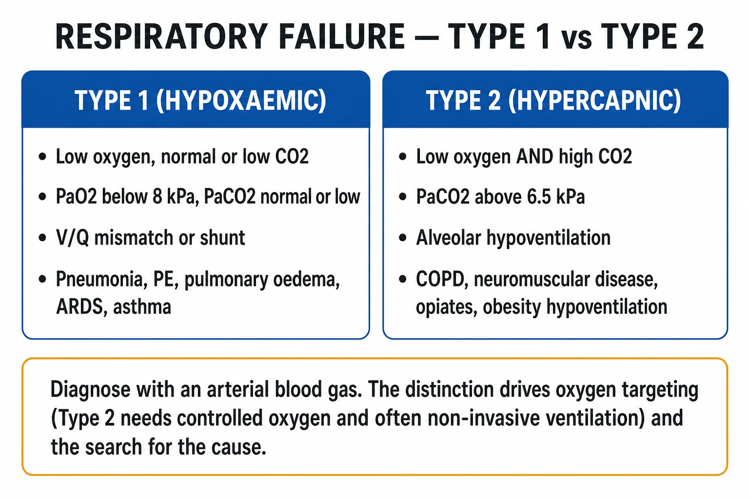
By blood gas — the classification that matters clinically:[1]
- Type 1 (hypoxaemic) — PaO2 below 60 mmHg (8 kPa) with normal or low PaCO2. Mechanism V/Q mismatch or shunt. Causes: pneumonia, ARDS, pulmonary oedema, pulmonary embolism, asthma (early), pneumothorax, interstitial lung disease, pulmonary haemorrhage.
- Type 2 (hypercapnic / ventilatory) — PaO2 below 60 mmHg (8 kPa) with PaCO2 above 45 mmHg (6 kPa). Mechanism alveolar hypoventilation (the lung itself may be normal). Causes: COPD exacerbation, neuromuscular weakness (Guillain–Barré, myasthenia, MND), obesity hypoventilation, opiate/sedative overdose, brainstem stroke, severe kyphoscoliosis, fatigue/end-stage respiratory failure. [1]
By time course: [1]
- Acute — sudden onset, no renal compensation (normal HCO3), pH falls quickly with rising CO2. The dangerous state.
- Chronic — slow onset, renal bicarbonate retention compensates, pH near-normal despite very high PaCO2 (the chronic COPD patient with PaCO2 8 kPa and HCO3 36).
- Acute-on-chronic — a chronically hypercapnic patient decompensates (e.g., a COPD exacerbation; HCO3 elevated but pH acidaemic) — the commonest hospital presentation. The most important bedside skill here is reading the pH: a normal/near-normal pH means chronic compensated hypercapnia; a low pH means acute decompensation that needs immediate support. [1]
By mechanism (the modern framework): [1]
- Oxygenation failure — the lung cannot transfer oxygen (Type 1).
- Ventilatory (pump) failure — the pump cannot move air, so CO2 accumulates (Type 2). The "pump" is the entire chain: CNS drive → spinal cord → phrenic and intercostal nerves → neuromuscular junction → respiratory muscles → chest wall → airways → alveoli. [1]
The extended Type 3 / Type 4 scheme (used in some texts) covers: Type 3 — perioperative / atelectasis-related hypoxaemia; Type 4 — shock / low perfusion with mixed venous desaturation. These are subsets of the same gas-exchange problem and are managed within the Type 1/2 framework. [1]
Epidemiology & Risk Factors
Respiratory failure is among the commonest reasons for ICU admission in adults. It is a syndrome, not a disease, so its epidemiology tracks the underlying causes. COPD is the single commonest cause of acute-on-chronic Type 2 respiratory failure presenting to hospital; pneumonia, cardiogenic pulmonary oedema, and ARDS dominate Type 1.[1]
Risk factors for developing respiratory failure: [1]
- Chronic lung disease — COPD, asthma, bronchiectasis, cystic fibrosis, interstitial lung disease.
- Smoking — the dominant modifiable risk factor for COPD and lung cancer.
- Immunocompromise — HIV, transplant, chemotherapy, biologics (broad opportunistic differential: PJP, CMV, fungal).
- Obesity — obesity hypoventilation syndrome, OSA overlap, restrictive physiology.
- Sedatives and opiates — the rising epidemic cause of Type 2 failure from CNS depression.
- Neuromuscular disease — GBS, myasthenia gravis, motor neuron disease, muscular dystrophy.
- Post-operative state — atelectasis, splinting, residual anaesthesia/opioid.
- Age extremes — neonatal immature lungs and immature immunity; elderly blunted respiratory drive and comorbidity.
- Aspiration risk — reduced GCS, dysphagia, stroke, seizure, alcohol. [1]
Pathophysiology
The five mechanisms of hypoxaemia
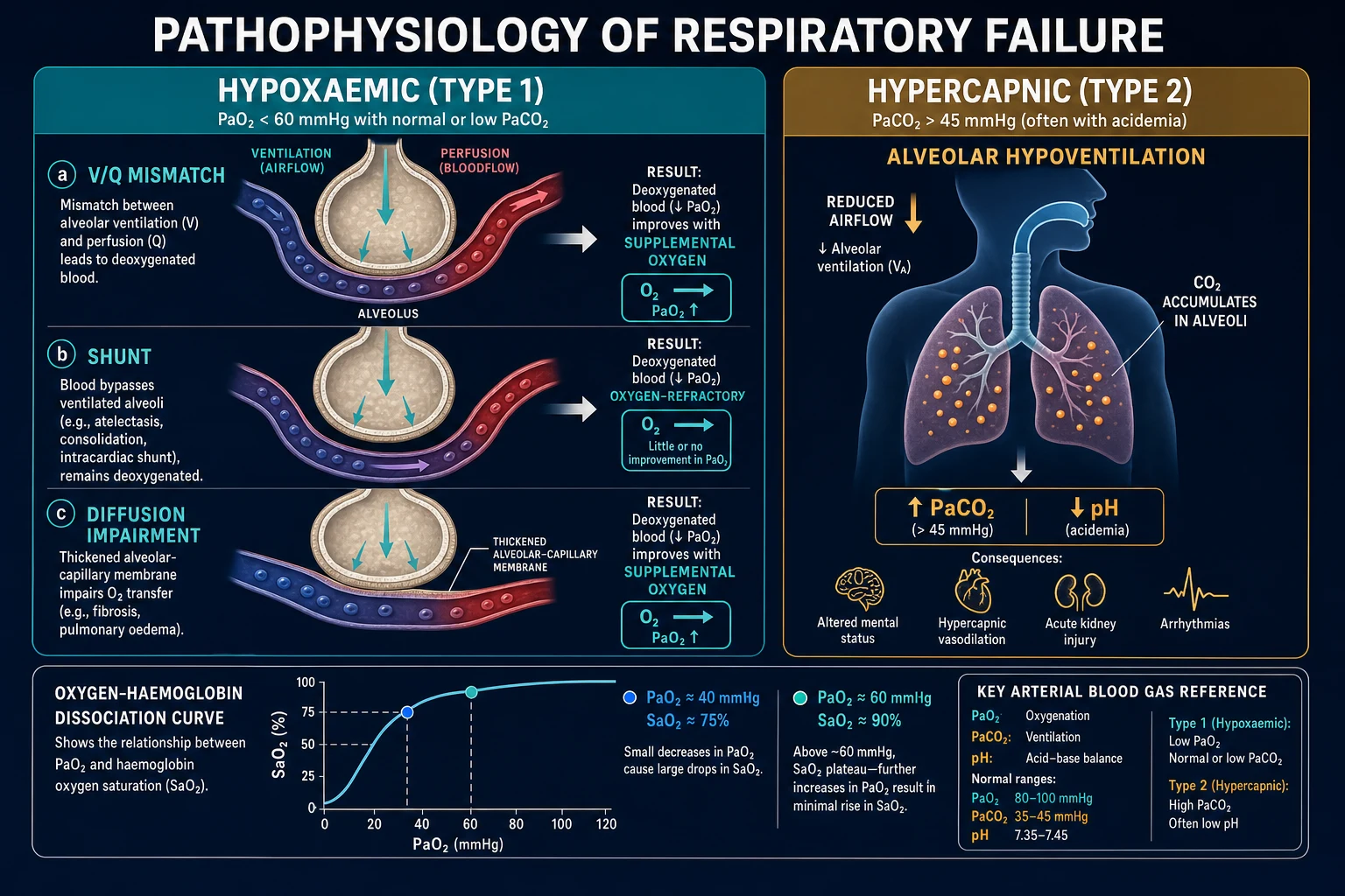
Every cause of hypoxaemia reduces to one (or more) of five mechanisms, and whether each responds to supplemental oxygen is the single most exam-worthy fact about them. [1]
Hypoventilation
- PaCO2 rises; alveolar gas equation lowers PAO2.
- A-a gradient is NORMAL — the lung itself is fine.
- Always corrects with oxygen, but the cause (CNS depression, neuromuscular, fatigue) must be fixed or ventilation supported.
- Causes: opiates, sedatives, brainstem stroke, neuromuscular weakness, obesity hypoventilation.
V/Q mismatch
- The commonest mechanism in disease.
- Blood perfuses under-ventilated alveoli (pneumonia, oedema, asthma, COPD, PE).
- Largely CORRECTS with supplemental oxygen (excess O2 in well-ventilated units compensates).
- The mechanism behind most Type 1 failure and most COPD exacerbations.
Shunt
- Blood bypasses ventilated alveoli entirely (ARDS, lobar pneumonia, pulmonary AV malformation, R-to-L cardiac shunt).
- Does NOT correct with 100% oxygen — the blood never sees the alveolus.
- The bedside test: refractory hypoxaemia on high FiO2 suggests shunt.
- Mechanism of severe ARDS and the refractory hypoxaemia of hepatopulmonary syndrome.
Diffusion impairment
- Thickened alveolar-capillary membrane (ILD, pulmonary fibrosis, pulmonary oedema).
- Corrects with supplemental oxygen (raises the gradient).
- Often worse on exercise (shorter capillary transit time).
Low inspired PaO2
- Altitude, hypoxic gas mixture, rebreathing.
- A-a gradient is normal.
- Corrects by restoring FiO2 (descent, supplemental oxygen).
Normal arterial blood gas values (room air, sea level)
The alveolar gas equation — why CO2 controls oxygen
The single most useful equation at the bedside is the alveolar gas equation: [1]
PAO2 = FiO2 × (Patm − PH₂O) − PaCO2 / RQ [1]
On room air at sea level this simplifies to PAO2 ≈ 150 − PaCO2/0.8. Two exam-worthy consequences: [1]
- Rising PaCO2 directly lowers alveolar (and therefore arterial) oxygen. A patient hypoventilating to a PaCO2 of 80 mmHg has a PAO2 of only 150 − 100 = 50 mmHg before any lung disease is considered. This is why Type 2 failure is always hypoxaemic.
- The A-a gradient (PAO2 − PaO2) tells you whether the hypoxaemia is purely from hypoventilation (A-a normal, lung fine) or from lung disease (A-a raised). Calculate it on every ABG. [1]
The A-a gradient (PAO2 − PaO2)
Oxygen-induced hypercapnia — the Haldane effect and loss of HPV
Giving high-flow oxygen to a CO2 retainer (COPD, obesity hypoventilation) worsens hypercapnia by two mechanisms:[1]
- The Haldane effect — deoxyhaemoglobin carries CO2 (as carbamino compounds) and buffers H plus far better than oxyhaemoglobin. Oxygenating venous blood in the lung releases CO2 from haemoglobin into plasma, raising mixed venous and arterial CO2. In a patient with already marginal ventilation, this extra CO2 load cannot be cleared.
- Loss of hypoxic pulmonary vasoconstriction (HPV) — in COPD, poorly ventilated lung units are kept locally vasoconstricted by hypoxia, diverting blood to better-ventilated units. Supplemental oxygen relieves the hypoxia, relaxes the vasoconstriction, and re-perfuses poorly ventilated units, worsening V/Q mismatch and increasing dead-space-equivalent CO2 retention. [1]
The practical consequence is the controlled-oxygen rule in COPD: target SpO2 88–92%, give oxygen via a Venturi mask (fixed-performance), and repeat the ABG at 30–60 minutes. [1]
CO2 narcosis — the cascade to coma
Rising PaCO2 produces CSF acidosis (CO2 crosses the blood–brain barrier freely). The clinical evolution is: cerebral vasodilatation → morning headache, papilloedema (chronic) → warm peripheries, bounding pulse, flapping tremor (asterixis) → confusion, drowsiness → depression of central respiratory drive → somnolence, coma, respiratory arrest. The drowsy hypercapnic patient is losing the drive to breathe — do not assume they are "settling down"; check the ABG and support ventilation. [1]
Pump failure — where in the chain?
Type 2 (ventilatory) failure is pump failure. Localising the level of failure drives the differential and the treatment: [1]
| Level of pump | Mechanism of failure | Examples |
|---|---|---|
| CNS drive | Depression of respiratory centre | Opiates, sedatives, brainstem stroke, raised ICP, sleep apnoea |
| Spinal cord | Anterior horn / phrenic pathway | Cervical cord injury, poliomyelitis, ALS |
| Peripheral nerve | Motor neuropathy | Guillain–Barré, critical-illness neuropathy |
| Neuromuscular junction | Transmission failure | Myasthenia gravis, botulism, organophosphates, residual neuromuscular blockade |
| Respiratory muscle | Weakness / fatigue | Myopathy, muscular dystrophy, fatigue, electrolyte disturbance |
| Chest wall | Restriction / load | Kyphoscoliosis, flail chest, obesity, pleural disease |
Clinical Presentation
Symptoms: [1]
- Hypoxaemia — dyspnoea, tachypnoea, agitation, restlessness, confusion (an early cerebral sign), cyanosis.
- Hypercapnia — headache (especially morning, from cerebral vasodilatation), drowsiness, lethargy, warm peripheries, bounding pulse, flapping tremor (asterixis), and — in chronic hypercapnia — papilloedema.
- Symptoms of the underlying cause — productive cough and fever (pneumonia), pleuritic chest pain (PE, pneumothorax), orthopnoea and frothy sputum (pulmonary oedema), ascending weakness (GBS), diplopia and fatigability (myasthenia). [1]
Signs on examination — the respiratory assessment: [1]
- Respiratory rate — the most sensitive single vital sign; tachypnoea is universal early, but a falling rate in an exhausted patient is a danger sign.
- Accessory-muscle use, tracheal tug, intercostal recession — increased work of breathing.
- Inability to speak in full sentences — severe breathlessness.
- Paradoxical (see-saw / abdominal) breathing — diaphragmatic fatigue; the abdomen moves in during inspiration. Pre-arrest sign.
- Cyanosis — central (blue tongue and lips; de-saturated arterial blood) is pathological and appears at around 2.5 g/dL of deoxyhaemoglobin; peripheral (cold blue fingers with normal SpO2) is usually circulatory.
- Pulsus paradoxus — an exaggerated fall in systolic BP on inspiration (> 10 mmHg), a sign of severe asthma/COPD.
- Signs of the cause — wheeze (asthma/COPD), consolidation (pneumonia), crackles and gallop (oedema), neurological deficit (stroke, GBS), pupillary changes (opiates — pin-point; brainstem — fixed). [1]
The fatiguing patient — the pre-arrest recognition
The single most important bedside judgement. The fatiguing patient first tachypnoeic and agitated, then becomes shallow, slower, and drowsy — paradoxical (see-saw) breathing appears, accessory-muscle use wanes as the muscles fail, and the patient drifts toward apnoea. Do not be reassured by a falling respiratory rate in a tachypnoeic patient. This is fatigue, not improvement: intubate before the arrest.[1]
Atypical presentations
- Elderly — confusion, falls, functional decline, anorexia rather than classical dyspnoea; blunted ventilatory response; high aspiration risk.
- Immunocompromised — subtle, often painless hypoxia with a broad differential (PJP, fungal, viral); rapid progression.
- Opiate overdose — pin-point pupils, bradypnoea, reduced GCS; classic Type 2 from CNS depression.
- Pregnancy — reduced functional residual capacity, raised diaphragm, higher oxygen consumption; rapid desaturation under any stress (intubate early, by an expert, with a smaller tube).
- Diabetic — may have blunted dyspnoea sensation; ketoacidosis drives a compensatory Kussmaul breathing that can mask a failing lung. [1]
Differential Diagnosis
An acidaemic, breathless, hypoxic patient is not always straightforward. Distinguish the Type 1 and Type 2 differentials. [1]
Type 1 (hypoxaemic) — causes and distinguishing features: [1]
| Cause | Distinguishing features |
|---|---|
| Pneumonia | Fever, productive cough, consolidation on CXR, raised CRP, infective source |
| ARDS | Bilateral infiltrates, refractory hypoxia, P/F ratio below 300, known trigger (sepsis, trauma, aspiration); not cardiac |
| Cardiogenic pulmonary oedema | Cardiac history, gallop, bibasal crackles, cardiomegaly, upper-lobe diversion, raised NT-proBNP, response to diuretic/CPAP |
| Pulmonary embolism | Sudden pleuritic dyspnoea, risk factors (immobility, malignancy, post-op), sinus tachycardia, sometimes S1Q3T3, raised D-dimer, CTPA fills a defect |
| Asthma (acute severe) | Wheeze, known asthma, poor peak flow, hyperinflation; usually Type 1 early, Type 2 late (fatigue) |
| Pneumothorax | Sudden pleuritic pain, reduced breath sounds, hyperresonance; tracheal deviation in tension; CXR/bedside USS confirms |
| Pulmonary haemorrhage / DAH | Haemoptysis, falling Hb, diffuse alveolar infiltrates, often vasculitic or immune cause |
| ILD exacerbation | Known ILD, basal crackles, reticulonodular/honeycomb CXR |
Type 2 (hypercapnic) — causes and distinguishing features: [1]
| Cause | Distinguishing features |
|---|---|
| COPD exacerbation | Known COPD, smoker, pursed-lip breathing, hyperinflation, infective or viral trigger |
| Neuromuscular (GBS, MG, MND) | Ascending/fluctuating weakness, cranial-nerve signs, falling FVC/MIP; GBS areflexia |
| Obesity hypoventilation | BMI above 30, sleep-disordered breathing, raised HCO3 from chronic compensation |
| Opiate/sedative toxicity | Pin-point pupils (opiates), reduced GCS, bradypnoea, drug history; reverses with naloxone |
| Brainstem stroke | Focal CNS signs, reduced consciousness, abnormal respiratory pattern (ataxic, apneustic) |
| Severe kyphoscoliosis | Restrictive chest-wall deformity, chronic compensated Type 2 |
How the A-a gradient narrows the differential: a normal A-a gradient with hypoxaemia means pure hypoventilation — the lung is fine, the problem is the pump (opiates, neuromuscular, brainstem). A raised A-a gradient means lung disease or right-to-left shunt. This single calculation redirects the entire work-up. [1]
Clinical & Bedside Assessment
Vital signs drive severity — respiratory rate, SpO2, blood pressure, heart rate, temperature, conscious level (GCS), and urine output. The most sensitive single marker of a failing respiratory system is a raised (or acutely falling) respiratory rate. Apply a recognised early-warning score (NEWS2 in the UK) — a rising score triggers urgent senior review.[1]
Bedside examination: [1]
- Hands — clubbing (chronic lung disease, ILD, bronchiectasis, malignancy), flapping tremor/asterixis (CO2 retention), peripheral cyanosis.
- Face and neck — central cyanosis, pursed-lip breathing (COPD), JVP (cor pulmonale, fluid overload), tracheal position (pneumothorax, collapse), cervical lymphadenopathy.
- Chest — expansion (reduced in consolidation/effusion/fatigue), percussion (dull consolidation/effusion; hyper-resonant pneumothorax/hyperinflation), auscultation (bronchial breath sounds and crackles consolidation; wheeze asthma/COPD; silent chest = life-threatening asthma), and pleural rub.
- Cardiovascular — rhythm (AF, arrhythmia from hypoxia), gallop (S3 failure), right-ventricular heave and loud P2 (pulmonary hypertension / cor pulmonale), murmurs.
- Abdomen — hepatomegaly and ascites (right-heart failure), paradoxical abdominal movement (diaphragmatic fatigue).
- Neurological — GCS, pupils (pin-point opiates; fixed brainstem), asterixis, focal deficit (stroke, GBS). [1]
Pulse oximetry — continuous SpO2 is standard, but its limits must be known: it is a late detector of hypoventilation (SpO2 holds until PaO2 falls onto the steep shoulder of the dissociation curve), it reads falsely normal in carbon-monoxide poisoning and methaemoglobinaemia (oximetry cannot distinguish these haemoglobins from oxyhaemoglobin — confirm with an ABG and co-oximetry), and it is unreliable in poor perfusion, anaemia, and nail-polish.[1]
Capnography — end-tidal CO2 (ETCO2) is mandatory in the intubated/ventilated patient to confirm tube placement and to trend PaCO2 (ETCO2 normally runs 2–5 mmHg below PaCO2; a widening gap signals dead space, e.g. PE, or a falling cardiac output). [1]
Investigations
The arterial blood gas is the diagnostic test. Take it on room air where possible (or record the FiO2), and repeat it after every change in oxygen or ventilatory support. [1]
The blood gas — what it tells you
First-line investigations to find the cause: [1]
- Chest X-ray — mandatory and immediate: consolidation, oedema, pneumothorax, collapse, effusion, ARDS pattern.
- ECG — arrhythmia, ischaemia, right-heart strain (S1Q3T3, right-axis, T inversion V1–3 in PE).
- Bloods — FBC (infection, anaemia), U&E (renal, electrolytes), CRP, LFT, troponin, lactate (hypoperfusion, sepsis), beta-hydroxybutyrate if DKA.
- NT-proBNP — to separate cardiac from non-cardiac dyspnoea.
- D-dimer — if PE plausible and Wells score low (rule-out only).
- Drug/toxin screen — opiate/sedative levels; consider in unexplained Type 2.
- Blood cultures — before antibiotics if septic.
- Sputum and viral PCR — influenza, COVID-19, RSV, atypicals.
- Continuous SpO2 and, in the ventilated patient, capnography + repeated ABGs. [1]
Second-line / cause-specific: CT chest (interstitial disease, occult PE, mass), CT pulmonary angiography (PE), echocardiography (heart failure, pulmonary hypertension, valve disease), bronchoscopy (immunocompromised, atelectasis, haemoptysis), lung-function tests (once stable — obstructive vs restrictive), and respiratory-muscle tests in suspected neuromuscular failure (FVC, maximum inspiratory/expiratory pressure MIP/MEP, sniff nasal pressure). [1]
Severity scores reproduced verbatim
The Berlin ARDS Definition (2012):[4]
- Timing — within 1 week of a known clinical insult or new/worsening respiratory symptoms.
- Chest imaging — bilateral opacities not fully explained by effusions, lobar/lung collapse, or nodules (CT or CXR).
- Origin of oedema — respiratory failure not fully explained by cardiac failure or fluid overload (objective assessment, e.g. echocardiography, if no risk factor present).
- Oxygenation (on PEEP or CPACP at least 5 cmH2O):
- Mild — PaO2/FiO2 (P/F) 200 to 300 mmHg
- Moderate — P/F 100 to 200 mmHg
- Severe — P/F below 100 mmHg [1]
The PaO2/FiO2 (P/F) ratio — a bedside severity index. Normal above 400; below 300 defines ARDS-range hypoxaemia; below 100 is severe. Use it to titrate FiO2, PEEP, and to judge escalation to prone/ECMO. [1]
NEWS2 (UK National Early Warning Score) — the six physiological parameters that escalate concern: respiratory rate, SpO2 (with a separate scale for patients on oxygen), systolic BP, pulse, consciousness (AVPU), temperature. A total of 5 or more, or any single "red" parameter, triggers urgent clinical review.[1]
Management — Resuscitation
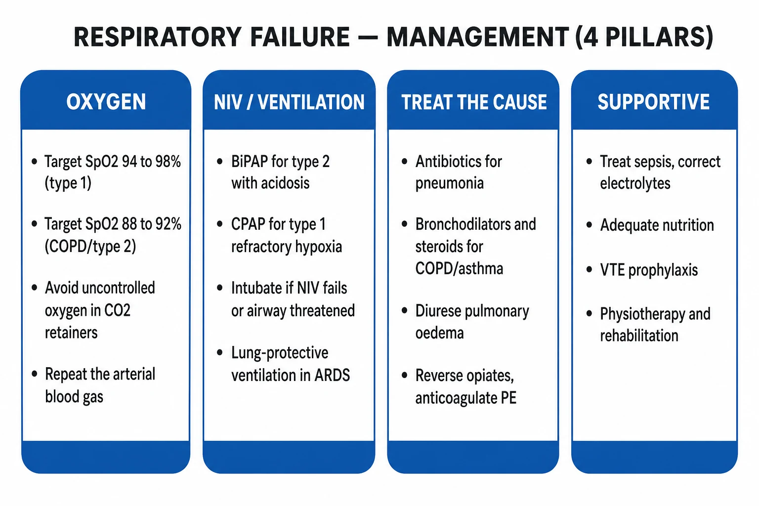
ABCDE first. The resuscitation phase buys time to find and treat the cause.[1]
- Airway — secure; position upright; suction if secretions; consider simple airway adjuncts; intubate if GCS below 8 or airway not protected.
- Breathing — give oxygen, target SpO2 94–98% for Type 1 and 88–92% for COPD/Type 2; reassess; repeat the ABG at 30–60 minutes.
- Circulation — IV access, fluid balance, treat shock; balance crystalloid cautiously (especially in ARDS — conservative fluid strategy).
- Disability — GCS, pupils, glucose, capnography if intubated; reverse reversible causes (naloxone, glucose, thiamine).
- Exposure / examine — find the cause (CXR, ECG, bloods, drug screen). [1]
The oxygen target — the single most tested decision. Uncontrolled high-flow oxygen in a CO2 retainer worsens hypercapnia (Haldane effect, loss of HPV) and precipitates CO2 narcosis. Use controlled oxygen — a Venturi mask (fixed-performance: 24% or 28% for known COPD) — and target SpO2 88–92%, then repeat the ABG. If PaCO2 rises and pH falls despite controlled oxygen, start BiPAP.[1][2]
Resuscitative bundle in COPD exacerbation with acidotic hypercapnic respiratory failure: controlled oxygen; salbutamol 5 mg and ipratropium 500 micrograms nebulised (driven by air if CO2 retainer, or oxygen with close monitoring); prednisolone 30–40 mg orally (or hydrocortisone 100 mg IV); antibiotics if infective (amoxicillin–clavulanate or doxycycline, local guidelines); repeat ABG at 30–60 min; start BiPAP if pH below 7.35 despite standard medical therapy.[1]
Resuscitative bundle in opiate overdose (Type 2): airway; bag-valve-mask ventilation with oxygen; naloxone 0.04–0.4 mg IV titrated to respiratory rate (not full wakefulness — avoid precipitating acute withdrawal and pulmonary oedema); repeat every 2–3 minutes up to ~2 mg; consider naloxone infusion for long-acting opiates. Observe for re-sedation — naloxone's half-life (1–1.5 h) is shorter than most opiates. [1]
Management — Definitive & Stepwise
Oxygen delivery devices
Standard / low-flow
- Nasal cannula 1–5 L/min (FiO2 ~24–40%).
- Simple face mask 5–10 L/min (40–60%).
- Non-rebreather mask 10–15 L/min with reservoir (60–90%).
- Variable performance — actual FiO2 depends on patient inspiratory flow.
- Suitable for most Type 1; use with caution in CO2 retainers.
Fixed-performance (Venturi)
- Delivers a precise, set FiO2 (24, 28, 31, 35, 40%).
- The controlled-oxygen tool for COPD / Type 2 retainers.
- High total flow entrains room air at a fixed ratio.
- Start 24% or 28%, target SpO2 88–92%, repeat the ABG.
High-flow nasal cannula (HFNC)
- Up to 60 L/min, FiO2 titrated 21–100%, heated humidified.
- Low-level PEEP (~3–5 cmH2O), washout of dead space, reduced work of breathing.
- FLORALI 2015: reduced 90-day mortality vs standard oxygen/NIV in P/F below 300.
- ROX index (SpO2/FiO2 divided by RR) at 2, 6, 12 h helps predict success (above 4.88 favourable).
Non-invasive ventilation
- BiPAP (IPAP/EPAP) — pressure support for ventilatory failure (COPD, OHS, neuromuscular).
- CPAP (continuous positive pressure) — splints alveoli; cardiogenic oedema, refractory Type 1, atelectasis.
- Start BiPAP IPAP 10–15 / EPAP 4–5; titrate IPAP to pH/CO2.
- Interfaces: full-face mask first; switch to nasal once stable.
Invasive mechanical ventilation
- Indicated when NIV fails, airway is threatened, or severe refractory hypoxia.
- Lung-protective in ARDS: tidal volume 6 mL/kg PBW, plateau below 30 cmH2O.
- Permissive hypercapnia (allow pH at least 7.20).
- Sedation, analgesia, paralysis as needed; wean with SBT.
NIV indications — the BTS/ICS framework
Reproduce the BTS/ICS 2016 indications for acute NIV in adults:[1]
- COPD exacerbation with pH 7.25–7.35 despite standard medical therapy (level-1 evidence; first-line). pH below 7.25 — NIV on the ICU, with a low threshold for intubation.
- Decompensated obesity hypoventilation syndrome (OHS) with acidotic hypercapnia.
- Neuromuscular weakness / chest-wall disease with hypercapnia.
- Cardiogenic pulmonary oedema — CPAP improves dyspnoea and physiology faster than standard oxygen (3CPO found no mortality difference, but CPAP/NIV remain standard for the breathless patient).
- Weaning and peri-extubation in high-risk hypercapnic patients. [1]
BiPAP setup — practical numbers
Start IPAP 10–15 cmH2O, EPAP 4–5 cmH2O, oxygen entrained to target SpO2 88–92%, full-face mask. Titrate IPAP up by 2–5 cmH2O every 10 minutes until pH above 7.35 or PaCO2 falls; reassess at 1, 4, 12 hours. Failures (rising CO2, falling pH, exhaustion) escalate to intubation. [1]
Intubation criteria
Intubate when any of: [1]
- Failure of NIV — rising PaCO2, falling pH, exhaustion, intolerance of the mask.
- Airway not protected — reduced GCS, copious secretions, vomiting risk.
- Severe refractory hypoxia despite NIV/CPAP/HFNC.
- Respiratory arrest / peri-arrest.
- Haemodynamic instability requiring vasopressors alongside respiratory failure. [1]
ARDS — lung-protective ventilation (the ARDS Network)
The ARDS Network 2000 trial transformed ARDS care: low tidal volume (6 mL/kg predicted body weight) versus traditional (12 mL/kg) reduced mortality from 40% to 31%.[3]
Reproduce the ARDS Network lung-protective bundle: [1]
- Tidal volume 6 mL/kg predicted (ideal) body weight (PBW: male = 50 + 2.3×(height inches − 60); female = 45.5 + 2.3×(height inches − 60)).
- Plateau pressure below 30 cmH2O.
- PEEP titrated to FiO2 (higher-PEEP strategy in moderate–severe ARDS — ALVEOLI/Express trial evidence; Brower 2004).[6]
- Permissive hypercapnia — allow PaCO2 to rise provided pH at least 7.20 (bicarbonate may be given if pH below 7.15).
- Oxygen target SpO2 88–95% / PaO2 55–80 mmHg — accept modest hypoxaemia to avoid toxic FiO2 and high pressures.
- Conservative fluid strategy — keep the lung dry.
- Prone positioning at least 16 hours/day in severe ARDS (P/F below 150) — PROSEVA reduced mortality from 33% to 16%.[5]
- ECMO for refractory hypoxaemia (P/F below 80) despite optimised ventilation, or uncontrollable respiratory acidosis (pH below 7.20 with PaCO2 above 80) — centre-of-excellence referral (CESAR/EOLIA framework).
High-flow nasal cannula (HFNC) — when to use it
FLORALI (2015) randomised hypoxaemic respiratory failure (P/F below 300, without hypercapnia) to HFNC, standard oxygen, or NIV: HFNC reduced 90-day mortality (driven by the P/F-at-or-below-200 subgroup) and was better tolerated.[7] Use HFNC as first-line for moderate hypoxaemic failure (P/F 150–300), bridge/intolerant of NIV, and post-extubation. Monitor closely — the ROX index (SpO2/FiO2 divided by respiratory rate) measured at 2, 6, and 12 hours stratifies success; a value below 3.85 at 2 hours or persistently low predicts intubation. Do not persist with HFNC in a deteriorating patient — intubate.
Treat the underlying cause — by name
| Cause | Specific therapy |
|---|---|
| Pneumonia | Antibiotics within 4 h (beta-lactam plus macrolide admitted; cover atypicals) |
| COPD / asthma | Bronchodilators (salbutamol, ipratropium), systemic steroids, antibiotics if infective (COPD), magnesium 2 g IV in severe asthma |
| Cardiogenic oedema | IV furosemide, nitrate, CPAP/NIV, treat the cardiac cause |
| Pulmonary embolism | Anticoagulation; thrombolysis in massive PE with shock |
| ARDS | Lung-protective ventilation, treat the trigger (sepsis, trauma, aspiration), prone in severe, ECMO if refractory |
| Opiate overdose | Naloxone titrated to respiratory rate |
| Neuromuscular (GBS, MG) | NIV early; treat the disease (IVIG, plasma exchange, pyridostigmine, immunosuppression); cough augmentation |
| Obesity hypoventilation | NIV (BiPAP) acutely; long-term nocturnal NIV; weight loss; CPAP for OSA overlap |
Specific Subtypes & Scenarios
- COPD exacerbation with acidotic hypercapnic respiratory failure — the classic exam scenario. Controlled oxygen 88–92%, repeat ABG at 30–60 min, bronchodilators + IV steroids + antibiotics if infective, early BiPAP if pH below 7.35. Plant (2000) showed early NIV on general respiratory wards reduced mortality, intubation, and length of stay.[1][2]
- ARDS — bilateral infiltrates, refractory hypoxia, P/F below 300 (Berlin). Lung-protective ventilation, conservative fluid, prone positioning (at least 16 h/day, severe), ECMO if refractory.
- Acute severe asthma — high-flow oxygen, nebulised salbutamol 5 mg repeatedly plus ipratropium 500 micrograms, IV hydrocortisone 100 mg or oral prednisolone 40 mg, IV magnesium sulphate 2 g over 20 min; consider IV salbutamol; intubate early if silent chest, exhaustion, or poor perfusion. Once intubated: permissive hypercapnia, slow rate, long expiratory time, low tidal volume, deep inhalational anaesthesia (volatile) often helps.
- Neuromuscular weakness (GBS, myasthenia, MND) — Type 2 from pump failure. Monitor FVC, MIP, MEP (not SpO2, which is late). Thresholds to support: FVC below 20 mL/kg (below 1 L), MIP below −30 cmH2O, MEP below 40 cmH2O, or a fall of FVC above 30%. Start NIV (BiPAP), arrange cough augmentation (mechanical insufflation–exsufflation), and intubate electively when bulbar or fatigue supervenes; treat the disease (IVIG/plasma exchange in GBS, cholinesterase inhibitors/immunosuppression in MG).
- Obesity hypoventilation syndrome (OHS) — decompensated OHS: start BiPAP (IPAP 10–15, EPAP 5–8) to target SpO2 88–92% and a falling PaCO2; long-term nocturnal NIV, weight loss, treat the OSA overlap with CPAP/pressure support.
- Cardiogenic pulmonary oedema — upright posture, CPAP/NIV 5–10 cmH2O (3CPO: faster physiological improvement, no mortality benefit, but standard for the breathless patient), IV furosemide 40–80 mg, nitrate if BP permits, treat the cardiac cause (ischaemia, AF, valve).
- Opiate overdose — naloxone 0.04–0.4 mg IV titrated to respiratory rate, airway, ventilation; observe for re-sedation; infusion for long-acting opiates (fentanyl patches, methadone).
Complications & Pitfalls
Complications of the disease: [1]
- Cardiac arrest from severe hypoxia and acidosis.
- CO2 narcosis and coma from uncontrolled oxygen or progressive hypercapnia.
- Multi-organ failure — AKI, hepatic, gut ischaemia from prolonged hypoxia and hypoperfusion.
- Cor pulmonale and pulmonary hypertension in chronic Type 2.
- Critical-illness polyneuromyopathy in prolonged ICU stay. [1]
Complications of treatment: [1]
- Ventilator-associated pneumonia (VAP) — the longer the intubation, the higher the risk; head-up 30 degrees, oral chlorhexidine, daily sedation holds, early extubation.
- Barotrauma and volutrauma — pneumothorax, pneumomediastinum from high pressures/volumes; the rationale for plateau below 30 cmH2O and 6 mL/kg.
- Oxygen toxicity — absorption atelectasis, tracheobronchitis, and free-radical injury with prolonged FiO2 above 0.6; aim for the lowest FiO2 that meets the target SpO2.
- Atelectrauma — repetitive opening and closing of alveoli; the rationale for adequate PEEP.
- NIV complications — pressure areas and nasal-bridge ulceration, gastric insufflation, patient intolerance, mask leak.
- Delirium and ICU-acquired weakness. [1]
Ventilator-induced lung injury (VILI) is the umbrella term: volutrauma (overdistension), barotrauma (high pressure), atelectrauma (recruitment–derecruitment), and biotrauma (inflammatory mediator release from injured alveoli). The whole rationale for lung-protective ventilation is to minimise VILI. [1]
Classic pitfalls
- Uncontrolled high-flow oxygen in a CO2 retainer — precipitates CO2 narcosis (Haldane + loss of HPV). The commonest single error.
- Delaying NIV in COPD acidosis — every hour of untreated acidotic hypercapnic failure increases intubation and mortality risk.
- Delayed intubation of the fatiguing patient — intubate before the arrest, not after.
- Failing to repeat the ABG after every change in oxygen or ventilatory support.
- Misreading a normal SpO2 in carbon-monoxide poisoning or methaemoglobinaemia — always correlate with PaO2 on the ABG and co-oximetry.
- Using too-large tidal volumes in ARDS — volutrauma and excess mortality (the ARDS Network lesson).
- Stopping at "respiratory failure" as the diagnosis — always find and treat the underlying cause. [1]
Prognosis & Disposition
Prognosis tracks the underlying cause, the severity (P/F ratio, pH), the comorbidity, the age, and the duration of hypoxia.[1]
- COPD exacerbations with acidosis do well with prompt NIV (NIV reduces mortality, intubation rate, and length of stay).
- ARDS mortality remains around 35% in severe disease, driven by the trigger (sepsis worst) and the degree of hypoxaemia.
- Opiate overdose survival is excellent with prompt naloxone and airway support.
- Neuromuscular respiratory failure prognosis tracks the disease (GBS good with supportive care; MND poor).
- Cardiogenic pulmonary oedema responds rapidly to CPAP/NIV and diuresis. [1]
Disposition: [1]
- Discharge — stable on standard oxygen or room air, treating the cause, safe social situation, safety-net to return.
- Ward — stable on standard oxygen, no acidosis, low NIV need.
- HDU/ICU — NIV or HFNC, organ failure, need for close monitoring.
- ICU — intubated/ventilated, vasopressors, severe refractory hypoxia, multi-organ failure. [1]
Weaning from NIV/ventilation: correct the underlying cause; reduce pressure support gradually; switch to HFNC or standard oxygen; monitor the ABG; structured spontaneous breathing trials (SBT) for invasive ventilation. [1]
Special Populations
- COPD (Type 2) — controlled oxygen (88–92%) and early NIV; the classic exam scenario.
- Pregnancy — reduced FRC, raised diaphragm, higher oxygen consumption cause rapid desaturation; treat aggressively, left lateral tilt, early intubation by an expert with a smaller (6.0–6.5 mm) tube; HFNC and NIV have a growing role.
- Elderly — blunted ventilatory response, comorbidity, atypical presentation (confusion, falls), high aspiration risk; lower threshold to admit.
- Paediatric — weight-based drug doses, small airway, rapid fatigue; bronchiolitis and asthma common; HFNC increasingly first-line; children desaturate fast.
- Immunocompromised — broad differential (PJP, CMV, fungal, bacterial), low threshold for early bronchoscopy, NIV often first-line, often rapid progression; avoid delay.
- Neuromuscular disease — FVC and MIP/MEP drive the intubation/NIV decision (not SpO2, which is late); cough augmentation; consider tracheostomy for long-term ventilatory failure.
- Anticoagulated — balance bleeding risk against PE/ACS treatment; reverse per protocol if intubation is urgent. [1]
Evidence, Guidelines & Regional Differences
The landmark trials and guidelines:[1]
- BTS/ICS 2016 (Davidson 2016) — the UK guideline for ventilatory management of acute hypercapnic respiratory failure in adults; the NIV indications and setup framework.[1]
- Plant 2000 (Lancet) — early NIV on general respiratory wards for COPD exacerbations reduced mortality, need for intubation, and length of stay — the foundation of ward-based NIV.[2]
- ARDS Network 2000 (NEJM) — low (6 mL/kg) vs traditional (12 mL/kg) tidal volumes; mortality 31% vs 40%.[3]
- Berlin ARDS Definition 2012 (JAMA) — the modern severity framework (P/F 200–300 mild, 100–200 moderate, below 100 severe).[4]
- PROSEVA 2013 (NEJM) — prone positioning at least 16 h/day in severe ARDS (P/F below 150); mortality 16% vs 33%.[5]
- ALVEOLI / Brower 2004 (NEJM) — higher vs lower PEEP strategies; higher PEEP benefits moderate–severe ARDS.[6]
- FLORALI 2015 (NEJM) — HFNC vs standard oxygen vs NIV in hypoxaemic failure (P/F below 300); HFNC reduced 90-day mortality.[7]
Regional oxygen-target practice: [1]
- UK (BTS oxygen guideline 2017) — target SpO2 94–98% for most adults, 88–92% for COPD / CO2 retainers; controlled oxygen via Venturi; oximetry mandatory in all breathless patients.
- US (Beacon/ICU, ATS/ACCP) — broadly similar targets; more permissive of HFNC as first-line.
- India (ICMR / NMC) — oxygen is an essential medicine; infrastructure (concentrators, cylinders, power, piped oxygen), cost, and supply drive delivery method; controlled-oxygen teaching emphasised for COPD; HFNC availability variable. [1]
Current controversies: [1]
- HFNC vs NIV in hypoxaemic failure — HFNC increasingly first-line for moderate; NIV for hypercapnia and cardiogenic oedema; avoid prolonged failed HFNC in the deteriorating patient.
- Conservative vs liberal oxygen — the LOCO2, HOT-ICU, and ICU-ROX trials raised concern about hyperoxia; most guidelines now favour normoxia (SpO2 92–96%) rather than maximal oxygen in the critically ill, with the COPD exception.
- Awake prone positioning in non-intubated hypoxaemic failure (matured in COVID-19) — adjunct, not a substitute for intubation in the deteriorating patient. [1]
Exam Pearls
The five mechanisms of hypoxaemia — HVS-DL
HVS-DL
Normal A-a gradient; opiates, neuromuscular, brainstem
The commonest in disease; corrects with O2; pneumonia, COPD, oedema, PE
Does NOT correct with 100% O2; ARDS, lobar pneumonia, R-to-L shunt
Thickened membrane; ILD, fibrosis; corrects with O2
Altitude; normal A-a; corrects by restoring FiO2
The thresholds that decide every answer
Oxygen-induced hypercapnia in COPD — two mechanisms
HH
O2 displaces CO2 from haemoglobin; releases Haldane CO2 into plasma that ventilation cannot clear
Relief of hypoxic pulmonary vasoconstriction re-perfuses poorly ventilated units, worsening V/Q mismatch
- "Respiratory failure = PaO2 below 60 mmHg on room air; type 1 vs type 2 by PaCO2."
- "Type 1 (hypoxaemic, normal/low CO2): V/Q mismatch/shunt — pneumonia, PE, oedema, ARDS, asthma, pneumothorax."
- "Type 2 (hypercapnic): hypoventilation — COPD, neuromuscular, opiates, obesity hypoventilation, brainstem."
- "Oxygen target: 94–98% type 1; 88–92% COPD/type 2; repeat the ABG."[1]
- "Type 2 plus acidosis → BiPAP (Plant 2000); intubate if NIV fails or airway threatened."[2]
- "Five mechanisms of hypoxaemia; only shunt fails to correct with 100% oxygen."
- "Normal A-a gradient with hypoxaemia = pure hypoventilation (opiates, neuromuscular, brainstem)."
- "ARDS: lung-protective ventilation (6 mL/kg PBW, plateau below 30 cmH2O); Berlin P/F severity; prone in severe (PROSEVA); ECMO if refractory."[3][5]
- "HFNC reduces intubation/mortality in P/F at or below 300 (FLORALI 2015); ROX index predicts success."[7]
- "Pulse oximetry is falsely reassuring in CO poisoning and methaemoglobinaemia — always confirm with an ABG/co-oximetry."
- "Always treat the underlying cause; oxygen and ventilation buy time, not cure."
Exam application bank (NEET-PG / INICET)
One-line answer
Respiratory failure is the failure of the respiratory system to maintain adequate gas exchange, defined by the arterial blood gas: PaO2 below 8 kPa (60 mmHg) at room air, with or without a raised PaCO2. Type 1 (hypoxaemic) is low oxygen with normal or low CO2, caused by ventilation-perfusion mismatch or shunt — pneumonia, pulmonary embolism, pulmonary oedema, ARDS, asthma. Type 2 (hypercapnic) is low oxygen AND high CO2 (PaCO2 above 6 kPa / 45 mmHg), caused by alveolar hypoventilation — COPD, neuromuscular disease, opiates, obesity hypoventilation, brainstem depression. The ABG diagnoses it and the type drives treatment. Management is oxygen (target SpO2 94–98% for type 1; 88–92% for COPD/type 2 to avoid CO2 narcosis), non-invasive ventilation (BiPAP) for type 2 with acidosis, CPAP/HFNC for refractory type 1 hypoxia, intubation if NIV fails or the airway is threatened, lung-protective ve
Worked stems (answer without another resource)
Stem 1 — Classic presentation. Map symptoms to mechanism; name the first investigation and first treatment step with dose/route if drug therapy is standard. [1]
Stem 2 — Unstable / complicated. List red flags that force immediate resuscitation, theatre, ICU, antidote, or reperfusion — and what you do in the first 15 minutes. [1]
Stem 3 — Atypical group. Elderly, pregnancy, child, or immunocompromised: how presentation and thresholds change. [1]
Stem 4 — Differential trap. Name the three closest mimics and one discriminator for each. [1]
Stem 5 — Disposition. Who goes home with safety-netting, who is admitted, who needs HDU/ICU/theatre, and what follow-up is mandatory. [1]
Rapid viva checklist
- Definition + classification
- Pathophysiology chain
- Bedside signs / criteria
- Score with exact components (if any)
- Emergency bundle
- Definitive therapy with doses
- Complications of disease and of treatment
- Special populations
- Guideline/trial name if classic
- Three exam traps
Coverage self-check
If you cannot answer any stem above from this page alone, re-read the matching section — the page is intended to be self-sufficient for final-prof and NEET-PG/INICET questions on Respiratory Failure.
References
- [1]Davidson AC, Banham S, Elliott M, et al. BTS/ICS guideline for the ventilatory management of acute hypercapnic respiratory failure in adults Thorax, 2016.PMID 26976648
- [2]Plant PK, Owen JL, Elliott MW. Early use of non-invasive ventilation for acute exacerbations of chronic obstructive pulmonary disease on general respiratory wards: a multicentre randomised controlled trial Lancet, 2000.PMID 10859037
- [3]The Acute Respiratory Distress Syndrome Network. Ventilation with lower tidal volumes as compared with traditional tidal volumes for acute lung injury and the acute respiratory distress syndrome N Engl J Med, 2000.PMID 10793162
- [4]ARDS Definition Task Force; Ranieri VM, Rubenfeld GD, Thompson BT, et al. Acute respiratory distress syndrome: the Berlin Definition JAMA, 2012.PMID 22797452
- [5]Guérin C, Reignier J, Richard JC, et al. Prone positioning in severe acute respiratory distress syndrome N Engl J Med, 2013.PMID 23688302
- [6]Brower RG, Lanken PN, MacIntyre N, et al. Higher versus lower positive end-expiratory pressures in patients with the acute respiratory distress syndrome N Engl J Med, 2004.PMID 15269312
- [7]Frat JP, Thille AW, Mercat A, et al. High-flow oxygen through nasal cannula in acute hypoxemic respiratory failure N Engl J Med, 2015.PMID 25981908