Respiratory · General Medicine
Solitary Pulmonary Nodule
Also known as Solitary pulmonary nodule · Pulmonary nodule · Coin lesion · Incidental pulmonary nodule · Brock risk model
A solitary pulmonary nodule (SPN) is a single, well-defined, roughly spherical opacity 3 cm or less in diameter, surrounded by aerated lung, with no associated atelectasis, adenopathy or pleural effusion — most often found incidentally on CT or on low-dose CT lung cancer screening. The clinical task is to distinguish benign from malignant using size, density (solid, part-solid, ground-glass), border (smooth vs spiculated), growth rate, calcification, and patient risk (age, smoking, prior cancer). Benign features favour a smooth border, dense central/popcorn calcification, and no growth over two years; malignant features are a spiculated or part-solid nodule that grows in an older smoker. A validated risk model (Brock, then Herder; Mayo/Swensen as the classical model) quantifies cancer probability and drives management — low risk: CT surveillance (Fleischner or BTS intervals); intermediate: PET-CT and biopsy; high risk: tissue diagnosis and surgical resection if malignant. A nodule over 3 cm is a mass, presumed malignant until proven otherwise.
On this page & tools
Your progress
Saved locally on this device.
Practise this topic
Exam tags
Red flags
Overview & Definition
A solitary pulmonary nodule (SPN) — historically a "coin lesion" — is a single, well-defined, roughly spherical opacity 3 cm or less in greatest diameter, surrounded by aerated lung, with no associated atelectasis, mediastinal adenopathy, or pleural effusion. The size threshold is the central exam fact: a lesion larger than 3 cm is, by definition, a pulmonary mass, which is presumed malignant until proven otherwise and is investigated and staged as probable lung cancer rather than worked up as an SPN.[1][5]
The clinical importance of the SPN is not that it is dangerous in itself — most are benign — but that a minority represent early, surgically curable lung cancer. The whole apparatus of nodule management (risk models, surveillance intervals, PET, biopsy) exists to catch the curable stage IA cancer without over-investigating the great majority of nodules that are benign. Because chest CT use has exploded (incidental imaging, COVID-era CT, and most importantly organised low-dose CT lung cancer screening), SPNs are detected in over 1.5 million adults in the United States each year, making the algorithm one of the highest-yield topics in thoracic medicine.[1][6]
Why the SPN matters
Classification
An SPN is classified along three axes that each change pre-test probability and drive the management pathway: (1) size, (2) radiographic density, and (3) aetiology. Density is the most examinable, because it tracks the underlying biology — a solid nodule behaves differently from a pure ground-glass nodule, and a part-solid nodule carries the highest malignant risk of all.[1]
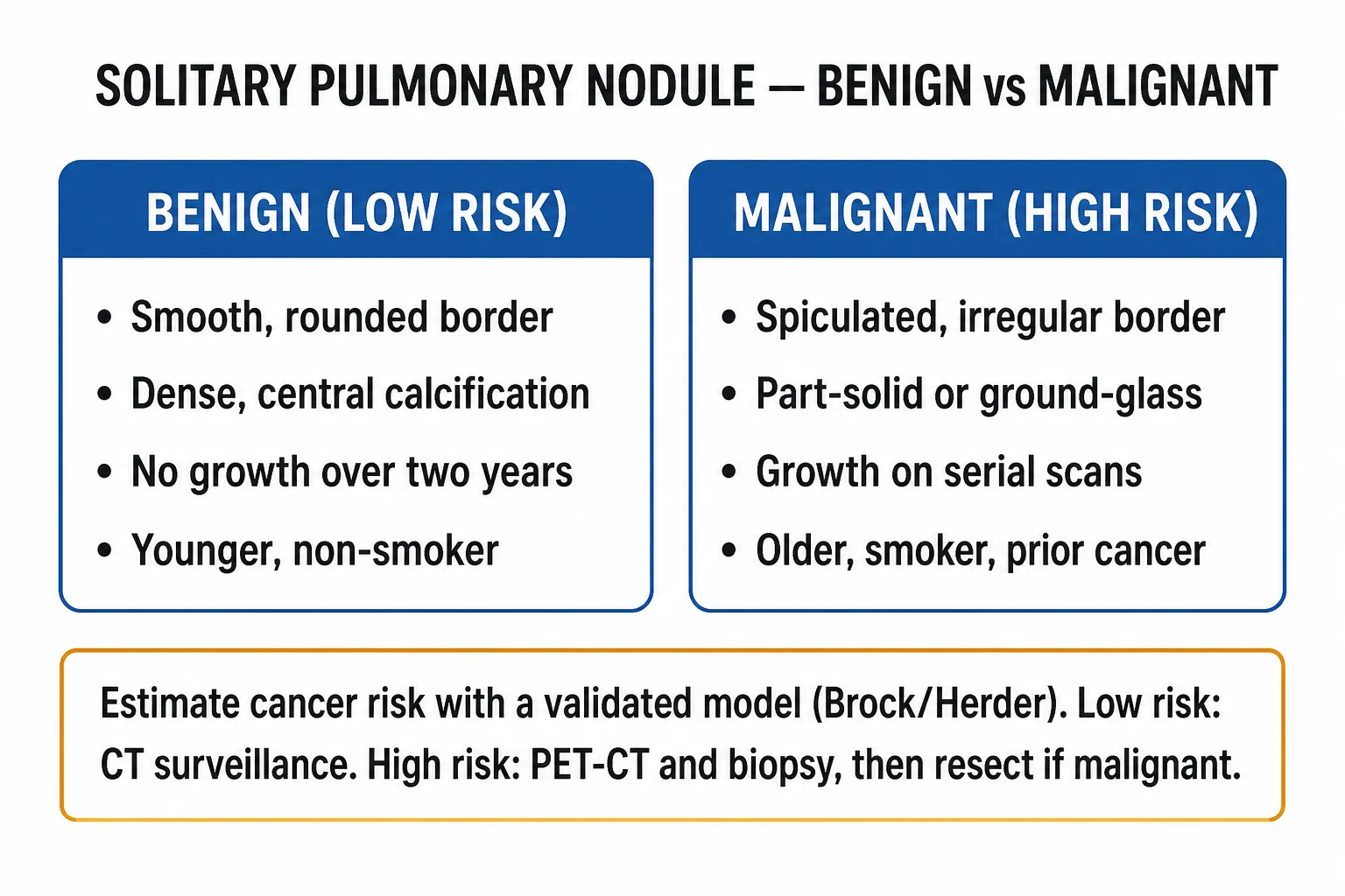
By density (the most examinable axis)
Solid nodule
Completely obscures lung parenchyma
- Commonest type; differential widest (granuloma, cancer, hamartoma)
- Doubling time 30-400 days suggests malignancy; under 30 d inflammatory; over 400 d benign
- Fleischner 2017: most under 6 mm need no follow-up; 6-8 mm CT at 6-12 and 18-24 mo; over 8 mm CT-PET/biopsy
Part-solid nodule
Solid component within ground-glass
- HIGHEST malignant risk per mm of any nodule type
- Solid component size is the key metric (≥6 mm solid = high risk)
- Represents invasive adenocarcinoma within a lepidic (in-situ) background
Pure ground-glass
Hazy density, vessels visible through it
- Often adenocarcinoma in situ / minimally invasive adenocarcinoma (AIS / MIA)
- Slow-growing; long surveillance (to 5 years if ≥6 mm)
- Often PET-negative and contrast-CT-negative — biology is lepidic, low-metabolic
By aetiology
Malignant
- Primary lung cancer (adenocarcinoma commonest, then squamous, small-cell, carcinoid)
- Solitary metastasis (renal, colorectal, breast, sarcoma, melanoma)
- Primary pulmonary lymphoma (rare)
Benign neoplastic
- Hamartoma — commonest benign lung tumour; fat + 'popcorn' calcification
- Inflammatory myofibroblastic tumour, chondroma, lipoma
Infectious / granulomatous
- Healed granuloma — TB, histoplasmosis, coccidioidomycosis (dense central/laminated calcification)
- Active infection — round pneumonia, lung abscess, septic embolus, hydatid, active TB
Inflammatory / vascular / congenital
- Rheumatoid nodule, granulomatosis with polyangiitis (GPA, Wegener's)
- Arteriovenous malformation (AVM), pulmonary infarct, rounded atelectasis, bronchogenic cyst
Epidemiology & Risk Factors
The prevalence of SPNs and their malignant probability both depend entirely on the population being scanned. In an incidental-CT population the malignant rate is low (often 1-12 percent); in a lung cancer screening (LDCT) population it is higher (around 1 in 10 baseline nodules eventually proves malignant, with many more false positives); in a patient with prior cancer the rate rises sharply.[6]
Patient-level risk factors for malignancy are the backbone of every risk model and every examiner's first follow-up question. They are, in roughly descending weight: age (risk rises steeply after 50, and an SPN in a person under 35 is much more likely benign); smoking (the dominant modifiable risk; current over former, pack-years matter); personal history of extrathoracic cancer (especially melanoma, sarcoma, breast, colon, renal — raises the metastasis differential); family history of lung cancer; chronic lung disease (COPD and pulmonary fibrosis both raise lung cancer risk independently of smoking); and occupational exposure (asbestos, uranium, radon, silica, chromium, nickel).[2][4]
Nodule-level imaging features that raise malignant probability are the other half of every risk model: size (cancer probability tracks diameter roughly linearly — under 10 percent for a 5 mm nodule, around 50 percent at 2 cm, and over 80 percent at 3 cm in a smoker); spiculated or lobulated border; upper-lobe location (lung cancer is more common in upper lobes); growth on serial imaging; part-solid morphology; and cavitation with a thick, irregular wall (a wall over 15 mm favours malignancy or infection, under 5 mm favours benign).[1][4]
In TB-endemic regions (large parts of India, sub-Saharan Africa, South-East Asia), the pre-test probability of a benign granuloma is much higher, which depresses the specificity of PET-CT (granulomas are FDG-avid) and shifts the algorithm toward biopsy with AFB/mycobacterial culture and IGRA before assuming cancer and embarking on staging.[1]
Pathophysiology
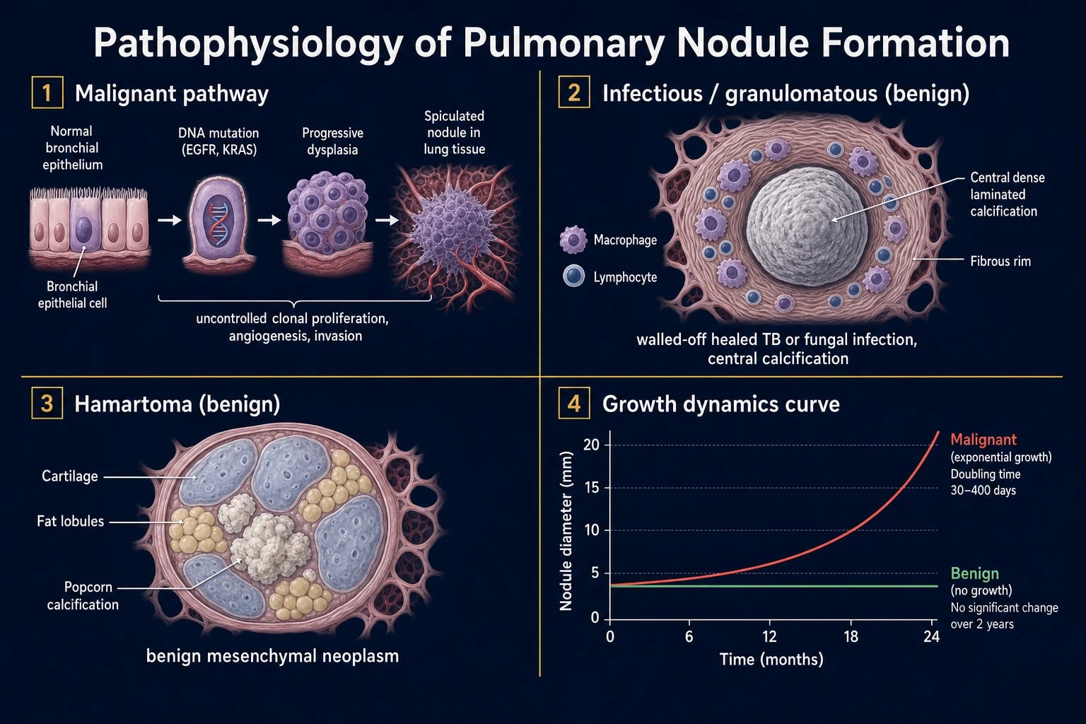
The malignant pathway: the adenocarcinoma sequence
A malignant SPN, in the modern CT era, is most often a pulmonary adenocarcinoma travelling along a defined histological and molecular sequence. The earliest lesion is atypical adenomatous hyperplasia (AAH) — a small (≤5 mm) ground-glass focus of Clara-cell/type-II-pneumocyte atypia. AAH can progress to adenocarcinoma in situ (AIS), a pure lepidic (along-alveolar-wall, non-invasive) growth that appears radiologically as a persistent pure ground-glass nodule. AIS can progress to minimally invasive adenocarcinoma (MIA) — a lepidic lesion with a small invasive component (≤5 mm) — and finally to invasive adenocarcinoma, where a solid component appears within the ground-glass as stroma-invasive tumour breaks through the alveolar wall. This sequence is the biological basis of the density axis: a pure ground-glass nodule sits early in the sequence and is slow, a part-solid nodule has developed an invasive component, and a solid nodule may have replaced the lepidic precursor entirely.[1]
The driver mutations are the examinable molecular detail. In non-smokers and women, EGFR mutations (and ALK / ROS1 rearrangements) are more common; in smokers, KRAS dominates. Each driver defines not only the biology but the systemic-therapy options (EGFR/ALK tyrosine-kinase inhibitors in advanced disease — relevant when an SPN turns out to be a more advanced cancer than hoped). [1]
The benign granuloma: a walled-off infection
The commonest benign SPN worldwide is the healed granuloma — a fibro-calcified scar of an old inhaled infection. The mechanism is the classic cell-mediated immune response: inhaled Mycobacterium tuberculosis (or, in endemic regions, Histoplasma capsulatum, Coccidioides immitis, or Blastomyces) is phagocytosed by alveolar macrophages, processed and presented to CD4+ T-helper-1 cells, which release interferon-γ and IL-2, recruiting and activating more macrophages. Activated macrophages fuse into epithelioid and Langhans giant cells and organise into a granuloma that walls off the organism. Over months to years the centre undergoes caseous necrosis and dystrophic calcification, producing the radiologically dense, central, laminated ("target") or diffuse calcification that is the exam hallmark of a benign healed granuloma. Importantly, this calcification is metabolically inactive — which is why a benign granuloma can still be mildly FDG-avid on PET (active inflammatory cells) but is negative for the high-SUV uptake of cancer.[4]
The hamartoma: a benign mesenchymal neoplasm
A pulmonary hamartoma is the commonest benign tumour of the lung. It is a disordered but mature mixture of cartilage, fat, smooth muscle and epithelial clefts. The radiological signature is fat within a nodule (negative Hounsfield units, −40 to −120 HU) with or without 'popcorn' chondroid calcification. When fat or popcorn calcification is present, no follow-up is required — the diagnosis is radiological and benign.[5]
Growth dynamics: the doubling-time rule
A tumour grows by exponential cell division, and the examinable metric is the volume doubling time — the time for a nodule to double its volume. A malignant nodule doubles in volume every 30 to 400 days; faster than 30 days is too quick for cancer and suggests infection or inflammation (e.g. a round pneumonia); slower than 400 days is too slow for most cancers and suggests benign disease (the lepidic adenocarcinomas are an important exception — they can be very slow).[1][5]
[1]Why part-solid is the highest-risk morphology
A part-solid nodule's ground-glass component is the lepidic (in-situ) tumour spreading along alveolar walls; its solid component is the invasive adenocarcinoma breaching those walls. Because invasion is what makes a cancer a cancer, the solid-component size (not the total nodule size) drives risk — a part-solid nodule with a solid component of 6 mm or more is high-risk even if the overall lesion is modest. This is why the Fleischner and BTS guidelines key off the solid component for subsolid lesions.[1]
Clinical Presentation
An SPN is almost always asymptomatic — that is the whole point of its detection by imaging. It is found in one of three contexts: (1) incidentally, on a CT done for another reason (trauma, cardiac work-up, COVID suspicion, abdominal pathology with chest slices); (2) on organised low-dose CT lung cancer screening in a high-risk smoker; or rarely (3) on a chest X-ray (the historic "coin lesion"), where thin-section CT is then mandatory to characterise it. [1]
The history is therefore not a symptom history but a risk-factor history, and it is where the marks are won in a viva or long case. Elicit, specifically and quantitatively: smoking (current vs former, pack-years, and for ex-smokers the time since quitting — recent quitters carry near-current risk and the "quitter's cough / cancer after quitting" pattern is classic); personal history of cancer (and for the Mayo/Herder models, whether it was extrathoracic and more than 5 years ago); family history of lung cancer; occupational exposure (asbestos, uranium mining, radon, silica, chromium, nickel); travel and residence in TB- or histoplasmosis-endemic regions; and constitutional symptoms (haemoptysis, weight loss, anorexia, bone pain, hoarseness from recurrent laryngeal nerve involvement) — any of which signals advanced or alternative disease rather than an incidental nodule.[4][5]
Symptoms, when present, change the question entirely. Haemoptysis with a nodule implies either a central endobronchial lesion (consider bronchoscopy), cavitation, or advanced disease. Weight loss, bone pain, jaundice, or neurological symptoms indicate metastatic disease and move the nodule from "incidental" to "the tip of the iceberg". Paraneoplastic syndromes (SIADH in small-cell, hypercalcaemia from PTHrP in squamous, hypertrophic pulmonary osteoarthropathy with clubbing) are rare but exam-favourite clues that a nodule is malignant.[5]
Atypical presentations are deliberately tested. In the elderly or immunocompromised patient a granuloma, an active fungal or mycobacterial infection, and a primary lung cancer can coexist — a single nodule cannot be assumed benign just because the patient is "well". In a patient with a prior extrathoracic cancer, an SPN is a solitary metastasis until tissue proves otherwise (especially melanoma, sarcoma, renal, breast, colon). In the Indian / TB-endemic patient, an apparently classic "spiculated growing nodule" can be an active tuberculoma — tissue (and AFB) is needed before staging as cancer. [1]
Differential Diagnosis
The differential of an SPN is wide, but a structured approach using imaging morphology collapses it rapidly. The exam answer requires at least three named differentials, each with at least three distinguishing features.[5]
Primary lung adenocarcinoma
Commonest malignant SPN
- Sericulated or lobulated solid/part-solid nodule; upper lobe; growth on serial CT
- FDG-avid on PET (SUV max typically >2.5); older smoker
- May carry an EGFR/KRAS/ALK driver mutation on tissue
Healed granuloma
TB, histoplasmosis, coccidioidomycosis
- Dense central / laminated / diffuse calcification; smooth border; no growth over 2 yr
- Residence in or travel to an endemic region; positive IGRA / tuberculin test
- Often multiple calcified nodules + hilar nodes
Pulmonary hamartoma
Commonest benign tumour
- Fat (−40 to −120 HU) within the nodule ± 'popcorn' chondroid calcification; smooth border
- Patient usually under 50 and a non-smoker
- No follow-up required when fat or popcorn calcification is definite
Carcinoid tumour
Typical / atypical
- Well-defined, often central and intensely FDG-avid; younger patient (40s), non-smoker
- May cause carcinoid syndrome or central obstruction; richly vascular on contrast CT
- Somatostatin-receptor-positive (Ga-68 DOTATATE PET positive)
Solitary metastasis
Renal, colorectal, breast, sarcoma, melanoma
- Smooth, round, often lower-lobe, often multiple when more are sought
- Known extrathoracic primary; tissue histology matches the primary
- The disease-free interval and primary histology determine resectability
Active infection
Round pneumonia, abscess, TB, fungal, hydatid, septic embolus
- Acute onset, fever, cough, sputum; cavity with thick irregular wall or air-fluid level
- Leukocytosis, raised CRP; resolves on follow-up imaging after antibiotics
- Sputum/blood cultures positive
Rheumatoid nodule / GPA (Wegener's)
Inflammatory
- Nodule(s) in seropositive rheumatoid arthritis or c-ANCA/PR3-positive GPA
- Often cavitating; may be migratory or multiply
- Biopsy: necrobiotic granuloma (rheumatoid) or necrotising granulomatous vasculitis (GPA)
Arteriovenous malformation (AVM)
Vascular
- Smooth, well-defined, intensely and homogeneously enhancing; feeding and draining vessels on CT
- Bruit on auscultation; history of hereditary haemorrhagic telangiectasia (HHT)
- Risk of haemoptysis and paradoxical embolus
Clinical & Bedside Assessment
The clinical assessment of an SPN is a focused history and a directed examination aimed at refining pre-test probability and finding signs of advanced or alternative disease — most patients have a completely normal examination, which does not exclude malignancy. [1]
The history must quantify, in this order: (1) smoking — current or former, pack-years ((cigarettes per day ÷ 20) × years smoked), and for ex-smokers the years since quitting (the NLST and most screening programmes treat quitting within 15 years as still screen-eligible); (2) prior malignancy — site, histology, time since diagnosis, and treatment; (3) family history of lung cancer in a first-degree relative; (4) occupational and environmental exposure — asbestos, uranium/radon, silica, chromium, nickel, arsenic, and indoor biomass-fuel smoke (a recognised lung-cancer risk in parts of India); (5) travel and residence in TB- or fungal-endemic regions; and (6) symptoms — haemoptysis, weight loss, anorexia, bone pain, hoarseness, headache, jaundice.[4]
The physical examination is directed at finding signs of: advanced local disease (wheeze or unilateral reduced breath sounds from obstruction, dullness from effusion, SVC syndrome from a right upper-lobe mass); metastatic spread (cervical and supraclavicular lymphadenopathy, hepatomegaly, bone tenderness, neurological signs, skin deposits); a primary extrathoracic cancer that has metastasised (breast examination, skin examination for melanoma, abdominal examination for renal or colorectal primary); and paraneoplastic signs (finger clubbing and hypertrophic pulmonary osteoarthropathy, gynaecomastia from hCG-secreting tumours, Cushingoid features from ectopic ACTH in small-cell).[5]
The pre-test probability — what every history is computing
Investigations
The investigative principle of the SPN is characterise first, then estimate probability, then choose surveillance, further imaging, or tissue. The single most important investigation is a thin-section (1 mm) CT of the nodule, ideally with prior imaging for comparison.[1]
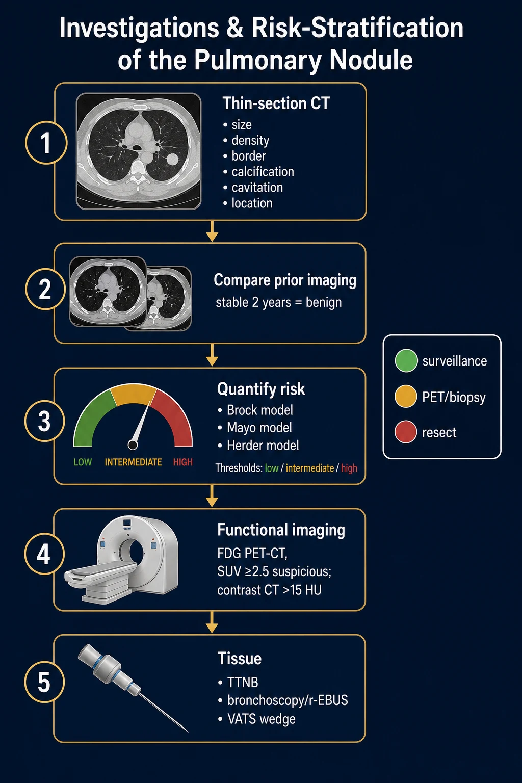
Step 1 — Characterise on thin-section CT
Thin-section CT defines the six features that drive every risk model and every guideline interval: [1]
- Size — the greatest diameter on axial, sagittal or coronal images (for subsolid nodules, volume is preferred because small diameter changes mean large volume changes).
- Density — solid, part-solid (a ground-glass nodule with a solid component), or pure ground-glass; presence of fat (−40 to −120 HU → hamartoma); presence of calcification and its pattern.
- Border — smooth (favours benign), lobulated (suggests uneven growth), spiculated (the classic malignant "sunburst" sign), with or without a halo (ground-glass — haemorrhage in invasive aspergillosis or angioinvasive tumour).
- Calcification pattern — the four benign patterns are central, laminated ("target"), diffuse, and "popcorn" (chondroid, hamartoma); the malignant-associated patterns are eccentric or stippled calcification (scarred cancer or tumour engulfing a granuloma).
- Cavitation and wall thickness — a wall over 15 mm favours malignancy or infection; under 5 mm favours benign.
- Location — upper lobe slightly raises malignant probability (lung cancer is upper-lobe predominant). [1]
Step 2 — Compare with prior imaging (the 2-year rule)
If a nodule has been stable in size for two years on serial CT (or, with caveats, on chest X-ray), it is benign by the stability rule and needs no further investigation. This single fact — having an old CT — can end the work-up; always hunt for prior imaging before PET or biopsy.[5]
Step 3 — Quantify pre-test probability with a validated risk model
This is the most examinable step. Three validated clinical prediction models are in active use, and the modern British Thoracic Society (BTS) 2015 algorithm chains the first two together.[2][3][4]
Mayo / Swensen (1997)
The classical model
- Six variables: age, smoking, extrathoracic cancer >5 yr ago, nodule diameter, spiculation, upper-lobe location
- Thresholds: under 5% low, 5-65% intermediate, over 65% high
- Validated, simple, paper-and-pencil; used in the ACCP (Gould 2013) guideline
Brock / PanCan (2013)
The modern screening model
- Developed in the PanCan / BCG screening cohort (McWilliams, NEJM 2013)
- Variables: sex (female), age, family history of lung cancer, emphysema, nodule size, nodule type (part-solid highest), upper-lobe, nodule count, spiculation
- Best-performing model for screening-detected nodules; first-line in BTS 2015
Herder (2005)
Brock + PET
- Adds FDG-PET uptake (none/faint/intense) to a modified Mayo model
- BTS 2015 second step: if Brock over 10%, run Herder
- Herder over 70% → treatment; 10-70% → PET/biopsy; under 10% → surveillance
VA model / ACPI
Alternatives
- VA: age, smoking, diameter, time since quitting, how discovered
- Useful where Brock variables unavailable
- ACPI is used in some UK centres
Step 4 — Further functional imaging
FDG PET-CT
Metabolic activity
- Sensitivity ~90-95%, specificity ~75-90% (lower in TB-endemic regions)
- SUV max ≥2.5 generally suspicious; faint uptake indeterminate; no uptake lowers risk
- False positive: TB, histoplasmosis, sarcoid, rheumatoid nodule, infection; false negative: AIS, MIA, nodules under 8-10 mm
- Adds the Herder step and simultaneously stages mediastinum and distant metastases
Contrast-enhanced CT
Vascularity
- Enhancement over 15 HU after IV contrast suggests malignancy (sensitivity ~80%, specificity ~80%)
- Largely supplanted by PET-CT but useful when PET unavailable or in very small nodules
- Also characterises AVM (intense homogeneous enhancement with feeding/draining vessels)
Step 5 — Tissue diagnosis
Tissue is required when the post-imaging probability is intermediate-high and the result will change management. The route depends on nodule location and patient fitness:[5]
- Percutaneous transthoracic needle biopsy (TTNB) — best for peripheral nodules under CT or fluoroscopic guidance. Sensitivity ~90 percent for malignancy; principal complication is pneumothorax (~20 percent, ~5 percent requiring chest tube), plus pulmonary haemorrhage and rare systemic air embolism. Avoid in the fully anticoagulated or in patients with severe emphysema and a deep lesion.
- Bronchoscopic biopsy — best for central nodules or those reachable by radial endobronchial ultrasound (r-EBUS) or electromagnetic navigation bronchoscopy (ENB) / robotic bronchoscopy, which can reach small peripheral lesions. Lower pneumothorax risk than TTNB; allows rapid on-site cytology and staging of mediastinal nodes in the same sitting.
- Surgical biopsy — when a nodule is high-risk and resectable, the diagnosis and treatment can be combined: a video-assisted thoracoscopic (VATS) wedge resection with intra-operative frozen section, proceeding to anatomic lobectomy + mediastinal lymph node dissection if malignant. This is both diagnostic and therapeutic and is preferred when pre-operative probability is very high.[5]
Management — Resuscitation
An SPN is rarely a time-critical emergency — the work-up spans weeks, not minutes. The "resuscitation" step is therefore a safety-net assessment, asking three questions at the first encounter: [1]
- Is the lesion actually a mass? A lesion over 3 cm is, by definition, a pulmonary mass presumed malignant — work it up and stage it as lung cancer, not as an SPN. This is the single most important reclassification at triage.[1]
- Is there an acute complication needing urgent care? Look for haemoptysis (especially with a central or cavitating lesion), post-obstructive pneumonia, SVC obstruction, hypercalcaemia (PTHrP), SIADH, or haemodynamic instability from a ruptured AVM or abscess. Any of these escalates the nodule from an outpatient CT pathway to an urgent inpatient one.
- Is the patient already symptomatic from advanced disease? Weight loss, bone pain, jaundice, neurological symptoms, or a palpable mass indicate metastatic disease and shift the pathway to urgent staging CT-PET and tissue diagnosis within two weeks (UK "2-week-wait" / suspected-cancer pathway).[5]
In the absence of any of these, the SPN enters the risk-stratified outpatient pathway below. [1]
Management — Definitive & Stepwise
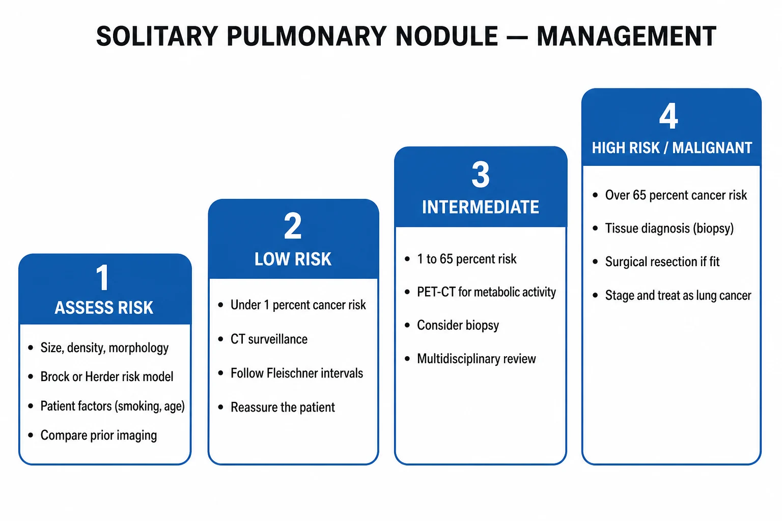
The overarching principle is risk-stratified, guideline-driven management, designed to minimise the harm of over-treatment (unnecessary PET, biopsy, surgery, anxiety, cost — the great majority of nodules are benign) while never delaying the curable early lung cancer. The two operating guideline families are Fleischner Society 2017 (incidental nodules, global) and British Thoracic Society 2015 (algorithmic, model-driven, UK), with ACCP (Gould 2013) used in North America.[1][3][5]
The four-step algorithm
Risk-stratified management of an SPN
Characterise and quantify risk
Thin-section CT (size, density, border, calcification, cavitation, growth vs priors). Apply a validated model (Brock first; Mayo if Brock variables unavailable; Herder if intermediate). Identify benign patterns (fat/popcorn = hamartoma; central/laminated calcification = granuloma; stable 2 yr = benign) that end the work-up.
Low-risk pathway — CT surveillance
Risk under 5% (Mayo) or under 10% (Brock/BTS): reassure and follow at Fleischner intervals. Most solid nodules under 6 mm need no follow-up at all in low-risk adults.
Intermediate-risk pathway — functional imaging and biopsy
Risk 5-65% (Mayo) or 10-70% (Herder): PET-CT to reclassify, then percutaneous or bronchoscopic biopsy of nodules whose result will change management. Multidisciplinary team (MDT) review.
High-risk pathway — tissue and treat
Risk over 65-85% in a fit patient: surgical resection (VATS wedge to lobectomy if malignant) without pre-operative biopsy. If medically inoperable, SBRT. Stage and treat as lung cancer.
Fleischner Society 2017 — incidental solid nodules (low-risk adults)
Under 6 mm (single)
- No routine follow-up in low-risk adults
- Optional follow-up at 12 months in high-risk patients
6-8 mm (single)
- CT at 6-12 months, then 18-24 months
- Consider earlier/additional follow-up in high-risk patients
Over 8 mm (single)
- Consider CT, PET, and/or tissue sampling at ~3 months
- All three modalities in play; management individualised by risk
Multiple nodules
- Use the size of the largest nodule, with additional follow-up at the second interval
- Reflects higher malignancy risk of multiple lesions (metastases / multifocal adenocarcinoma)
Caveats to Fleischner 2017: the recommendations apply to adults 35 years and older with incidental nodules; they do not apply to (1) lung cancer screening patients (use Lung-RADS instead), (2) immunocompromised patients, (3) patients with known primary cancer (metastasis in play), or (4) children. The intervals are shorter in high-risk (heavy-smoking) patients and no follow-up is needed for definite benign patterns (fat, popcorn calcification, central/laminated calcification).[1]
Fleischner Society 2017 — incidental subsolid nodules
Subsolid nodules follow longer surveillance because the lepidic biology is slow but the malignant potential is real, and the rules key off the solid component.[1]
Pure ground-glass, under 6 mm
- No routine follow-up in low-risk adults
Pure ground-glass, 6 mm or more
- CT at 6-12 months, then every 2 years to 5 years
- Reflects the slow growth of AIS/MIA
Part-solid, under 6 mm
- No routine follow-up in low-risk adults
- High-risk: CT at 6-12 and 18-24 months
Part-solid, 6 mm or more, solid under 6 mm
- CT at 6-12 months, then every 2 years to 5 years
Part-solid, solid component 6 mm or more
- High-risk: CT at 3-6 months, then PET and/or tissue sampling if persistent or growing
- Solid component of 6 mm or more is the lepidic-to-invasive threshold
British Thoracic Society (BTS) 2015 — the model-driven algorithm
The BTS algorithm is explicitly chained on the risk models, and is the cleanest single examinable pathway:[2][3]
BTS 2015 algorithm for the pulmonary nodule
Apply Brock model
To every nodule 5 mm or more (and 80 mm³ or more volume). If Brock risk under 10%, proceed to CT surveillance at BTS intervals.
Apply Herder model if Brock over 10%
Herder = Brock + PET uptake. If Herder over 70%, offer treatment (tissue diagnosis and/or resection if fit; SBRT if inoperable).
Intermediate Herder 10-70%
PET-CT ± biopsy, MDT discussion.
Low Herder under 10%
CT surveillance at BTS intervals (3-12 months initially, lengthening to 2-4 years).
Lung cancer screening (NLST / NCCN / Lung-RADS) — the third pathway
When the nodule is found on organised low-dose CT screening in a high-risk smoker, neither Fleischner nor BTS applies — instead use the Lung-RADS scheme. Screening is offered to adults aged 55-80 (USPSTF) / 55-74 (NCCN) who have 30 pack-years or more of smoking and quit within 15 years, with annual low-dose CT. The National Lung Screening Trial (NLST, NEJM 2011) showed a 20 percent reduction in lung-cancer mortality and a 6.7 percent all-cause mortality reduction with three rounds of annual LDCT vs chest X-ray.[6]
NLST — the trial that legitimised screening
Surgical treatment of the malignant SPN
Once a malignant SPN is confirmed (or a high-risk nodule is resected diagnostically), the standard operation is anatomic lobectomy with mediastinal lymph node sampling or dissection, which provides both cure and accurate pathological staging.[5]
- Sublobar resection (segmentectomy or wedge) — acceptable for small (under 2 cm), peripheral stage IA tumours, especially in patients with limited pulmonary reserve. Sublobar must achieve a 2 cm or greater margin or a margin-to-tumour ratio of 1 or more and include hilar and mediastinal node sampling (CALGB 140503 and JCOG0802/WJOG4607L showed non-inferior survival vs lobectomy for small peripheral tumours; segmentectomy preferred over wedge for adequate margins and nodal yield).
- VATS / robotic lobectomy — minimally-invasive approach is now standard for early-stage disease, with lower morbidity, shorter stay, equivalent oncologic outcome to thoracotomy.
- SBRT (stereotactic body radiotherapy) — for medically inoperable early-stage (T1-2aN0) lung cancer; delivers a biologically potent dose in a few fractions; local control over 90 percent and 3-year survival ~50-60 percent in unfit patients who would otherwise receive best supportive care. [1]
Escalation and de-escalation triggers
The triggers to step up from surveillance to biopsy/surgery are: growth on serial CT (especially a diameter rise consistent with volume doubling — recall the 26 percent diameter rule); rising or new FDG avidity on PET; positive biopsy; or a Herder/Mayo probability crossing the high-risk threshold in a fit patient. The trigger to step down is a definite benign pattern (fat, popcorn calcification, dense central/laminated calcification, stability over 2 years) or a patient who is not fit for any treatment even if the nodule is malignant (in whom surveillance is appropriate and invasive work-up is futile).[1][5]
Specific Subtypes & Scenarios
Pure ground-glass nodule (the lepidic spectrum)
A persistent pure ground-glass nodule (GGN) of 6 mm or more sits along the AAH → AIS → MIA pathway and grows slowly (often over years). Management is long surveillance (Fleischner: CT at 6-12 months then every 2 years to 5 years). Surgery (sublobar resection) is reserved for a growing lesion, a developing solid component (transition to part-solid = invasion), or a lesion that persists and grows beyond 5 years. PET and contrast-CT are often negative in pure GGN because the lesion is lepidic and low-metabolic — a negative PET does not exclude AIS/MIA.[1]
Part-solid nodule
The highest-risk morphology. The solid component size drives risk: a solid component of 6 mm or more (or any growth of the solid component) is high-risk and warrants PET ± tissue. The ground-glass component is the lepidic part; the solid component is the invasive part.[1]
Hamartoma
Fat (−40 to −120 HU) ± 'popcorn' calcification in a smooth, well-defined nodule = radiological diagnosis, no follow-up required, no treatment. The commonest benign lung tumour; typically in patients under 50 and non-smokers.[5]
Carcinoid tumour
A well-defined, often central, intensely FDG-avid (or rather, DOTATATE-avid) nodule in a younger, non-smoking patient. May cause central obstruction, haemoptysis, or carcinoid syndrome. Surgical resection (lobectomy or sleeve resection) is curative for typical carcinoid; excellent prognosis (5-year survival over 90 percent for typical carcinoid). Atypical carcinoid behaves more aggressively.[5]
Solitary metastasis from an extrathoracic primary
In a patient with a known extrathoracic cancer (especially melanoma, sarcoma, renal, breast, colon), an SPN is a solitary metastasis until proven otherwise. Tissue histology decides (compare with the primary). Pulmonary metastasectomy is curative in selected cases (single lesion, controlled primary, long disease-free interval, no extrathoracic disease).[5]
Nodule in a TB-endemic region (the Indian context)
In TB/histoplasmosis-endemic regions, the pre-test probability of a benign granuloma is much higher and PET specificity drops sharply (granulomas are FDG-avid). The approach is: correlate with IGRA / tuberculin test, send sputum for AFB, mycobacterial culture and Xpert MTB-RIF, and have a lower threshold for tissue biopsy with stains for AFB and fungi before assuming cancer and embarking on staging.[1]
Screening-detected nodule (Lung-RADS)
Use Lung-RADS categories (1-4) rather than Fleischner. A solid nodule of 15 mm or more (or 8 mm or more new/growing at annual screening) is Lung-RADS 4B (suspicious) and warrants PET-CT and/or biopsy. The baseline round of screening generates more false positives than annual rounds.[6]
Complications & Pitfalls
Complications of over-investigation
- Unnecessary PET, biopsy and surgery for benign disease — cost, anxiety, morbidity
- TTNB pneumothorax (~20%) and haemorrhage in a patient with a benign nodule
- Surgical mortality (~1-2% for lobectomy) for a benign nodule that should have been surveilled
- Patient anxiety and the 'scanxiety' of long surveillance protocols
Complications of under-investigation
- Missed or delayed diagnosis of a curable stage IA lung cancer — the cardinal sin
- Progression to incurable disease because a growing nodule was not re-imaged
- Failure to recognise a mass (over 3 cm) or a multiple-nodules picture (metastases) as a different entity
Procedural complications
- TTNB: pneumothorax (~20%, ~5% needing chest tube), pulmonary haemorrhage, systemic air embolism (rare, fatal)
- Bronchoscopic biopsy: bleeding, post-procedure fever, pneumothorax (lower than TTNB)
- Surgical resection: mortality ~1-2%, prolonged air leak, infection, arrhythmia, loss of lung function
Interpretation pitfalls
- False-positive PET in TB, histoplasmosis, sarcoid, rheumatoid nodule, infection (especially in endemic regions)
- False-negative PET in AIS, MIA, and nodules under 8-10 mm (low metabolic activity)
- Mis-reading a small diameter change as trivial (recall the 26% diameter = 100% volume rule)
- Applying Fleischner (incidental) intervals to a screening or symptomatic patient; or vice versa
Prognosis & Disposition
Most SPNs are benign and need no treatment — the goal of management is to catch the curable early lung cancer among them. A resected stage IA lung cancer detected as a nodule has a 5-year survival of over 80-90 percent, compared with under 20 percent for symptomatic late-stage disease — this survival gap is the entire justification for the surveillance-and-tissue apparatus, and is the one-liner to give the examiner.[5][6]
Outcome is driven by four factors: (1) malignant vs benign (the dominant determinant); (2) histological subtype and stage (adenocarcinoma, squamous, small-cell, carcinoid, metastasis); (3) fitness for surgery (comorbidity, pulmonary reserve, age); and (4) the timeliness of detection (whether caught at stage IA or allowed to progress).[5]
Disposition follows the risk band: low-risk nodules return to primary care with a structured CT surveillance plan and reassurance; intermediate-risk nodules are referred to the respiratory / lung-cancer multidisciplinary team (MDT) for PET ± biopsy; high-risk nodules are referred to the thoracic surgery / lung-cancer MDT for tissue diagnosis and resection (or SBRT if inoperable). The safety-net is a clear, written surveillance plan with named CT dates and a named responsible clinician — patients lost to follow-up during surveillance are the classic preventable failure.[1]
Special Populations
Current or former heavy smoker
- Highest pre-test probability; apply Brock then Herder
- Eligible for LDCT screening if 55-80 yr, ≥30 pack-yr, quit within 15 yr (USPSTF/NCCN)
- Use Lung-RADS (not Fleischner) if the nodule was found on screening
Severe COPD / poor pulmonary reserve
- Higher lung-cancer risk (COPD is an independent risk factor on top of smoking)
- Lower threshold for tissue diagnosis, but prefer non-surgical sampling
- If malignant and inoperable → SBRT rather than lobectomy
Pregnant patient
- Avoid CT with iodinated contrast and PET-CT (radiation) where possible
- Use low-dose non-contrast CT if characterisation is essential; otherwise defer to post-partum if low-risk
- Multidisciplinary (obstetrics + respiratory) decision; never delay investigation of a high-risk nodule in late pregnancy
Immunocompromised
- Higher rates of infection (fungal, mycobacterial, nocardia), post-transplant lymphoproliferative disorder, and primary lung cancer
- Lower threshold for tissue diagnosis; surveillance intervals may need shortening
- Fleischner 2017 explicitly excludes immunocompromised patients — individualise
Prior extrathoracic cancer
- Treat the nodule as a solitary metastasis until proven otherwise
- Tissue histology decides (compare with primary); consider pulmonary metastasectomy in selected cases
- Also still a candidate for a new primary lung cancer (especially after head-and-neck, bladder, breast cancer)
Indian / TB-endemic patient
- Higher benign granuloma prevalence shifts pre-test probability; PET false-positive
- Correlate with IGRA / tuberculin, send sputum AFB / Xpert MTB-RIF; consider biopsy with AFB and fungal stains before staging as cancer
- Use a regionally calibrated threshold; do not reflexively treat as cancer
Evidence, Guidelines & Regional Differences
The SPN literature is built on a small number of landmark prediction models and trials that every examiner rewards.[2][3][4][6]
NLST — National Lung Screening Trial
National Lung Screening Trial Research Team. NEJM 2011;365:395-409
RCT of ~53,000 heavy smokers: 3 rounds of annual low-dose CT vs chest X-ray
Key finding
20 percent reduction in lung-cancer mortality and 6.7 percent reduction in all-cause mortality with LDCT. Established LDCT screening as standard for high-risk smokers.
Practice change
The single trial that legitimised organised lung-cancer screening; basis of USPSTF, NCCN and CMS recommendations.
Brock / PanCan model — McWilliams et al.
McWilliams A, et al. NEJM 2013;369:910-919 (PanCan / BCG cohort)
Prospective screening cohorts (~12,000+ nodules) building a multivariable cancer-prediction model from patient + nodule features
Key finding
Best-performing prediction model for screening-detected nodules (AUC ~0.90+); part-solid type and upper-lobe location are strong predictors. Adopted as first-line model in BTS 2015.
Practice change
Replaced the older Mayo model as the default in modern, screening-era algorithms.
Mayo / Swensen model — Swensen et al.
Swensen SJ, et al. Arch Intern Med 1997;157:849-855
639 patients with SPNs 4-30 mm; logistic regression on six clinical and radiographic variables
Key finding
Six-variable model (age, smoking, extrathoracic cancer over 5 yr ago, diameter, spiculation, upper-lobe) with good discrimination. The classical model underpinning ACCP (Gould 2013).
Practice change
Still valid and used where Brock variables unavailable; thresholds under 5, 5-65, over 65 percent.
Herder model — Herder et al.
Herder GJ, et al. Chest 2005;128:2490-2496
Modified Mayo model + FDG-PET uptake (none/faint/intense) in 106 patients with intermediate-risk nodules
Key finding
Adding PET correctly reclassified ~15% of intermediate-risk nodules into the low- or high-risk band. The second step in the BTS 2015 algorithm.
Practice change
PET is most useful in the intermediate-risk band; little added value at the extremes.
Guideline landscape and regional deltas
- Fleischner Society 2017 (MacMahon, Radiology 2017) — the global default for incidental solid and subsolid nodules; simplified and reduced follow-up versus the 2005 version; does not apply to screening, immunocompromised, or known-cancer patients.[1]
- British Thoracic Society (BTS) 2015 (Callister, Thorax 2015) — the UK algorithm; model-driven (Brock → Herder), with explicit risk thresholds (under 10, 10-70, over 70 percent).
- ACCP (Gould 2013) — the US/North American guideline; uses the Mayo/VA models and clinical judgement; thresholds similar to BTS.[5]
- NCCN / Lung-RADS — for screening only; categorises nodules 1-4 and drives biopsy thresholds.
- ICMR / Indian context — no single national SPN guideline; in practice Fleischner/BTS is applied with regional calibration because the high prevalence of TB and granulomatous disease lowers PET specificity and raises the benign pre-test probability.[1]
Controversies
The two live controversies are overdiagnosis of indolent ground-glass / lepidic adenocarcinomas (treating AIS/MIA that may never have harmed the patient — the downside of sensitive CT) and PET false-positives in TB-endemic regions (treating a tuberculoma as cancer). Both push toward structured, model-driven, biopsy-before-staging pathways rather than reflex PET-CT or reflex surgery.[1][3]
Exam Pearls
The benign triad vs the malignant triad
3 cm
the cut-off: a nodule is up to 3 cm; over 3 cm = a MASS (presumed malignant). The single most examinable number.
stable on serial CT = benign (the stability rule).
central, laminated ('target'), diffuse, and 'popcorn' (chondroid/hamartoma). Malignant: eccentric, stippled.
malignant 30-400 d; under 30 d = infection/inflammation; over 400 d = benign (lepidic exceptions).
= 100% volume — the cube-root-of-2 rule. Never dismiss a small diameter rise.
Exam application bank (NEET-PG / INICET)
One-line answer
A solitary pulmonary nodule (SPN) is a single, well-defined, roughly spherical opacity 3 cm or less in diameter, surrounded by aerated lung, with no associated atelectasis, adenopathy or pleural effusion — most often found incidentally on CT or on low-dose CT lung cancer screening. The clinical task is to distinguish benign from malignant using size, density (solid, part-solid, ground-glass), border (smooth vs spiculated), growth rate, calcification, and patient risk (age, smoking, prior cancer). Benign features favour a smooth border, dense central/popcorn calcification, and no growth over two years; malignant features are a spiculated or part-solid nodule that grows in an older smoker. A validated risk model (Brock, then Herder; Mayo/Swensen as the classical model) quantifies cancer probability and drives management — low risk: CT surveillance (Fleischner or BTS intervals); intermediat
Worked stems (answer without another resource)
Stem 1 — Classic presentation. Map symptoms to mechanism; name the first investigation and first treatment step with dose/route if drug therapy is standard. [1]
Stem 2 — Unstable / complicated. List red flags that force immediate resuscitation, theatre, ICU, antidote, or reperfusion — and what you do in the first 15 minutes. [1]
Stem 3 — Atypical group. Elderly, pregnancy, child, or immunocompromised: how presentation and thresholds change. [1]
Stem 4 — Differential trap. Name the three closest mimics and one discriminator for each. [1]
Stem 5 — Disposition. Who goes home with safety-netting, who is admitted, who needs HDU/ICU/theatre, and what follow-up is mandatory. [1]
Rapid viva checklist
- Definition + classification
- Pathophysiology chain
- Bedside signs / criteria
- Score with exact components (if any)
- Emergency bundle
- Definitive therapy with doses
- Complications of disease and of treatment
- Special populations
- Guideline/trial name if classic
- Three exam traps
Coverage self-check
If you cannot answer any stem above from this page alone, re-read the matching section — the page is intended to be self-sufficient for final-prof and NEET-PG/INICET questions on Solitary Pulmonary Nodule.
References
- [1]MacMahon H, Naidich DP, Goo JM, et al. Guidelines for Management of Incidental Pulmonary Nodules Detected on CT Images: From the Fleischner Society 2017 Radiology, 2017.PMID 28240562
- [2]McWilliams A, Tammemagi MC, Mayo JR, et al. Probability of cancer in pulmonary nodules detected on first screening CT N Engl J Med, 2013.PMID 24004118
- [3]Herder GJ, van Tinteren H, Golding RP, et al. Clinical prediction model to characterize pulmonary nodules: validation and added value of 18F-fluorodeoxyglucose positron emission tomography Chest, 2005.PMID 16236914
- [4]Swensen SJ, Silverstein MD, Ilstrup DM, et al. The probability of malignancy in solitary pulmonary nodules. Application to small radiologically indeterminate nodules Arch Intern Med, 1997.PMID 9129544
- [5]Gould MK, Donington J, Lynch WR, et al. Evaluation of individuals with pulmonary nodules: when is it lung cancer? Diagnosis and management of lung cancer, 3rd ed: American College of Chest Physicians evidence-based clinical practice guidelines Chest, 2013.PMID 23649456
- [6]National Lung Screening Trial Research Team; Aberle DR, Adams AM, Berg CD, et al. Reduced lung-cancer mortality with low-dose computed tomographic screening N Engl J Med, 2011.PMID 21714641