Respiratory · General Medicine
Tuberculosis
Also known as Tuberculosis · TB · Pulmonary tuberculosis · Latent TB infection · MDR-TB · XDR-TB · Consumption · Phthisis
Tuberculosis (TB) is infection by Mycobacterium tuberculosis complex, transmitted by the airborne route via droplet nuclei. It is usually pulmonary but can affect any organ. Distinguish latent TB infection (LTBI) — contained, non-infectious bacilli — from active TB disease — multiplying, transmissible infection causing chronic cough (often with haemoptysis), weight loss, night sweats and fever, with apical cavitation on chest X-ray. Diagnosis is by sputum smear and culture, with Xpert MTB/RIF Ultra providing rapid molecular detection and rifampicin-resistance testing. Standard drug-susceptible TB is treated with a 6-month, four-drug regimen — 2 months of HRZE (rifampicin, isoniazid, pyrazinamide, ethambutol) then 4 months of HR, given as directly observed therapy (DOTS). Multidrug-resistant TB (MDR-TB) needs prolonged individualised therapy or the shorter BPaLM regimen. Drug toxicity is distinctive: rifampicin — orange secretions, CYP induction; isoniazid — neuropathy, hepatitis; pyrazinamide — hyperuricaemia; ethambutol — optic neuritis.
On this page & tools
Your progress
Saved locally on this device.
Practise this topic
Exam tags
Red flags
Overview & Definition
Tuberculosis (TB) is a chronic granulomatous infection caused by the Mycobacterium tuberculosis complex — principally Mycobacterium tuberculosis, an acid-fast, obligate aerobe with a waxy cell wall rich in mycolic acid (the basis of acid-fastness on Ziehl-Neelsen staining). The complex also includes M. bovis (unpasteurised milk), M. africanum (West Africa), and the vaccine strain M. bovis BCG. They are distinguished from non-tuberculous mycobacteria (NTM) such as M. avium complex, M. kansasii, and M. abscessus, which cause pulmonary and disseminated disease in the immunocompromised but are not transmitted person-to-person.[1]
TB is transmitted by the airborne route: infectious patients generate droplet nuclei (1 to 5 micrometres) by coughing, sneezing or singing, and a single inhaled bacillus can seed infection in a susceptible contact. The clinical skill in TB is twofold: recognising active, infectious disease in a patient with chronic respiratory and constitutional symptoms (and isolating it promptly to protect staff and other patients), and understanding the latent-to-active spectrum so that preventive therapy is targeted at those who will benefit. The public-health reflex — isolate, notify, trace contacts, and treat under direct observation — is as central to TB as the pharmacology.[1]
Classification
TB is classified along three axes: by anatomical site, by natural-history stage, and by drug-susceptibility pattern. Each axis changes the management and the public-health response. [1]
By anatomical site: [1]
- Pulmonary TB (about 80 percent of cases) — parenchymal lung disease; the only form that is contagious. Includes pleural TB by convention.
- Extrapulmonary TB (about 20 percent, higher in HIV and children) — lymph node, pleural, skeletal (Pott disease), meningeal, miliary (disseminated), peritoneal, pericardial, genitourinary, and intestinal. Not contagious in most forms, but often paucibacillary and harder to diagnose. [1]
By natural-history stage: [1]
Exposure
No infection, no disease
- Contact with an infectious case, negative tests, no symptoms
- No treatment needed; surveillance only
- Risk stratified by intensity and duration of exposure
Latent TB infection (LTBI)
Infected, not diseased
- Live bacilli contained in granulomas by cell-mediated immunity
- Asymptomatic, non-infectious, normal CXR, negative smear
- Positive tuberculin skin test (Mantoux) or IGRA
- Lifetime reactivation risk about 5 to 10 percent; treated with preventive therapy in high-risk groups
Active TB disease
Multiplying, transmissible
- Symptomatic (cough, weight loss, fever, night sweats) or subclinical with positive cultures
- Infectious if pulmonary or laryngeal; requires full multidrug therapy and notification
- Smear and culture may be positive or negative (paucibacillary)
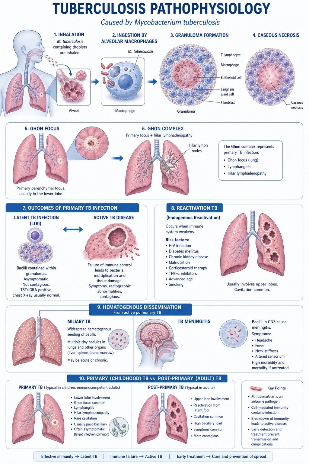
Primary versus post-primary (reactivation) disease: in primary TB the bacilli multiply in the subpleural upper-lobe or lower-lobe alveolar space (the Ghon focus) with regional lymphadenopathy (the Ghon complex), which usually heals by fibrosis and calcification to form the Ranke complex. Most primary infection is subclinical in immunocompetent adults. Post-primary (reactivation) TB arises years later when waning cell-mediated immunity allows dormant bacilli in a Ghon focus or apical focus (seeded by lymphohaematogenous spread at primary infection) to multiply. It preferentially affects the upper-lobe apices and posterior segments — oxygen-rich zones where the obligate-aerobe bacillus thrives — producing caseating granulomas that liquefy and rupture into a bronchus, forming a cavity teeming with bacilli (high infectivity).[1]
First-line anti-TB drugs (drug-susceptible TB)
Standard 6-month regimen: 2HRZE intensive then 4HR continuation
Dose
Rifampicin 10 mg/kg (max 600 mg); Isoniazid 5 mg/kg (max 300 mg) plus pyridoxine 25 mg; Pyrazinamide 25 mg/kg (max 2 g); Ethambutol 15 mg/kg
By drug-susceptibility pattern (WHO definitions): [1]
- Drug-susceptible TB — susceptible to all first-line drugs.
- Mono-resistance — resistant to one first-line drug (other than rifampicin and isoniazid together).
- Poly-resistance — resistant to more than one first-line drug, but not both rifampicin and isoniazid.
- Multidrug-resistant TB (MDR-TB) — resistant to at least rifampicin and isoniazid.
- Pre-XDR-TB — MDR-TB plus resistance to any fluoroquinolone (e.g. levofloxacin, moxifloxacin).
- Extensively drug-resistant TB (XDR-TB) — MDR-TB plus resistance to any fluoroquinolone and at least one additional Group A drug (bedaquiline or linezolid).[3]
Epidemiology & Risk Factors
TB is among the world's commonest infectious killers and remains the leading cause of death from a single infectious agent, surpassing HIV and malaria in most years. The WHO Global Tuberculosis Report estimates about 10.6 million incident cases and 1.3 million deaths annually (2022 data), with India carrying the highest burden of any single country (about a quarter of all global cases), followed by Indonesia, China, the Philippines, Pakistan, Nigeria, Bangladesh and South Africa. About two-thirds of cases are in eight high-burden countries; sub-Saharan Africa has the highest per-capita incidence, driven by the HIV epidemic.[8]
TB-HIV co-infection is the single most important interaction in TB epidemiology. TB is the leading cause of death in people with HIV worldwide, responsible for about a third of HIV-related mortality. The risk of progression from latent infection to active disease is amplified tenfold or more in HIV, and HIV changes the clinical phenotype towards smear-negative, extrapulmonary and disseminated forms that are harder to diagnose.[1]
Risk factors for progression from latent infection to active disease: [1]
| Risk factor | Relative risk of reactivation |
|---|---|
| HIV co-infection | Highest — about 5 to 15 percent annual risk (versus 5 to 10 percent lifetime in HIV-negative) |
| Recent infection (within 1 to 2 years) | High — risk concentrated in the first year |
| Young children (under 5 years) | High — immature cell-mediated immunity; risk of severe forms (TBM, miliary) |
| Anti-TNF therapy, transplant immunosuppression | High — TNF-alpha is central to granuloma containment |
| Silicosis | About 30-fold increased lifetime risk |
| Chronic kidney disease / dialysis | Several-fold increased risk |
| Diabetes mellitus | About 3-fold increased risk; worsens TB outcomes |
| Malnutrition, malignancy, alcohol misuse, smoking | Each adds incremental risk |
| Young age and age extremes | The very young and very old |
The greatest single risk factor is HIV co-infection (CD4 depletion cripples the granuloma), which is why every patient diagnosed with TB must be tested for HIV (bidirectional screening).[1]
Pathophysiology
TB pathogenesis is a dance between the bacillus and the cell-mediated immune response — the disease is shaped by where and how effectively the host walls off the organism. [1]
Transmission and implantation. Infectious patients expel droplet nuclei (1 to 5 micrometres) that remain airborne for hours. A single inhaled bacillus reaches a terminal airway and is phagocytosed by an alveolar macrophage. The bacillus survives inside the macrophage by inhibiting phagolysosome fusion; it multiplies intracellularly, kills the macrophage, and is taken up by recruited macrophages and dendritic cells that carry it to the draining hilar and mediastinal lymph nodes.[1]
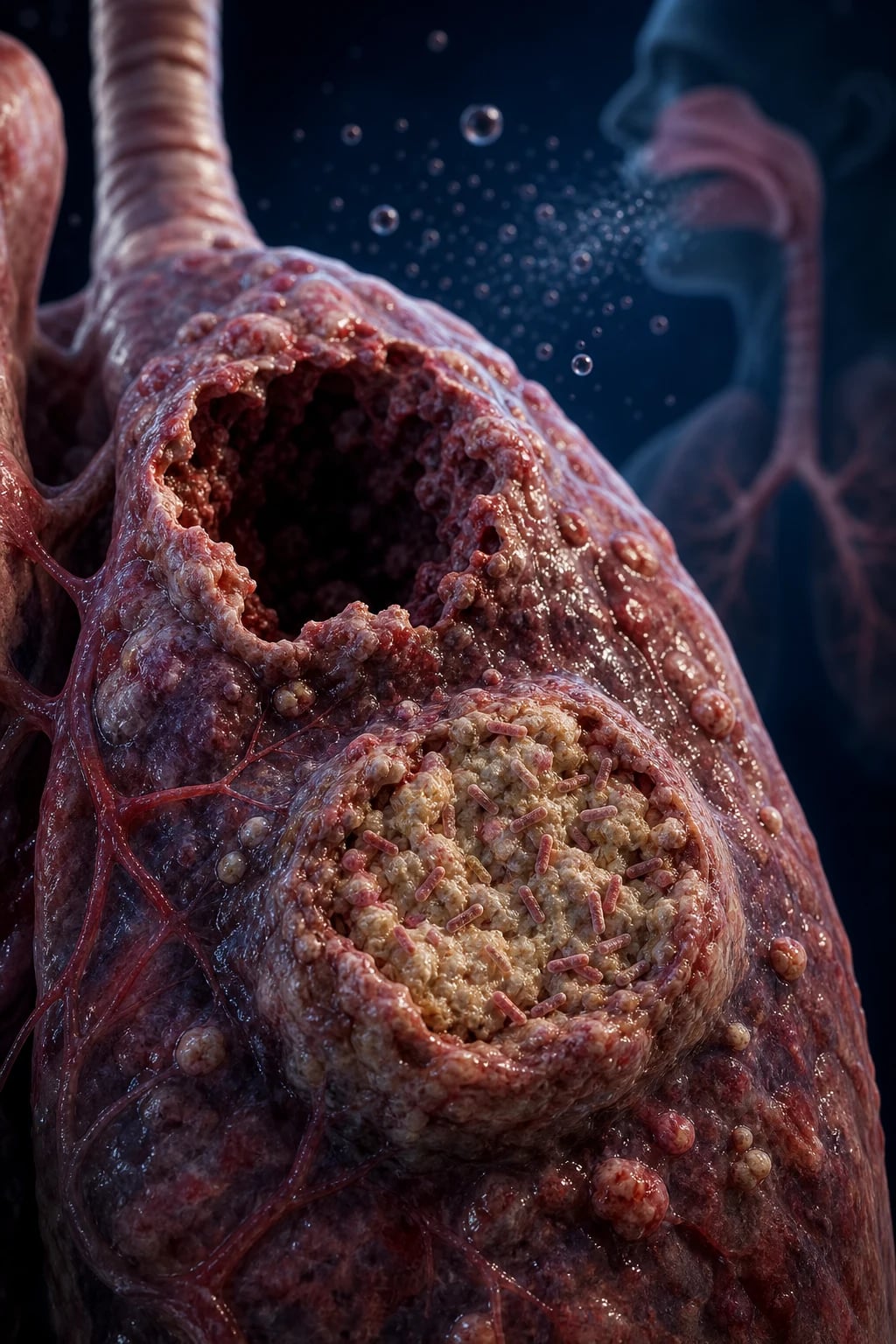
The granuloma and the Ghon complex. Over 2 to 8 weeks a T-helper-1 (CD4+) cell-mediated immune response develops: macrophages activated by IFN-gamma and TNF-alpha transform into epithelioid cells and fuse into Langhans giant cells (multinucleate, with peripheral horseshoe nuclei), organising a granuloma with central caseous necrosis (cheesy, amorphous, acellular). The subpleural parenchymal focus plus the draining lymph node is the Ghon complex; once it calcifies and fibroses it becomes the Ranke complex — a radiological marker of healed primary infection. Antigen from this primary response drives the tuberculin skin test (Mantoux) positive.[1]
Latency. Most bacilli are killed or walled off; a few survive in a dormant, non-replicating state inside macrophages in the centre of the granuloma or in old apical foci (seeded by lymphohaematogenous spread at primary infection). This is latent TB infection (LTBI) — live but contained, non-infectious, asymptomatic, with normal imaging. Loss of immune containment (HIV, anti-TNF, malnutrition, diabetes, ageing, immunosuppression) allows reactivation: the granuloma caseates, liquefies (cavity formation), and caseous material rich in bacilli erodes into a bronchus, seeding the airways and producing the post-primary apical cavity that is the hallmark of contagious adult pulmonary TB.[1]
Why the upper lobes? M. tuberculosis is an obligate aerobe and the upper lobes have the highest oxygen tension (higher ventilation-perfusion ratio — less perfusion, more ventilation), favouring bacillary growth there. This is why reactivation TB classically appears as apical and posterior upper-lobe infiltrates with cavitation, and why miliary spread seeds the well-perfused organs (liver, spleen, bone marrow, meninges, kidneys).[1]
HIV and the granuloma. CD4+ T-cell depletion cripples granuloma formation. In advanced HIV the granulomas are poorly formed, the bacillary load is higher, smears are more often negative (paucibacillary in some forms, overwhelming in others), and disease is more often extrapulmonary and disseminated (miliary, meningeal). This is why any patient with TB atypical in site or severity must be tested for HIV.[1]
Clinical Presentation
The presentation of TB depends on site, immune status, and age. The classic pulmonary presentation is a chronic (weeks to months) illness; extrapulmonary disease presents by organ system; and the immunocompromised host breaks every rule. [1]
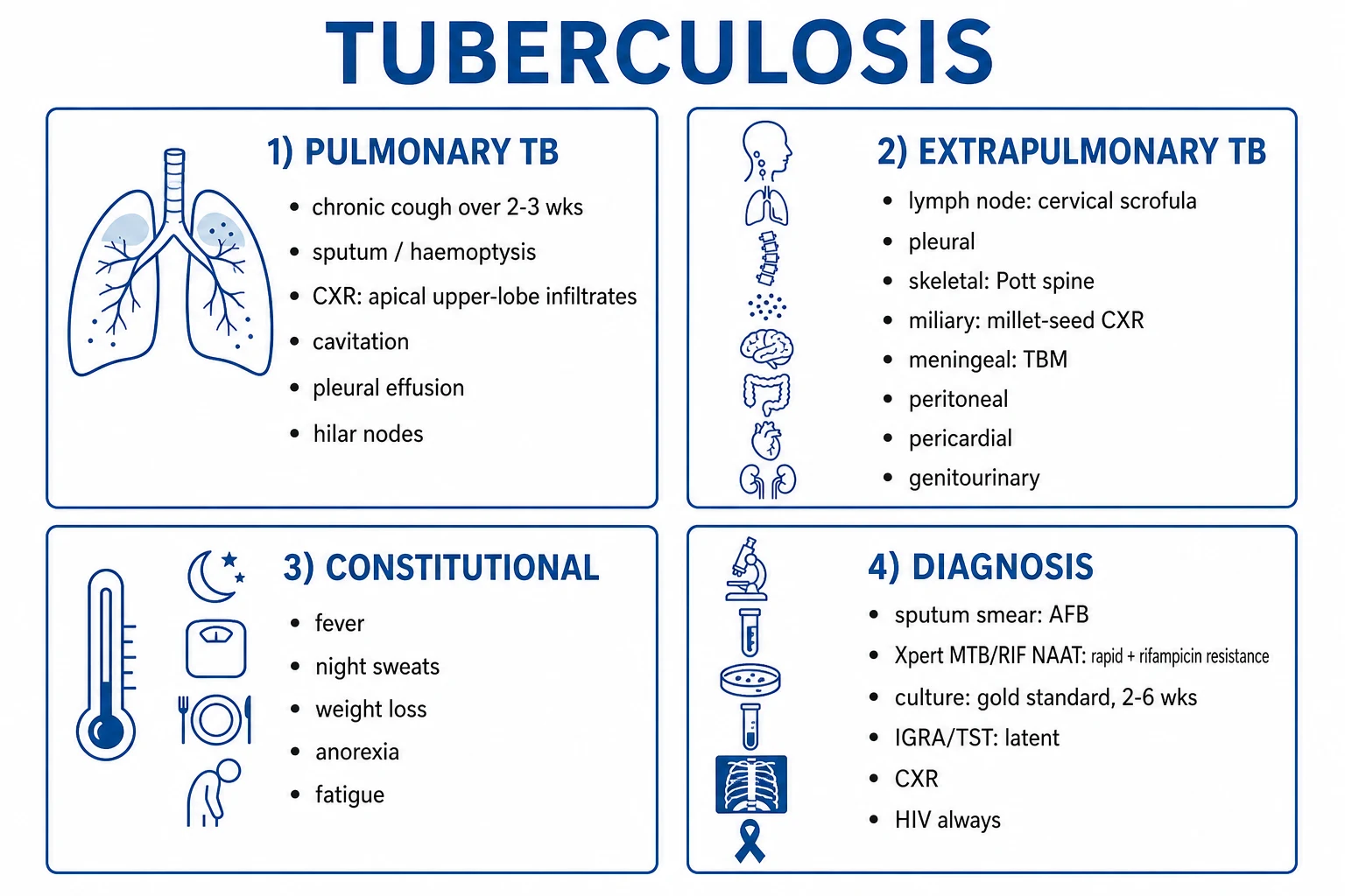
Pulmonary TB — the classic presentation. The cardinal symptom is a chronic cough lasting more than 2 to 3 weeks, initially dry then purulent or mucopurulent, with haemoptysis in about a third (streaks to massive). Constitutional symptoms — low-grade fever (often with an evening rise), drenching night sweats, weight loss, anorexia, fatigue and malaise — are nearly invariable. Pleuritic chest pain and exertional dyspnoea occur with extensive disease, effusion or pneumothorax. The duration (weeks, not days) is the key discriminator from acute bacterial pneumonia.[1]
Extrapulmonary TB — by site: [1]
TB lymphadenitis (scrofula)
Commonest extrapulmonary form
- Painless, slowly enlarging cervical lymph nodes (posterior triangle, supraclavicular)
- Classically matted, firm, non-tender; may form a cold abscess or discharging sinus
- Biopsy shows caseating granulomas; culture/FNA for AFB and Xpert
- Children and young women; HIV co-infection common
Pleural TB
Unilateral effusion
- Unilateral pleural effusion, often with acute febrile presentation (pleurisy)
- Lymphocytic exudate by Light criteria; ADA above 40 U/L supports TB
- Pleural biopsy (caseating granuloma) is more sensitive than fluid culture
- Usually self-limited but represents active disease — treat fully
TB meningitis (TBM)
Most lethal form
- Subacute onset: 1 to 3 weeks of headache, low-grade fever, malaise, then meningism, confusion, cranial-nerve palsies (III, VI, VII)
- Stage 1: conscious, no neuro deficit; Stage 2: confusion or cranial nerve palsy; Stage 3: coma or dense hemiplegia
- CSF: high lymphocytes, high protein (often over 1 g/L), low glucose, cobweb coagulum
- Requires 12 months of therapy plus corticosteroids; mortality 20 to 30 percent even with treatment
Miliary TB
Haematogenous dissemination
- Diffuse 1 to 3 mm nodules on chest X-ray (millet-seed appearance)
- Fever, weight loss, hepatosplenomegaly, anaemia of chronic disease
- Choroidal tubercles on fundoscopy (pathognomonic, seen in about 15 percent)
- Higher in children, HIV, elderly; can cause TBM, adrenal insufficiency (Addisonian)
Skeletal TB (Pott disease)
Spine
- Lower thoracic and upper lumbar spine; gradual back pain, gibbus deformity, paravertebral (psoas) cold abscess
- Pott paraplegia from cord compression; a surgical emergency
- X-ray/MRI: vertebral body destruction, disc-space narrowing, paravertebral abscess
- Treat medically for 9 to 12 months; surgery for cord compression or instability
Genitourinary TB
Kidney, epididymis, Fallopian tube
- Sterile pyuria (pus cells on microscopy, negative routine culture)
- Epididymitis (beaded vas deferens), pelvic inflammatory disease, infertility
- Renal calcification on imaging; moth-eaten renal pelvis
- Send early-morning urine for mycobacterial culture and Xpert
Abdominal / peritoneal TB
Intestinal, peritoneal
- Ascites (lymphocytic exudate, high ADA), doughy abdomen, abdominal masses
- Intestinal TB mimics Crohn disease (terminal ileum, skip lesions)
- Distinguishing from Crohn is a classic exam dilemma; biopsy and culture are essential
Pericardial TB
Effusion, constriction
- Pericardial effusion (tamponade physiology in severe cases)
- May progress to constrictive pericarditis over months to years
- Pericardiocentesis and biopsy; adjunctive corticosteroids debated
Atypical presentations (examiners test these deliberately): [1]
- HIV co-infection — smear-negative pulmonary TB, extrapulmonary and disseminated disease, rapidly progressive, can present as immune reconstitution inflammatory syndrome (IRIS) after ART starts.
- Diabetes — alters the immune response; lower-lobe disease is more common than the classic apical pattern.
- Elderly — non-specific decline, anorexia, weight loss, low-grade fever; TB may be missed or attributed to malignancy.
- Children — paucibacillary, more often extrapulmonary; severe forms (TBM, miliary) are commoner; present with failure to thrive, cough, or non-specific febrile illness.
- Pregnancy — TB can present in any trimester; untreated TB harms mother and fetus; first-line therapy is safe in pregnancy.[1]
Differential Diagnosis
The differential of chronic cough with constitutional symptoms or apical cavitation is broad, and the discriminator is usually tempo, risk factors, and the response (or lack of it) to empirical antibiotics.[1]
| Differential | Distinguishing features |
|---|---|
| Bacterial pneumonia | Acute (days, not weeks), high fever, rigors, productive sputum, responds to antibiotics within 48 to 72 hours; lobar consolidation. TB does not respond to standard antibiotics. |
| Lung cancer | Older smoker, weight loss, haemoptysis; mass (not cavity, though cavitation occurs); no fever resolution; bronchoscopy and biopsy. Squamous cell can cavitate and mimic TB. |
| Lung abscess | Foul-smelling (anaerobic) sputum, air-fluid level in a thick-walled cavity; aspiration risk (alcohol, seizure, swallow disorder). |
| Fungal infection | Histoplasmosis, coccidioidomycosis, blastomycosis in endemic areas (Ohio/Mississippi valleys, US Southwest); can produce apical cavitation identical to TB. Serology, antigen, biopsy. |
| Sarcoidosis | Bilateral hilar lymphadenopathy, non-caseating granulomas on biopsy, erythema nodosum, uveitis; negative TB tests; ACE may be raised. |
| Bronchiectasis | Daily copious purulent sputum from childhood; tram-track bronchi on CT; recurrent infections; whooping cough or TB in childhood. |
| Pneumoconiosis (silicosis) | Occupational history; diffuse nodular/interstitial pattern; huge TB-reactivation risk. |
| Wegener granulomatosis (GPA) | Cavitating lung nodules, sinusitis, glomerulonephritis, c-ANCA positive. |
Tuberculous versus malignant pleural effusion. A TB pleural effusion is a unilateral, lymphocytic exudate with ADA above 40 U/L and often low glucose; mesothelioma and metastatic malignancy can mimic it. Pleural biopsy (caseating granulomas) is more sensitive than fluid culture for TB.[1]
Clinical & Bedside Assessment
Bedside findings of post-primary pulmonary TB are often subtle. Focused respiratory examination may reveal apical crackles (post-tussive, often only on cough), signs of consolidation (bronchial breath sounds, dullness), amphoric breath sounds over a large cavity, or diffuse crackles in miliary disease. Cachexia and muscle wasting reflect the chronic catabolic illness. Look specifically for the extrapulmonary signs that the chest will not show: [1]
- Cervical lymph nodes — matted, firm, non-tender (scrofula); look in the posterior triangle and supraclavicular fossa.
- Spine — gibbus deformity (acute angular kyphosis of Pott disease), a paravertebral (psoas) cold abscess that may point in the groin.
- Abdomen — ascites (peritoneal TB), a doughy abdomen, palpable masses.
- Cardiovascular — a pericardial rub or signs of tamponade (TB pericarditis); later, raised JVP, hepatomegaly, ascites of constrictive pericarditis.
- Neurological — meningism (neck stiffness, photophobia), cranial-nerve palsies (III, VI, VII), altered conscious level, focal deficits (TBM).
- Fundoscopy — choroidal tubercles (small, pale, multiple lesions near the optic disc) — pathognomonic of miliary TB and easy to miss.[1]
Bedside observations that flag severity: SpO2, respiratory rate, BP, GCS, temperature. The public-health reflex — isolate any suspected open pulmonary TB (negative-pressure room, N95 or FFP2/FFP3 respirator) — is activated at the bedside, not after the smear result.[4]
Investigations
The diagnosis of TB rests on demonstrating the organism (smear, molecular test, or culture) in a clinical sample, supported by imaging and the immune-based tests for latent infection. [1]
First-line investigations for suspected pulmonary TB: [1]
- Sputum Xpert MTB/RIF Ultra (NAAT / CBNAAT) — the WHO-recommended initial test for all presumptive TB. Detects M. tuberculosis complex DNA and rifampicin resistance within about 2 hours; far more sensitive than smear (especially in HIV and paucibacillary disease). Two to three spot or early-morning samples.[4][5]
- Sputum smear microscopy (Ziehl-Neelsen or auramine fluorescence for AFB) — rapid and cheap but insensitive (detects about 5,000 to 10,000 bacilli per mL); a negative smear does not exclude TB. Grades scanty to 3-plus.
- Mycobacterial culture — the gold standard and the only test that confirms viability and allows full drug-susceptibility testing. Liquid MGIT culture is faster (1 to 3 weeks) than solid Lowenstein-Jensen (2 to 6 weeks). Always send for culture even when Xpert is positive.
- Chest X-ray — supports the diagnosis and defines extent; apical and posterior upper-lobe infiltrates with cavitation are classic. See patterns below.
- HIV test — mandatory in every patient with TB.[4]
Chest X-ray patterns and what they imply: [1]
- Apical/posterior upper-lobe infiltrates with cavitation — reactivation (post-primary) TB; the classic pattern.
- Hilar or mediastinal lymphadenopathy with a subpleural focus — primary TB (Ghon complex), commoner in children.
- Diffuse, fine, uniform (millet-seed) nodules — miliary TB (disseminated).
- Unilateral pleural effusion — pleural TB.
- Lower-lobe disease — consider diabetes, HIV, or primary progressive TB. [1]
CT chest is reserved for finer detail, for extrapulmonary disease (cavities, tree-in-bud bronchiolar spread, lymph nodes), and to distinguish TB from malignancy. [1]
Tests for latent TB infection (LTBI) — do not diagnose active disease: [1]
- Tuberculin skin test (Mantoux / PPD) — intradermal PPD, read induration at 48 to 72 hours. Thresholds: 5 mm for HIV, immunosuppression, recent contacts, transplant; 10 mm for healthcare workers, migrants from endemic areas, IV drug users, prisoners, diabetes, CKD, silicosis; 15 mm for low-risk individuals. Positive means infection, not disease. False-negative in HIV, malnutrition, miliary TB, corticosteroids (anergy). Cross-reacts with BCG and NTM.
- Interferon-gamma release assay (IGRA, e.g. QuantiFERON-TB Gold, T-SPOT.TB) — measures IFN-gamma release from T-cells exposed to M. tuberculosis antigens (ESAT-6, CFP-10). More specific than TST (not affected by BCG), single visit, but does not distinguish latent from active TB and cannot replace smear/culture for active disease.[4]
Light's criteria for pleural fluid (classifies exudate versus transudate) — fluid is an exudate if any of: pleural-to-serum protein ratio above 0.5; pleural-to-serum LDH ratio above 0.6; or pleural LDH above two-thirds the upper limit of normal for serum. A TB pleural effusion is a lymphocytic exudate with ADA above 40 U/L and often low glucose.[1]
Special situations: [1]
- TB meningitis — CSF. High lymphocytic pleocytosis (10 to 500 cells), high protein (often above 1 g/L, can reach several g/L), low glucose (typically below 2.2 mmol/L or under 40 percent of simultaneous blood glucose), and a cobweb coagulum that forms on standing. Xpert MTB/RIF on CSF is the rapid test; CSF culture confirms. ADA above 10 U/L in CSF supports TB.
- Urine lipoarabinomannan (LF-LAM) — useful in HIV with CD4 below 100, where it detects disseminated TB at the bedside; insensitive in HIV-negative or high-CD4 patients.
- Line-probe assay (LPA) and whole-genome sequencing — for resistance patterns after rifampicin resistance is found on Xpert; LPA detects fluoroquinolone and injectable resistance within a day.[4]
Management — Resuscitation
The immediate priorities in suspected TB are infection control first, then supportive care for any life-threatening complication. [1]
Immediate priorities in suspected open pulmonary TB
1 — Airborne isolation
Place the patient in a **negative-pressure** (or, failing that, single) room with **N95 or FFP2/FFP3 respirator** protection for staff and a surgical mask on the patient during transport. Do NOT wait for the smear result — isolate on suspicion. Keep isolated until the patient is improving and smear-negative (typically after 2 weeks of effective therapy in drug-susceptible disease).
2 — ABCDE and supportive care
**Oxygen** for hypoxia (target SpO2 94 to 98 percent, or 88 to 92 percent in COPD). IV access, bloods (FBC, ESR, CRP, renal and liver function, glucose, HIV test). Manage **sepsis** (miliary or extensive disease) with the Surviving Sepsis hour-1 bundle if hypotensive.
3 — Send diagnostic samples immediately
**Two to three sputum samples** (spot, early-morning, spot) for **Xpert MTB/RIF Ultra**, **AFB smear**, and **mycobacterial culture**. If the patient cannot produce sputum, use **induced sputum** (hypertonic saline nebuliser) or **bronchoscopy** (BAL).
4 — Notify and trace contacts
TB is a **notifiable disease** (Ni-kshay in India, local public health in the UK/ANZ). Initiate **contact screening** for household and close contacts (symptom screen, chest X-ray, IGRA/TST, and CXR for those positive).
5 — Recognise the emergencies
**Massive haemoptysis** (over 200 mL in 24 hours) — protect the airway, place affected-side-down, urgent **bronchial-artery embolisation** or thoracic surgery. **TB meningitis** — empirical anti-TB therapy plus **dexamethasone**, urgent CT then LP. **Tension pneumothorax** from ruptured cavity — needle decompression and chest drain.
Massive haemoptysis is a surgical emergency: the bleeding source is usually a bronchial artery eroded by a cavity. Place the patient affected-side-down (to protect the uninvolved lung from drowning in blood), secure the airway (large-bore endotracheal tube, possible selective main-bronchus intubation), and arrange urgent bronchial-artery embolisation by interventional radiology. Surgery (lobectomy) is reserved for embolisation failure.[1]
Management — Definitive & Stepwise
The standard treatment of drug-susceptible TB is a 6-month regimen of four drugs for the intensive phase (2 months) followed by two drugs for the continuation phase (4 months), given daily and under directly observed therapy (DOTS).[2]
The regimen — 2 HRZE / 4 HR: [1]
| Phase | Duration | Drugs | Doses |
|---|---|---|---|
| Intensive | 2 months (8 weeks) | HRZE — rifampicin, isoniazid, pyrazinamide, ethambutol | Daily, all four |
| Continuation | 4 months (16 weeks) | HR — rifampicin, isoniazid | Daily, two drugs |
| Total | 6 months |
Weight-based daily doses (adults): [1]
| Drug | Daily dose (mg/kg) | Maximum | Toxicity and monitoring |
|---|---|---|---|
| Rifampicin (R) | 10 mg/kg | 600 mg | Orange secretions (harmless), hepatitis, potent CYP3A4 inducer (contraceptives, warfarin, ART, azoles fail). Monitor LFTs. |
| Isoniazid (H) | 5 mg/kg | 300 mg | Hepatitis, peripheral neuropathy (give pyridoxine/B6 25 mg daily prophylactically); slow acetylators at higher risk. Monitor LFTs. |
| Pyrazinamide (Z) | 20 to 30 mg/kg (usual 25) | 2 g | Hepatitis (the most hepatotoxic), hyperuricaemia and gout, arthralgia. Monitor LFTs and uric acid. |
| Ethambutol (E) | 15 mg/kg (range 15 to 20) | 1.6 g | Optic (retrobulbar) neuritis — red-green colour blindness, reduced acuity, central scotoma. Baseline acuity and colour vision; stop immediately if any visual change. |
DOTS — Directly Observed Treatment, Short-course. The five components of DOTS (the WHO strategy, adopted by India's NTEP/RNTCP): [1]
- Political commitment with sustained financing.
- Case detection by quality-assured bacteriology (now Xpert-first).
- Standardised treatment with direct observation — a trained observer watches the patient swallow each dose.
- An effective drug supply and management system (free fixed-dose combinations).
- A monitoring and evaluation system with impact measurement (Ni-kshay in India).[2]
Fixed-dose combinations (FDCs). India's NTEP supplies free AKURIT-4 (RHZE) and AKURIT-2 (RH) FDC tablets, dosed by weight band. FDCs reduce pill burden and improve adherence. [1]
When to extend therapy. Cavitary pulmonary TB with positive 2-month culture, TB meningitis (12 months), bone and joint TB (9 to 12 months), miliary TB (consider 9 months), and pericardial TB (6 months) all warrant extended or modified durations. [1]
Adjunctive corticosteroids. Dexamethasone improves survival in TB meningitis (Thwaites 2004 RCT) and is given for the first 6 to 8 weeks in tapering doses. Steroids are also considered in TB pericarditis (reduce constriction) and severe TB-IRIS; they do not have a role in routine pulmonary TB.[6]
Dexamethasone in TB meningitis (Thwaites et al.)
New England Journal of Medicine
Randomised, double-blind, placebo-controlled trial in 545 Vietnamese adolescents and adults with TB meningitis.
Key finding
Adjunctive dexamethasone (intravenous then oral, tapered over 8 weeks) significantly reduced mortality, especially in Grade I and II disease. The benefit was greatest in those without HIV; the HIV-subgroup benefit was less clear.
Practice change
Corticosteroids are now a standard adjunct in TB meningitis, given alongside a 12-month anti-TB regimen (HRZE intensive with an ethionamide or fluoroquinolone substituted for ethambutol).
Specific Subtypes & Scenarios
TB meningitis (TBM)
The most lethal form of TB. Subacute onset (1 to 3 weeks): headache, low-grade fever, malaise, then meningism, confusion, cranial-nerve palsies (III, VI, VII), and focal deficits. Basal exudate causes hydrocephalus, vasculitis (causing stroke), and cranial-nerve entrapment. The Rich focus — a subpial or subependymal focus that ruptures into the subarachnoid space — is the pathogenesis. CSF: lymphocytic pleocytosis, high protein, low glucose, cobweb coagulum. Treatment is empirical and urgent — do not wait for confirmation if the clinical picture fits: [1]
- 12-month regimen: intensive phase of 2 months HRZE (some centres substitute ethionamide or a fluoroquinolone for ethambutol for better CSF penetration) plus 10 months HR.
- Adjunctive dexamethasone (Thwaites): IV then oral, tapered over 6 to 8 weeks. Improves survival.[6]
- Mortality 20 to 30 percent even with optimal therapy; worse in HIV and in Grade III (comatose) disease.
Miliary TB
Haematogenous dissemination producing 1 to 3 mm nodules throughout both lung fields (and seeding liver, spleen, bone marrow, meninges). Presents with fever, weight loss, hepatosplenomegaly, anaemia of chronic disease, and choroidal tubercles on fundoscopy (pathognomonic). Higher in children, HIV, and the elderly. Treat with the standard 6-month regimen (consider extending to 9 months); look for and treat coexistent TBM.[1]
TB pleural effusion
A unilateral lymphocytic exudate with ADA above 40 U/L and low glucose; pleural biopsy (caseating granulomas) is more sensitive than fluid culture. Represents a hypersensitivity reaction to bacillary antigens as much as direct infection, so it often resolves spontaneously — but the patient still has active TB and must receive the full 6-month regimen.[1]
Skeletal TB — Pott disease
Lower thoracic and upper lumbar spine. Gradual back pain, a gibbus deformity (acute angular kyphosis), a paravertebral (psoas) cold abscess that may track into the groin, and Pott paraplegia from cord compression (a surgical emergency). Imaging shows vertebral-body destruction, disc-space narrowing, and paravertebral abscess. Treat medically for 9 to 12 months; surgery (decompression, stabilisation) for cord compression, neurological deficit, or spinal instability.[1]
TB lymphadenitis (scrofula)
The commonest extrapulmonary form. Painless, slowly enlarging cervical lymph nodes (often posterior triangle and supraclavicular), classically matted, firm, non-tender; may form a cold abscess or discharging sinus. Biopsy shows caseating granulomas; FNA for AFB and Xpert. Treat with the standard 6-month regimen; surgical excision reserved for diagnostic uncertainty or persistent draining sinuses.[1]
Genitourinary TB
Sterile pyuria — urine full of leucocytes on microscopy but negative on routine bacterial culture — is the classic clue (M. tuberculosis does not grow on standard media). Renal calcification, a moth-eaten renal pelvis, epididymitis (beaded vas deferens), pelvic inflammatory disease, and infertility. Send early-morning urine (three samples) for mycobacterial culture and Xpert; treat with the standard 6-month regimen.[1]
Intestinal and peritoneal TB
Intestinal TB (often terminal ileum) mimics Crohn disease — a classic exam dilemma. Features favouring TB: transverse ulcers, caseating granulomas, ascites with high ADA, peritoneal thickening, chest X-ray changes, positive TB tests. Features favouring Crohn: longitudinal (aphthous) ulcers, non-caseating granulomas, fistulae, perianal disease, skip lesions. Biopsy and culture are decisive.[1]
TB-HIV co-infection
Every patient with TB must be tested for HIV, and vice versa. HIV transforms TB: more smear-negative, extrapulmonary, and disseminated disease, higher mortality. Start ART early — within 2 weeks of anti-TB therapy for CD4 under 50 cells per microlitre, and within 8 weeks for higher CD4. Rifamycin-ART interactions are critical: rifampicin is a potent CYP3A4 inducer and lowers levels of most protease inhibitors (use efavirenz-based or rifabutin-based regimens). Watch for TB-IRIS (immune reconstitution inflammatory syndrome) — paradoxical worsening of TB symptoms 1 to 8 weeks after ART starts, treated with corticosteroids if severe.[1]
Childhood TB
Paucibacillary (smear often negative), more often extrapulmonary (lymph node, TBM, miliary). Severe forms (TBM, miliary) are commoner because of immature cell-mediated immunity. Weight-based dosing is essential. The primary complex (Ghon focus plus hilar lymphadenopathy) is the radiological hallmark. BCG is given at birth and protects against disseminated/childhood TB and TBM.[1]
Complications & Pitfalls
Local complications of pulmonary TB: massive haemoptysis (cavity erosion into a bronchial artery — emergency), aspergilloma (Aspergillus colonising a chronic cavity — causes further haemoptysis), post-TB bronchiectasis and pulmonary fibrosis (lung destruction), pleural empyema, pneumothorax (ruptured cavity), cor pulmonale from chronic destructive disease. [1]
Extrapulmonary complications: Pott paraplegia (cord compression), constrictive pericarditis (TB pericarditis), hydrocephalus and cranial-nerve palsy (TBM), adrenal insufficiency (Addison disease) from adrenal TB, infertility (genital TB), renal failure (destructive GU TB). [1]
Drug toxicity is the commonest reason to interrupt therapy: [1]
- Hepatotoxicity — rifampicin, isoniazid, pyrazinamide all hepatotoxic. Transient asymptomatic transaminitis is common and monitored; if ALT above 3 times the upper limit of normal with symptoms (jaundice, nausea, right-upper-quadrant pain) or above 5 times without symptoms, stop the offending drug(s) — pyrazinamide is usually stopped first (most hepatotoxic), then rifampicin, then isoniazid; reintroduce sequentially once LFTs normalise.
- Ethambutol optic neuritis — check acuity and red-green colour vision at baseline and monthly; stop immediately on any change (often irreversible if not caught early).
- Isoniazid peripheral neuropathy — prevented by pyridoxine (B6) 25 mg daily.
- Rifampicin — orange secretions (harmless), CYP induction (contraceptive and ART failure, warfarin dose adjustment).[2]
TB-IRIS (immune reconstitution inflammatory syndrome) in HIV: paradoxical worsening of TB symptoms 1 to 8 weeks after ART starts, driven by recovering immunity. Distinguish from treatment failure (positive cultures, worsening on therapy) and paradoxical reaction (similar in HIV-negative patients on anti-TB therapy). Treat mild IRIS symptomatically; severe IRIS (respiratory distress, TBM, severe lymphadenitis) with prednisolone 1 to 1.5 mg/kg/day tapered over weeks.[1]
Classic pitfalls: missing extrapulmonary or atypical TB (especially in HIV, elderly, diabetic); treating smear-negative chronic cough with repeated courses of antibiotics without sending Xpert; delaying MDR-TB detection by not sending Xpert on every patient; missing ethambutol optic neuritis by not testing vision; default and lost-to-follow-up fuelling resistance; giving rifampicin with protease inhibitors without dose adjustment; failing to test for HIV in every TB patient. [1]
Prognosis & Disposition
Drug-susceptible TB is curable in over 85 percent of patients with a full 6-month regimen under DOTS. Outcome is determined by adherence, drug susceptibility, host factors (HIV, diabetes, malnutrition), site (TBM and miliary worse), and the timeliness of diagnosis. Untreated, TB kills roughly half of patients within 5 years; latent TB carries a roughly 1-in-10 lifetime reactivation risk (much higher in HIV).[1]
Ending isolation. A patient with drug-susceptible pulmonary TB becomes non-infectious after 2 weeks of effective therapy provided they are clinically improving (less cough, less fever) and have negative follow-up smears. Drug-resistant TB requires negative cultures before de-isolation — a much longer period. [1]
WHO treatment-outcome categories: cured (smear/culture negative at end of treatment), treatment completed (full course, no end-of-treatment bacteriology), treatment failure (positive culture at 5 months or beyond), died, lost to follow-up (default), not evaluated.[2]
Mortality of severe forms. TB meningitis mortality is 20 to 30 percent even with optimal therapy; miliary TB mortality is 15 to 20 percent, higher in HIV and the elderly. These forms warrant inpatient initiation of therapy. [1]
Special Populations
TB in HIV
The highest-risk group. Screen all people with HIV for TB at every encounter (symptom screen — cough, fever, weight loss, night sweats; if positive, Xpert and CXR). Treat TB first, then start ART early: within 2 weeks of anti-TB therapy for CD4 under 50, within 8 weeks for CD4 50 to 200, and by 8 weeks for higher CD4. Use efavirenz-based ART with rifampicin (avoid protease inhibitors); or substitute rifabutin for rifampicin if a protease-inhibitor regimen is needed. Watch for TB-IRIS. Give cotrimoxazole prophylaxis. Treat latent TB in all HIV-positive patients without active disease.[1]
TB in pregnancy
Treat TB in pregnancy — first-line drugs are safe. The regimen is 2 HRZE plus 4 HR; do NOT give streptomycin (ototoxicity to the fetus — causes congenital deafness). Pyridoxine is given with isoniazid. Untreated TB harms mother and fetus (prematurity, low birth weight, congenital TB). Treat latent TB in pregnancy if high-risk (HIV, recent contact); otherwise defer to the post-partum period. Breastfeeding is safe on first-line therapy (small drug amounts in milk, not harmful).[1]
TB in children
Paucibacillary, more often extrapulmonary, severe forms (TBM, miliary) commoner. Weight-based dosing of all four drugs. Gastric aspirate or induced sputum for samples (children cannot expectorate). BCG protects against disseminated/childhood TB and TBM. Treat with the standard 6-month regimen; ethambutol is sometimes omitted in young children due to difficulty testing vision.[1]
TB in chronic kidney disease and dialysis
Higher risk of reactivation. Adjust ethambutol and pyrazinamide dosing for renal function (ethambutol 15 mg/kg three times weekly after haemodialysis; pyrazinamide 25 mg/kg three times weekly). Rifampicin and isoniazid do not need renal adjustment. Treat latent TB in all dialysis patients with positive IGRA.[1]
TB before immunosuppression (anti-TNF, transplant)
Screen with IGRA and chest X-ray before starting anti-TNF therapy (infliximab, adalimumab, etanercept), transplant immunosuppression, or high-dose corticosteroids. Treat latent TB first (3HP, 4R, or 9H) — TNF-alpha is central to granuloma containment, and anti-TNF reactivates latent TB at 5 to 10 times the baseline rate. Do not start anti-TNF until at least one month of LTBI therapy is complete.[11]
Evidence, Guidelines & Regional Differences
The WHO publishes the Consolidated Guidelines on Tuberculosis (Modules 1 to 5), updated annually, covering diagnosis, treatment, prevention, and drug resistance. Key recommendations:[4]
- Diagnosis: Xpert MTB/RIF Ultra is the initial test for all presumptive TB (replacing smear as the first test), and for extrapulmonary samples (CSF, lymph-node, pleural, urine in HIV).
- Drug-susceptible TB treatment: 6 months of HRZE/HR; daily dosing; no intermittent therapy in the intensive phase.
- MDR-TB: the shorter all-oral BPaLM regimen (bedaquiline, pretomanid, linezolid, plus moxifloxacin for 6 months) for eligible MDR/pre-XDR cases; longer individualised all-oral regimens for the rest. No more injectables as first-line MDR therapy (otoxicity).
- Preventive therapy: 3HP (weekly rifapentine plus isoniazid for 3 months), 4R (daily rifampicin for 4 months), or 6H/9H (isoniazid). [1]
Nix-TB: bedaquiline, pretomanid, linezolid for XDR/MDR-TB (Conradie et al.)
New England Journal of Medicine
Single-arm, open-label trial of the 6-month all-oral BPaL regimen in 109 patients with extensively drug-resistant or treatment-intolerant MDR-TB in South Africa.
Key finding
Favourable outcome in 90 percent at 6 months after the end of therapy — a transformational result in a disease with historical cure rates under 50 percent and 18-month injectable regimens. Linezolid toxicity (myelosuppression, neuropathy) required dose reduction in many patients.
Practice change
The BPaL and BPaLM regimens are now WHO-recommended for eligible MDR/pre-XDR-TB cases, replacing prolonged injectable-containing regimens.
Regional program differences: [1]
WHO consolidated guidelines govern global practice: Xpert-first diagnosis, 6-month HRZE/HR, BPaLM for eligible MDR, and a full preventive-therapy programme. The WHO End TB Strategy targets a 90 percent reduction in incidence and 95 percent reduction in mortality by 2035 (baseline 2015).
Australia / New Zealand: similar to the UK — IGRA-based, 6-month HRZE/HR, notifiable, contact tracing by public health. Neonatal BCG given selectively to at-risk groups (Aboriginal and Torres Strait Islander neonates in some regions, migrant neonates).
India — NTEP / RNTCP
Daily DOTS, free FDCs, Ni-kshay
- **National TB Elimination Programme (NTEP)**, formerly RNTCP — daily DOTS, free AKURIT-4/2 FDCs by weight band
- **Ni-kshay** — the national web-based notification and tracking system (mandatory notification of every case)
- **Bidirectional TB-HIV screening** — every TB patient tested for HIV and vice versa
- Daily regimen (changed from the older thrice-weekly intermittent regimen); Xpert-first diagnosis
- **India End TB** targets TB elimination (incidence under 1 per million) by 2025 — five years ahead of the WHO target
UK — NICE NG33
IGRA-based, entry screening
- IGRA (not TST) for latent TB testing in adults and children over 5; TST acceptable under 5
- Entry screening for migrants from high-incidence countries (over 40 per 100,000)
- 6-month HRZE/HR; 4R or 3HP for latent TB
- Notifiable; contact tracing by public health; selective neonatal BCG
US — ATS/CDC/IDSA
IGRA preferred, targeted BCG
- IGRA preferred over TST for latent TB in most settings
- 6-month HRZE/HR; 3HP, 4R, 9H for latent TB
- No universal BCG; targeted testing of high-risk groups instead
- Mandatory notification to state health departments; public-health-funded treatment
Prevention
BCG vaccine
Bacille Calmette-Guerin (BCG) is a live attenuated strain of M. bovis. Given intradermally at birth (in high-burden countries, including India — universal) or selectively (UK, ANZ, US — high-risk neonates only). The Colditz meta-analysis established that BCG: [1]
- Protects against disseminated and childhood TB and TB meningitis (efficacy about 70 to 80 percent for these severe forms) — the major benefit.
- Variable and modest protection against adult pulmonary TB (0 to 80 percent across studies; latitude-dependent — less protection nearer the equator, possibly due to NTM exposure).
- Contraindicated in immunocompromised (HIV with symptoms, primary immunodeficiency) — risk of disseminated BCG infection.[12]
Infection control
Airborne precautions for all suspected or confirmed pulmonary/laryngeal TB: negative-pressure isolation (or single room), N95 or FFP2/FFP3 respirators for staff (surgical masks are insufficient for the airborne route — they protect the patient from the environment, not the staff from the patient), and UV germicidal irradiation and natural ventilation in high-burden settings. De-isolate after 2 weeks of effective therapy with clinical improvement and negative smears (drug-susceptible disease). [1]
Latent TB infection (LTBI) preventive therapy
Targeted at those most likely to progress. Regimens: [1]
- 3HP — weekly rifapentine plus isoniazid for 3 months (12 doses); preferred for most adults and children over 2.
- 4R — daily rifampicin for 4 months; fewer hepatotoxic events than isoniazid.
- 6H or 9H — daily isoniazid for 6 to 9 months (the traditional regimen, still used in pregnancy and children); give pyridoxine. [1]
Indications: recent contact of an infectious case; HIV-positive; before anti-TNF or transplant immunosuppression; end-stage CKD/dialysis; silicosis; fibrotic lesions on chest X-ray from old TB. Exclude active TB first (symptom screen, CXR) — giving monotherapy isoniazid to a patient with active disease breeds resistance.[10][11]
Non-tuberculous mycobacteria (NTM)
NTM — Mycobacterium avium complex (MAC), M. kansasii, M. abscessus, M. fortuitum — cause chronic pulmonary disease (especially in COPD, bronchiectasis, older women with nodular bronchiectatic pattern — Lady Windermere syndrome), cervical lymphadenitis in children, and disseminated disease in advanced HIV (MAC bacteraemia with CD4 under 50). They are not contagious (environmental source — water, soil). Diagnosis requires meeting clinical, radiographic, and microbiological criteria (at least two positive cultures from sputum, or one from BAL or biopsy) — a single positive can be colonisation. Treatment differs from TB: MAC needs macrolide (azithromycin/clarithromycin) plus rifampicin plus ethambutol for at least 12 months with culture negativity; M. abscessus needs multidrug IV therapy (amikacin, imipenem, cefoxitin, tigecycline, macrolide) and often surgical resection.[1]
Exam Pearls
The four first-line drugs — RIPE
RIPE
RNA polymerase inhibitor; orange secretions; CYP3A4 inducer; hepatitis
Mycolic-acid inhibitor; neuropathy (give pyridoxine/B6 25 mg); hepatitis
Sterilises dormant bacilli (the drug that shortened the regimen to 6 months); hyperuricaemia; hepatitis
Arabinosyl-transferase inhibitor; optic neuritis (red-green colour vision); dropped after intensive phase
High-yield one-liners examiners reward: [1]
- Ghon complex = primary subpleural focus plus draining lymph node; Ranke complex = the healed, calcified form.
- Caseating granuloma with Langhans giant cells is the histological hallmark; acid-fast bacilli stain with Ziehl-Neelsen (carbol fuchsin, acid-alcohol decolourisation); the cord factor (trehalose dimycolate) causes serpentine cording in liquid culture.
- Apical or posterior upper-lobe cavitation = reactivation TB (high oxygen); orange-red urine = rifampicin (harmless); red-green colour blindness or visual change = ethambutol optic neuritis — stop the drug.
- Pyridoxine (B6) 25 mg daily prevents isoniazid neuropathy; isoniazid and pyrazinamide are the most hepatotoxic; rifampicin induces CYP3A4 (oral contraceptive and ART failure, warfarin dose needs doubling).
- Sterile pyuria (pus cells, negative routine culture) = genitourinary TB; gibbus deformity plus cold abscess = Pott disease; Rich focus = the subpial focus that ruptures to cause TBM; choroidal tubercles = miliary TB.
- BCG prevents childhood disseminated TB and TBM (not adult pulmonary TB reliably); 3HP = weekly rifapentine plus isoniazid for 12 doses; BPaLM = bedaquiline, pretomanid, linezolid, plus moxifloxacin for MDR-TB.
- DOTS five components (sustained political commitment; case detection by bacteriology; standardised supervised treatment; effective drug supply; monitoring and evaluation); Ni-kshay = India's notification system; always test HIV in every TB patient; isolate (airborne) suspected open TB.
- MDR-TB = resistant to at least rifampicin plus isoniazid; XDR-TB = MDR plus fluoroquinolone plus bedaquiline or linezolid; streptomycin is NOT used in pregnancy (fetal ototoxicity); dexamethasone improves survival in TBM (Thwaites 2004).[1][2]
Exam application bank (NEET-PG / INICET)
One-line answer
Tuberculosis (TB) is infection by Mycobacterium tuberculosis complex, transmitted by the airborne route via droplet nuclei. It is usually pulmonary but can affect any organ. Distinguish latent TB infection (LTBI) — contained, non-infectious bacilli — from active TB disease — multiplying, transmissible infection causing chronic cough (often with haemoptysis), weight loss, night sweats and fever, with apical cavitation on chest X-ray. Diagnosis is by sputum smear and culture, with Xpert MTB/RIF Ultra providing rapid molecular detection and rifampicin-resistance testing. Standard drug-susceptible TB is treated with a 6-month, four-drug regimen — 2 months of HRZE (rifampicin, isoniazid, pyrazinamide, ethambutol) then 4 months of HR, given as directly observed therapy (DOTS). Multidrug-resistant TB (MDR-TB) needs prolonged individualised therapy or the shorter BPaLM regimen. Drug toxicity is
Worked stems (answer without another resource)
Stem 1 — Classic presentation. Map symptoms to mechanism; name the first investigation and first treatment step with dose/route if drug therapy is standard. [1]
Stem 2 — Unstable / complicated. List red flags that force immediate resuscitation, theatre, ICU, antidote, or reperfusion — and what you do in the first 15 minutes. [1]
Stem 3 — Atypical group. Elderly, pregnancy, child, or immunocompromised: how presentation and thresholds change. [1]
Stem 4 — Differential trap. Name the three closest mimics and one discriminator for each. [1]
Stem 5 — Disposition. Who goes home with safety-netting, who is admitted, who needs HDU/ICU/theatre, and what follow-up is mandatory. [1]
Rapid viva checklist
- Definition + classification
- Pathophysiology chain
- Bedside signs / criteria
- Score with exact components (if any)
- Emergency bundle
- Definitive therapy with doses
- Complications of disease and of treatment
- Special populations
- Guideline/trial name if classic
- Three exam traps
Coverage self-check
If you cannot answer any stem above from this page alone, re-read the matching section — the page is intended to be self-sufficient for final-prof and NEET-PG/INICET questions on Tuberculosis.
References
- [1]Pai M, Behr MA, Dowdy D, et al. Tuberculosis Nat Rev Dis Primers, 2016.PMID 27784885
- [2]Sotgiu G, Nahid P, Loddenkemper R, et al. The ERS-endorsed official ATS/CDC/IDSA clinical practice guidelines on treatment of drug-susceptible tuberculosis Eur Respir J, 2016.PMID 27587550
- [3]Nahid P, Mase SR, Migliori GB, et al. Treatment of Drug-Resistant Tuberculosis. An Official ATS/CDC/ERS/IDSA Clinical Practice Guideline Am J Respir Crit Care Med, 2019.PMID 31729908
- [4]Lewinsohn DM, Leonard MK, LoBue PA, et al. Official American Thoracic Society/Infectious Diseases Society of America/Centers for Disease Control and Prevention Clinical Practice Guidelines: Diagnosis of Tuberculosis in Adults and Children Clin Infect Dis, 2017.PMID 28052967
- [5]Horne DJ, Kohli M, Zifodya JS, et al. Xpert MTB/RIF Ultra assay for pulmonary tuberculosis and rifampicin resistance in adults and adolescents Cochrane Database Syst Rev, 2025.PMID 40728034
- [6]Thwaites GE, Nguyen DB, Nguyen HD, et al. Dexamethasone for the treatment of tuberculous meningitis in adolescents and adults N Engl J Med, 2004.PMID 15496623
- [7]Conradie F, Diacon AH, Ngubane N, et al. Treatment of Highly Drug-Resistant Pulmonary Tuberculosis N Engl J Med, 2020.PMID 32130813
- [8]Murray CJ, Ortblad KF, Guinovart C, et al. Global, regional, and national incidence and mortality for HIV, tuberculosis, and malaria during 1990-2013: a systematic analysis for the Global Burden of Disease Study 2013 Lancet, 2014.PMID 25059949
- [9]Boehme CC, Nabeta P, Hillemann D, et al. Rapid molecular detection of tuberculosis and rifampin resistance N Engl J Med, 2010.PMID 20825313
- [10]Sterling TR, Njie G, Zenner D, et al. Guidelines for the Treatment of Latent Tuberculosis Infection: Recommendations from the National Tuberculosis Controllers Association and CDC, 2020 MMWR Recomm Rep, 2020.PMID 32053584
- [11]Getahun H, Matteelli A, Abubakar I, et al. Management of latent Mycobacterium tuberculosis infection: WHO guidelines for low tuberculosis burden countries Eur Respir J, 2015.PMID 26405286
- [12]Colditz GA, Brewer TF, Berkey CS, et al. Efficacy of BCG vaccine in the prevention of tuberculosis. Meta-analysis of the published literature JAMA, 1994.PMID 8309034