Rheumatology · General Medicine
Adult-Onset Still's Disease
Also known as Adult Still disease · Adult-onset Still disease · AOSD · Wissler-Fanconi syndrome
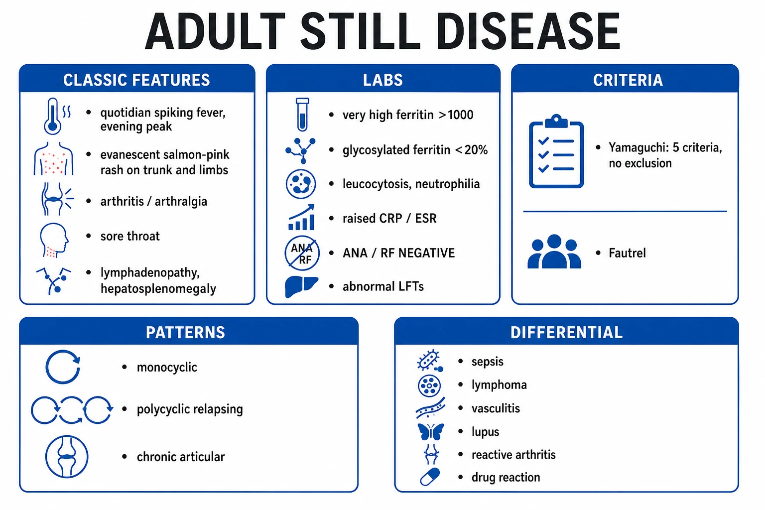
Adult-onset Still's disease (AOSD) is a rare systemic autoinflammatory disorder (the adult counterpart of systemic juvenile idiopathic arthritis) presenting with quotidian spiking fevers (evening peak), an evanescent salmon-pink rash, arthritis, sore throat, and markedly raised inflammatory markers (ferritin often very high) with negative autoantibodies (ANA, RF negative). It affects young adults (16 to 35). Three patterns: self-limited monocyclic, polycyclic (relapsing-remitting), or chronic articular. The feared complication is macrophage activation syndrome (MAS) / secondary HLH — a life-threatening hyperinflammatory emergency. Diagnosis is clinical (Yamaguchi or Fautrel criteria), after excluding infection, malignancy and other rheumatic disease. Treatment: NSAIDs and glucocorticoids first-line; methotrexate; IL-1 inhibitors (anakinra, canakinumab) for refractory systemic disease and IL-6 inhibitor (tocilizumab) for the chronic articular pattern.
On this page & tools
Your progress
Saved locally on this device.
Exam tags
Red flags
Overview & Definition
Adult-onset Still's disease (AOSD) is a rare, multisystem, autoinflammatory disorder of unknown cause and the adult counterpart of systemic juvenile idiopathic arthritis (sJIA). It is defined by the clinical tetrad of quotidian (daily) spiking fever, an evanescent salmon-pink rash, arthritis or arthralgia, and sore throat, occurring in the setting of markedly raised inflammatory markers and ferritin with negative autoantibodies (ANA and rheumatoid factor both negative).[1][6]
The defining clinical skill at the bedside is recognising that AOSD is a diagnosis of exclusion. It mimics sepsis (spiking fevers, rash, neutrophilic leucocytosis, very high inflammatory markers) but the patient has negative blood cultures, negative autoantibodies, and a dramatically elevated ferritin. The diagnostic task is therefore the systematic exclusion of three categories of mimic — infection, malignancy (especially lymphoma), and other rheumatic disease — and only then applying clinical classification criteria (Yamaguchi or Fautrel).[1][2]
The single most important conceptual point is that AOSD sits on the autoinflammatory side of the immune spectrum, not the autoimmune side. There is no high-titre adaptive autoantibody response — which is why ANA and RF are negative and why the disease responds so dramatically to cytokine-blocking biologics (IL-1, IL-6 and IL-18 inhibitors) rather than to B-cell depletion. The feared complication is macrophage activation syndrome (MAS), synonymous here with secondary haemophagocytic lymphohistiocytosis (HLH) — a hyperinflammatory emergency signalled by falling blood counts and fibrinogen, rising ferritin, LDH, triglycerides and transaminases, and persistent fever, carrying significant mortality.[2][6]
Classification & Clinical Patterns
AOSD is best classified by its clinical course pattern, because the pattern predicts prognosis and guides the choice of biologic. Three patterns are recognised, plus the feared MAS-complicated course:[2][6]
- Monocyclic pattern (about one-third of cases): a single systemic episode that fully remits within a year (usually within nine months) and does not recur. This carries the best prognosis and often needs only NSAIDs and a short, tapering course of glucocorticoids.
- Polycyclic (relapsing-remitting) pattern (about one-fifth): recurrent systemic flares separated by remissions of variable length; between flares the patient is well and inflammatory markers normalise. Joint damage is usually modest because the flares are systemic rather than relentlessly articular.
- Chronic articular pattern (about one-third to half): persistent polyarthritis that dominates the course and carries the greatest structural morbidity — erosive, deforming disease of the wrists, knees, ankles and small joints of the hands. This pattern most needs methotrexate and is the pattern in which IL-6 blockade (tocilizumab) is most useful. [1]
A fourth, feared pattern is AOSD complicated by macrophage activation syndrome (secondary HLH) — a medical emergency that can complicate any of the three patterns, most often early in the disease course or during a flare.[2]
Epidemiology & Risk Factors
AOSD is genuinely rare, which is itself a diagnostic clue — a common disease presenting this way is far more likely to be infection or lymphoma than AOSD.[1]
- Incidence: about 0.16 to 0.4 new cases per 100,000 adults per year; prevalence roughly 1 to 34 per million depending on the population studied. Reported worldwide, with no definite ethnic predilection.
- Age: a characteristic bimodal distribution — a first peak at 15 to 25 years and a second peak at 36 to 46 years. Most patients are between 16 and 35.
- Sex: roughly equal, with a slight female preponderance (M = F in most series).
- Genetic predisposition: associations with HLA-DRB1 alleles and, in some populations, HLA-B17 and HLA-B35 — consistent with a polygenic, innate-immune disorder rather than a classic HLA-linked autoimmune disease.
- Environmental trigger: an infective or stressor stimulus in a susceptible host is frequently reported before onset — rubella, parvovirus B19, Epstein-Barr virus, Coxsackie, cytomegalovirus, Yersinia, Chlamydia pneumoniae, and Mycoplasma among others. None is causative in the Koch sense; the model is that an infection ignites a dysregulated innate-inflammatory response that then becomes self-sustaining.
- Mortality: overall around 1 to 5 percent, driven mainly by macrophage activation syndrome, liver failure, and systemic thrombosis; MAS complicates about 10 to 15 percent of cases and is the leading cause of death.[2][6]
Pathophysiology
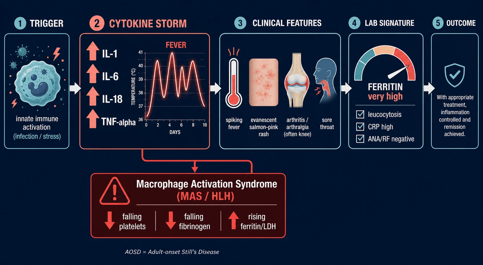
AOSD is a polygenic autoinflammatory disease of innate immunity. Activated macrophages, neutrophils and natural killer cells generate a cytokine storm dominated by interleukin (IL)-1β, IL-6, IL-18 and TNF-α, with a marked interferon-γ (IFN-γ) signature in the most severe disease. There is no high-titre adaptive autoantibody response, which is precisely why ANA and RF are negative and why the disease responds so dramatically to cytokine-blocking biologics.[1][2][9]
The cytokine biology explains each clinical feature and is worth memorising because it is directly examined: [1]
- IL-1β is the pivotal early cytokine. It drives the fever, rash and systemic inflammation and underlies the often-dramatic, within-hours response to IL-1 receptor blockade (anakinra, canakinumab). IL-1 blockade is now the first-line biologic for systemic disease.[7]
- IL-6 is the key driver of the chronic articular pattern, the acute-phase response (CRP, ESR, ferritin synthesis by the liver) and the reactive thrombocytosis. This is why tocilizumab (anti-IL-6 receptor) is the preferred biologic when the chronic articular pattern dominates.[8]
- IL-18 is produced by activated macrophages and Kupffer cells and is the cytokine most closely linked to macrophage activation syndrome. Markedly elevated serum IL-18 (often over 10,000 pg/mL) identifies patients at risk of MAS, and anti-IL-18 biologics (tadekinig alfa) are an emerging therapy.[6]
- IFN-γ amplifies the macrophage activation loop and, with IL-18, produces the uncontrolled haemophagocytosis of MAS; JAK inhibitors (ruxolitinib) are increasingly used in refractory MAS for exactly this reason.[9]
- TNF-α plays a smaller role than in rheumatoid arthritis; TNF inhibitors (etanercept, adalimumab) are less effective for systemic AOSD than IL-1/IL-6 blockade but are used in selected chronic articular cases.
Clinical Presentation
The classic tetrad (the exam answer) and its nuances:[1][2]
- Quotidian spiking fever — peak in the late afternoon or evening, usually 39 °C or higher (most over 39.5 °C), returning towards normal in the morning so that the patient is often well between spikes. This quotidian (once-daily) pattern, with one or two daily spikes, is the most consistent feature and often the presenting symptom. A fever that is unremitting and does not dip towards normal should make you question the diagnosis.
- Evanescent salmon-pink rash — non-pruritic, macular or maculopapular, salmon-pink, distributed over the trunk, proximal limbs and areas of pressure or rubbing (Koebner phenomenon). It appears with the fever and fades without scarring as the fever settles, so it is frequently missed — examine the patient during a fever spike and ask the patient or family to photograph it.
- Arthritis / arthralgia — usually polyarticular and symmetric, affecting the wrists, knees, ankles, metacarpophalangeal and proximal interphalangeal joints, shoulders, elbows and hips. In the chronic articular pattern the arthritis can become erosive and deforming, sometimes indistinguishable from rheumatoid arthritis except for the negative RF/anti-CCP and the preceding systemic illness.
- Sore throat — a persistent, non-exudative pharyngitis reported by over 90 percent of patients; a frequently under-valued minor criterion that, in the right context, strongly supports the diagnosis. [1]
Other systemic features: marked malaise, fatigue and weight loss (often several kilograms over weeks); severe myalgia; generalised lymphadenopathy (often cervical); splenomegaly and hepatomegaly; abdominal pain (mesenteric lymphadenopathy or serositis); and serositis — pleuritis and pericarditis are the commonest cardiac/thoracic manifestations, while myocarditis, tamponade, pleural effusion, interstitial pneumonitis and acute respiratory distress syndrome are rarer but life-threatening.[2][6]
The laboratory pattern is as characteristic as the clinical picture: very high ferritin (often over 1000 μg/L, sometimes over 10,000), neutrophilic leucocytosis (often over 15 ×10⁹/L, with neutrophils over 80 percent), raised CRP and ESR, reactive thrombocytosis, anaemia of chronic disease, ANA and RF negative, glycosylated ferritin under 20 percent, and abnormal liver function tests (raised AST/ALT, sometimes a cholestatic picture or, rarely, acute liver failure).[2]
Atypical presentations — the examiner-tested pitfalls: predominantly articular disease mimicking rheumatoid arthritis (but RF/anti-CCP negative); a fever of unknown origin with no rash (the rash missed because no one examined during a spike); an older patient presenting as a fever of unknown origin; acute liver injury or hepatic failure as the presenting feature (mimicking viral or drug-induced hepatitis); myocarditis or cardiac tamponade from serositis; and the dramatic debut of macrophage activation syndrome as the first manifestation of disease.[1]
Differential Diagnosis
Because AOSD is a diagnosis of exclusion, the differential is the diagnostic process: each mimic must be actively ruled out before criteria are applied.[1][2]
- Infection — sepsis, infective endocarditis, occult abscess, disseminated TB, malaria, enteric fever. Spiking fever, leucocytosis, high inflammatory markers all overlap completely. Exclude with multiple blood cultures, echocardiography, and targeted imaging. This is the cardinal exclusion: a missed infected source is the most dangerous error.
- Lymphoma / leukaemia / malignancy — fever of unknown origin, lymphadenopathy, hepatosplenomegaly, and a high ferritin can all occur in lymphoma. Exclude with LDH, CT chest/abdomen/pelvis, lymph-node biopsy, and bone-marrow biopsy when cytopenias or atypical features are present.
- Systemic lupus erythematosus — fever, rash, arthritis overlap. Distinguished by positive ANA and anti-dsDNA, cytopenias (especially lymphopenia), renal disease, low complement, and a lower ferritin. AOSD has negative ANA/RF and a much higher ferritin.
- Acute rheumatic fever — young patient, fever, arthritis, sore throat. Distinguished by migratory polyarthritis, carditis, raised ASO titre, evidence of preceding streptococcal infection, and Jones criteria.
- Reactive arthritis / viral arthritis (parvovirus B19, hepatitis B and C, rubella, EBV, HIV) — preceding infection, asymmetric oligoarthritis, usually self-limited; serology distinguishes these.
- Rheumatoid arthritis — symmetric small-joint polyarthritis with positive RF and anti-CCP, lower ferritin, no evanescent rash or quotidian fever.
- ANCA-associated vasculitis / polyarteritis nodosa — constitutional features plus organ-specific disease (renal, pulmonary, neuropathy); distinguish by ANCA, angiography, biopsy.
- Periodic fever syndromes / familial autoinflammatory disorders (familial Mediterranean fever, PFAPA, TRAPS) — recurrent fevers, younger age, ethnic predilection, genetic testing of the relevant genes.
- DRESS (drug reaction with eosinophilia and systemic symptoms) / serum sickness / severe drug reaction — rash, fever, eosinophilia, and a clear recent drug exposure with a characteristic latency (two to eight weeks for DRESS). [1]
Clinical & Bedside Assessment
Approach the patient as a fever of unknown origin and a diagnosis of exclusion: rule out infection first, then malignancy, then other rheumatic disease, and only then apply the classification criteria.[1]
- Confirm the quotidian fever pattern (one to two daily spikes, usually evening, over 39 °C) with a nursing temperature chart over several days — the quotidian rhythm is itself diagnostic information and must be documented, not assumed.
- Examine during a fever spike. The salmon-pink rash is evanescent and is reliably seen only when the patient is febrile; examine the trunk, proximal limbs and pressure areas, and ask the patient or family to photograph the rash between reviews.
- Examine the throat (non-exudative pharyngitis), palpate for lymphadenopathy and hepatosplenomegaly, auscultate the lungs and heart for a pericardial or pleural rub, and examine all joints (wrists and small joints of the hands most often).
- Look actively for MAS at every review: new persistent fever with falling white-cell and platelet counts, falling fibrinogen, and rising ferritin, LDH and transaminases — this changes the patient from a diagnostic puzzle to an emergency.
- Document the systemic score (the Pouchot normalised score) if used locally, and re-examine serially: AOSD evolves over days to weeks.
- Examine the rash for Koebner phenomenon — linear streaks of rash along lines of scratching or tight clothing (under waistbands, bra-straps, watch-straps) — a useful confirmatory sign, because the rash favours areas of mild mechanical trauma.[1]
- Listen for a pericardial or pleural friction rub and look for a pericardial effusion or pleural effusion clinically (muffled heart sounds, raised JVP, reduced breath sounds at a base); serositis is common and usually silent until large.
- Build a fever-and-ferritin chart: plot temperature, CRP and ferritin daily. AOSD shows a quotidian fever with parallel spikes in CRP/ferritin; MAS shows a ferritin that climbs while fibrinogen and platelets fall — the divergence is the diagnostic signal.
- Remember the two diagnoses that must not be missed at the bedside: infective endocarditis (new murmur, splinter haemorrhages, embolic phenomena — get an echo and cultures) and lymphoma (hard, fixed or matted nodes, hepatosplenomegaly, night sweats — biopsy).
Investigations
There is no single diagnostic test for AOSD. Investigations serve two purposes — to support the diagnosis and to exclude the mimics — and the second is the more important.[1][2]
Supportive bloods: [1]
- Full blood count: neutrophilic leucocytosis (over 15 ×10⁹/L, neutrophils over 80 percent), reactive thrombocytosis, anaemia of chronic disease. In MAS the counts fall — a falling neutrophil or platelet count in a febrile AOSD patient is a red flag.
- Ferritin: markedly raised — over 1000 μg/L in the great majority, frequently over 5000 and sometimes over 10,000 μg/L. A normal ferritin essentially excludes active AOSD.
- Glycosylated ferritin: fraction under 20 percent is supportive (specificity over 90 percent; sensitivity around 70 to 80 percent). Normally 50 to 80 percent is glycosylated.
- CRP and ESR: both markedly raised (CRP often over 100 mg/L); they track disease activity and are used to monitor response.
- Liver function tests: raised AST and ALT (often two to five times the upper limit), sometimes a cholestatic picture; acute liver failure is rare but recognised.
- ANA and RF (and anti-CCP): negative — one of the cardinal diagnostic features. If positive, reconsider the diagnosis.
- LDH: raised, and a marker of MAS when it climbs steeply. [1]
Exclude infection: multiple blood cultures (including for fastidious organisms), urine culture, serology for HIV, hepatitis B and C, EBV, CMV, parvovirus B19, Mycoplasma, Chlamydia and Yersinia, echocardiography (exclude endocarditis), TB screen (IGRA ± chest X-ray), and a malaria film in endemic areas.[1]
Exclude malignancy: LDH, CT chest/abdomen/pelvis, lymph-node biopsy if nodes are present, and bone-marrow biopsy if cytopenias or atypical cells are found; age-appropriate cancer screening (mammography, colonoscopy) in older patients.[1]
MAS / secondary HLH workup (HLH-2004 criteria, adapted): the diagnosis of HLH/MAS requires either a molecular diagnosis or five of eight criteria — fever, splenomegaly, cytopenias (at least two lineages), hypertriglyceridaemia (over 3 mmol/L) and/or hypofibrinogenaemia (under 1.5 g/L), haemophagocytosis on marrow/spleen/lymph node, low or absent NK-cell activity, ferritin over 500 μg/L (in AOSD-MAS often over 10,000), and elevated soluble CD25 (soluble IL-2 receptor alpha). Haemophagocytosis on marrow is supportive but not required — it is frequently absent early.[2][6]
Other investigations: synovial fluid (if an effusion is tapped) is inflammatory, sterile, culture- and crystal-negative; imaging — chest X-ray and CT for serositis, lymphadenopathy and occult malignancy; ultrasound or MRI of symptomatic joints; and liver ultrasound for hepatosplenomegaly. Bone-marrow biopsy is performed to exclude malignancy or to demonstrate haemophagocytosis, not to diagnose AOSD itself. [1]
Interpreting a very high ferritin: the hyperferritinaemia differential
A ferritin in the thousands is the single most suggestive single test in AOSD, but a very high ferritin is not diagnostic by itself — the differential of a markedly raised ferritin must be worked through, because each of these is also a diagnosis of exclusion of AOSD:[1][2]
- Adult-onset Still's disease — typically over 1000 μg/L, often over 5000, with glycosylated fraction under 20 percent and the clinical tetrad.
- Macrophage activation syndrome / secondary HLH — ferritin often over 10,000 μg/L, with falling counts and fibrinogen; this can complicate AOSD itself.
- Septic shock and severe bacterial infection — ferritin can be very high, but the clinical picture and positive cultures distinguish it; the glycosylated fraction is usually above 20 percent.
- Lymphoma / leukaemia / disseminated malignancy — high ferritin and LDH; biopsy is diagnostic.
- Massive hepatocellular necrosis / acute liver failure — ferritin released from dying hepatocytes; the transaminases (often over 1000 U/L) dominate.
- Hereditary haemochromatosis and iron overload — chronically high ferritin with high transferrin saturation (over 45 percent), not an acute febrile illness.
- Chronic alcohol misuse, metabolic dysfunction–associated steatotic liver disease, and adult-onset Still's-unrelated chronic inflammation — moderate elevation, no quotidian fever. [1]
The practical rule: a ferritin over 1000 μg/L with a quotidian fever, salmon-pink rash, arthritis, negative ANA/RF, and no infection or malignancy is AOSD until proven otherwise — but the exclusions are mandatory, not optional.[1]
Yamaguchi classification criteria (reproduced verbatim)[3]
The Yamaguchi criteria are the most widely used and most examinable. A diagnosis requires five or more criteria in total, of which at least two must be major, AND no exclusion criterion may be present. [1]
Major criteria (4): [1]
- Fever of 39 °C or more, lasting one week or longer.
- Arthralgia lasting two weeks or longer.
- Typical rash — non-pruritic, salmon-pink, macular or maculopapular, on the trunk and extremities with the febrile phase.
- Leucocytosis — 10 ×10⁹/L or more, with neutrophils 80 percent or more. [1]
Minor criteria (5): [1]
- Sore throat.
- Lymphadenopathy and/or splenomegaly.
- Liver dysfunction (abnormal transaminases or LDH).
- Negative rheumatoid factor and negative antinuclear antibody.
- Abnormal LFTs (raised transaminases). [1]
Exclusion criteria (mandatory): infection — especially sepsis and Epstein-Barr virus; malignancy — especially lymphoma; and other rheumatic disease — especially polyarteritis nodosa and rheumatoid arthritis with extra-articular features. No exclusion criterion may be present. The Yamaguchi criteria have a sensitivity of about 96 percent and a specificity of about 92 percent.[3]
Fautrel criteria (alternative set)[4]
Fautrel's criteria do not require exclusion criteria and add ferritin and glycosylated ferritin as major items. The diagnosis requires four major criteria, OR three major plus two minor criteria. [1]
Major criteria (6): fever over 39 °C, arthralgia/arthritis, transient erythema, pharyngitis, neutrophil polymorphonuclear count over 80 percent, and glycosylated ferritin fraction under 20 percent. [1]
Minor criteria (2): maculopapular rash, and ferritin over 1000 μg/L. [1]
Management — Resuscitation
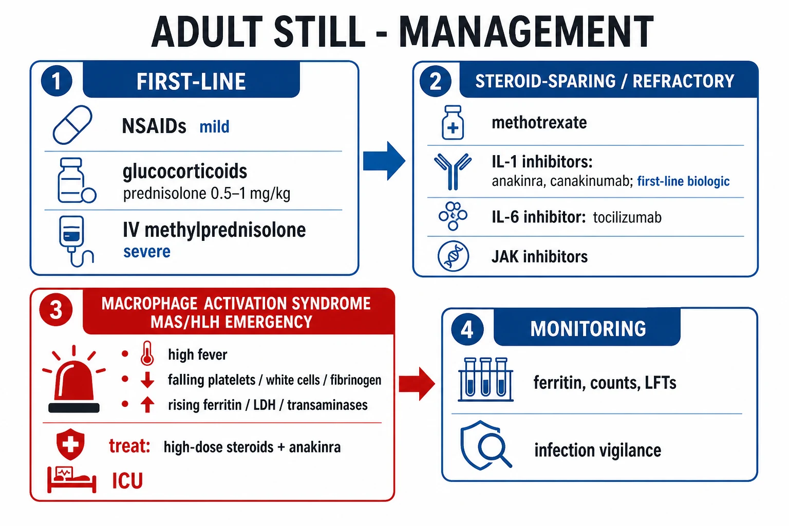
Most AOSD presentations are haemodynamically stable, and the immediate priority is not fluids or pressors but recognising macrophage activation syndrome / secondary HLH, which is the resuscitation problem:[1][6]
- If MAS is suspected (new persistent fever with falling platelets and white cells, falling fibrinogen, rising ferritin, LDH and transaminases), treat it as an emergency: urgent hospital admission, usually to ICU/HDU; high-dose IV methylprednisolone pulses (e.g. methylprednisolone 500 mg to 1 g IV once daily for one to three days) followed by high-dose oral prednisolone (1 mg/kg/day), plus anakinra (IL-1 receptor blockade, 100 mg subcutaneously daily); identify and treat any trigger (infection, drug); and give supportive care for cytopenias, coagulopathy, hepatic and multi-organ failure. Refractory MAS may require ciclosporin, IV immunoglobulin, ruxolitinib (JAK inhibition), or etoposide-based HLH-2004 protocols under haematology guidance.[9]
- If cardiac tamponade (large pericardial effusion with clinical compromise) or acute liver failure complicates the presentation, manage in the usual emergency fashion (pericardiocentesis; hepatology/liver-unit referral) while initiating disease control.
- Sepsis must be excluded empirically even as you treat: take cultures first, give broad-spectrum antibiotics if there is any diagnostic doubt, and de-escalate once infection is ruled out. Never diagnose AOSD in a patient who is still being treated for uncontrolled sepsis.
Macrophage activation syndrome in depth
MAS is a cytokine-driven, hyperinflammatory syndrome of uncontrolled T-cell and macrophage activation producing haemophagocytosis, cytopenias, coagulopathy and multi-organ failure. In AOSD the IL-18 / IFN-γ axis is the dominant driver — very high IL-18 primes cytotoxic T cells and macrophages, and defective cytotoxic function prevents the immune response from switching off, so activated macrophages accumulate and phagocytose blood-cell precursors in the marrow. This is the same biology as primary (genetic) HLH and as MAS in sJIA.[6][9]
Two overlapping diagnostic frameworks are used. The HLH-2004 criteria (above) require five of eight items. The H-score (Fardet 2014) is a weighted score (0 to 337) that captures the degree of abnormality — fever, organomegaly, number of cytopenic lineages, ferritin level, AST, triglycerides, fibrinogen, age, immunosuppression, and haemophagocytosis on marrow — with a score over 169 best identifying MAS. In clinical practice, the H-score is the more sensitive bedside tool because it weights the trend (a climbing ferritin and falling fibrinogen count even when absolute values are still "normal").[2]
The treatment of MAS is empirical and immediate — do not wait for a marrow result. The standard backbone is IV methylprednisolone pulses (500 mg to 1 g daily for up to three days) followed by oral prednisolone 1 mg/kg/day, plus anakinra 100 mg subcutaneously daily; ciclosporin (3 to 5 mg/kg/day) or IV immunoglobulin (2 g/kg over two to five days) are added for refractory disease, and ruxolitinib or etoposide-based HLH-2004 therapy is reserved for the most refractory cases under haematology guidance. Supportive care — cytopenia support, coagulation correction, treatment of any trigger, and organ support — runs in parallel.[9]
Management — Definitive & Stepwise
Definitive treatment is stepwise and pattern-directed, treating to remission (no fever, normal inflammatory markers, no active arthritis) and then tapering. The choice of agent depends on disease severity and on whether systemic or articular features dominate.[1][2][5]
Step 1 — Mild systemic disease (first-line). Start an NSAID for fever, rash and arthritis: naproxen 500 mg orally twice daily (preferred) or ibuprofen 400 to 800 mg orally three times daily, or indometacin 25 to 50 mg orally two to three times daily, with gastric protection. Add a glucocorticoid for systemic features that fail NSAIDs or that are moderate at the outset — oral prednisolone 0.5 to 1 mg/kg once daily (typically 30 to 60 mg), tapering slowly once fever and inflammatory markers normalise (usually over weeks to months). For severe or organ-threatening disease, give IV methylprednisolone pulses (e.g. methylprednisolone 500 mg to 1 g IV daily for one to three days) before transitioning to oral prednisolone.[1]
Step 2 — Chronic articular pattern or steroid-sparing. Introduce a conventional synthetic DMARD, almost always methotrexate 10 to 25 mg orally once weekly with folic acid 5 mg once weekly (on a different day), as the first steroid-sparing agent for the chronic articular pattern or when steroids cannot be tapered. Leflunomide 10 to 20 mg orally once daily is an alternative. Hydroxychloroquine has a limited role and is generally inadequate alone.[2]
Step 3 — Refractory systemic disease (the first-line biologic is an IL-1 inhibitor). For systemic disease that fails glucocorticoids and DMARDs, the 2024 EULAR/ACR recommendations and current evidence support IL-1 blockade first:[5][7]
- Anakinra (recombinant IL-1 receptor antagonist) — 100 mg subcutaneously once daily — the first-line biologic for systemic disease and for MAS, with a rapid (often within 24 to 72 hours) defervescence. Weight-based dosing (2 mg/kg/day, max 100 mg) in selected cases.
- Canakinumab (anti-IL-1β monoclonal antibody) — 150 mg subcutaneously every eight weeks (300 mg every four weeks for patients over 100 kg) — longer dosing interval, useful for maintenance and for patients on anakinra who respond but want less frequent injections.[7]
Step 4 — Chronic articular pattern (the first-line biologic is an IL-6 inhibitor). When chronic erosive polyarthritis dominates: [1]
- Tocilizumab (anti-IL-6 receptor monoclonal antibody) — 8 mg/kg intravenously every four weeks (or 162 mg subcutaneously every one to two weeks) — the preferred biologic for the chronic articular pattern, with good efficacy on both joint and systemic disease.[8]
Step 5 — Less common or refractory options. TNF inhibitors — etanercept 50 mg subcutaneously weekly, adalimumab 40 mg subcutaneously every two weeks — are less effective for systemic disease but may help selected chronic articular cases. JAK inhibitors (tofacitinib, ruxolitinib) and anti-IL-18 therapy (tadekinig alfa) are emerging for refractory systemic disease and MAS. IV immunoglobulin (2 g/kg over two to five days) and ciclosporin (3 to 5 mg/kg/day) are used in refractory MAS.[6][9]
Before any biologic: screen for TB (IGRA ± chest X-ray), hepatitis B and C, and HIV; ensure vaccinations are up to date (influenza, pneumococcal, hepatitis B) before starting immunosuppression; avoid live vaccines during therapy; and arrange baseline FBC, LFTs and renal function for monitoring.[5]
Specific Subtypes & Scenarios (Clinical Patterns in Practice)
Each clinical pattern has a different management emphasis and prognosis:[2][6]
- Monocyclic pattern — a single systemic episode remitting within a year; needs NSAIDs and a short tapering course of steroids; the best prognosis, and patients can usually be told that they are likely to make a full recovery. Do not over-treat with long-term biologics.
- Polycyclic (relapsing-remitting) pattern — recurrent flares separated by remissions; treat each flare with NSAIDs ± short steroid bursts, and use a DMARD or a long-acting IL-1 inhibitor (canakinumab) if flares are frequent or steroid-dependent. Joints are usually spared.
- Chronic articular pattern — persistent polyarthritis with erosive potential; this is the pattern needing methotrexate and the IL-6 inhibitor tocilizumab; monitor joints clinically and with imaging, and aim for treat-to-target (low disease activity or remission).[8]
- AOSD complicated by MAS (secondary HLH) — a medical emergency at any point in the course; treat with high-dose IV methylprednisolone plus anakinra (see Management — Resuscitation), monitor ferritin/LDH/fibrinogen/counts daily, and involve haematology/critical care.
- Hepatic phenotype — patients presenting with marked transaminitis or acute liver failure need aggressive disease control (steroids, IL-1 blockade) and a hepatology/liver-unit referral; AOSD is a recognised cause of acute liver failure requiring transplant assessment.
Complications & Pitfalls
- Macrophage activation syndrome / secondary HLH — the leading cause of death (complicates about 10 to 15 percent of cases); cytopenias, coagulopathy (low fibrinogen, high PT), liver failure and multi-organ failure. The earliest laboratory signature — falling platelets and white cells, falling fibrinogen, rising ferritin, LDH, triglycerides and transaminases with persistent fever — must trigger emergency treatment, not further investigation.[2][6]
- Chronic erosive arthritis — in the chronic articular pattern, joint destruction, deformity and disability resembling rheumatoid arthritis (but RF-negative); wrists and small joints of the hands are most affected.
- Secondary (AA) amyloidosis — a long-term complication of uncontrolled chronic inflammation, presenting as nephrotic-range proteinuria and progressive renal impairment; now rare with effective IL-1/IL-6 blockade.
- Cardiopulmonary disease — pericarditis and pleuritis are common and usually benign; myocarditis, cardiac tamponade, interstitial pneumonitis and ARDS are rare but life-threatening.
- Liver disease — transaminitis is common; acute liver failure and hepatic rupture are rare but recognised.
- Treatment-related toxicity — glucocorticoid toxicity (diabetes, osteoporosis, hypertension, infection), NSAID gastropathy and renal effects, methotrexate hepatotoxicity and pneumonitis, and biologic-related infection (re-activation of TB, hepatitis B).
- Diagnostic pitfalls — over-calling AOSD before excluding lymphoma or endocarditis (the two most dangerous errors); missing MAS behind a "fever of the underlying disease"; and treating chronic articular AOSD as rheumatoid arthritis with hydroxychloroquine and sulfasalazine, which are generally inadequate.
The single most useful prognostic signal is the clinical pattern, established over the first year. Patients who go a full year without relapse are likely to remain in long-term remission; those who develop a persistent polyarthritis within months of onset are the group at risk of chronic joint damage and need early DMARD and IL-6 blockade. A persistently elevated ferritin and CRP despite treatment predict relapse and should prompt intensification rather than reassurance. Mortality, when it occurs, is concentrated in three scenarios — MAS, fulminant hepatic failure, and cardiac involvement — so vigilance for these dominates follow-up.[2][6]
Prognosis & Disposition
Overall prognosis is favourable with modern therapy, but it is pattern-dependent:[1][2]
- The monocyclic pattern remits completely in about 60 to 70 percent of patients within a year and rarely recurs.
- The polycyclic pattern flares but rarely damages joints; long-term morbidity is mainly from cumulative steroid toxicity, so steroid-sparing strategies matter.
- The chronic articular pattern carries the greatest long-term morbidity from joint destruction, analogous to seronegative rheumatoid arthritis; early DMARD and IL-6 blockade limits damage.
- Mortality is about 1 to 5 percent overall, driven by MAS, cardiac involvement and liver failure.
- Disposition: newly diagnosed or flaring AOSD is usually admitted for diagnostic workup and initiation of treatment; discharge once fever has settled, inflammatory markers are falling, and an oral regimen is tolerated. Any suspicion of MAS mandates ICU/HDU admission. Long-term follow-up is under a rheumatologist with monitoring of inflammatory markers, ferritin, FBC and LFTs, and active screening for MAS at every flare. [1]
Special Populations
- Pregnancy: plan conception during remission with multidisciplinary care. Glucocorticoids are the safest agent (prednisolone at the lowest effective dose); avoid NSAIDs after 20 weeks gestation (oligohydramnios, premature ductus arteriosus closure); methotrexate is teratogenic and must be stopped at least three months before conception (and in men); leflunomide needs a cholestyramine washout. Anakinra and tocilizumab have limited safety data — avoid where possible and counsel individually, although the benefit in life-threatening MAS may outweigh the risk.[1]
- Children and adolescents: the paediatric counterpart is systemic juvenile idiopathic arthritis (sJIA) — clinically and biologically the same disease; the management principles and the risk of MAS are identical, and anakinra/canakinumab are first-line biologics. [5]
- The elderly patient presenting as a fever of unknown origin: a higher threshold for diagnosis — exclude malignancy and endocarditis more aggressively, and lower the threshold for lymph-node and bone-marrow biopsy. Drug doses: start prednisolone at 0.5 mg/kg (not 1 mg/kg) and watch for steroid toxicity, infection and fluid balance.
- The immunocompromised or post-transplant patient: AOSD is rare here, and the differential (lymphoma, infection, drug reaction, haemophagocytic syndrome) must be pursued harder before attributing the picture to AOSD.
Evidence, Guidelines & Regional Differences
- Yamaguchi 1992 (J Rheumatol) — the Yamaguchi classification criteria (sensitivity about 96 percent, specificity about 92 percent): the most widely used criteria in exams and in practice worldwide.[3]
- Fautrel 2002 (Medicine) — the Fautrel criteria, the only set to incorporate glycosylated ferritin under 20 percent as a major item; similar accuracy to Yamaguchi and no exclusion criteria.[4]
- Australian Rheumatology Association / PBS — PBS authority required for biologics (anakinra, tocilizumab, canakinumab) after failure of conventional therapy; IL-1 blockade preferred for systemic/MAS, tocilizumab for chronic articular disease. MAS managed jointly with haematology/critical care.
Landmark evidence and recent advances:[6][7][8][9]
- The 2024 EULAR/ACR/PRINTO/PReS recommendations unify the management of Still's disease across the age spectrum (sJIA and AOSD), with IL-1 inhibitors as first-line biologics for systemic disease and IL-6 blockade for chronic articular disease, and explicit guidance on MAS recognition and treatment.[5]
- Systematic reviews and meta-analyses confirm that canakinumab is effective and reasonably safe in AOSD, with high rates of fever clearance and steroid-sparing.[7]
- Tocilizumab multicentre retrospective data show durable control of refractory disease, particularly for the chronic articular pattern.[8]
- IL-18 blockade (tadekinig alfa) and JAK inhibitors (ruxolitinib, tofacitinib) are emerging therapies for refractory systemic disease and MAS, reflecting the central role of the IL-18/IFN-γ axis.[9]
Exam Pearls & High-Yield Minutiae
- YOUNG ADULT + QUOTIDIAN SPIKING FEVER + SALMON-PINK EVANESCENT RASH + ARTHRITIS + SORE THROAT + VERY HIGH FERRITIN + NEGATIVE ANA/RF = Adult Still's disease.
- Yamaguchi: 5 criteria, at least 2 major, NO exclusion — the single most examinable fact. Major = fever, arthralgia, rash, leucocytosis.[3]
- Glycosylated ferritin under 20 percent is the laboratory pearl — highly specific, and a Fautrel major criterion.[4]
- IL-1 for systemic, IL-6 for articular — the biologic-rule-of-thumb that decides a treatment MCQ.[5]
- MAS = falling counts and fibrinogen, rising ferritin/LDH — the emergency; treat with high-dose IV steroids plus anakinra.[2]
- AOSD is the adult counterpart of systemic JIA — same biology, same cytokines, same MAS risk.[6]
- Examine during the fever spike — the rash is evanescent and is otherwise missed.[1]
- Exclusion of lymphoma and endocarditis is part of the diagnostic criteria, not an afterthought.[3]
- Bimodal age (15 to 25 and 36 to 46) and M = F are the epidemiology facts worth knowing.[1]
- The disease is named after George Still (1897, systemic JIA in children); the adult form was characterised by Bywaters in 1971.
Self-test: which biologic for which pattern? (tap to reveal)
Systemic refractory disease and MAS → IL-1 blockade (anakinra first-line; canakinumab for maintenance). Chronic articular (erosive polyarthritis) pattern → IL-6 blockade (tocilizumab). TNF inhibitors are third-line for selected chronic articular disease. The logic is the dominant cytokine: IL-1β drives systemic inflammation, IL-6 drives the chronic synovitis and acute-phase response.[5][7][8]
Self-test: state the Yamaguchi major criteria (tap to reveal)
Four majors — fever over 39 °C for at least one week, arthralgia for at least two weeks, the typical non-pruritic salmon-pink rash, and a neutrophilic leucocytosis over 10 ×10⁹/L with neutrophils over 80 percent. Diagnosis needs five criteria with at least two major, and no exclusion (infection, malignancy, other rheumatic disease).[3]
Key Facts & Mnemonics
Adult Still's disease
autoinflammatory, ferritin very high
- Young adult; quotidian spiking fever + evanescent salmon-pink rash + arthritis + sore throat
- Ferritin very high (often over 1000); glycosylated fraction under 20 percent
- ANA and RF NEGATIVE; neutrophilic leucocytosis
- IL-1 inhibitors (anakinra, canakinumab) for systemic; tocilizumab for chronic articular
Systemic lupus erythematosus
autoimmune, positive antibodies
- Malar rash, renal disease, cytopenias
- ANA POSITIVE; anti-dsDNA and anti-Smith positive
- Low complement during flares; lymphopenia
- Hydroxychloroquine base; steroids/immunosuppressants for flares
Lymphoma
malignancy, must exclude
- Fever of unknown origin, lymphadenopathy, B-symptoms
- LDH markedly raised; biopsy diagnostic
- Ferritin may be high but no evanescent rash or quotidian fever pattern
- Treat the underlying malignancy
Adult Still's disease — key numbers
Adult Still's disease — FEVER
FEVER
ferritin often over 1000; glycosylated under 20 percent; autoinflammatory not autoimmune
trunk and proximal limbs; appears with fever, fades between spikes; Koebner-positive
diagnosis of exclusion — rule out infection, malignancy, SLE first
over 39 degrees C, late-afternoon or evening peak
anakinra/canakinumab for systemic; tocilizumab for chronic articular; MAS = falling counts + fibrinogen
Exam application bank (NEET-PG / INICET)
One-line answer
Adult-onset Still's disease (AOSD) is a rare systemic autoinflammatory disorder (the adult counterpart of systemic juvenile idiopathic arthritis) presenting with quotidian spiking fevers (evening peak), an evanescent salmon-pink rash, arthritis, sore throat, and markedly raised inflammatory markers (ferritin often very high) with negative autoantibodies (ANA, RF negative). It affects young adults (16 to 35). Three patterns: self-limited monocyclic, polycyclic (relapsing-remitting), or chronic articular. The feared complication is macrophage activation syndrome (MAS) / secondary HLH — a life-threatening hyperinflammatory emergency. Diagnosis is clinical (Yamaguchi or Fautrel criteria), after excluding infection, malignancy and other rheumatic disease. Treatment: NSAIDs and glucocorticoids first-line; methotrexate; IL-1 inhibitors (anakinra, canakinumab) for refractory systemic disease and
Worked stems (answer without another resource)
Stem 1 — Classic presentation. Map symptoms to mechanism; name the first investigation and first treatment step with dose/route if drug therapy is standard. [1]
Stem 2 — Unstable / complicated. List red flags that force immediate resuscitation, theatre, ICU, antidote, or reperfusion — and what you do in the first 15 minutes. [1]
Stem 3 — Atypical group. Elderly, pregnancy, child, or immunocompromised: how presentation and thresholds change. [1]
Stem 4 — Differential trap. Name the three closest mimics and one discriminator for each. [1]
Stem 5 — Disposition. Who goes home with safety-netting, who is admitted, who needs HDU/ICU/theatre, and what follow-up is mandatory. [1]
Rapid viva checklist
- Definition + classification
- Pathophysiology chain
- Bedside signs / criteria
- Score with exact components (if any)
- Emergency bundle
- Definitive therapy with doses
- Complications of disease and of treatment
- Special populations
- Guideline/trial name if classic
- Three exam traps
Coverage self-check
If you cannot answer any stem above from this page alone, re-read the matching section — the page is intended to be self-sufficient for final-prof and NEET-PG/INICET questions on Adult-Onset Still's Disease.
References
- [1]Efthimiou P, Kontzias A, Hur P. Adult-onset Still's disease in focus: Clinical manifestations, diagnosis, treatment, and unmet needs in the era of targeted therapies Semin Arthritis Rheum, 2021.PMID 34175791
- [2]Giacomelli R, Ruscitti P, Shoenfeld Y. A comprehensive review on adult onset Still's disease J Autoimmun, 2018.PMID 30077425
- [3]Yamaguchi M, Ohta A, Tsunematsu T, et al. Preliminary criteria for classification of adult Still's disease J Rheumatol, 1992.PMID 1578458
- [4]Fautrel B, Zing E, Golmard JL, et al. Proposal for a new set of classification criteria for adult-onset still disease Medicine (Baltimore), 2002.PMID 11997716
- [5]Fautrel B, Goola A, Otten MH, et al. EULAR/PReS recommendations for the diagnosis and management of Still's disease, comprising systemic juvenile idiopathic arthritis and adult-onset Still's disease Ann Rheum Dis, 2024.PMID 39317417
- [6]Ruscitti P, Cipriani P, Giacomelli R. Recent advances and evolving concepts in Still's disease Nat Rev Rheumatol, 2024.PMID 38212542
- [7]Liang H, Xu WD, Pan HF, Ye DQ. Efficacy and safety of canakinumab in the treatment of adult-onset Still's disease: A systematic review Semin Arthritis Rheum, 2021.PMID 34493394
- [8]Li F, Lin D, Hou Y, et al. Tocilizumab in patients with adult-onset still's disease refractory to glucocorticoid treatment: a randomised, double-blind, placebo-controlled phase III trial Ann Rheum Dis, 2018.PMID 30279267
- [9]Fraenkel MA, Utz JP, Pittock SJ, Matteson EL. Adult-Onset Still's Disease (AOSD): Advances in Understanding Pathophysiology, Genetics and Emerging Treatment Options Drugs, 2024.PMID 38441807