Rheumatology · Rheumatology
Calcium Pyrophosphate Deposition Disease (CPPD / Pseudogout)
Also known as CPPD · CPPD disease · Pseudogout · Pyrophosphate arthropathy · Chondrocalcinosis · Acute CPP crystal arthritis
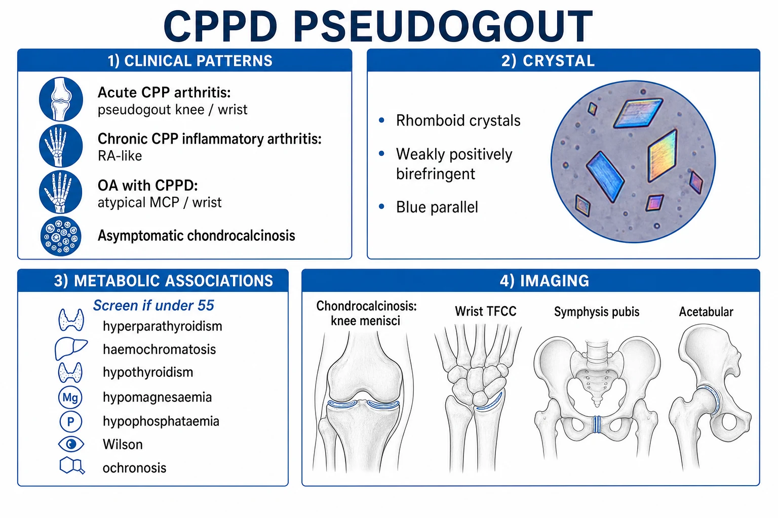
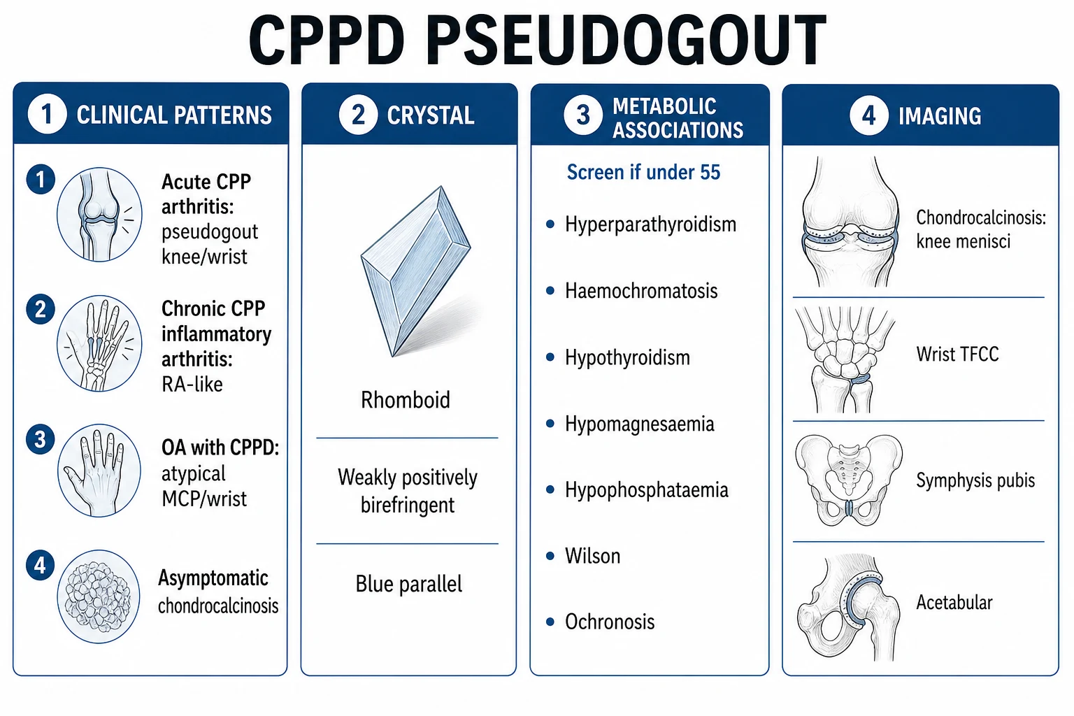
Calcium pyrophosphate deposition (CPPD) disease is a crystal-deposition arthritis caused by calcium pyrophosphate dihydrate (CPP) crystals forming in articular cartilage and fibrocartilage (chondrocalcinosis) and shedding into joints. It is the commonest crystal arthritis of the elderly and the second commonest overall after gout. Prevalence rises sharply with age, reaching 30 to 60 per cent in those over 85. Presentation is a spectrum: acute CPP arthritis ('pseudogout') mimicking gout (classically the knee, wrist); chronic CPP crystal inflammatory arthritis (RA-like); osteoarthritis with CPPD (pseudo-OA); or asymptomatic chondrocalcinosis. Associations are ageing, osteoarthritis and metabolic triggers (hyperparathyroidism, haemochromatosis, hypothyroidism, hypomagnesaemia) — screen for these in younger (under 55) patients. Diagnosis: rhomboid, weakly positively birefringent crystals plus chondrocalcinosis on X-ray. Treatment mirrors gout (NSAIDs, colchicine, intra-articular or oral corticosteroid); treat the metabolic cause; always aspirate acute monoarthritis to exclude sepsis. Unlike gout, no drug dissolves CPP crystals.
On this page & tools
Your progress
Saved locally on this device.
Exam tags
Red flags
Overview & Definition
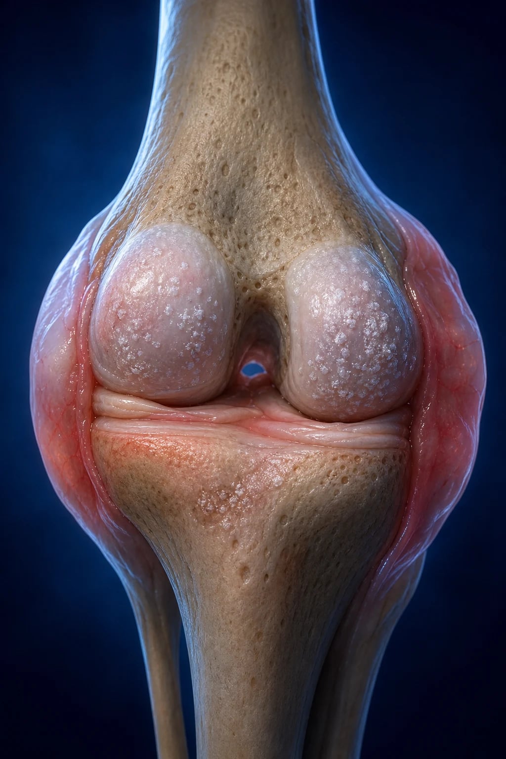
Calcium pyrophosphate deposition (CPPD) disease is the umbrella term for the whole spectrum of joint disease caused by the local formation of calcium pyrophosphate dihydrate (CPP) crystals in and around articular cartilage. It is the second commonest crystal arthritis (after gout) and the commonest crystal arthritis of older adults, yet it is the one most often missed or mislabelled as gout, infection or osteoarthritis because it presents in elderly patients and overlaps clinically with all three.[1][3]
The 2011 EULAR task force fixed the terminology so that examiners, clinicians and radiologists mean the same thing by each term — these distinctions are themselves a high-yield exam topic:[5]
- CPP crystal deposition — the biochemical/histological fact of CPP crystals forming in tissues. The disease is what happens when this causes trouble.
- Chondrocalcinosis — strictly a radiographic or pathological term: calcification of articular cartilage or fibrocartilage by CPP (or, rarely, other) deposits. It may be entirely asymptomatic ("lanthanide").
- Acute CPP crystal arthritis — the preferred modern name for the clinical entity historically called 'pseudogout' (Zitnan and Sitaj 1960): an acute mono- or oligo-articular inflammatory arthritis caused by CPP crystals shedding into a joint.
- Chronic CPP crystal inflammatory arthritis — a chronic, often polyarticular inflammatory arthritis driven by CPP crystals, mimicking rheumatoid arthritis.
- Osteoarthritis (OA) with CPPD — a chronic degenerative pattern in joints unusual for primary OA (e.g. MCPs, wrists, shoulders), where CPP deposition coexists with structural OA change. [1]
The diagnostic skill that decides every CPPD question is joint aspiration with compensated polarised-light microscopy of synovial fluid: the crystal shape and birefringence distinguish CPP (rhomboid, weakly positive) from gout (needle-shaped, negative). In any younger patient (under 55) with CPPD, screen for metabolic triggers — finding a hyperparathyroidism, haemochromatosis, hypomagnesaemia or hypothyroidism changes management. Treatment mirrors gout (NSAIDs, colchicine, corticosteroids), but there is no crystal-lowering equivalent of allopurinol; the only disease-modifying lever is treating the metabolic cause.[3][4]
Classification
CPPD is classified by aetiology into three groups, because the cause determines the work-up and (sometimes) the management.[5]
Sporadic (common)
- The overwhelming majority — age-related
- No single gene; multifactorial
- Strongly linked with OA and joint trauma
- Diagnosis of exclusion after ruling out metabolic causes
Hereditary (familial)
- Autosomal dominant familial chondrocalcinosis
- ANKH gene (CCAL2) mutations — early onset, severe
- Also CCAL1 locus; some families described in France, Sicily, Argentina
- Onset often 3rd-5th decade, generalised chondrocalcinosis
Metabolic-associated
- A treatable subgroup — always look
- Hyperparathyroidism, haemochromatosis, hypothyroidism, hypomagnesaemia
- Also hypophosphataemia, Wilson disease, ochronosis
- Screen any patient under 55 or with atypical disease
The clinical pattern (how the disease behaves) is then described separately from the aetiology (why the crystals formed). A single patient can move between patterns over time — a person with asymptomatic chondrocalcinosis may later develop acute pseudogout, then a chronic inflammatory arthritis in the same joint. Recognising the current pattern drives the investigation and counselling: the acute pattern demands aspiration to exclude sepsis; the chronic inflammatory pattern demands differentiation from rheumatoid arthritis; the pseudo-OA pattern demands identification of atypical joints; and asymptomatic chondrocalcinosis demands only reassurance (and, if the patient is young, a metabolic screen).[1][5]
The crystal arthropathies — situating CPPD
CPPD is one member of a family of crystal-induced arthropathies, each defined by the chemistry and morphology of its crystal. Knowing the family helps the differential and the polarised-light reading.[3]
CPPD (pseudogout)
- Calcium pyrophosphate dihydrate (Ca2P2O7.2H2O)
- RHOMBOID; weakly POSITIVELY birefringent
- Fibrocartilage + articular cartilage; knee, wrist
- Ageing, OA, metabolic (4H)
Gout
- Monosodium urate (MSU)
- NEEDLE; strongly NEGATIVELY birefringent
- First MTP, mid-foot, olecranon bursa
- Hyperuricaemia; allopurinol lowers urate
Basic calcium phosphate
- Hydroxyapatite / octacalcium phosphate
- Too small for light microscopy — need alizarin red
- Milwaukee shoulder; calcific periarthritis/tendinitis
- Older women; destructive shoulder effusion
Calcium oxalate
- Bipyramidal; strongly POSITIVELY birefringent
- Primary oxalosis or end-stage renal disease
- Small and large joints
- Rare; seen in dialysis patients
The practical point: under the polarised-light microscope, the only crystals visible to routine light microscopy are CPP and MSU. Basic calcium phosphate (apatite) crystals are sub-micron and cannot be seen by routine light microscopy — they are inferred from a negative crystal search in an inflammatory fluid (or confirmed with alizarin red staining), and classically present as a Milwaukee shoulder (destructive shoulder arthropathy in older women) or calcific periarthritis/tendinitis. Oxalate is rare and confined to primary hyperoxaluria or end-stage renal disease.[3]
Epidemiology & Risk Factors
CPPD is overwhelmingly a disease of ageing cartilage. The prevalence of radiographic chondrocalcinosis rises steeply and linearly with age.[7]
CPPD — the numbers examiners reward
The major risk factors are non-modifiable and explain almost all cases:[1][3][7]
- Age — by far the strongest. Cartilage PPi production rises with chondrocyte senescence; prevalence roughly doubles each decade after 50.
- Pre-existing osteoarthritis — CPPD and OA coexist so frequently that some authors consider them part of the same degenerative continuum. OA joints (especially knees) are the typical seedbed for CPP deposition.
- Joint trauma and overload — meniscectomy, occupational repetitive loading and prior joint injury all increase local risk.
- Female sex — slight predominance in the very elderly, partly because women live longer and have more OA.
- Metabolic disease — hyperparathyroidism, haemochromatosis, hypothyroidism, hypomagnesaemia, hypophosphataemia, Wilson disease and ochronosis. These are uncommon overall but common among young CPPD patients, which is exactly why every patient under 55 (or with atypical disease) must be screened. [1]
CPPD is the commonest crystal arthritis in the elderly and the second commonest crystal arthritis overall (after gout). It carries significant morbidity through joint destruction, recurrent attacks and acceleration of OA, and is associated with increased cardiovascular comorbidity, partly because the same elderly population carries vascular risk and partly through shared metabolic drivers.[3][7]
Pathophysiology
CPPD begins with an imbalance of inorganic pyrophosphate (PPi) metabolism in cartilage. PPi is a small anion (P₂O₇⁴⁻, written here as PPi) produced when cells break down ATP — specifically, extracellular PPi is generated by ecto-nucleotide pyrophosphatase/phosphodiesterase 1 (ENPP1, also called PC-1), an enzyme that cleaves ATP to AMP and PPi at the chondrocyte surface. PPi is then exported from the cell by the ANKH transporter (encoded by the ANKH gene), a multipass transmembrane channel that pumps PPi from the cytoplasm into the pericellular matrix. Under normal conditions PPi is a potent inhibitor of hydroxyapatite (basic calcium phosphate) mineralisation: it keeps cartilage and soft tissues uncalcified.[3]
The counter-regulator is tissue-nonspecific alkaline phosphatase (TNAP), which hydrolyses PPi to inorganic phosphate (Pi), allowing controlled mineralisation (as in growth plate and bone). The local PPi:Pi ratio therefore governs whether cartilage stays unmineralised (high PPi), forms CPP crystals (very high PPi), or forms apatite (low PPi, high Pi). In CPPD, excess PPi accumulates in the cartilage matrix, and CPP crystals — calcium pyrophosphate dihydrate, Ca₂P₂O₇·2H₂O — precipitate preferentially in the avascular fibrocartilage and the mid-zone of articular hyaline cartilage, where PPi transporters are most active.[3]
Several forces raise cartilage PPi and drive crystallisation: [1]
- Ageing chondrocytes progressively produce more PPi and acquire an "hypertrophic" phenotype resembling mineralising growth-plate chondrocytes.
- TGF-β (released in OA cartilage) stimulates PPi production — explaining the OA–CPPD link.
- Growth factors and cytokines in damaged cartilage further shift chondrocytes toward a mineralising phenotype.
- Hypomagnesaemia — magnesium is an essential cofactor for TNAP; low magnesium disables PPi hydrolysis, raises PPi, and is one of the most directly treatable metabolic drivers.
- Hypercalcaemia / hyperparathyroidism — excess calcium provides the cation for crystal nucleation.
- Iron overload (haemochromatosis) — iron is deposited in cartilage and disturbs chondrocyte metabolism, predisposing to CPPD (and a distinctive MCP arthropathy).
- Hypothyroidism, hypophosphataemia, Wilson disease (copper), ochronosis (homogentisic acid) — rarer drivers that alter cartilage biochemistry. [1]
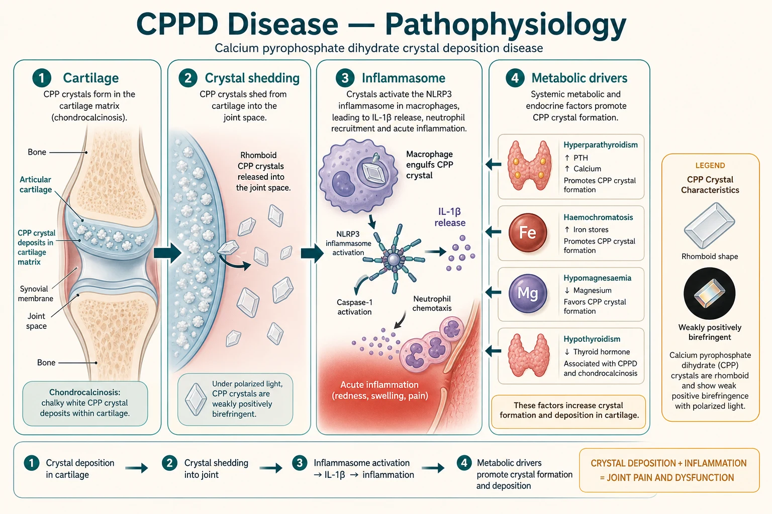
The acute attack is the downstream consequence of crystal shedding. When CPP crystals — large, hard rhomboids — are physically dislodged from cartilage into the synovial cavity (by minor trauma, joint stress, surgery, or rapid changes in joint fluid chemistry during intercurrent illness), they are phagocytosed by macrophages and neutrophils. Phagolysosomal rupture releases crystal surfaces and cathepsin B, which activate the NLRP3 inflammasome, driving caspase-1 cleavage of pro-IL-1β to active IL-1β. IL-1β then drives a cascade of IL-6, IL-8, TNF-α and neutrophil chemoattraction — exactly the same inflammasome pathway activated by urate in gout, which is why the two attacks are clinically so similar. This shared final pathway also explains why IL-1β blockade (anakinra, canakinumab) works for refractory CPPD, just as it does for refractory gout.[3][6]
[1]Clinical Presentation
CPPD is a spectrum, and a patient may occupy a different point on it at different times. The 2011 EULAR task force recognised several overlapping presentations; recognising the pattern drives both investigation and counselling.[1][5]
Acute CPP crystal arthritis ('pseudogout')
The classic and most examinable presentation. An older adult (typically over 65, often with established OA) develops rapid-onset, severe pain, swelling, warmth and stiffness of a single large joint over hours to one to two days. The joint is hot, erythematous, swollen and exquisitely tender with a large effusion and markedly restricted range — clinically indistinguishable from gout or septic arthritis. Fever and even a systemic inflammatory response are common, which is why aspiration is mandatory.[1]
The classic joints are the knee (commonest by far) and the wrist (especially the radiocarpal joint, the most characteristic site after the knee). Other large joints — shoulder, elbow, ankle, hip — can be involved; oligo- or poly-articular attacks occur but are less common. A characteristic and high-yield trigger is precipitation by intercurrent illness or surgery — a patient who develops a hot swollen knee on day three to seven after a general anaesthetic, a respiratory infection, a stroke, or a myocardial infarction is a classic pseudogout stem. Direct joint trauma and medical procedures (arthroscopy, intra-articular injection) can also trigger shedding.[1][3]
Chronic CPP crystal inflammatory arthritis
A subacute or chronic, often polyarticular, inflammatory arthritis with prolonged morning stiffness, fatigue, synovitis and elevated inflammatory markers — clinically mimicking rheumatoid arthritis, especially when it involves the wrists, MCPs and shoulders symmetrically. Distinguishing features: it occurs in an older patient with chondrocalcinosis, often with coexisting OA, and the synovial fluid shows CPP crystals. Synovial fluid analysis (and rheumatoid factor / anti-CCP) resolves the confusion.[1]
Osteoarthritis with CPPD (pseudo-OA)
A chronic degenerative pattern in joints unusual for primary OA — the 2nd/3rd MCPs, wrists, elbows, shoulders, ankles — alongside typical OA in the knees and spine. Coarse crepitus, bony swelling, varus deformity and restricted movement predominate, with relative sparing of the inflammatory features. The MCP involvement with hook-like osteophytes in a patient who also has chondrocalcinosis should prompt iron studies — this is the classical haemochromatosis arthropathy.[8]
Asymptomatic (lanthanide) chondrocalcinosis
The commonest form in the very elderly: incidental chondrocalcinosis on an X-ray taken for another reason, with no joint symptoms attributable to it. Requires no treatment beyond reassurance, unless the patient is young enough that a metabolic screen is indicated.[1]
Rare destructive (neuropathic-like) arthropathy
A rare but examinable pattern — a rapidly progressive, destructive, Charcot-like arthropathy of a large joint (classically the knee or shoulder) with fragmentation, subchondral collapse and disorganisation resembling a Charcot joint, but in a neurologically intact patient. It is associated with recurrent haemorrhagic effusions and marked bone destruction; management is difficult and often ends in arthroplasty.[3]
Atypical presentations (examiners test these deliberately)
Examiners love the atypical stem because it tests whether you can recognise CPPD outside the textbook pattern. In the very elderly or frail patient, fever, confusion and even systemic upset may dominate over joint pain, so a hot swollen joint may be missed until the knee is examined; delirium with a swollen knee in a nursing-home resident is a recognised presentation. Oligo- or poly-articular attacks can mimic RA or reactive arthritis, especially in the small joints of the hand or in the shoulders bilaterally. Axial involvement — inflammatory back pain from CPP deposition in the intervertebral disc annulus or facet joints — is uncommon but described, particularly in the elderly. Tendon and bursal deposition (e.g. calcific deposits in the Achilles insertion or subacromial bursa) can mimic enthesitis or tendinopathy. After an acute stroke or prolonged immobilisation, a flaccid hemiplegic limb may develop pseudogout in the shoulder or knee, complicating rehabilitation. The lesson is uniform: any acute inflammatory mono- or oligo-arthritis in an older adult warrants aspiration, regardless of whether the clinical story is "typical".[1][3]
Differential Diagnosis
The decisive step in any acute monoarthritis is synovial fluid aspiration and analysis — clinical features alone cannot reliably distinguish CPPD from gout, sepsis or a haemarthrosis. The crystal shape and birefringence, the Gram stain, the cell count and the culture together settle the diagnosis.[1][5]
CPPD (pseudogout)
- RHOMBOID crystals, WEAKLY POSITIVELY birefringent (blue parallel)
- Knee or wrist in older patient
- Chondrocalcinosis on X-ray
- Treatment mirrors gout; no crystal-lowering drug
Gout
- NEEDLE crystals, STRONGLY NEGATIVELY birefringent (yellow parallel)
- First MTP (podagra); middle-aged men, metabolic syndrome
- Serum urate high; tophi in chronic disease
- Allopurinol/febuxostat lower urate long-term
Septic arthritis
- MUST exclude — can coexist with CPPD
- Gram stain + culture positive; WBC often over 50,000
- Higher fever, rigors, very high inflammatory markers
- Urgent washout + IV antibiotics (e.g. flucloxacillin)
Rheumatoid arthritis
- Symmetric small-joint polyarthritis, prolonged morning stiffness
- RF / anti-CCP positive
- Erosions on X-ray; no crystals
- DMARDs (methotrexate first-line)
Reactive / SpA
- Oligoarthritis, lower limb, enthesitis, dactylitis
- Recent infection (GI/GU); HLA-B27
- No crystals; inflammatory fluid
- NSAIDs; antibiotics do not help arthritis
The two must-not-miss mimics are gout (the crystal differential — distinguished on polarised-light microscopy) and septic arthritis (the dangerous differential — distinguished on Gram stain, culture and a high clinical suspicion). The crucial teaching point is that sepsis and CPPD can coexist in the same joint: the presence of CPP crystals does NOT exclude infection, so culture must always be sent and the joint reassessed if there is any clinical doubt. Other differentials include traumatic haemarthrosis (blood in the fluid, history of trauma), a flare of osteoarthritis (less inflammatory, no crystals), psoriatic arthritis and, in the chronic pattern, polymyalgia rheumatica.[1][3]
Clinical & Bedside Assessment
The bedside assessment is built around three questions: is this joint septic?, which crystal is it?, and is there a metabolic cause? [1]
Examine the affected joint for warmth, erythema, swelling, effusion, tenderness and range of movement. A hot, swollen knee or wrist in an older adult with a large effusion and severe pain on passive motion is the pseudogout stem; perform a ballotable patella test for a knee effusion and look for bulge sign in the wrist. Document the tempo (hours = crystal/sepsis; weeks = chronic arthritis), any fever or rigors (do NOT be reassured — pseudogout causes fever too), and the number and distribution of joints involved.[1]
Then look beyond the hot joint for clues: [1]
- Coexisting osteoarthritis — Heberden's and Bouchard's nodes, MCP squaring, knee deformity, crepitus.
- Metabolic stigmata — bronze/slate-grey skin (haemochromatosis), goitre, bradycardia, delayed reflexes (hypothyroidism), ** Kayser–Fleischer ring** and hepatic/neurological signs (Wilson disease), ochronotic darkening of pinna/sclera (alkaptonuria), proximal muscle weakness, hypertension, renal stones (hyperparathyroidism), tetany/Chvostek sign (hypomagnesaemia/hypocalcaemia).
- Precipitants — recent surgery, infection, trauma, hospitalisation, stroke or MI in the preceding days (the classic pseudogout trigger).
- Joint-specific pattern — 2nd/3rd MCP involvement with hook osteophytes points to haemochromatosis arthropathy, which is treatable and easily missed.[8]
Assess the whole patient: comorbidities govern drug choice (renal function dictates NSAID vs steroid; cardiovascular risk dictates NSAID caution; diabetes dictates steroid caution; anticoagulation dictates joint injection timing). Document baseline observations, and always consider whether this joint could be septic — the bedside exam is not the place to be falsely reassured. [1]
Investigations
Synovial fluid analysis — the definitive test
Aspiration of synovial fluid with compensated polarised-light microscopy is the diagnostic investigation. Send the fluid for crystal identification, cell count, Gram stain and culture on every acute sample.[5]
[1]The fluid is inflammatory: white cell count commonly 2,000 to 100,000 per microlitre (often 15,000 to 30,000), neutrophil-predominant. It is typically clear to slightly turbid and yellow (unlike the bloody fluid of haemarthrosis or the opaque, strongly purulent fluid of sepsis). The mucin clot is poor, and viscosity is low. Crucially — the presence of crystals does NOT exclude sepsis: Gram stain and culture are always sent, and a positive culture overrides a crystal diagnosis. Cell counts above 50,000 per microlitre with a neutrophil majority should heighten suspicion of sepsis even if crystals are present.[1][5]
Plain radiographs
Chondrocalcinosis — calcification of articular cartilage and fibrocartilage by CPP deposits — is the radiographic hallmark. Look for fine, linear or punctate calcification in characteristic sites:[1][5]
Site
- Knee — medial AND lateral menisci (linear, parallel to joint line)
- Wrist — triangular fibrocartilage complex (TFCC), radiocarpal
- Symphysis pubis — fibrocartilaginous disc
- Acetabular labrum (hip), glenoid labrum (shoulder)
Also seen
- Annulus fibrosus of intervertebral discs
- Pubic symphysis articular surface
- Tendon insertions and bursae (rare)
- May coexist with OA changes (joint-space loss, sclerosis, cysts)
The knee radiograph is the single most useful film — order anteroposterior and lateral views of the symptomatic joint, plus a wrist and pelvis (for symphysis pubis) when screening. Chondrocalcinosis plus compatible crystals confirms CPPD, but chondrocalcinosis alone (especially in the very elderly) does not prove that the current arthritis is CPPD — crystals in the fluid do.[5]
Advanced imaging
The 2023 EULAR imaging recommendations place ultrasound as the first-line imaging modality for crystal detection, because CPP deposits within fibrocartilage produce a characteristic hyperechoic, dotted or chunky deposition (distinct from the urate 'double-contour sign' on hyaline cartilage). Ultrasound is more sensitive than plain film for early deposition, is radiation-free and can be done at the bedside. CT is highly sensitive for cartilage and tendon calcification and is useful in deep joints (hip, spine). MRI shows structural damage, synovitis and bone marrow lesions but is less specific for crystal deposition. Dual-energy CT can distinguish urate from calcium-containing crystals in research settings but is not routine.[2]
Metabolic screen
In any patient under 55 or with atypical, severe, polyarticular or recurrent disease, screen for the metabolic associations. The screen is cheap, treatable, and the single most useful thing an examiner wants you to remember:[1][3]
CPPD metabolic screen — the 4H + others
4H+
Serum calcium, PTH, alkaline phosphatase
Iron studies, ferritin, transferrin saturation, LFTs, HFE gene
TSH, free T4
Serum magnesium (treatable — give oral magnesium)
Phosphate, glucose, copper/ceruloplasmin (Wilson), and consider ochronosis clinically
A pragmatic screen covers calcium, magnesium, phosphate, alkaline phosphatase, TSH, iron studies and ferritin, glucose, and LFTs; add copper and caeruloplasmin if Wilson disease is plausible (younger patient with hepatic and neurological features). Hypomagnesaemia and haemochromatosis are the two most examinable because both are treatable and change management — magnesium replacement can reduce flares, and venesection (or iron chelation) for haemochromatosis slows extra-articular organ damage even though it does not dissolve existing crystals.[8]
Other blood tests
CRP and ESR are typically elevated (CRP often 50 to 150 mg/L in acute pseudogout), FBC shows a neutrophilic leucocytosis, and urate may be modestly high or normal (do NOT overcall gout on a serum urate — the fluid decides). Urea, electrolytes and eGFR govern drug choice (NSAID caution). Rheumatoid factor and anti-CCP are useful when the chronic inflammatory pattern mimics RA. Blood cultures if sepsis is suspected. Urinalysis (renal stones in hyperparathyroidosis).[1]
Management — Resuscitation
CPPD is not, in itself, a medical emergency — but the differential is. The only "resuscitation" dimension is the rule that any acute hot swollen joint is septic arthritis until proven otherwise, and the immediate action is joint aspiration.[1]
The acute hot swollen joint — the first 60 minutes
Recognise the hot swollen joint as a diagnostic emergency — septic arthritis destroys cartilage within hours to days
Aspirate the joint promptly (knee: suprapatellar, lateral to quadriceps tendon; wrist: dorsal radiocarpal) using aseptic technique
Send fluid immediately for: cell count, Gram stain, culture, and polarised-light microscopy for crystals
If sepsis cannot be excluded (high WBC, positive Gram stain, systemically unwell) — admit, start IV antibiotics (e.g. flucloxacillin 2 g IV QDS, vancomycin if MRSA risk), and arrange urgent orthopaedic washout
If CPP crystals confirmed and sepsis excluded — begin specific acute CPPD treatment (see below)
Always send culture even when crystals are seen — pseudogout and sepsis can coexist
The principle is simple: aspirate first, treat second. Empiric antibiotics before aspiration (except in a systemically septic patient you cannot stabilise) contaminate culture and obscure the diagnosis; corticosteroid injection into a septic joint is catastrophic. Once the fluid confirms CPP crystals and culture is set up (or Gram stain is negative and sepsis is clinically unlikely), specific CPPD therapy can begin.[1][5]
Management — Definitive & Stepwise
Acute CPP arthritis is treated identically to gout, because the two diseases share the same inflammasome-driven neutrophilic inflammatory mechanism. The 2011 EULAR Part II recommendations and the 2021 systematic review underpin the ladder below.[4][6]
Acute CPP arthritis
Acute pseudogout — drug ladder (choose one, tailor to comorbidity)
The choice is governed by comorbidity, not by efficacy differences (the options are broadly equally effective):[4][6]
- Single large joint, no sepsis → intra-articular corticosteroid (triamcinolone acetonide 40 mg for a knee, 20 to 30 mg for a wrist) — the cleanest option, minimal systemic effect.
- Elderly, renal impairment, heart failure, anticoagulated, or multiple joints → oral glucocorticoid (prednisolone 30 to 40 mg OD tapering) — preferred because NSAIDs are risky.
- Fit younger patient, no contraindication → NSAID at full anti-inflammatory dose (naproxen 500 mg BD is evidence-based and has the best cardiovascular profile; add a PPI).
- Presenting within 24 h → colchicine 500 micrograms BD to QID works well, but reduce the dose in CKD and avoid combinations with clarithromycin, statins or strong CYP3A4 inhibitors. [1]
Chronic and recurrent disease
The goal shifts from aborting individual attacks to reducing flare frequency and controlling chronic synovitis, since no drug dissolves existing CPP crystals.[4][6]
- Colchicine prophylaxis — 500 micrograms (0.5 mg) BD, long-term, is the best-supported option for recurrent attacks (especially peri-procedural).
- Low-dose NSAID with a PPI — for chronic inflammatory symptoms; chosen cautiously for cardiovascular, renal and GI risk.
- Methotrexate — 7.5 to 15 mg orally once weekly (with folic acid) — has limited but real evidence in refractory chronic CPP crystal inflammatory arthritis.
- IL-1β blockade — anakinra 100 mg subcutaneously daily or canakinumab — for refractory disease unresponsive to colchicine and methotrexate, exploiting the shared NLRP3/IL-1β pathway. Emerging, off-label, and reserved for specialist use.
- Intra-articular steroid — for refractory chronic monoarticular disease, repeated injections (with imaging) for the same joint, spaced at least three months apart.
- Joint replacement — for end-stage destructive arthropathy (especially knee and shoulder). [1]
Treat the metabolic cause
This is the only disease-modifying lever available, and the one most often forgotten. Treating the underlying metabolic disorder does not dissolve crystals already deposited, but may slow progression and reduce flare frequency.[3][8]
Haemochromatosis
- Venesection (or iron chelation) to reduce iron overload
- Does not reverse established arthropathy/crystals
- May slow further deposition; treat extra-articular organ damage
- Family screen + HFE genotype
Hyperparathyroidism
- Parathyroidectomy for primary hyperPTH (adenoma)
- Correct hypercalcaemia; bisphosphonates if severe
- Re-check calcium, PTH, vitamin D post-operatively
Hypomagnesaemia
- Oral magnesium (chloride or glycerophosphate) chronically
- Identify cause (PPIs, diuretics, malabsorption, Gitelman)
- Most directly treatable PPi-axis driver
Hypothyroidism
- Levothyroxine replacement to euthyroid
- TSH-guided titration
- May reduce flares
Supportive and rehabilitative care
Physiotherapy, joint protection, weight management and assistive devices address the OA component and prevent deconditioning between flares. Patient education — explaining the episodic, non-life-threatening nature of CPPD, the importance of early treatment of flares, and the rationale for metabolic screening — improves adherence and outcomes. There is no role for long-term oral colchicine toxicity monitoring beyond renal/hepatic awareness, and no role for urate-lowering drugs (allopurinol, febuxostat) in pure CPPD — a frequent exam trap.[4]
Specific Subtypes & Scenarios
Which CPPD scenarios must I be able to handle?
The five scenarios examiners probe are: (1) acute monoarthritis post-surgery — the classic pseudogout stem, aspirate first, then intra-articular steroid; (2) CPPD in renal impairment — avoid NSAIDs, prefer steroid; (3) CPPD with a positive metabolic screen — treat the cause (venesection, parathyroidectomy, magnesium, levothyroxine); (4) refractory chronic CPPD inflammatory arthritis — escalate to methotrexate then IL-1β blockade; and (5) CPPD and gout coexisting — the fluid shows both crystal types; treat the dominant crystal but aspirate to confirm.
The post-operative pseudogout flare deserves special mention because it is the most common SAQ stem. A patient develops a hot swollen knee on day three to seven after a general surgical procedure, a fracture fixation, or a joint replacement. Triggers include the surgical stress response, fluid shifts, immobilisation, and acute phase changes that disturb crystal equilibrium. Aspiration confirms CPP crystals (and excludes the must-not-miss prosthetic joint infection); management is intra-articular corticosteroid (if the joint is not a recent replacement) or short-course oral steroid. Colchicine prophylaxis (500 micrograms BD) in the peri-operative period is reasonable for high-risk patients with a known history of CPPD.[1][4]
Haemochromatosis arthropathy is the most examinable metabolic subtype. Classically it affects the 2nd and 3rd MCP joints (often with hook-like osteophytes on X-ray), is associated with chondrocalcinosis, and may be the presenting feature of the disease. Always check iron studies and ferritin (transferrin saturation over 45 per cent, ferritin markedly elevated) and HFE genotyping in any young patient with chondrocalcinosis or this distinctive arthropathy. Venesection treats the systemic iron overload but does not reverse the joint disease — the arthritis can progress even after iron depletion.[8]
Complications & Pitfalls
CPPD causes substantial morbidity through structural joint damage and through iatrogenic harm from its treatment. The major complications are:[1][3]
- Joint damage and secondary osteoarthritis — recurrent crystal shedding accelerates cartilage loss, especially in the knee and wrist; many patients end up with severe OA requiring arthroplasty.
- Recurrent acute attacks — episodic, often triggered by illness or surgery, with cumulative disability, chronic pain and stiffness between flares.
- Destructive (Charcot-like) arthropathy — rare but severe; rapidly progressive joint destruction, fragmentation and disorganisation.
- Iatrogenic harm — NSAID-induced acute kidney injury, gastrointestinal bleeding and cardiovascular events (especially in the elderly); colchicine toxicity in renal impairment (myelosuppression, neuromyopathy, diarrhoea); corticosteroid hyperglycaemia, hypertension, osteoporosis and infection risk.
- Missed septic arthritis — the single most dangerous pitfall. Crystals do not exclude infection; never inject a steroid without excluding sepsis.
- Cardiovascular comorbidity — through association with OA, metabolic disease and age. [1]
Exam application bank (NEET-PG / INICET)
One-line answer
Calcium pyrophosphate deposition (CPPD) disease is a crystal-deposition arthritis caused by calcium pyrophosphate dihydrate (CPP) crystals forming in articular cartilage and fibrocartilage (chondrocalcinosis) and shedding into joints. It is the commonest crystal arthritis of the elderly and the second commonest overall after gout. Prevalence rises sharply with age, reaching 30 to 60 per cent in those over 85. Presentation is a spectrum: acute CPP arthritis ('pseudogout') mimicking gout (classically the knee, wrist); chronic CPP crystal inflammatory arthritis (RA-like); osteoarthritis with CPPD (pseudo-OA); or asymptomatic chondrocalcinosis. Associations are ageing, osteoarthritis and metabolic triggers (hyperparathyroidism, haemochromatosis, hypothyroidism, hypomagnesaemia) — screen for these in younger (under 55) patients. Diagnosis: rhomboid, weakly positively birefringent crystals plu
Worked stems (answer without another resource)
Stem 1 — Classic presentation. Map symptoms to mechanism; name the first investigation and first treatment step with dose/route if drug therapy is standard. [1]
Stem 2 — Unstable / complicated. List red flags that force immediate resuscitation, theatre, ICU, antidote, or reperfusion — and what you do in the first 15 minutes. [1]
Stem 3 — Atypical group. Elderly, pregnancy, child, or immunocompromised: how presentation and thresholds change. [1]
Stem 4 — Differential trap. Name the three closest mimics and one discriminator for each. [1]
Stem 5 — Disposition. Who goes home with safety-netting, who is admitted, who needs HDU/ICU/theatre, and what follow-up is mandatory. [1]
Rapid viva checklist
- Definition + classification
- Pathophysiology chain
- Bedside signs / criteria
- Score with exact components (if any)
- Emergency bundle
- Definitive therapy with doses
- Complications of disease and of treatment
- Special populations
- Guideline/trial name if classic
- Three exam traps
Coverage self-check
If you cannot answer any stem above from this page alone, re-read the matching section — the page is intended to be self-sufficient for final-prof and NEET-PG/INICET questions on Calcium Pyrophosphate Deposition Disease (CPPD / Pseudogout).
[1]Prognosis & Disposition
Acute attacks are self-limiting, resolving over days to two to three weeks even without treatment, and most respond promptly to NSAIDs, colchicine or corticosteroid within 24 to 72 hours. Chronic forms, however, cause progressive joint damage for which no disease-modifying therapy exists; morbidity is driven by joint destruction, recurrent attacks and coexisting OA in the elderly. Treating an underlying metabolic cause may reduce flare frequency and slow further deposition, but does not reverse existing damage.[1][3]
The natural history of CPPD reflects the interplay between crystal deposition and structural joint damage. Once CPP crystals have formed in cartilage they persist for life; chondrocalcinosis is, in most elderly patients, a permanent radiographic finding. Acute attacks tend to cluster — a patient may have two or three flares in one year and then none for several years — and the frequency and severity of flares do not reliably predict the rate of structural damage, which depends more on the burden of coexisting OA, joint loading and metabolic control. Over years, recurrent inflammation and crystal-driven matrix degradation accelerate cartilage loss, subchondral sclerosis, cyst formation and joint-space narrowing, producing the radiographic picture of "OA with CPPD" and, in a minority, the destructive arthropathy that ends in arthroplasty. Patients with a treatable metabolic driver (especially haemochromatosis and hypomagnesaemia) may experience a genuine reduction in flare frequency once the driver is corrected, even though existing crystals and damage remain — this is the single clearest prognostic lever and the reason the metabolic screen matters.[3][6]
Disposition for acute pseudogout: most patients are managed as outpatients with intra-articular steroid or oral therapy and follow-up in rheumatology or primary care. Admit when sepsis cannot be excluded, the patient is systemically unwell, the joint is a recent prosthesis, or comorbidity prevents safe outpatient management (e.g. severe CKD, anticoagulation, frailty). The safety-net is clear: any patient whose joint pain, swelling or systemic features worsen, or who develops fever or rigors after discharge, must return urgently for re-aspiration and reassessment — sepsis can declare itself days into a presumed crystal arthritis.[1][5]
Special Populations
- Elderly patients — the highest-prevalence group and the most vulnerable to drug harm. Prefer intra-articular or low-dose oral corticosteroid over NSAIDs (renal, gastrointestinal and cardiovascular risk), and reduce the colchicine dose in renal impairment. Screen for falls risk during a flare, and address the OA component with physiotherapy and joint protection.[1]
- Younger patients (under 55) with CPPD — always screen metabolic causes; haemochromatosis is treatable and easily missed, and familial chondrocalcinosis (ANKH) should be considered with early-onset generalised disease and a family history.[8]
- Renal impairment — avoid NSAIDs; prefer intra-articular steroid for a single joint or a short course of oral prednisolone. Halve the colchicine dose when eGFR is under 30, and avoid it entirely with strong CYP3A4 inhibitors.[4]
- Post-surgical / critically ill patients — pseudogout flares classically follow surgery, sepsis, stroke or MI; consider colchicine prophylaxis in known CPPD patients undergoing major surgery, and aspirate any new hot joint in this group to exclude prosthetic/iatrogenic infection.[1]
- Anticoagulated patients — joint aspiration is generally safe with INR under therapeutic range (under about 3.0) but discuss timing with haematology for high-INR patients; intra-articular steroid injection carries a small bleeding risk.
- Pregnancy — CPPD is rare in this group; NSAIDs are avoided after 20 weeks (and especially after 28 weeks), colchicine is avoided (foetal chromosomal risk), making intra-articular corticosteroid the preferred acute option.
Evidence, Guidelines & Regional Differences
CPPD evidence is thinner than gout evidence — there are no large randomised trials and management is largely extrapolated from gout and from small observational studies.[6]
2011 EULAR recommendations — CPPD Parts I and II
Ann Rheum Dis 2011
PMID 21216817, 21257614
Key finding
Standardised CPPD terminology (Part I) and a management ladder (Part II) that mirrors gout: NSAIDs, colchicine, intra-articular or oral corticosteroids for acute attacks; colchicine prophylaxis and treat-the-metabolic-cause for chronic disease. Foundational for exam answers.
2023 EULAR imaging recommendations for crystal arthropathies
Ann Rheum Dis 2024
PMID 38320811
Key finding
Ultrasound is first-line imaging for crystal detection; CPP deposits appear as hyperechoic dotted/chunky deposition in fibrocartilage (distinct from the urate double-contour sign on hyaline cartilage). CT and MRI aid structural assessment.
Management of CPPD — systematic review
Semin Arthritis Rheum 2021
PMID 33360232
Key finding
Trial evidence for CPPD therapy is scarce; NSAIDs, colchicine and corticosteroids remain first-line by extrapolation; low-quality evidence supports methotrexate and IL-1β blockade for refractory disease; no drug dissolves CPP crystals.
There are no major regional divergences in CPPD management. The 2011 EULAR (European) recommendations are adopted globally, including by the American College of Rheumatology and British Society for Rheumatology. NSAID choice varies regionally (naproxen and ibuprofen are preferred for cardiovascular safety; etoricoxib is available in the UK/Europe and India but not the US; diclofenac is restricted in some countries for cardiovascular risk). Anakinra and canakinumab for refractory CPPD are off-label everywhere and accessed via specialist centres.
Exam Pearls
The CPPD topic lives or dies on a handful of high-yield discriminators. Master these and you answer any MCQ, SAQ or viva stem:[1][3]
- CPPD crystals = rhomboid, weakly POSITIVELY birefringent (blue parallel to the compensator). Gout = needle-shaped, strongly NEGATIVELY birefringent (yellow parallel). This is the single most tested fact.
- Pseudogout = acute monoarthritis of the KNEE or WRIST in an older adult (gout = first MTP / podagra).
- Chondrocalcinosis on X-ray: knee menisci (medial and lateral), wrist TFCC, symphysis pubis, acetabular and glenoid labra.
- Screen metabolic causes if under 55 — the 4H: Hyperparathyroidism, Haemochromatosis, Hypothyroidism, Hypomagnesaemia — plus hypophosphataemia, Wilson disease, ochronosis.
- Aspirate any acute monoarthritis — distinguish CPPD from gout AND exclude septic arthritis (they can coexist).
- Treatment mirrors gout: NSAIDs, colchicine, intra-articular or oral corticosteroid — but no urate-lowering drug (no allopurinol/febuxostat for pure CPPD).
- No drug dissolves CPP crystals; colchicine prophylaxis for recurrent or peri-operative flares; methotrexate or IL-1β blockade for refractory chronic disease.
- Haemochromatosis arthropathy: 2nd/3rd MCP with hook-like osteophytes plus chondrocalcinosis — check iron studies and HFE genotype.
- Post-operative or intercurrent-illness flare is a classic pseudogout trigger.
- Hypomagnesaemia is the elegant, treatable, mechanistically clean metabolic answer — Mg is the TNAP cofactor. [1]
Quick self-test — cover the answer
Q. A 76-year-old woman develops a hot swollen right knee on day four after a hemicolectomy. eGFR 45. What is the diagnosis, the definitive investigation, and the preferred treatment? A. Acute CPP crystal arthritis (pseudogout) precipitated by surgery. Aspiration with polarised-light microscopy — rhomboid, weakly positively birefringent crystals, with Gram stain and culture to exclude sepsis. Intra-articular triamcinolone acetonide 40 mg after sepsis excluded (avoids NSAID nephrotoxicity in CKD); oral prednisolone 30 mg tapering is the alternative.
CPPD — the core facts (CPPD)
CPPD
X-ray calcification of knee menisci, wrist TFCC, symphysis pubis
Rhomboid, weakly POSITIVELY birefringent (blue parallel)
Knee or wrist in older patients (NOT first MTP)
NSAIDs, colchicine, intra-articular/oral steroids — no crystal-lowering drug
References
- [1]Ferrone C, Andracco R, Cimmino MA. Calcium pyrophosphate deposition disease: clinical manifestations Reumatismo, 2012.PMID 22303531
- [2]Mandl P, Agostino MA, Navarro-Compán V, et al. 2023 EULAR recommendations on imaging in diagnosis and management of crystal-induced arthropathies in clinical practice Ann Rheum Dis, 2024.PMID 38320811
- [3]Pascart T, Latourte A, Richette P. Calcium pyrophosphate deposition disease Lancet Rheumatol, 2024.PMID 39089298
- [4]Zhang W, Doherty M, Pascual E, et al. EULAR recommendations for calcium pyrophosphate deposition. Part II: management Ann Rheum Dis, 2011.PMID 21257614
- [5]Zhang W, Doherty M, Pascual E, et al. European League Against Rheumatism recommendations for calcium pyrophosphate deposition. Part I: terminology and diagnosis Ann Rheum Dis, 2011.PMID 21216817
- [6]Parperis K, Myers R, Fuchs H, et al. Management of calcium pyrophosphate crystal deposition disease: A systematic review Semin Arthritis Rheum, 2021.PMID 33360232
- [7]Ciancio G, Bortoluzzi A, Modena V, Govoni M. Epidemiology of gout and chondrocalcinosis Reumatismo, 2012.PMID 22303527
- [8]Oppl B, Kaser-Eichberger A, Muhr A, Varga F. [Hemochromatosis] Z Rheumatol, 2015.PMID 26197713