Rheumatology · Rheumatology
Giant Cell Arteritis
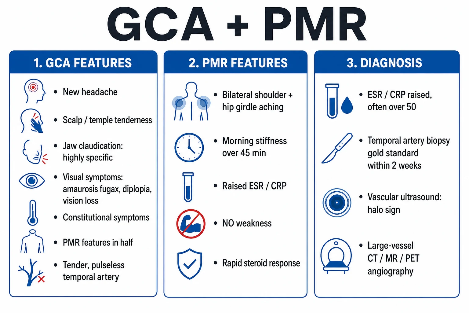
Giant cell arteritis (GCA, temporal arteritis) is the most common primary vasculitis in adults over 50, affecting large and medium arteries (especially the extracranial branches of the carotid, particularly the temporal arteries, and the aorta and its branches). Symptoms: new-onset headache, scalp tenderness, jaw claudication, visual disturbance (ischaemic optic neuropathy, amaurosis fugax, diplopia), constitutional symptoms, polymyalgia rheumatica. Diagnosis: ESR/CRP markedly elevated; temporal artery biopsy confirms. Treatment: high-dose corticosteroids started immediately on clinical suspicion (do not wait for biopsy); tocilizumab for relapse or steroid-sparing.
On this page & tools
Your progress
Saved locally on this device.
Exam tags
Red flags
Overview & Definition
Giant cell arteritis (GCA), also called temporal arteritis or Horton disease, is the most common primary systemic vasculitis of adults and the single most common cause of acute, permanent visual loss in this age group that is entirely preventable.[3][4] It is a granulomatous vasculitis of medium and large arteries, defined anatomically by involvement of vessels with an elastic lamina and a well-developed vasa vasorum. The clinical hallmark is involvement of the extracranial branches of the carotid artery — most often the superficial temporal artery, but also the ophthalmic, posterior ciliary, vertebral, occipital, facial and maxillary arteries. In roughly a third of patients the disease extends to the aorta and its primary branches (subclavian, axillary, brachial, carotid, iliac), the so-called large-vessel or extracranial GCA, which carries a distinct risk of aneurysm, dissection and limb or cerebral ischaemia.[1]
Pathologically the arterial wall is infiltrated by CD4+ T lymphocytes, macrophages and multinucleated giant cells, with fragmentation of the internal elastic lamina, intimal hyperplasia and luminal occlusion — the structural correlate of ischaemic symptoms such as jaw claudication, scalp necrosis, tongue ischaemia and anterior ischaemic optic neuropathy (AION). GCA is intimately linked with polymyalgia rheumatica (PMR): about half of patients with GCA have PMR, and roughly a fifth of patients presenting with isolated PMR will go on to develop GCA.[3][5]
The unifying clinical principle — and the single most examined fact — is that GCA is a treatable medical emergency in which irreversible blindness is preventable only by immediate empirical glucocorticoids. Biopsy confirms; it never delays treatment. [1]
Classification
GCA is classified by the distribution of vessel involvement, because this changes the presentation, the diagnostic strategy, and the long-term complication profile.[1][4]
Cranial (classic) GCA
- Temporal, occipital, facial and ophthalmic branches of the external carotid
- Headache, scalp tenderness, jaw claudication, visual loss (anterior ischaemic optic neuropathy)
- Temporal artery biopsy positive in 60 to 90 percent of cases
- The form most undergraduates and vivas describe — 70 to 80 percent of cases
- Primary risk: permanent blindness if steroids delayed
Large-vessel GCA
- Aorta and primary branches: subclavian, axillary, carotid, vertebral, iliac
- Limb claudication, asymmetric blood pressure (over 15 mmHg between arms), bruits, absent radial pulse
- Cranial features may be absent — 'fever of unknown origin' or occult presentation
- Temporal artery biopsy often negative; PET-CT or MRA required
- Long-term risk: thoracic aortic aneurysm, dissection, aortic regurgitation
- 20 to 30 percent of cases; up to 50 percent on imaging
GCA with PMR overlap
- Bilateral shoulder and pelvic-girdle pain and morning stiffness greater than 45 minutes
- 40 to 60 percent of GCA patients; up to 20 percent of isolated PMR later develop GCA
- Inflammatory markers raised; normal muscle enzymes
- Responds to lower-dose prednisolone (15 mg) once the GCA component is controlled
- Must be actively asked about in every suspected GCA case
Isolated ocular GCA
- Sudden painless visual loss as the first symptom, minimal systemic features
- Posterior ciliary artery occlusion → anterior ischaemic optic neuropathy
- Highest risk of bilateral involvement within days
- Treat as emergency: IV methylprednisolone 500 mg to 1 g daily for 3 days
- Often missed; the rare presentation every examiner loves
GCA is also classified pathologically by the dominant inflammatory pattern (granulomatous with giant cells versus a neutrophil-poor pan-arteritis), but this distinction has little clinical consequence beyond confirming the diagnosis on biopsy. [1]
Epidemiology & Risk Factors
GCA is exclusively a disease of older adults: it is virtually never seen under 50 years, and incidence rises steeply with each decade. The peak incidence is between 70 and 80 years of age. There is a striking female predominance of about 3:1, and the highest global rates are reported in Northern European and Scandinavian populations — Olmsted County, Minnesota (a Scandinavian-descended cohort) reports the world's highest incidence at 18 to 30 per 100 000 persons over 50. The disease is uncommon in Black, Hispanic, Asian and Arab populations, although it is increasingly recognised worldwide as life expectancy rises.[3][4]
Recognised risk factors are age, female sex, Northern European ancestry, a personal or family history of PMR or GCA, HLA-DRB1*04 alleles (particularly *0401), and cigarette smoking in women. A seasonal and cyclical clustering of cases has been described, supporting a hypothesised environmental (possibly infectious, e.g. Mycoplasma pneumoniae, parvovirus B19, Chlamydia pneumoniae) trigger in genetically susceptible hosts, although no pathogen has been consistently confirmed. [1]
The single most important clinical association is polymyalgia rheumatica: roughly 40 to 60 percent of patients with GCA have PMR symptoms, and a patient presenting with isolated PMR has a 15 to 20 percent lifetime risk of developing clinical GCA. This is why every PMR patient is counselled to report new headache, jaw pain or visual change immediately.[5]
Pathophysiology
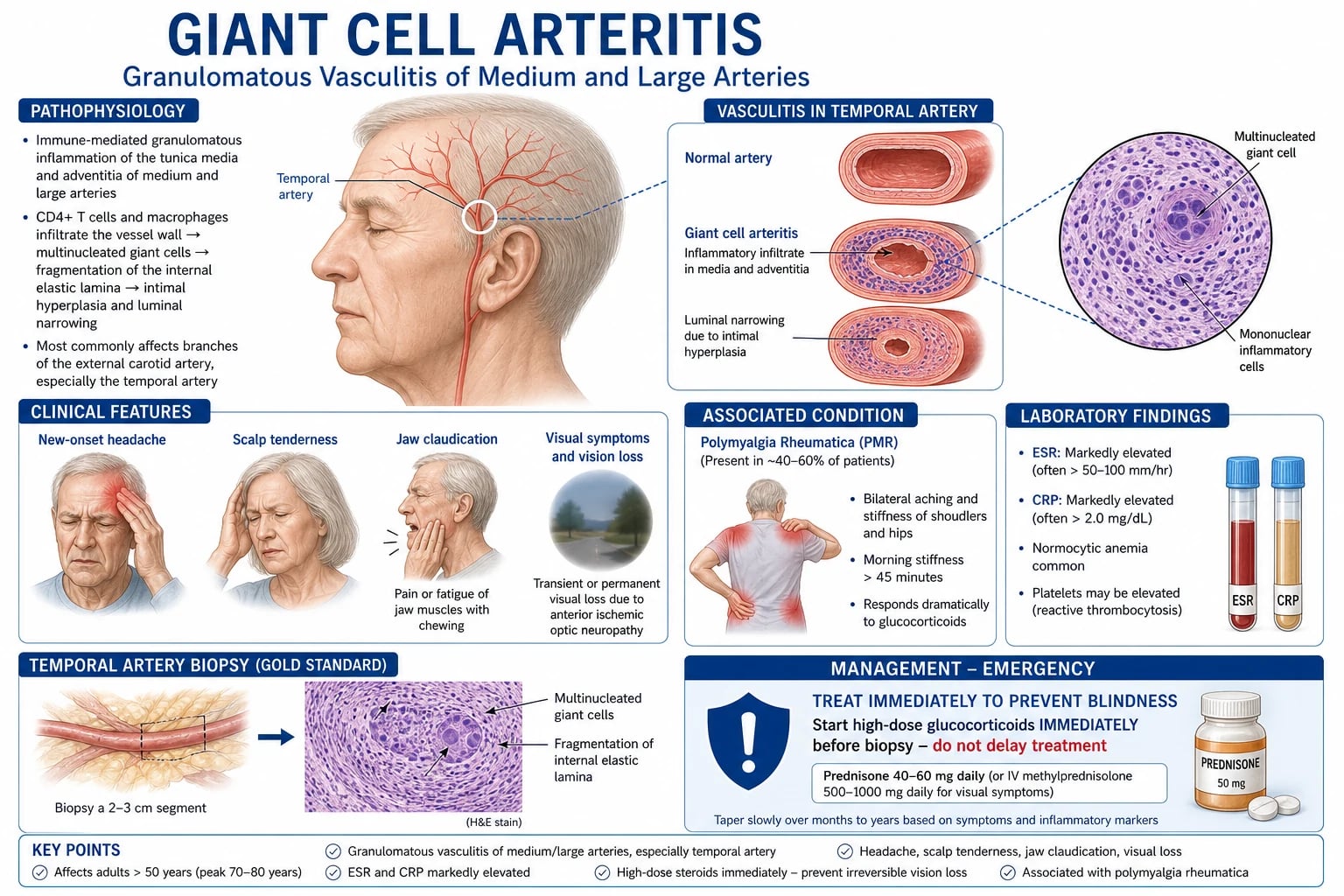
GCA is a T-cell-driven granulomatous vasculitis of the arterial wall. The disease begins at the vasa vasorum in the adventitia, where dendritic cells resident in the arterial wall — normally quiescent sentinels — become activated by an unknown trigger and recruit circulating CD4+ T cells.[4]
Two helper T-cell axes dominate the response. Th1 cells produce interferon-gamma, which activates macrophages that aggregate along the elastic lamina, fuse into multinucleated giant cells, and drive the granulomatous destruction of the internal elastic lamina. Th17 cells produce IL-17 and are recruited and maintained by IL-6 and IL-23. Macrophages at the media release reactive oxygen species and metalloproteinases that fragment the elastic lamina, while macrophages in the intima secrete growth factors (PDGF, VEGF, TGF-beta) that drive intimal hyperplasia — the structural cause of luminal narrowing and tissue ischaemia.[4]
The downstream consequences are ischaemic: the posterior ciliary arteries (terminal branches of the ophthalmic artery with no collateral supply) are most vulnerable, so anterior ischaemic optic neuropathy is the dominant ocular lesion. The masseteric and temporalis branches of the external carotid cause jaw claudication. The vertebral arteries are involved in serious cranial disease and underlie GCA-related posterior circulation stroke. In large-vessel GCA, granulomatous aortitis weakens the aortic media and predisposes to thoracic aortic aneurysm and dissection — the chief cause of excess late mortality. [1]
The central pathogenic role of IL-6 is the rationale for tocilizumab, the anti-IL-6 receptor monoclonal antibody licensed for GCA on the strength of the GiACTA trial.[2]
VISUAL
Why the temporal artery and the aorta, and why almost exclusively in older adults? The answer lies in vascular ageing and immunosenescence. GCA targets only vessels that retain a well-developed internal elastic lamina and an adventitial vasa vasorum — features that distinguish the cranial branches of the carotid and the aorta from intracranial vessels (which are spared, explaining why GCA rarely causes parenchymal brain lesions). The elastic lamina carries oxidised lipids and stress antigens that, in an aged artery, constitutively activate wall-resident dendritic cells via Toll-like receptors. With age, the T-cell repertoire contracts (thymic involution) and senescent CD4+ T cells accumulate; these are precisely the autoreactive clones that infiltrate the arterial wall in GCA. The same immune-ageing explains the female predominance (women lose thymic output earlier) and the exclusive occurrence after 50.[4]
The clinical consequence is a tight clinicopathological correlation: the vessel territory involved dictates the symptom. Posterior ciliary artery occlusion causes anterior ischaemic optic neuropathy; masseteric branch ischaemia causes jaw claudication; vertebral artery involvement causes posterior-circulation stroke; aortic inflammation causes aneurysm and dissection. This is why a careful vessel-by-vessel history and examination outperforms any single blood test at the bedside. [1]
Clinical Presentation
GCA typically presents subacutely over days to weeks in an older patient, although catastrophic visual loss can be the first symptom. The classic triad is new headache, scalp tenderness and jaw claudication, often accompanied by PMR and constitutional upset. The presentation divides into cranial, ischaemic-ocular, large-vessel and constitutional patterns, and these overlap freely.[3][4]
Cranial symptoms
A new-onset headache is the most common symptom, present in 70 to 80 percent. It is classically temporal and unilateral but may be frontal, occipital or diffuse; it is often worse at night and on cold exposure or combing the hair. Scalp tenderness (over the temporal artery, or pain on brushing hair, lying on a pillow or wearing glasses) affects about half. Jaw claudication — aching pain in the masseter and temporalis muscles provoked by prolonged chewing (such as eating meat or crusty bread) and relieved by rest — is the most specific symptom and is present in 30 to 50 percent. Tongue claudication and rarely tongue or scalp necrosis are highly specific but uncommon. [1]
Visual symptoms — the emergency
Visual involvement occurs in 15 to 30 percent of untreated patients and is the cardinal reason to treat empirically and urgently. Presentations include: [1]
- Amaurosis fugax — transient monocular visual loss lasting seconds to minutes, the warning sign of impending infarction.
- Diplopia — from ischaemic cranial nerve palsies (III, IV, VI) or brainstem involvement.
- Sudden, painless, permanent visual loss — most often from anterior ischaemic optic neuropathy (AION) due to posterior ciliary artery occlusion, producing a pale, swollen optic disc with flame haemorrhages and an altitudinal field defect (classically inferior). Central retinal artery occlusion and posterior ischaemic optic neuropathy are less common.
- Bilateral blindness can develop within days of the first eye if steroids are not started. [1]
Polymyalgia rheumatica
Bilateral aching and morning stiffness (more than 45 minutes) of the shoulder and pelvic-girdle musculature — often severe enough to prevent the patient from lifting their arms, combing their hair or rising from a chair — is present in 40 to 60 percent and may be the dominant complaint. Muscle strength is relatively preserved (the patient complains of pain and stiffness, not true weakness), and creatine kinase is normal, distinguishing it from myositis. [1]
Constitutional and systemic features
About half of patients have low-grade fever, fatigue, malaise, anorexia and weight loss. GCA is an important, under-recognised cause of fever of unknown origin in the elderly; a patient with large-vessel GCA and normal cranial arteries may present with fever, weight loss and a high ESR but no headache. Night sweats, depression and a general feeling of being unwell are common. [1]
Large-vessel involvement
Arm or leg claudication, asymmetry of blood pressure between arms (more than 15 mmHg), absent or weak radial pulses, and bruits over the subclavian, axillary, carotid or abdominal arteries suggest extracranial disease. Aortic root dilatation may produce a new aortic regurgitation murmur. Rarely the first presentation is a thoracic aortic aneurysm or dissection, which is why GCA patients carry lifelong aortic imaging surveillance. [1]
Atypical and occult presentations
Examiners test the corners. Atypical presentations include cough (rare; from ischaemia of the recurrent laryngeal nerve or cough receptors), sore throat, depression or cognitive decline mistaken for ageing, neck pain, peripheral neuropathy, pericarditis or pleural effusion, and recurrent falls. Occult large-vessel GCA presents as fever of unknown origin with a normal cranial examination — the diagnosis is missed unless large-vessel imaging is performed. [1]
Differential Diagnosis
The differential divides into other vasculitides, the headache mimics, the ocular mimics, and the systemic-inflammation mimics. Each must be excluded because the treatment of GCA — prolonged high-dose steroids — is itself dangerous if given for the wrong diagnosis.[1][3]
Polymyalgia rheumatica
- Same age and sex distribution, raised ESR/CRP
- Proximal girdle pain and morning stiffness, but no cranial symptoms, no visual change, normal temporal artery
- PMR alone responds to low-dose prednisolone 15 mg; if GCA features develop, switch to high dose
- 15 to 20 percent of isolated PMR later develop GCA
Takayasu arteritis
- Same large-vessel pathology, but under 40 years (typically 15 to 40), 'pulseless disease'
- Subclavian/renal/aortic involvement, bruits, asymmetric BP, hypertension from renal artery stenosis
- Cranial symptoms rare; ESR may be normal
- Treat with steroids ± tocilizumab; revascularisation often needed
Migraine / tension / cluster headache
- Migraine: throbbing, photophobia, aura, normal ESR/CRP
- Tension: bilateral pressing, no systemic features, normal inflammatory markers
- Cluster: unilateral peri-orbital, lacrimation, rhinorrhoea, lasting 15 to 180 minutes
- None cause scalp tenderness, jaw claudication or visual loss from AION
Non-arteritic AION
- Sudden painless visual loss in older patient with vascular risk factors (small disc 'disc at risk')
- ESR/CRP normal, no headache, no jaw claudication
- No temporal artery abnormality
- NOT a steroid-responsive emergency — the mimic that traps the unwary
Dental / TMJ / trigeminal neuralgia
- Jaw pain from temporomandibular joint dysfunction, dental abscess, parotitis
- Pain not claudicant (not provoked by prolonged chewing then relieved by rest)
- Normal ESR/CRP, normal temporal artery, no visual symptoms
Malignancy / infection / other vasculitis
- Lymphoma, metastatic RCC, multiple myeloma, disseminated TB, infective endocarditis all raise ESR/CRP
- ANCA-associated vasculitis (GPA, MPA, EGPA) — sinus, lung, renal involvement
- Polyarteritis nodosa — medium-vessel, neuropathy, mesenteric ischaemia, hepatitis B
- Exclude with blood cultures, ANCA, urinalysis, imaging
The high-yield discriminator at the bedside is the combination of age over 50, new cranial pain, raised inflammatory markers and an abnormal temporal artery — when all four coexist, treat as GCA. [1]
Clinical & Bedside Assessment
A focused GCA assessment takes ten minutes and answers three questions: is this GCA, is vision threatened, and is there large-vessel disease? [1]
History
Establish age (must be over 50), the tempo and character of any headache (new, persistent, often temporal), scalp and hair-combing pain, jaw claudication on prolonged chewing, and visual symptoms in detail — amaurosis fugax, diplopia, blurring and the time course of any visual loss (sudden painless loss in AION). Ask about PMR (bilateral shoulder and pelvic-girdle stiffness, morning stiffness more than 45 minutes, difficulty rising from a chair), constitutional symptoms (fever, weight loss, night sweats, anorexia), limb claudication, and risk factors (PMR history, family history, smoking, Northern European ancestry). Ask specifically about carotidynia (focal neck tenderness) and cough, an under-recognised GCA symptom. [1]
Examination
Vital signs and both arms
Temporal arteries
Visual acuity and fields
Fundoscopy
Cranial nerves and eye movements
Cardiovascular
Peripheral pulses
Musculoskeletal
Named signs and pearls
Jaw claudication is reproduced by asking the patient to chew or clench for one to two minutes. Temporal artery tenderness and thickening is the single most useful bedside sign. Fundal pallor and swelling (AION) is the ophthalmological emergency that mandates IV steroids. Carotidynia — focal carotid tenderness — is a recognised but underappreciated GCA sign. [1]
Investigations
Investigations support but never delay treatment. The strategy is: bloods for inflammatory burden, biopsy for confirmation, and imaging for large-vessel extent.[1][6]
Bloods
- ESR — markedly raised, typically 50 to 100 mm/h or higher; however 5 to 10 percent of biopsy-proven GCA have a normal or near-normal ESR, so a normal ESR never excludes GCA.
- CRP — almost always raised; more sensitive than ESR and the preferred marker for monitoring response and relapse. Combining ESR and CRP raises sensitivity to over 95 percent.
- Full blood count — normocytic anaemia of chronic disease, thrombocytosis (platelets often over 400 × 10⁹/L).
- Liver function tests — mildly raised alkaline phosphatase in a third; hypoalbuminaemia.
- Creatine kinase — normal (helps exclude myositis in a patient with proximal pain).
- UEC, glucose, lipids, TSH — baseline before steroids.
- Autoimmune and infection screen — ANA, ANCA, RF, complement, immunoglobulins, blood cultures, hepatitis B/C, HIV, quantiferon — to exclude mimics (ANCA-associated vasculitis, endocarditis, TB, malignancy).
- Urinalysis — proteinuria or haematuria suggests an alternative or overlap vasculitis. [1]
Temporal artery biopsy — the (imperfect) gold standard
The biopsy shows granulomatous transmural inflammation with multinucleated giant cells and fragmentation of the internal elastic lamina. Key principles:[5][6]
- Unilateral, 1.5 to 2 cm length, ideally within 2 weeks of starting steroids (steroids do not abolish diagnostic changes for 2 to 4 weeks, so a delayed biopsy is still worthwhile).
- Skip lesions cause a false-negative rate of 10 to 15 percent (some quote up to 30 percent). A negative biopsy never excludes GCA in the right clinical context; consider contralateral biopsy if suspicion remains high.
- Sensitivity 60 to 90 percent depending on operator, length and timing. [1]
Colour duplex ultrasound — the halo sign
Non-invasive, operator-dependent, best within the first week of steroids. The halo sign — a dark, hypoechoic, non-compressible circumferential wall thickening around the arterial lumen — represents oedema and inflammatory infiltrate. The TABUL study reported a sensitivity of about 54 percent and specificity of 81 percent for ultrasound alone; combined clinical and ultrasound assessment improves yield.[6] A compression sign (loss of arterial compressibility) is a useful adjunct. Halo sign in axillary arteries is highly specific for large-vessel GCA.
Large-vessel imaging
- PET-CT — the most sensitive modality for active large-vessel inflammation; uptake in the aortic wall and branches confirms large-vessel GCA, particularly when the cranial picture is occult or the biopsy is negative.
- MR angiography of the temporal arteries, aorta and branches — wall thickening, mural oedema and luminal stenosis; useful for diagnosis and surveillance.
- CT angiography — luminal anatomy, stenoses, aneurysms; the modality of choice for acute dissection or aneurysm.
- High-resolution MRI of the temporal arteries — wall enhancement; an emerging alternative to biopsy.
- Axillary/subclavian duplex — halo sign or wall thickening; useful where PET is unavailable. [1]
Other
Chest X-ray (aortic widening, calcification), echocardiogram (aortic root dimension, regurgitation), ECG (coexisting ischaemia). Fundoscopy by an ophthalmologist is mandatory in any patient with visual symptoms to document AION, CRAO or other ocular lesion and to baseline the unaffected eye. [1]
[1]Management — Resuscitation
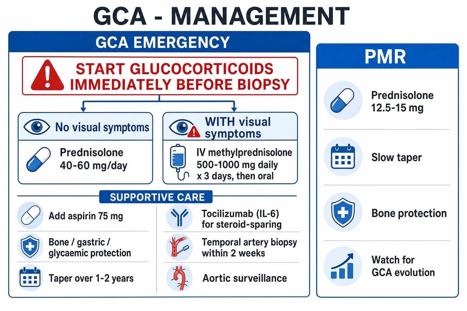
The single most important act in suspected GCA is to give high-dose glucocorticoids immediately on clinical suspicion, before biopsy or any test. The aim is to prevent irreversible visual loss and to abort ischaemia of the optic nerve and other territories.[1]
[1]For suspected GCA without visual symptoms, give oral prednisolone 40 to 60 mg once daily (60 mg preferred; 1 mg/kg, maximum 60 to 80 mg) immediately. The classical, almost diagnostic response within 24 to 72 hours — relief of headache and PMR stiffness and a fall in ESR/CRP — supports the diagnosis while awaiting biopsy.[5]
Adjunctive measures started at the same time: [1]
- Gastric protection: proton-pump inhibitor (omeprazole 20 mg daily) for the steroid course.
- Bone protection: calcium 1000 mg and vitamin D 800 to 1000 IU daily from day 1; add a bisphosphonate (alendronate 70 mg weekly, risedronate 35 mg weekly) if FRAX risk is high or steroids will exceed 3 months. Baseline DEXA scan.
- Antiplatelet: low-dose aspirin 75 mg daily if no contraindication — widely used given the increased ischaemic risk, although evidence is observational rather than randomised.
- Vaccinations: pneumococcal, influenza, COVID-19 and shingles, ideally before or at steroid initiation.
- Glucose monitoring, low-sodium diet, weight, blood pressure. [1]
Approach to the suspected case
The first-hour decision is binary and time-critical. A patient over 50 with a compatible history (new cranial pain, scalp tenderness, jaw claudication, PMR, or unexplained fever with high ESR/CRP) is treated as GCA while the work-up proceeds in parallel.[1]
Stratify for ischaemia
Treat before testing
Draw baseline bloods
Arrange biopsy within 2 weeks
Image for large-vessel disease
Specialist and ophthalmology review
The classical, almost diagnostic response within 24 to 72 hours — relief of headache, scalp and PMR stiffness, and a fall in CRP — itself supports the diagnosis. A patient who fails to improve in 72 hours should be reassessed for an alternative diagnosis (infection, malignancy, non-arteritic AION, other vasculitis). [1]
Management — Definitive & Stepwise
The aim is disease remission on the lowest possible steroid dose, sustained over 12 to 24 months, using tocilizumab as the licensed steroid-sparing agent and managing steroid toxicity proactively.[1][2]
Initial steroid regimen
No visual symptoms
- Prednisolone 40 to 60 mg orally once daily (60 mg preferred)
- Equivalent IV if unable to take orally or very systemic
- Expect dramatic response in 24 to 72 hours
- Continue full dose for 2 to 4 weeks until symptoms and CRP controlled
Visual or evolving ischaemic symptoms
- IV methylprednisolone 500 mg to 1 g daily for 3 days
- Then oral prednisolone 1 mg/kg (typically 60 mg) daily
- Urgent ophthalmology review and baseline fundus findings
- Consider tocilizumab at induction to reduce cumulative steroid
Relapse on taper
- Increase prednisolone to the last effective dose (often plus 10 mg)
- Add or resume tocilizumab
- Re-image for large-vessel progression if new or atypical symptoms
- Re-check for mimics — infection, malignancy — if relapse is atypical
Steroid taper
Taper begins once symptoms and CRP are controlled — usually after 2 to 4 weeks at full dose — and proceeds slowly over 12 to 24 months. A typical EULAR/BSR-aligned schedule:[1][5]
About 30 to 50 percent of patients relapse on tapering, especially in the first 18 months. Relapse is defined by recurrent symptoms with rising ESR/CRP — never by a rising marker alone (chase the patient, not the number). [1]
Tocilizumab — the GiACTA evidence
GiACTA
Population: 251 patients with new-onset or relapsing GCA
Key finding
Sustained remission at 12 months: 56 percent (weekly) versus 53 percent (fortnightly) versus 14 percent (26-week steroid taper) and 18 percent (52-week steroid taper). Cumulative steroid exposure roughly halved.
Tocilizumab (anti-IL-6 receptor) is given at 162 mg subcutaneously once weekly (or 8 mg/kg IV every 4 weeks where SC is unsuitable). Indications per EULAR:[1][2]
- At induction in patients at high risk of steroid toxicity (age over 70, diabetes, osteoporosis, BMI over 30, prior steroid complications, fracture history).
- On relapse despite adequate steroids.
- For refractory disease or to allow faster taper. [1]
Continue tocilizumab for at least 12 months (typically 1 to 2 years), with gradual withdrawal under monitoring. Note tocilizumab masks CRP and ESR (it blocks IL-6); monitor clinically and consider imaging for large-vessel activity. Live vaccines are contraindicated; screen for latent TB and hepatitis B before starting. [1]
Other immunosuppressants — methotrexate 10 to 15 mg weekly, azathioprine, mycophenolate — have a weaker evidence base and are reserved for tocilizumab failure, intolerance, or pregnancy. Cyclophosphamide is reserved for severe refractory disease. Methotrexate remains a reasonable option where biologics are unavailable.[1]
Adjunctive and long-term measures
- Aspirin 75 mg daily for vascular protection (observational evidence).
- Bone protection: calcium/vitamin D, bisphosphonate, DEXA surveillance.
- PPI for the steroid course.
- Statins as indicated for cardiovascular risk.
- Annual aortic imaging (CT or MR angiography) for life in large-vessel GCA — thoracic aortic aneurysm and dissection are the chief cause of excess late mortality.
- Patient education: report any new visual symptom, headache, jaw pain or limb claudication immediately. [1]
Follow-up and monitoring
Active GCA disease activity is monitored clinically, not by blood test alone. The standard review — every 4 to 8 weeks during taper, then every 3 to 6 months for at least two years — covers five domains.[1][3]
Specific Subtypes & Scenarios
Cranial GCA with threatened vision
Treat as above — IV methylprednisolone 500 mg to 1 g daily for 3 days, then oral prednisolone 1 mg/kg, with urgent ophthalmology review, baseline fundus findings and temporal artery biopsy within 2 weeks. Add tocilizumab if steroid toxicity is likely. Counsel that vision lost before steroids is started rarely recovers; the priority is protecting the second eye. [1]
Large-vessel GCA
Diagnosed by PET-CT, MRA or CT angiography showing aortic and branch wall inflammation, stenosis or aneurysm. Treat with high-dose steroids and tocilizumab; biopsy may be negative. Revascularisation (angioplasty, stenting or bypass) for critical limb or cerebral ischaemia should be deferred until inflammation is controlled, because operating on an inflamed vessel carries high complication rates. Long-term annual aortic imaging is mandatory. [1]
GCA with polymyalgia rheumatica
Treat the GCA component with high-dose steroids (40 to 60 mg). Once the cranial and ischaemic features settle, the PMR component usually responds to the same taper. In isolated PMR (no GCA features), start prednisolone 15 mg daily with a rapid taper over 12 months; educate the patient to report any new cranial or visual symptom immediately. A PMR patient whose inflammatory markers rise disproportionately, or who develops systemic features, should be re-evaluated for GCA.[5]
Isolated PMR with high inflammatory markers
Monitor for GCA — 15 to 20 percent will develop it. Counsel about red-flag symptoms. Consider low-dose prednisolone 15 mg and review at one week (a poor response or atypical features should prompt GCA assessment). [1]
Ocular (anterior ischaemic optic neuropathy) GCA
The highest-stakes scenario. Sudden painless visual loss in an older patient with raised ESR/CRP is AION from GCA until proven otherwise. IV methylprednisolone within hours, urgent ophthalmology, and biopsy within days. Prognosis for the affected eye is guarded; the goal is to prevent contralateral involvement. [1]
Refractory and relapsing GCA
Relapse on taper is managed by increasing prednisolone to the last effective dose and adding or resuming tocilizumab. Refractory disease (failure of steroids plus tocilizumab) is rare; consider methotrexate, azathioprine, mycophenolate, or trial agents (JAK inhibitors, abatacept). Re-image for occult large-vessel progression and re-examine for mimics. [1]
GCA in pregnancy and premenopausal women
GCA before 50 is exceptional; true pregnancy-related GCA is rare. Prednisolone is safe in pregnancy and lactation (fluorinated steroids cross the placenta less). Tocilizumab is avoided in pregnancy and lactation; prefer steroids alone, with methotrexate contraindicated. [1]
Why must steroids be started before biopsy?
Because the diagnostic inflammatory changes persist in the arterial wall for at least 2 to 4 weeks after steroid initiation, and irreversible blindness can develop within hours of the first symptom. The tiny reduction in biopsy yield from a few days of steroids is dwarfed by the catastrophic cost of a delay. Treat first, biopsy within 2 weeks.
Complications & Pitfalls
Disease-related complications
- Permanent visual loss — anterior ischaemic optic neuropathy is the most feared complication; 15 to 30 percent of untreated patients lose vision, reduced to under 5 percent with prompt steroids. Once established, vision rarely recovers.
- Stroke — particularly posterior circulation from vertebral artery involvement; rare but devastating.
- Aortic aneurysm and dissection — predominantly thoracic; the risk is increased 2 to 4 fold and persists for years after diagnosis. This is the chief cause of excess late mortality.
- Limb claudication and critical ischaemia from subclavian, axillary or iliac stenosis.
- Scalp and tongue necrosis — rare, dramatic, ischaemic.
- Myocardial infarction, mesenteric and renal ischaemia — rare but reported. [1]
Treatment-related complications
Long-term steroids cause osteoporosis and vertebral fractures, diabetes, hypertension, fluid retention, weight gain, mood disturbance, insomnia, peptic ulcer, skin thinning and bruising, cataract, glaucoma, adrenal suppression, and increased infection risk. Bisphosphonate, PPI, glucose monitoring, vaccination and DEXA surveillance mitigate but do not abolish these harms — the rationale for steroid-sparing with tocilizumab. Tocilizumab carries risks of infection, neutropenia, transaminitis, hyperlipidaemia, bowel perforation (rare) and masks inflammatory markers.[2]
Classic pitfalls
- Waiting for biopsy before treating — the cardinal error.
- Attributing the headache to migraine, tension or dental disease in an older patient — always check ESR/CRP and the temporal artery.
- Excluding GCA on a normal ESR — 5 to 10 percent of biopsy-proven cases have a normal ESR; combine with CRP and the clinical picture.
- Excluding GCA on a negative biopsy — skip lesions give a 10 to 15 percent false-negative rate.
- Missed large-vessel GCA in the patient with fever of unknown origin, normal cranial examination and negative biopsy — image with PET-CT.
- Chasing the marker, not the patient — relapse is defined by recurrent symptoms, not by a rising number alone.
- Failing to image the aorta during follow-up — the silent development of thoracic aneurysm kills GCA patients years after remission. [1]
Prognosis & Disposition
Most patients respond dramatically to glucocorticoids within 24 to 72 hours, with normalisation of inflammatory markers over 2 to 4 weeks. Relapse occurs in 30 to 50 percent on tapering, most often in the first 18 months, and is managed by dose escalation and tocilizumab. Once visual loss occurs it is usually permanent, which is why empirical treatment is non-negotiable. [1]
Overall mortality is similar to the age-matched general population, with a modest excess from cardiovascular disease, aortic aneurysm and dissection, and steroid complications. Large-vessel involvement, particularly thoracic aortic aneurysm, is the chief driver of excess late mortality — hence lifelong aortic imaging. Quality of life is dominated by steroid toxicity; tocilizumab has substantially improved this.[2][3]
Disposition:
- Admit any patient with visual symptoms for IV methylprednisolone and ophthalmology review; also admit severe systemic disease, diagnostic uncertainty needing imaging, frailty or inability to manage complex outpatient therapy.
- Outpatient management is appropriate for uncomplicated GCA without visual symptoms, with a 72-hour review to confirm response and arrange biopsy within 2 weeks.
- Same-day ophthalmology for any visual symptom — to baseline the eye and to coordinate IV steroids. [1]
Special Populations
- Elderly (over 75) — the largest group; highest steroid-toxicity risk. Strongly consider tocilizumab at induction, aggressive bone protection, and falls assessment. Watch for steroid-induced delirium.
- Diabetes — high-dose steroids destabilise glucose control; involve the diabetes team, monitor capillary glucose, adjust insulin/oral therapy. Tocilizumab early to minimise cumulative steroid.
- Established osteoporosis or prior fracture — bisphosphonate from day 1, DEXA, calcium/vitamin D, teriparatide or denosumab if severe; favour tocilizumab.
- Immunocompromised — exclude infection (TB, endocarditis) before committing to high-dose steroids; co-ordinate with infectious diseases.
- Pregnancy (rare, as GCA is rare under 50) — prednisolone preferred (safe); avoid tocilizumab, methotrexate, mycophenolate. Monitor for gestational diabetes and hypertension.
- Visual loss in one eye — protect the contralateral eye with IV steroids and aspirin; counsel that second-eye involvement can follow within days.
- Aortic involvement — lifelong annual aortic imaging; strict blood pressure control; surgical or endovascular repair when thresholds are met. [1]
Evidence, Guidelines & Regional Differences
GCA management rests on EULAR, the British Society for Rheumatology (BSR), the American College of Rheumatology/Vasculitis Foundation (ACR/VF) and NICE guidance. Their core principles converge — empirical high-dose steroids on suspicion, biopsy within 2 weeks, imaging for large-vessel disease, tocilizumab for steroid-sparing, bone and gastric protection, and aortic surveillance.[1][5]
EULAR 2018 update (Hellmich 2020)
Population: Large-vessel vasculitis (GCA and Takayasu)
Key finding
Recommends immediate glucocorticoids; tocilizumab as first-line adjunct for high-risk or relapsing GCA; imaging (PET-CT, MRA, ultrasound) for large-vessel assessment; long-term glucocorticoid-sparing.
EULAR 2018 (Hellmich 2020) is the most widely cited international standard: empirical high-dose glucocorticoids, structured taper, tocilizumab as adjunctive steroid-sparing, large-vessel imaging with PET-CT or MRA, ultrasound halo sign as adjunct or alternative to biopsy, and long-term aortic surveillance.
Controversies and emerging evidence
- Ultrasound versus biopsy — the TABUL study showed ultrasound alone is less sensitive than hoped; combined clinical-and-ultrasound pathways are emerging as the fastest route to diagnosis. High-resolution MRI of the temporal arteries is gaining ground.
- IV versus oral induction for visual symptoms — no randomised trial has definitively shown IV methylprednisolone superior to high-dose oral for visual outcomes, but expert consensus and guidelines favour IV for threatened or established vision loss.
- Aspirin — observational data only; widely used but unproven by randomised trial.
- Duration of tocilizumab — 1 to 2 years is typical; relapse after withdrawal is common and the optimal duration is unclear.
- Earlier and faster steroid taper with biologics — modern regimens aim for a 26-week taper (GiACTA style) rather than the older 12 to 24 month courses. [1]
Exam Pearls
- GCA = large-vessel vasculitis of the over-50s; think temporal headache, scalp tenderness, jaw claudication, visual loss, PMR.
- Start high-dose steroids IMMEDIATELY on suspicion — never wait for biopsy or any test. Visual symptoms → IV methylprednisolone 500 mg to 1 g daily for 3 days.
- Jaw claudication (pain in the masseter on prolonged chewing, relieved by rest) is the most specific symptom.
- Visual loss is from anterior ischaemic optic neuropathy (posterior ciliary artery occlusion) — pale swollen disc, altitudinal field defect, usually permanent once it occurs.
- ESR typically over 50 mm/h, but 5 to 10 percent of biopsy-proven cases have a normal ESR — combine with CRP.
- Temporal artery biopsy within 2 weeks of starting steroids; skip lesions cause 10 to 15 percent false negatives.
- Ultrasound halo sign — hypoechoic wall oedema; PET-CT for large-vessel GCA.
- Tocilizumab (anti-IL-6, 162 mg SC weekly) — GiACTA trial — is the licensed steroid-sparing agent.
- Polymyalgia rheumatica in 40 to 60 percent; PMR alone responds to prednisolone 15 mg.
- Aortic aneurysm and dissection (especially thoracic) are late, often fatal complications — annual aortic imaging for life.
- Taper steroids over 12 to 24 months; relapse in 30 to 50 percent.
- Takayasu is the differential in those under 40 — same pathology, different age and territory. [1]
Exam application bank (NEET-PG / INICET)
One-line answer
Giant cell arteritis (GCA, temporal arteritis) is the most common primary vasculitis in adults over 50, affecting large and medium arteries (especially the extracranial branches of the carotid, particularly the temporal arteries, and the aorta and its branches). Symptoms: new-onset headache, scalp tenderness, jaw claudication, visual disturbance (ischaemic optic neuropathy, amaurosis fugax, diplopia), constitutional symptoms, polymyalgia rheumatica. Diagnosis: ESR/CRP markedly elevated; temporal artery biopsy confirms. Treatment: high-dose corticosteroids started immediately on clinical suspicion (do not wait for biopsy); tocilizumab for relapse or steroid-sparing.
Worked stems (answer without another resource)
Stem 1 — Classic presentation. Map symptoms to mechanism; name the first investigation and first treatment step with dose/route if drug therapy is standard. [1]
Stem 2 — Unstable / complicated. List red flags that force immediate resuscitation, theatre, ICU, antidote, or reperfusion — and what you do in the first 15 minutes. [1]
Stem 3 — Atypical group. Elderly, pregnancy, child, or immunocompromised: how presentation and thresholds change. [1]
Stem 4 — Differential trap. Name the three closest mimics and one discriminator for each. [1]
Stem 5 — Disposition. Who goes home with safety-netting, who is admitted, who needs HDU/ICU/theatre, and what follow-up is mandatory. [1]
Rapid viva checklist
- Definition + classification
- Pathophysiology chain
- Bedside signs / criteria
- Score with exact components (if any)
- Emergency bundle
- Definitive therapy with doses
- Complications of disease and of treatment
- Special populations
- Guideline/trial name if classic
- Three exam traps
Coverage self-check
If you cannot answer any stem above from this page alone, re-read the matching section — the page is intended to be self-sufficient for final-prof and NEET-PG/INICET questions on Giant Cell Arteritis.
[1] [1]References
- [1]Hellmich B, Agueda A, Monti S, et al. 2018 Update of the EULAR recommendations for the management of large vessel vasculitis Ann Rheum Dis, 2020.PMID 31270110
- [2]Stone JH, Tuckwell K, Dimonaco S, et al. Trial of Tocilizumab in Giant-Cell Arteritis N Engl J Med, 2017.PMID 28745999
- [3]Salvarani C, Cantini F, Hunder GG Polymyalgia rheumatica and giant-cell arteritis Lancet, 2008.PMID 18640460
- [4]Weyand CM, Goronzy JJ Clinical practice. Giant-cell arteritis and polymyalgia rheumatica N Engl J Med, 2014.PMID 24988557
- [5]Dasgupta B, Borg FA, Hassan N, et al. BSR and BHPR guidelines for the management of polymyalgia rheumatica Rheumatology (Oxford), 2010.PMID 19910443
- [6]Luqmani R, Lee E, Singh S, et al. The Role of Ultrasound Compared to Biopsy of Temporal Arteries in the Diagnosis and Treatment of Giant Cell Arteritis (TABUL): a diagnostic accuracy and cost-effectiveness study Health Technol Assess, 2016.PMID 27925577