Rheumatology · Rheumatology
Gout
Also known as Gout · Gouty arthritis · Podagra · Monosodium urate crystal deposition disease · Tophaceous gout
Gout is a crystal-deposition disease caused by monosodium urate (MSU) crystal accumulation in joints and soft tissues, the end result of chronic hyperuricaemia. It is the commonest inflammatory arthritis in adults. The clinical spectrum runs from asymptomatic hyperuricaemia to explosive acute monoarthritis (classically the first MTP — podagra), through intercritical periods, to chronic tophaceous gout with erosive joint destruction, urate nephropathy and uric acid urolithiasis. Risks: male sex, obesity, metabolic syndrome, CKD, alcohol (especially beer), high-purine diet, fructose, diuretics, cyclosporin, lead. The diagnostic hallmark is the negatively birefringent, needle-shaped monosodium urate crystal on polarised-light microscopy of synovial fluid. Acute attacks are treated with an anti-inflammatory-dose NSAID, low-dose colchicine or corticosteroid (all equally first-line, chosen by comorbidity). Long-term urate-lowering therapy (allopurinol first-line) is titrated to a target serum urate below 6 mg/dL (360 micromol/L), with low-dose colchicine prophylaxis against mobilisation flares.
On this page & tools
Your progress
Saved locally on this device.
Exam tags
Red flags
Overview & Definition
Gout is a crystal-deposition disease: a disease caused by the deposition of monosodium urate (MSU) crystals in and around joints and soft tissues, itself the consequence of persistent hyperuricaemia (serum urate above about 6.8 mg/dL or 400 micromol/L — the saturation point of urate at physiological pH and temperature). It is the commonest inflammatory arthritis in adults and its prevalence is rising globally in parallel with obesity, metabolic syndrome and chronic kidney disease.[1][5]
The natural history unfolds in four phases: [1]
- Asymptomatic hyperuricaemia — raised serum urate without symptoms or crystal deposition (most hyperuricaemic people never develop gout, so urate alone does not diagnose gout).
- Acute gouty arthritis — the first and recurrent flares: an explosive monoarthritis.
- Intercritical gout — asymptomatic intervals between attacks; over time these intervals shorten.
- Chronic tophaceous gout — persistent deposition forming tophi, erosive arthritis, and extra-articular disease (urate nephropathy, uric acid stones). [1]
The key clinical insight is that gout is curable — unlike most arthritides, sustained urate-lowering dissolves crystals, stops attacks and shrinks tophi. The single biggest reason gout remains poorly controlled is undertreatment: failure to start urate-lowering therapy, failure to titrate to a serum-urate target, and failure to give flare prophylaxis. The ACR 2020 guideline and EULAR 2016 recommendations both endorse a treat-to-target strategy.[2][3]
Classification
Gout is classified clinically by phase (asymptomatic hyperuricaemia, acute, intercritical, chronic tophaceous) and formally by the 2015 ACR/EULAR classification criteria, which are used in research and to confirm a diagnosis when crystal identification is unavailable.[4]
GOUT PHASES — the natural history
GOUT
Explosive monoarthritis, often podagra; reaches max in 12 to 24 hours
Asymptomatic interval between attacks; intervals shorten over time
Chronic tophaceous gout with subcutaneous tophi and erosive arthritis
Treat-to-target serum urate below 6 mg/dL (below 5 mg/dL if tophi) — gout is curable
Epidemiology & Risk Factors
Gout is the commonest inflammatory arthritis in men. Prevalence is 3 to 4 percent in adults in high-income countries and rising, driven by obesity, metabolic syndrome, CKD and longer survival. Male sex predominates: oestrogens are uricosuric, so gout is rare in premenopausal women and rises after the menopause. The peak onset in men is the fourth to sixth decade; in women it is the sixth to eighth decade, often in the setting of diuretic use, CKD or hypertension.[1][5]
Gout — key numbers
Risk factors cluster around urate overproduction or, far more commonly, urate underexcretion: [1]
- Hyperuricaemia — the central, modifiable driver; the higher the serum urate, the higher the risk.
- Metabolic: obesity (and weight gain in young adulthood), metabolic syndrome, type 2 diabetes, dyslipidaemia, hypertension — gout is now understood as a cardiometabolic disease, and gout confers an independent cardiovascular risk.
- Diet: high-purine foods (organ meats, red meat, shellfish, sardines, anchovies, game), beer and spirits (beer is purine-rich from yeast and alcohol suppresses urate excretion), and fructose / sugar-sweetened drinks (fructose metabolism generates AMP and raises urate). Low-fat dairy is uricosuric and protective.
- Drugs that raise urate: thiazide and loop diuretics, low-dose aspirin, cyclosporin, tacrolimus, niacin, pyrazinamide, ethambutol, lead.
- Kidney disease — underexcretion via reduced filtered load.
- High cell turnover: psoriasis, myeloproliferative disorders, malignancy, tumour lysis syndrome, haemolytic anaemia.
- Genetics: polymorphisms in urate transporters — SLC2A9 (GLUT9) and ABCG2 — explain a large share of serum-urate variance; rare X-linked enzyme defects cause severe early-onset gout (HGPRT deficiency = Lesch-Nyhan; PRPP synthetase overactivity).
- Lead exposure — saturnine gout, classically from moonshine or occupational lead, causes underexcretion and coexists with hypertension and CKD. [1]
Pathophysiology
Urate is the end product of purine metabolism in humans. Most mammals convert urate to allantoin via the enzyme uricase (urate oxidase), but humans (and higher apes) lost uricase during evolution, so urate accumulates. Purines are converted by xanthine oxidase (from hypoxanthine to xanthine to uric acid), which is the target of allopurinol and febuxostat.[1][5]
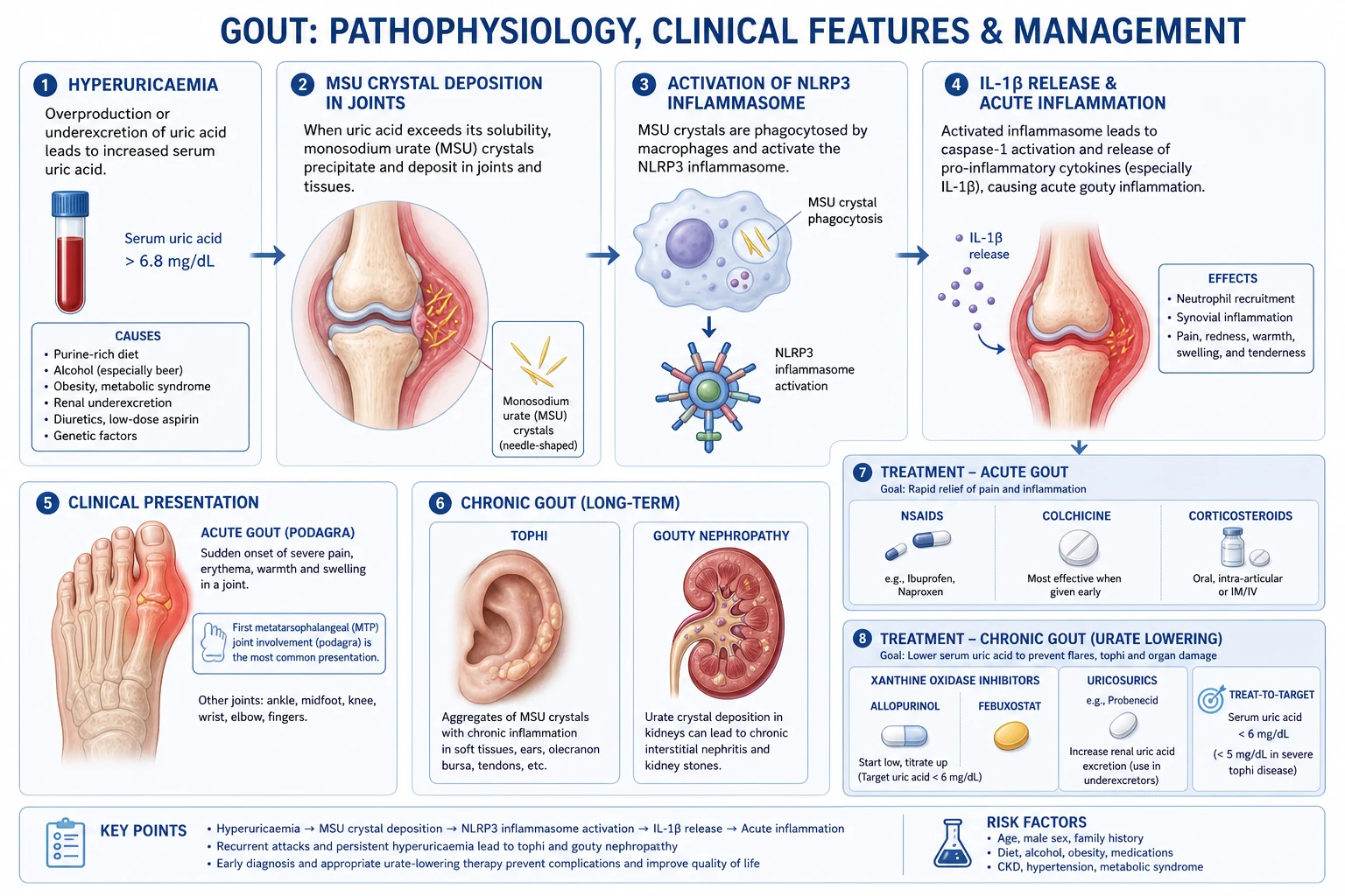
Hyperuricaemia arises from an imbalance between production and excretion, and in over 90 percent of patients the dominant fault is renal underexcretion, not overproduction. This is the single most important pathophysiological fact about gout, because it is why allopurinol and febuxostat (which cut production) and uricosurics (which boost excretion) can both work, and why renal function and urate-raising drugs dominate the risk profile. [1]
The four-step renal handling of urate. Urate is freely filtered at the glomerulus, then handled in the proximal tubule by a choreographed sequence of transporters that the exam rewards:[5]
- Glomerular filtration of unbound urate.
- Reabsorption (nearly complete) via URAT1 (SLC22A12) and GLUT9 (SLC2A9) on the apical (luminal) membrane — the targets of uricosurics (probenecid, lesinurad and benzbromarone all block URAT1).
- Secretion into the lumen via ABCG2 and NPT1 (SLC17A1).
- Post-secretory reabsorption, again largely via URAT1/GLUT9. [1]
Net excretion is therefore reabsorption-dominated, so any gain in reabsorptive capacity or loss of secretory capacity tips the patient into hyperuricaemia. Common ABCG2 dysfunction is a major contributor: a loss-of-function variant (the ABCG2 Q141K allele) impairs renal and intestinal urate excretion and is found in a large fraction of gout patients. Genetic variation in SLC2A9 (GLUT9) is the strongest common determinant of serum-urate concentration in the general population.[5]
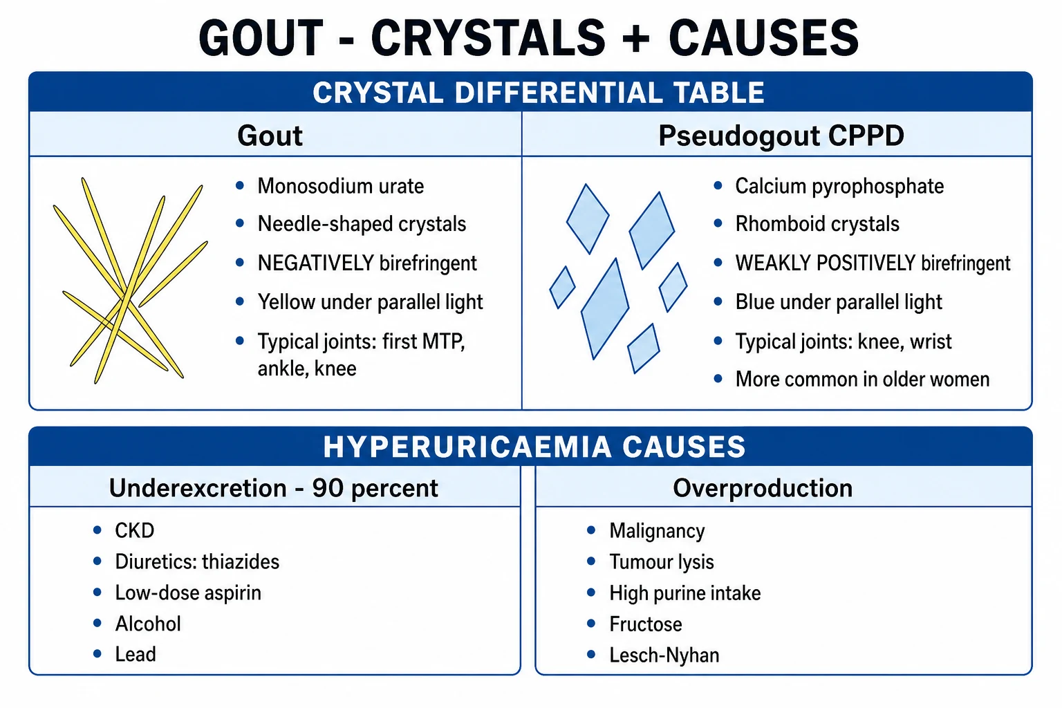
Underexcretion — about 90 percent
- Renal transporter polymorphisms: SLC2A9 (GLUT9) and ABCG2 (Q141K) variants
- Chronic kidney disease — reduced filtered load
- Drugs: thiazide and loop diuretics, ciclosporin, tacrolimus, low-dose aspirin, niacin, pyrazinamide, ethambutol
- Volume depletion / dehydration; ketoacidosis and lactic acidosis (organic acids compete with urate for secretion)
- Lead exposure (saturnine gout) — impairs tubular handling
- Hypothyroidism; pre-eclampsia
- Mechanism: enhanced URAT1/GLUT9 reabsorption or impaired ABCG2/NPT1 secretion
Overproduction — about 10 percent
- High cell turnover: myeloproliferative disorders, malignancy, haemolytic anaemia, psoriasis
- Tumour lysis syndrome — massive acute purine release after chemotherapy
- Enzyme defects: HGPRT deficiency (Lesch-Nyhan, X-linked); PRPP synthetase overactivity; glycogen storage disease type I
- Alcohol (beer is purine-rich from yeast; ethanol raises urate via lactate and purine turnover)
- Fructose / sugar-sweetened drinks — fructose phosphorylation depletes ATP, generating AMP and urate
- High-purine diet: organ meats, shellfish, sardines, anchovies, red meat, game
- Starvation / fasting — ATP depletion raises urate
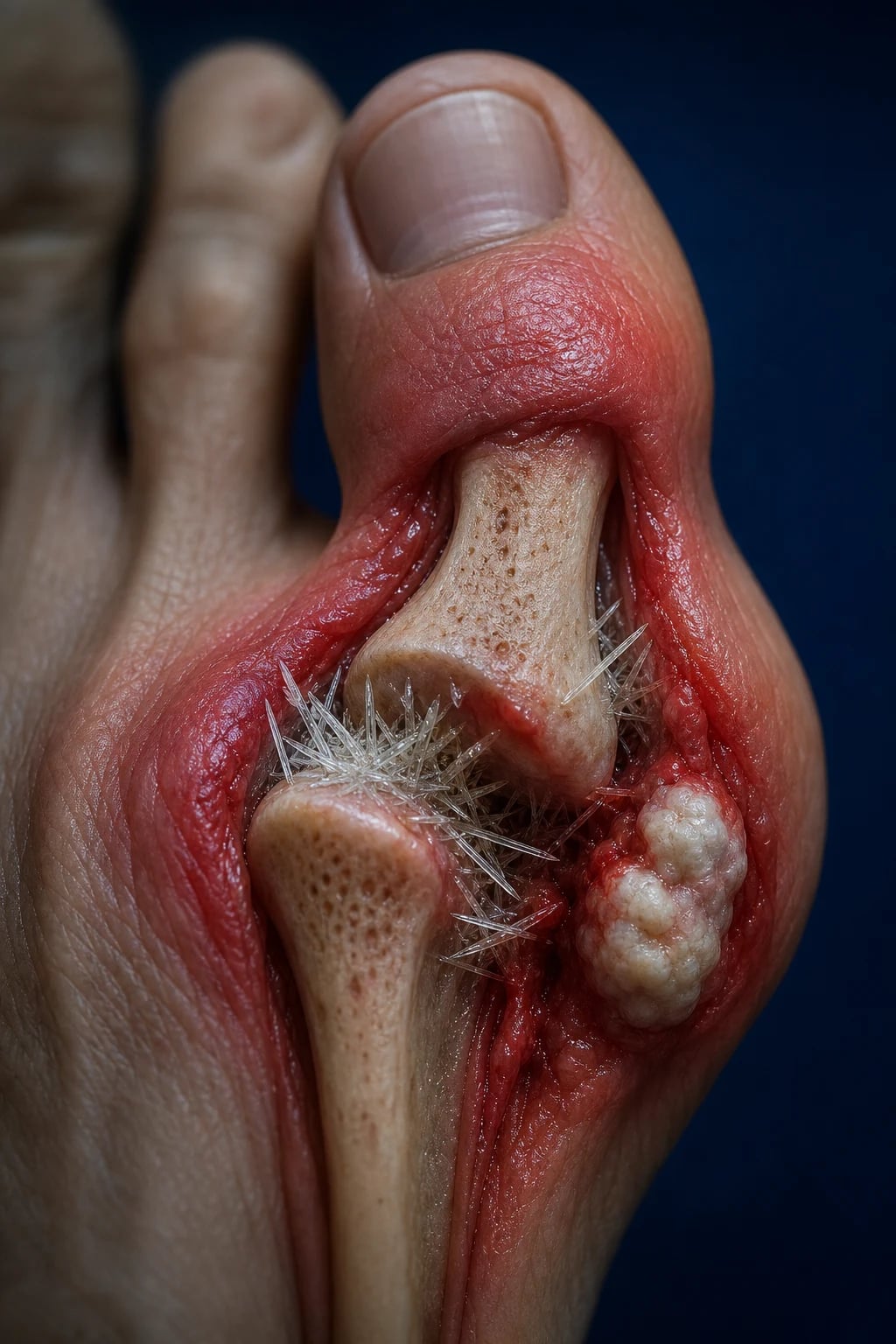
Why it crystallises where it does. When serum urate exceeds its solubility threshold (about 6.8 mg/dL / 400 micromol/L at 37 degrees C), monosodium urate precipitates. Crystallisation is favoured by low temperature — which is why the cool, peripheral first MTP and the helix of the ear are the classical sites — by low pH, dehydration, and pre-existing joint damage (osteoarthritic joints are preferentially involved, hence Heberden-node gout in elderly women). At lower pH urate converts to the more soluble uric acid form in urine, which is why acidic concentrated urine favours uric acid urolithiasis rather than joint disease. [1]
Crystal structure and birefringence. Monosodium urate forms slender needle-shaped monohydrate crystals (2 to 10 micrometres long) that are negatively birefringent under compensated polarised light — they appear yellow when their long axis is parallel to the compensator's slow axis and blue when perpendicular. This is the opposite of CPPD, which forms rhomboid crystals that are weakly positively birefringent (blue parallel, yellow perpendicular). The colour depends on whether the crystal's refractive index along its length exceeds that perpendicular to it; the exam pearl PGYP (Piss Yellow Parallel = Gout) encodes it precisely. [1]
The acute attack is a NLRP3-inflammasome-driven neutrophilic inflammation. Shed MSU crystals are phagocytosed by synovial macrophages and neutrophils. Crystal uptake causes lysosomal rupture and activates the NLRP3 inflammasome, which cleaves pro-caspase-1 to active caspase-1; caspase-1 in turn cleaves pro-IL-1 beta to active IL-1 beta. IL-1 beta signals through the IL-1 receptor to drive NF-kB, TNF-alpha, IL-6 and CXCL8, producing a massive neutrophil influx — the clinical red-hot joint. This is why IL-1 inhibitors (anakinra, canakinumab) are effective in refractory gout, and why colchicine (which disrupts microtubules and neutrophil chemotaxis) works. [1]
Why attacks are self-limiting: neutrophil apoptosis and clearance by CD14+ macrophages, apolipoprotein B and apolipoprotein E coating of crystals (neutralising their inflammatory potential), rising anti-inflammatory mediators (IL-37, TGF-beta, alpha-1 antitrypsin) and a neutrophil-to-macrophage phenotype shift. This explains the typical 3 to 14 day natural course of an untreated attack. [1]
Chronic disease: persistent deposition drives a granulomatous foreign-body reaction — macrophages and multinucleated giant cells around crystalline deposits forming tophi. Tophi secrete RANKL, activating osteoclasts and producing the characteristic punched-out erosions with overhanging sclerotic margins (the "rat-bite" / Martel sign) and erosive, deforming arthritis. [1]
Clinical Presentation
The acute gout attack is one of the most recognisable presentations in medicine: [1]
- Onset: explosive, typically overnight; reaches maximum intensity within 12 to 24 hours.
- Joint: classically a monoarthritis. The first MTP (podagra) is the site in over 50 percent of first attacks. Other sites, in descending frequency: instep/midfoot, ankle, knee, wrist, fingers, elbow.
- Appearance: the joint is red, hot, swollen, shiny and exquisitely tender — the patient cannot bear a sock, shoe or a bedsheet touching it. There may be desquamation of the overlying skin as the attack resolves.
- Systemic: low-grade fever and malaise in severe attacks; high fever should raise concern about sepsis.
- Triggers: alcohol binges (especially beer), dietary excess of purine-rich foods, dehydration or fasting, intercurrent illness, surgery or trauma, starting a urate-lowering drug (mobilisation flare), and diuretic change. [1]
The intercritical period is asymptomatic; left untreated, attacks become more frequent, last longer, involve more joints (oligo-/polyarticular) and may no longer fully remit, merging into chronic gout. [1]
Chronic tophaceous gout presents with firm, yellow-white, subcutaneous nodules (tophi) in characteristic sites: [1]
- Helix and antihelix of the ear
- Olecranon bursa and ulnar surface of the forearm
- Achilles tendon
- Finger pads and extensor surfaces of the fingers
- Heberden nodes of the hands (in elderly women) [1]
Tophi can ulcerate, discharge chalky material, deform joints and mimic rheumatoid nodules or xanthomata. [1]
Atypical presentations: [1]
- Elderly women — often on thiazides, with polyarticular, Heberden-node involvement and hand osteoarthritis as a substrate; less dramatic inflammation.
- Organ transplant recipients — cyclosporin or tacrolimus cause aggressive, early, polyarticular tophaceous gout, often within months of transplantation.
- CKD patients — attenuated inflammation, atypical joints, restricted drug options.
- Immunocompromised — the inflammatory response may be blunted, so do not be falsely reassured. [1]
Bursitis and enthesitis: olecranon and prepatellar bursitis, Achilles tenosynovitis and enthesitis can all be gouty and should be aspirated. [1]
[1]Differential Diagnosis
The pivotal decisions are (1) exclude sepsis in any acutely hot joint, and (2) distinguish gout from pseudogout by crystal analysis. Aspirate any acutely hot swollen joint.[1][5]
Gout
- Explosive monoarthritis, max in 12 to 24 hours; podagra (first MTP) over 50 percent
- Middle-aged male, metabolic syndrome, alcohol, diuretics, CKD
- Synovial fluid: NEEDLE-shaped, NEGATIVELY birefringent monosodium urate crystals; WBC 15,000 to 50,000
- X-ray late: punched-out erosions with overhanging sclerotic margins (rat-bite)
- Acute: NSAID/colchicine/steroid; ULT allopurinol to target below 6 mg/dL
Pseudogout (CPPD)
- Older patient (over 65); knee or wrist classically; mimics gout or OA
- Metabolic associations: hyperparathyroidism, haemochromatosis, hypothyroidism, hypomagnesaemia
- Synovial fluid: RHOMBOID, weakly POSITIVELY birefringent CPPD crystals
- X-ray: chondrocalcinosis (knee menisci, wrist triangular fibrocartilage, symphysis pubis)
- Acute: NSAID/colchicine/steroid; treat the metabolic cause; no urate-lowering
Septic arthritis
- Single hot, very tender joint; high fever, rigors; rapid joint destruction if untreated
- Synovial WCC over 50,000, neutrophilic; POSITIVE Gram stain and culture
- Staphylococcus aureus commonest; gonococcus in young adults; urgent washout plus IV antibiotics
- Always aspirate; crystals present do NOT exclude sepsis (can coexist)
- Emergency: do not delay antibiotics if aspiration pending — take cultures first
Reactive arthritis
- Younger male; 1 to 4 weeks after STI (Chlamydia) or gastroenteritis (Salmonella, Shigella, Campylobacter, Yersinia)
- Asymmetric oligoarthritis of lower limbs; enthesitis, dactylitis (sausage digit)
- Extra-articular: conjunctivitis, urethritis, circinate balanitis, keratoderma blennorrhagicum
- HLA-B27 associated; sterile synovial fluid, negative cultures
- NSAIDs first-line; consider sulfasalazine/DMARD if persistent
Psoriatic arthritis
- Asymmetric oligoarthritis with psoriasis and nail dystrophy (pitting, onycholysis)
- DIP involvement, dactylitis, enthesitis; younger onset
- No preceding infection; HLA-B27 in axial disease
- DMARDs (methotrexate) and biologics (anti-TNF, anti-IL-17) early
- Negative RF; may have raised CRP
Rheumatoid arthritis
- Symmetric small joints (MCP, PIP, wrists, MTP); DIP SPARED
- Prolonged morning stiffness over 1 hour; synovial (soft-tissue) swelling; fatigue
- Raised ESR/CRP; RF and anti-CCP positive; periarticular osteopenia, marginal erosions early
- Rarely acute monoarticular at onset — aspirate to exclude sepsis/crystals
- Early DMARDs: methotrexate first-line; treat-to-target
Other considerations: trauma / haemarthrosis (history; bloody synovial fluid), cellulitis or erysipelas (skin predominant, no joint effusion, aspirate normal), acute calcific periarthritis (shoulder; hydroxyapatite crystals, non-birefringent), and palindromic rheumatism (recurrent short-lived flits of arthritis). [1]
Clinical & Bedside Assessment
A focused joint and systemic assessment: [1]
- The affected joint: redness, swelling, warmth, severe tenderness, restricted and painful active and passive movement. Compare with the contralateral joint. Palpate for an effusion (patellar tap, ballottement).
- Tophi: examine the helix and antihelix of the ear, olecranon bursa, Achilles tendon, extensor finger surfaces, finger pads, ulnar forearm. Tophi are firm, mobile or fixed, yellow-white, and may discharge chalky material.
- Screen for the metabolic syndrome: BMI and waist circumference, blood pressure, signs of dyslipidaemia (xanthelasma, arcus), evidence of diabetes, and check for psoriasis (coexists and complicates the differential).
- Drug review — this is decisive: identify thiazide and loop diuretics, low-dose aspirin, cyclosporin, tacrolimus, niacin, pyrazinamide, ethambutol, and any allopurinol, febuxostat, azathioprine or colchicine the patient may already take.
- Cardiovascular risk assessment — gout is an independent cardiovascular risk factor; calculate a risk score and assess for ischaemic heart disease.
- Fluid status and renal assessment — for diuretic decisions and drug dosing. [1]
Investigations
The definitive investigation is synovial fluid crystal analysis. Always aspirate any acutely hot swollen joint.[1][4]
Synovial fluid: [1]
- Polarised-light microscopy — needle-shaped, negatively birefringent monosodium urate crystals, often intracellular within neutrophils during an acute attack. This confirms gout.
- Cell count — inflammatory, typically 15,000 to 50,000 cells per cubic millimetre, neutrophil-predominant.
- Gram stain and culture — mandatory on every sample to exclude septic arthritis; remember crystals present do not exclude coexisting sepsis. [1]
Blood tests: [1]
- Serum urate — may be normal during an acute attack (urate partitions into the inflamed joint and acute-phase response increases renal excretion); never use a single acutely-drawn urate to exclude gout. Repeat 2 weeks after the attack settles. A target serum urate below 6 mg/dL (360 micromol/L) confirms the diagnosis in chronic gout when sustained.
- FBC, CRP and ESR — raised neutrophils and inflammatory markers in acute attacks (helps distinguish from non-inflammatory arthritides but does not distinguish gout from sepsis).
- U&E, creatinine and eGFR — renal function governs every drug choice (allopurinol start dose, colchicine dose, NSAID safety, uricosuric eligibility).
- LFTs, fasting glucose, HbA1c and lipid profile — metabolic syndrome and cardiovascular risk screen.
- 24-hour urinary uric acid (on a normal diet) — distinguishes overexcretion (over 800 mg/day, supports overproduction) from underexcretion (the majority); helps select a uricosuric when allopurinol fails or is contraindicated. [1]
Imaging: [1]
- X-ray — normal in early gout; in chronic disease shows punched-out erosions with overhanging sclerotic (sclerosed) margins ("rat-bite" / Martel sign), soft-tissue tophi, and relative preservation of joint space until late (distinguishing feature from OA/RA).
- Ultrasound — the double-contour sign (an irregular hyperechoic band of urate on the surface of hyaline cartilage, non-floating) and synovial tophi (hyperechoic heterogeneous aggregates with an anechoic rim); useful when aspiration fails or in intercritical gout.
- Dual-energy CT (DECT) — colour-codes urate deposits (green) against calcium (red), detecting subclinical tophi and confirming gout non-invasively; uses ionising radiation and contrast. [1]
Diagnostic accuracy and the test hierarchy. Synovial fluid crystal identification is the gold standard — when performed by an experienced observer, identifying intracellular needle-shaped negatively birefringent MSU crystals has a sensitivity of about 85 percent and a specificity near 100 percent for gout, and it is the only bedside test that simultaneously excludes (or confirms) sepsis. A normal serum urate during an acute attack does not exclude gout — urate partitions into the inflamed joint, and the acute-phase cytokine response transiently increases renal urate excretion, so a single acutely-drawn level is normal in up to 40 percent of proven attacks. Always repeat the serum urate two or more weeks after the flare settles. The ultrasound double-contour sign has a sensitivity of roughly 80 percent in established gout and is the best non-invasive marker when aspiration fails; DECT is highly specific but less sensitive early in disease and exposes the patient to radiation.[5]
Gout — diagnostic thresholds and targets
Self-test: a 58-year-old man has an acutely hot swollen first MTP and a serum urate of 5.2 mg/dL drawn in the ED. Does this exclude gout?
No. Serum urate is normal during an acute attack in up to 40 percent of proven gout flares, because urate partitions into the inflamed joint and the acute-phase response increases renal excretion. Aspirate the joint for crystals (and culture to exclude sepsis), and repeat the serum urate two or more weeks after the attack settles. The diagnosis rests on crystal identification, not on a single urate level.
2015 ACR/EULAR classification criteria (used when crystal identification is unavailable): entry criterion = at least one swollen joint; sufficient criterion = MSU crystals in synovial fluid or a tophus. Otherwise score across clinical (joint involvement pattern, time-to-peak under 24 hours, tophi, erythema, number of attacks), laboratory (serum urate) and imaging (double-contour sign / tophi on ultrasound; urate on DECT) domains; a total score of at least 8 classifies as gout.[4]
Management — Resuscitation
Gout is rarely a true resuscitation emergency, but two principles apply acutely:[1][3]
- Exclude sepsis — any acutely hot swollen joint must be aspirated and cultured. If septic arthritis is suspected on clinical grounds (very high fever, rigors, very high synovial WCC over 50,000 to 100,000, positive Gram stain), start empirical intravenous antibiotics (e.g. flucloxacillin 2 g IV every 6 hours, or vancomycin if MRSA risk) and arrange urgent orthopaedic washout without waiting for crystal results — crystals present do not exclude sepsis.
- Relieve pain promptly — begin an anti-inflammatory-dose NSAID, low-dose colchicine or corticosteroid within 12 to 24 hours of onset (the earlier, the better; colchicine is most effective if given within 12 hours). Ice and joint rest help.
- Identify and treat acute triggers — sepsis, dehydration, acute kidney injury, or a recent drug change. [1]
Management — Definitive & Stepwise
Management splits into (A) treatment of the acute attack and (B) long-term urate-lowering therapy (ULT) with flare prophylaxis.[2][3]
A. Acute attack — three equally effective first-line options, chosen by comorbidity, started within 12 to 24 hours: [1]
- Oral NSAID at anti-inflammatory dose + PPI — naproxen 500 mg twice daily; indometacin 50 mg three times daily (then reduce); diclofenac 50 mg three times daily. Continue until symptoms settle (typically 5 to 7 days). Avoid in CKD, heart failure, active peptic ulcer, anticoagulation and significant cardiovascular disease.
- Low-dose colchicine — 1 mg at diagnosis then 500 micrograms one hour later, then 500 micrograms two to three times daily until symptoms settle (maximum 6 mg per course). Most effective within 12 hours of onset. Mechanism: binds tubulin and disrupts microtubules, impairing neutrophil chemotaxis and NLRP3 activation. Dose-reduce in CKD, avoid in CKD on statins (myotoxicity), and beware P-glycoprotein and CYP3A4 interactions (clarithromycin, ciclosporin, statins).
- Corticosteroid — oral prednisolone 30 to 40 mg daily for 5 days then taper over 7 to 10 days; intra-articular methylprednisolone or triamcinolone for confirmed monoarticular gout (after sepsis excluded); intramuscular or intravenous for polyarticular disease or when oral is unsuitable. Preferred when NSAIDs are contraindicated (CKD, heart failure).
- IL-1 inhibitor (anakinra, canakinumab) — reserve for refractory gout or when NSAID, colchicine and steroid are all contraindicated. [1]
The acute gout attack — a decision algorithm
**Step 1 — Aspirate the hot joint (always).** Send synovial fluid for polarised-light microscopy (needle-shaped, negatively birefringent MSU crystals), cell count, Gram stain and culture. Crystals present do NOT exclude sepsis.
**Step 2 — Exclude septic arthritis first.** If high fever, rigors, synovial WCC over 50,000, or positive Gram stain, start empirical IV antibiotics (e.g. flucloxacillin 2 g IV every 6 hours) and arrange urgent washout without waiting for crystal results.
**Step 3 — Start an anti-inflammatory within 12 to 24 hours.** Three equally effective first-line options; the choice is dictated by comorbidity, not by efficacy. Ice and joint rest help all.
**Step 4 — NSAID + PPI if renal, cardiac and GI function are normal.** Naproxen 500 mg twice daily or indometacin 50 mg three times daily (then reduce). Avoid in CKD, heart failure, active ulcer, anticoagulation.
**Step 5 — Low-dose colchicine if early (within 12 hours) and no interaction.** 1 mg then 500 micrograms one hour later, then 500 micrograms twice daily. Avoid in CKD on statins and with P-gp / CYP3A4 inhibitors.
**Step 6 — Corticosteroid if NSAIDs and colchicine are contraindicated.** Oral prednisolone 30 to 40 mg daily for 5 days then taper; intra-articular triamcinolone 40 mg for confirmed monoarticular gout (after sepsis excluded).
**Step 7 — IL-1 inhibitor for refractory or multiply contraindicated gout.** Anakinra 100 mg subcutaneously daily or canakinumab 150 mg subcutaneously (single dose) — targets the NLRP3 / IL-1 beta pathway directly.
**Step 8 — Decide on long-term ULT.** Do NOT stop existing ULT (continue and treat the flare). If ULT is indicated, start it 2 to 4 weeks after the attack settles, with low-dose colchicine prophylaxis for 3 to 6 months.
Colchicine
B. Long-term urate-lowering therapy (ULT) — indicated for: [1]
- 2 or more attacks per year, OR
- tophi, OR
- radiographic erosions, OR
- urate nephropathy or uric acid stones, OR
- CKD stage 2 or worse with hyperuricaemia, OR
- first attack with significant hyperuricaemia and comorbidity (the ACR 2020 conditionally recommends ULT even after the first attack in selected patients). [1]
ULT first-line: allopurinol (xanthine oxidase inhibitor): [1]
- Start LOW: 100 mg daily (50 mg daily in CKD stage 4 or worse).
- Titrate SLOWLY: increase every 2 to 5 weeks by 50 to 100 mg, monitoring serum urate, to a target below 6 mg/dL (360 micromol/L), or below 5 mg/dL (300 micromol/L) in tophaceous gout.
- Maximum 900 mg daily.
- Do NOT apply the outdated CrCl-based dose cap — modern evidence shows that the allopurinol hypersensitivity risk is mitigated by start-low-go-slow rather than by a hard ceiling, even in CKD.
- Screen HLA-B*5801 in Han Chinese, Korean, Thai and some African-ancestry populations before starting; if positive, use an alternative. [1]
Allopurinol (xanthine oxidase inhibitor; purine analogue)
ULT alternatives: [1]
- Febuxostat (non-purine xanthine oxidase inhibitor) — 80 mg daily, uptitrated to 120 mg. Cardiovascular caution in established cardiovascular disease (the CARES trial showed higher cardiovascular and all-cause mortality than allopurinol in this group). Useful in CKD (hepatic metabolism) and HLA-B*5801-positive patients.
- Uricosurics — probenecid (250 to 500 mg twice daily; increases renal urate excretion via URAT1 inhibition; contraindicated in urolithiasis and CKD; requires high fluid intake), benzbromarone (potent but hepatotoxicity, restricted use), lesinurad (combined with a xanthine oxidase inhibitor; risk of acute kidney injury).
- Pegloticase — PEGylated recombinant uricase, 8 mg IV every 2 weeks, for refractory tophaceous gout; immunogenic with infusion reactions and loss of efficacy; co-prescribe methotrexate (or another immunomodulator) to extend response. [1]
C. Flare prophylaxis when initiating ULT — give low-dose colchicine 500 micrograms once to twice daily for at least 3 to 6 months (or a low-dose NSAID if colchicine is contraindicated). This reduces mobilisation flares caused by the initial mobilisation of tissue urate as serum urate falls. [1]
Two rules that are non-negotiable: [1]
- Do NOT stop ULT during an acute attack — continue allopurinol and treat the flare.
- Treat to a target — serum urate below 6 mg/dL (360 micromol/L) generally, below 5 mg/dL (300 micromol/L) in tophaceous gout; monitor every 2 to 5 weeks during titration, then 6-monthly once at target. [1]
D. Lifestyle and comorbidity management (address in every patient): [1]
- Lose weight; limit alcohol (especially beer and spirits); reduce purine-rich foods (organ meats, shellfish, sardines, red meat, game); avoid fructose-sweetened drinks; encourage low-fat dairy (uricosuric), cherries (modest evidence) and adequate hydration.
- Review drugs: stop thiazides (switch antihypertensive to losartan, which is uricosuric, or amlodipine); review low-dose aspirin (continue if there is a cardiovascular indication — the urate effect is small and outweighed); consider fenofibrate if dyslipidaemia (mildly uricosuric); vitamin C 500 mg daily gives a modest urate reduction. [1]
Specific Subtypes & Scenarios
- Acute gouty arthritis — monoarticular, oligoarticular or polyarticular (polyarticular more common in chronic disease and transplant recipients).
- Intercritical gout — asymptomatic; attacks increase in frequency and duration without ULT.
- Chronic tophaceous gout — tophi, erosive arthritis, deformity; treat aggressively to a serum urate below 5 mg/dL (300 micromol/L) to dissolve deposits.
- Gouty nephropathy — three distinct renal patterns that examiners reward for separating: (1) chronic urate nephropathy — medullary interstitial urate deposition with a giant-cell reaction and crystal-related tubular injury, presenting as slowly progressive CKD (the commonest pattern, and a reason to treat hyperuricaemia even asymptomatic in CKD); (2) acute uric acid nephropathy — massive intratubular uric acid precipitation causing obstructive acute kidney injury, classically in tumour lysis syndrome after chemotherapy for high-grade lymphoma or leukaemia; prevent with aggressive hydration, rasburicase (recombinant uricase, degrades uric acid to soluble allantoin) or allopurinol before cytotoxic therapy; and (3) uric acid urolithiasis — radiolucent stones favoured by acidic, concentrated urine (urate precipitates as undissociated uric acid at pH under 5.5), managed by alkalinising the urine (potassium citrate) and a high fluid intake plus allopurinol. Distinguishing uric acid stones (radiolucent on plain film but visible on CT) from calcium stones is a classic exam discriminator.
- Saturnine gout — chronic lead exposure (occupational, or moonshine); causes urate underexcretion and coexists with hypertension and CKD; consider a blood lead level and EDTA chelation in confirmed exposure.
- Lesch-Nyhan syndrome — complete HPRT deficiency (X-linked recessive); presents in boyhood with self-mutilation (lip/finger biting), choreoathetosis, intellectual disability, macrocytic anaemia and severe hyperuricaemia with gout and uric acid stones. Allopurinol reduces urate but does not improve the neurological disease.
- Transplant gout — cyclosporin or tacrolimus cause aggressive, early, tophaceous gout. Avoid NSAIDs (transplant kidney); prefer colchicine and steroids for flares. Never combine allopurinol with azathioprine without 70 to 75 percent azathioprine dose reduction (xanthine oxidase inhibits azathioprine metabolism, causing fatal pancytopenia) — switch azathioprine to mycophenolate or reduce its dose. Febuxostat has the same azathioprine interaction. [1]
Complications & Pitfalls
- Joint destruction and disability in untreated chronic tophaceous gout; tophi can ulcerate, discharge and become infected.
- Recurrent flares with increasing frequency, duration and joint count without ULT.
- Urate nephropathy and uric acid stones — chronic kidney disease progression.
- Increased cardiovascular morbidity and mortality — gout is an independent risk factor for myocardial infarction and stroke; treat cardiometabolic risk aggressively.
- Allopurinol hypersensitivity syndrome (DRESS / Stevens-Johnson / TEN) — rash, fever, eosinophilia, hepatic and renal dysfunction; mortality up to 25 percent. Higher risk in CKD and HLA-B*5801 carriers (especially Han Chinese, Korean, Thai, some African ancestry). Screen HLA-B*5801 before starting in high-risk groups.
- Febuxostat cardiovascular caution — CARES trial showed higher CV and all-cause mortality than allopurinol in patients with established cardiovascular disease.
- NSAID and colchicine toxicity — renal injury, gastrointestinal bleeding, fluid retention; colchicine neuromyotoxicity and myelosuppression, especially in CKD, with statins or with P-gp/CYP3A4 inhibitors (clarithromycin, ciclosporin).
- The azathioprine–allopurinol interaction — fatal pancytopenia; reduce azathioprine to 25 to 33 percent of the standard dose or switch to mycophenolate.
- Frequent mobilisation flares if ULT is started without prophylaxis — the commonest reason patients abandon allopurinol.
- Pitfall: diagnosing "gout" on a single acutely-drawn serum urate (which may be normal) — always repeat 2 weeks after the attack. [1]
Prognosis & Disposition
Gout has an excellent prognosis with treat-to-target ULT — tophi dissolve, flares cease and joint damage halts once serum urate is sustained below target. Untreated, gout progresses to chronic tophaceous destructive arthritis, accelerated CKD and increased cardiovascular mortality. Adherence to ULT is the single biggest determinant of long-term outcome; serum urate is the biomarker to follow at every visit. Most acute attacks are managed in the community; admit for suspected sepsis, polyarticular disease needing parenteral therapy, systemic illness, or social isolation. Refer to rheumatology for refractory or tophaceous gout, recurrent flares despite ULT, or for pegloticase.[3][5]
Treat-to-target and nurse-led care. Sustaining a serum urate below target is the single most reliable predictor of a good outcome: at a urate below 6 mg/dL, crystal deposits dissolve and attacks cease within months to a year, and existing tophi shrink over 1 to 2 years at a target below 5 mg/dL. The best real-world results come from nurse-led gout services, which educate the patient, monitor the serum urate at each visit, titrate allopurinol to target, and give flare prophylaxis — an approach proven to roughly double the proportion of patients reaching target compared with usual primary care. The trap is the opposite: allopurinol started but never titrated above the starting dose ("allopurinol 100 mg forever"), which leaves the patient urate-high, still flaring, and labelled "treatment-resistant" when the only failure was titration. [1]
Special Populations
- Chronic kidney disease — start allopurinol at 50 mg daily in CKD stage 4 or worse, titrate slowly; avoid NSAIDs; dose-reduce colchicine; febuxostat requires less dose adjustment; avoid probenecid and lesinurad below eGFR 30.
- Pregnancy and breastfeeding — gout is rare. Treat acute attacks with corticosteroid (prednisolone; avoid NSAIDs after 20 weeks due to fetal renal and ductus effects). Allopurinol is probably safe in pregnancy and breastfeeding (limited data); colchicine crosses the placenta.
- Elderly — atypical, often polyarticular, with Heberden-node involvement; renal impairment and polypharmacy dictate a steroid preference; review the drug list for diuretics.
- Organ transplant recipients — cyclosporin/tacrolimus-induced; avoid NSAIDs (transplant kidney); prefer colchicine and steroids; switch azathioprine to mycophenolate or reduce by 70 to 75 percent before allopurinol/febuxostat.
- HLA-B*5801 carriers (Han Chinese, Korean, Thai, some African ancestry) — screen before allopurinol; if positive, use febuxostat.
- Anticoagulated or heart failure — avoid NSAIDs; prefer colchicine (check interactions) or steroid.
- Diabetes and metabolic syndrome — address the cardiometabolic cluster; consider SGLT2 inhibitors (modest urate lowering) and metformin; losartan for hypertension (uricosuric). [1]
Evidence, Guidelines & Regional Differences
- ACR 2020 Guideline for the Management of Gout (FitzGerald et al, Arthritis & Rheumatology) — start-low-go-slow allopurinol as first-line ULT including in CKD; allopurinol preferred over febuxostat; flare prophylaxis with colchicine for 3 to 6 months; treat-to-target below 6 mg/dL; ULT indicated for tophi, radiographic damage and frequent flares; conditionally favours starting ULT at first attack with significant hyperuricaemia and comorbidity.[3]
- EULAR 2016 recommendations (Richette et al) — target serum urate below 6 mg/dL, below 5 mg/dL in tophaceous gout; allopurinol first-line; do not stop ULT during flares.
- 2015 ACR/EULAR classification criteria (Neogi et al, Annals of the Rheumatic Diseases) — threshold score at least 8; high sensitivity and specificity; incorporates imaging (DECT urate deposits; ultrasound double-contour sign).[4]
- CARES trial (White et al, NEJM 2018) — febuxostat was non-inferior for the composite CV outcome but showed higher cardiovascular and all-cause mortality than allopurinol in patients with established cardiovascular disease; the FDA added a boxed warning. FAST (MacDonald et al, Lancet 2022) showed febuxostat non-inferior to allopurinol in a lower-risk European population without prior CV disease, suggesting the CV signal is confined to higher-risk groups.
CARES
N Engl J Med (White et al.)
6,190 gout patients with established cardiovascular disease randomised to febuxostat vs allopurinol; primary composite of cardiovascular death, non-fatal MI, non-fatal stroke, unstable angina with revascularisation
Key finding
Febuxostat was non-inferior for the composite MACE endpoint BUT showed significantly higher cardiovascular mortality (4.3 percent vs 3.2 percent) and all-cause mortality (7.8 percent vs 6.4 percent) than allopurinol
Practice change
FDA added a boxed warning for febuxostat; allopurinol is preferred first-line, especially in patients with established cardiovascular disease
FAST
Lancet (MacDonald et al.)
6,128 European gout patients (mostly without prior cardiovascular disease) randomised to febuxostat vs allopurinol
Key finding
Febuxostat was non-inferior to allopurinol for the composite cardiovascular outcome, with no excess mortality — suggesting the CARES cardiovascular signal is confined to higher-risk populations
Practice change
Reassures febuxostat safety in lower-risk populations; febuxostat remains a valid alternative where allopurinol is contraindicated or not tolerated
- NICE NG219 (2022) — allopurinol first-line, start 100 mg; offer ULT after first attack with comorbidity or urate over 480 micromol/L; offer ULT to all with 2 or more flares per year, tophi or CKD.
- Controversies: whether to start ULT during the first acute attack; the place of febuxostat given the CARES signal; the role of nurse-led gout care (proven to improve ULT adherence and outcomes). [1]
Exam Pearls
- Negatively birefringent NEEDLE-shaped crystals (urate); pseudogout = POSITIVELY birefringent RHOMBOID crystals (CPPD). Mnemonic: PGYP — Piss Yellow Parallel (gout crystal is yellow when parallel to the compensator).
- Podagra = first MTP is the site of over 50 percent of first attacks.
- Acute attack: NSAID OR colchicine OR steroid — all equally first-line, chosen by comorbidity; start within 12 to 24 hours.
- Colchicine: microtubule inhibitor that impairs neutrophil chemotaxis; most effective within 12 hours.
- ULT first-line = allopurinol (xanthine oxidase inhibitor); febuxostat alternative.
- Start allopurinol LOW (100 mg) and titrate SLOWLY to target — do not apply the outdated CrCl-based dose cap.
- Always give flare prophylaxis (low-dose colchicine for 3 to 6 months) when starting ULT.
- Target serum urate: below 6 mg/dL (360 micromol/L); below 5 mg/dL (300 micromol/L) if tophi.
- Serum urate can be NORMAL in an acute attack — do not use it to exclude gout; repeat after 2 weeks.
- Allopurinol hypersensitivity (DRESS/SJS) — screen HLA-B*5801 in Han Chinese, Korean, Thai before starting.
- NEVER combine allopurinol with azathioprine/6-mercaptopurine without 70 to 75 percent azathioprine dose reduction (fatal pancytopenia).
- Tophi: helix/antihelix of ear, olecranon bursa, Achilles tendon, finger pads.
- Aspirate ANY acutely hot joint to exclude septic arthritis (can coexist with gout).
- Saturnine gout = chronic lead exposure; coexists with hypertension and CKD.
- Lesch-Nyhan = complete HGPRT deficiency (X-linked): self-mutilation, choreoathetosis, childhood hyperuricaemia and gout.
- Drugs that RAISE urate: thiazides, loop diuretics, low-dose aspirin, cyclosporin, niacin, pyrazinamide, ethambutol, lead.
- Drugs that LOWER urate (useful swaps): losartan (uricosuric antihypertensive), fenofibrate (mildly uricosuric), vitamin C (modest).
- Pegloticase = PEGylated uricase; for refractory tophaceous gout; IV every 2 weeks; co-prescribe methotrexate to extend response.
- NLRP3 inflammasome -> caspase-1 -> IL-1 beta is the molecular core of the acute attack — the reason IL-1 inhibitors (anakinra, canakinumab) work. [1]
Exam application bank (NEET-PG / INICET)
One-line answer
Gout is a crystal-deposition disease caused by monosodium urate (MSU) crystal accumulation in joints and soft tissues, the end result of chronic hyperuricaemia. It is the commonest inflammatory arthritis in adults. The clinical spectrum runs from asymptomatic hyperuricaemia to explosive acute monoarthritis (classically the first MTP — podagra), through intercritical periods, to chronic tophaceous gout with erosive joint destruction, urate nephropathy and uric acid urolithiasis. Risks: male sex, obesity, metabolic syndrome, CKD, alcohol (especially beer), high-purine diet, fructose, diuretics, cyclosporin, lead. The diagnostic hallmark is the negatively birefringent, needle-shaped monosodium urate crystal on polarised-light microscopy of synovial fluid. Acute attacks are treated with an anti-inflammatory-dose NSAID, low-dose colchicine or corticosteroid (all equally first-line, chosen by
Worked stems (answer without another resource)
Stem 1 — Classic presentation. Map symptoms to mechanism; name the first investigation and first treatment step with dose/route if drug therapy is standard. [1]
Stem 2 — Unstable / complicated. List red flags that force immediate resuscitation, theatre, ICU, antidote, or reperfusion — and what you do in the first 15 minutes. [1]
Stem 3 — Atypical group. Elderly, pregnancy, child, or immunocompromised: how presentation and thresholds change. [1]
Stem 4 — Differential trap. Name the three closest mimics and one discriminator for each. [1]
Stem 5 — Disposition. Who goes home with safety-netting, who is admitted, who needs HDU/ICU/theatre, and what follow-up is mandatory. [1]
Rapid viva checklist
- Definition + classification
- Pathophysiology chain
- Bedside signs / criteria
- Score with exact components (if any)
- Emergency bundle
- Definitive therapy with doses
- Complications of disease and of treatment
- Special populations
- Guideline/trial name if classic
- Three exam traps
Coverage self-check
If you cannot answer any stem above from this page alone, re-read the matching section — the page is intended to be self-sufficient for final-prof and NEET-PG/INICET questions on Gout.
References
- [1]Neogi T. Clinical practice. Gout N Engl J Med, 2011.PMID 21288096
- [2]Afinogenova Y, Danve A, Neogi T. Update on gout management: what is old and what is new Curr Opin Rheumatol, 2022.PMID 34907116
- [3]FitzGerald JD, Dalbeth N, Mikuls T, et al. 2020 American College of Rheumatology Guideline for the Management of Gout Arthritis Rheumatol, 2020.PMID 32390306
- [4]Neogi T, Jansen TL, Dalbeth N, et al. 2015 Gout classification criteria: an American College of Rheumatology/European League Against Rheumatism collaborative initiative Ann Rheum Dis, 2015.PMID 26359487
- [5]Dalbeth N, Gosling AL, Gaffo A, Abhishek A. Gout Lancet, 2021.PMID 33798500