Rheumatology · General Medicine
IgG4-Related Disease
Also known as IgG4-related disease · IgG4-RD · IgG4-related sclerosing disease · IgG4-associated disease · Type 1 autoimmune pancreatitis
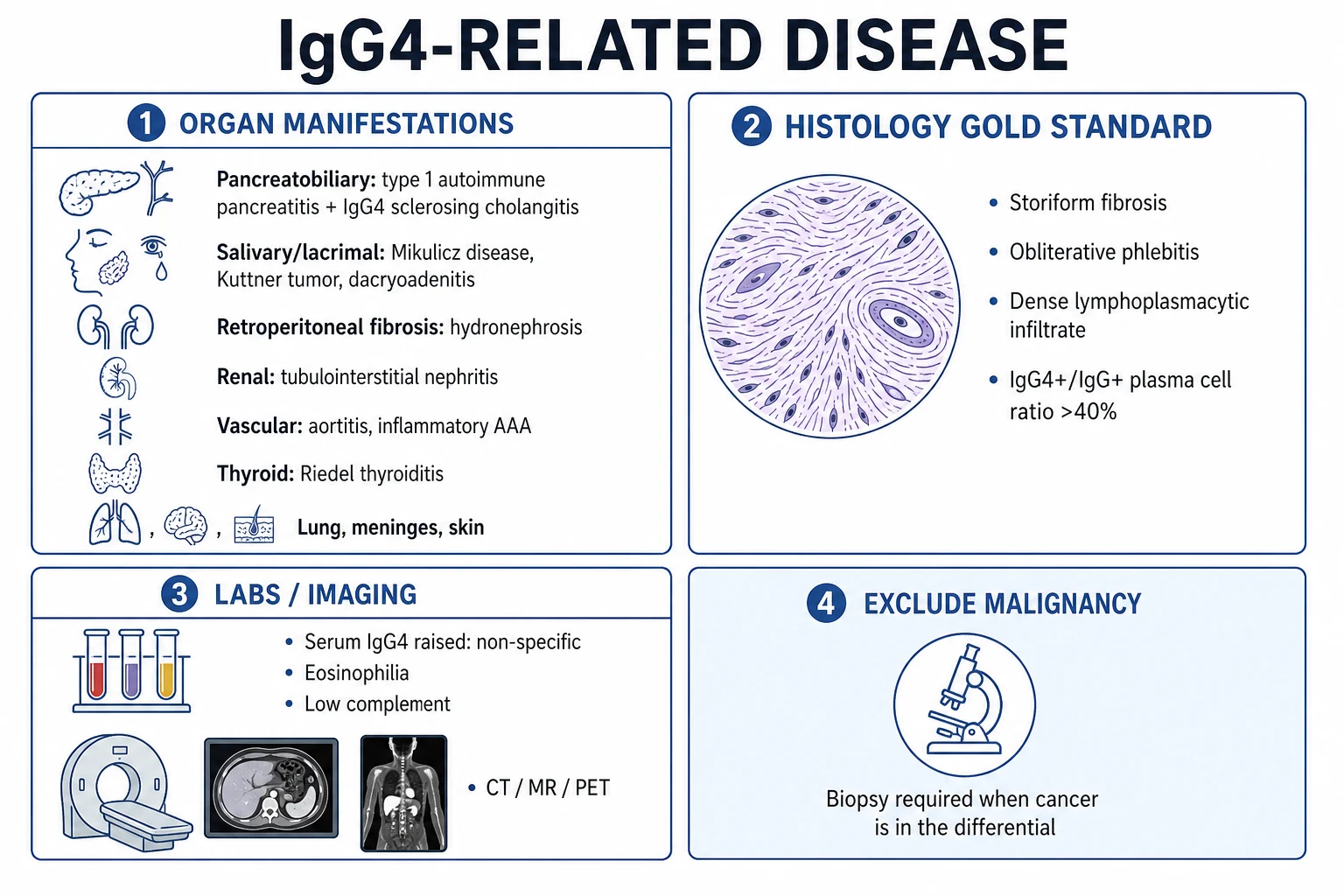
IgG4-related disease (IgG4-RD) is a fibro-inflammatory immune-mediated disorder characterised by tumefactive (mass-like) lesions, a dense lymphoplasmacytic infiltrate rich in IgG4-positive plasma cells, storiform fibrosis and obliterative phlebitis, and often elevated serum IgG4. It can affect nearly any organ — type 1 autoimmune pancreatitis, IgG4-related sclerosing cholangitis, sialadenitis (Mikulicz syndrome), dacryoadenitis, retroperitoneal fibrosis, tubulointerstitial nephritis, aortitis, Riedel thyroiditis and hypertrophic pachymeningitis. It predominantly affects older men and often presents as painless organ enlargement or a mass that mimics malignancy. Diagnosis rests on histology (the gold standard) plus serum IgG4 and imaging. Treatment gives a dramatic response to glucocorticoids; rituximab is the key steroid-sparing and refractory agent. The cardinal rule: biopsy mass-like lesions before assuming cancer or proceeding to surgery.
On this page & tools
Your progress
Saved locally on this device.
Practise this topic
Exam tags
Red flags
Overview & Definition
IgG4-related disease is a unifying diagnosis that re-explains what were once regarded as separate, organ-specific conditions — type 1 autoimmune pancreatitis, Mikulicz syndrome, Riedel thyroiditis, idiopathic retroperitoneal fibrosis, Kuttner tumour, inflammatory abdominal aortic aneurysm, hypertrophic pachymeningitis and idiopathic tubulointerstitial nephritis — as one fibro-inflammatory process linked by a shared histology and a shared tendency to form mass-like lesions.[1] Its importance to the clinician rests on two pillars.
First, it mimics malignancy. A painless mass in the pancreas, bile duct, salivary or lacrimal glands, kidney or retroperitoneum of an older man is precisely the presentation of cancer, so biopsy is mandatory before resection; a proportion of lesions resected as 'cancer' prove to be IgG4-RD, and the Whipple or nephrectomy was therefore avoidable. Second, it is one of the few diseases with a dramatic, near-diagnostic response to glucocorticoids — masses shrink, obstruction resolves, and unnecessary surgery is averted. Histology remains the gold standard; serum IgG4 supports but never confirms, because it is normal in a substantial minority of biopsy-proven cases and can rise in malignancy.[1][2]
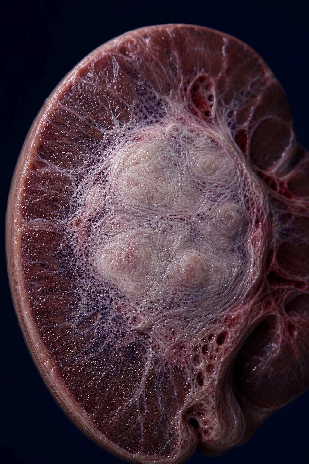
How IgG4-related disease was recognised
Classification
IgG4-RD is classified by two parallel criteria systems that are used together rather than in competition. The 2019 ACR/EULAR classification criteria are international and weighted toward histopathology, characteristic imaging, other-organ involvement and exclusion of mimics; the 2020 revised comprehensive diagnostic (RCD) criteria (Japanese, Umehara) are simpler and lean on the serum IgG4 threshold and the IgG4-positive/IgG-positive plasma-cell ratio. For type 1 autoimmune pancreatitis specifically, the older HISORt criteria (Mayo) still circulate: Histology, Imaging, Serology, Other organ involvement, and Response to steroids. The shared diagnostic tripod is (1) typical organ involvement, (2) serum IgG4 elevation, (3) characteristic histopathology.[5]
Epidemiology & Risk Factors
IgG4-RD is under-recognised rather than rare. Six epidemiological points dominate an examiner's expectations:[1][2]
- Older men, median age around 60: peak onset in the 6th to 7th decade (most patients older than 50 years), with an overall male-to-female predominance of roughly three to one; the skew is most pronounced for pancreaticobiliary disease, where type 1 autoimmune pancreatitis approaches a four-to-one male excess, whereas head-and-neck disease (Mikulicz, dacryoadenitis) is more evenly distributed between the sexes.[1]
- Worldwide, but best characterised in Japan and East Asia: the disease has no clear racial restriction, yet the Japanese literature and the revised comprehensive diagnostic criteria originated there, and reported series are more numerous and more readily recognised in Asian populations than in Western ones, where the diagnosis is still frequently missed or delayed. Estimates of the incidence of type 1 autoimmune pancreatitis are around 0.8 to 1.0 per 100,000 per year in Japan, with a rising prevalence everywhere as awareness grows.[1][2]
- Allergy and atopy are over-represented: roughly 40 percent of patients have allergic rhinitis, asthma, eczema or peripheral eosinophilia, supporting a Th2-biased background. This is an exam-favoured association that also provides a clinical hint when an atopic older man presents with a pancreatic or salivary mass.
- Triggers are incompletely defined: allergens, occupational exposures and possible microbial or autoantigen triggers (galectin-3, annexin A11) are suspected, but no single infectious agent is established and the disease is not considered contagious.
- Genetics: HLA-DRB1 associations (notably HLA-DRB1 star-04:05) are described in Japanese cohorts, but the genetic contribution is uncertain, the disease is not a classic hereditary condition, and no single gene is causal.
- Malignancy: a debated, modestly increased risk of pancreatic cancer and lymphoma has been reported in some cohorts, particularly in longstanding, fibrosis-heavy disease; this does not make IgG4-RD a frank premalignant condition, but ongoing surveillance for pancreatobiliary and haematological malignancy is advised.[2]
Pathophysiology
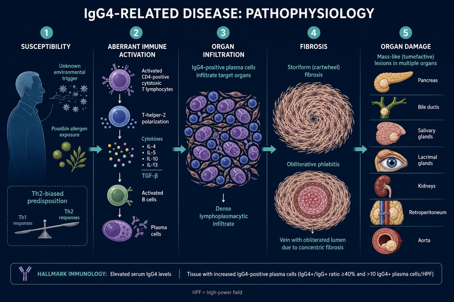
The aetiology is unknown. The working model is an aberrant immune response to an unknown antigenic or allergen trigger in a susceptible, Th2-biased, older host, producing tissue infiltration, IgG4-class switching and fibrosis. Three interlocking mechanisms drive the disease.[1][6]
Adaptive immunity — CD4-positive cytotoxic T lymphocytes (CD4 CTLs) are the dominant effector. Clonally expanded CD4 CTLs infiltrate the tissue and secrete IFN-gamma, TGF-beta, IL-1beta, IL-5 and IL-13. They express cytotoxic granules (granzyme B, perforin) and surface markers such as SLAMF7, and their clonality implies antigen-driven expansion. This is accompanied by a Th2 polarisation that raises IL-4, IL-5, IL-13 and IL-10, and by oligoclonal T-follicular-helper (Tfh) cells that drive class switching in germinal centres. The Th2 cytokine milieu also explains the eosinophilia and atopy seen in a substantial subset. [1]
B-cell and plasma-cell axis — class-switched IgG4-positive plasma cells infiltrate target tissue. Activated B cells mature into IgG4-producing plasma cells that accumulate in organs. Circulating plasmablasts (CD19-positive, CD20-negative, CD27-bright, CD38-bright) are a particularly informative biomarker — they correlate with disease activity better than serum IgG4 itself, because they reflect ongoing active class-switching and tissue recruitment. [1]
The IgG4 paradox. IgG4 is structurally unique: it undergoes Fab-arm exchange, forming functionally monovalent 'half-antibodies' that cannot cross-link antigens into large immune complexes, and it binds C1q poorly and therefore fixes complement poorly via the classical pathway. IgG4 is thus considered a down-regulatory or 'blocking' antibody (it appears in desensitisation and in helminth/allergic responses). This is why serum IgG4 correlates imperfectly with disease activity, and why IgG4 is regarded as a bystander of an ongoing immune process rather than the direct pathogen — the tissue damage is inflicted by CD4 CTLs and by fibrosis, not by antibody-mediated complement injury. [1]
Fibrosis. The profibrotic cytokine TGF-beta drives fibroblast activation into the characteristic storiform (cartwheel) fibrosis and obliterative phlebitis — veins are destroyed within the fibrotic swirl while arteries are characteristically spared. The end result is mass-like (tumefactive) fibrotic lesions that compress ducts and structures: biliary obstruction, ureteric entrapment with hydronephrosis, aortic wall weakening with aneurysm, and endocrine or exocrine organ failure. [1]
Why does serum IgG4 not predict disease activity?
Because IgG4 is a blocking, anti-inflammatory antibody that fixes complement poorly and forms monovalent half-antibodies by Fab-arm exchange. It is a down-regulatory bystander, not the pathogen. The real damage is done by CD4-positive cytotoxic T lymphocytes and by TGF-beta-driven fibrosis. Circulating plasmablasts actually track activity better than serum IgG4 — which is why serum IgG4 can be normal in up to 30 to 40 percent of biopsy-proven cases, and can rise in pancreatic cancer and other mimics.
IGG4 — the five axes of the disease
IGG4
Dense lymphoplasmacytic infiltrate; storiform fibrosis and obliterative phlebitis on histology (gold standard)
Tumefactive lesions in pancreas (type 1 AIP), bile ducts, salivary/lacrimal (Mikulicz), kidney, retroperitoneum, aorta
Prednisolone 30-40 mg tapering; dramatic, near-diagnostic response; rituximab for relapsing/organ-threatening disease
2020 RCD histology cut-off (more than 10 IgG4-positive cells/HPF); serum IgG4 above 135 mg/dL supportive, not diagnostic
Clinical Presentation
IgG4-RD presents as painless organ enlargement or a mass with variable constitutional symptoms (fatigue, weight loss, mild fever) — features that fuel the suspicion of malignancy. It is multifocal in about 60 to 90 percent at presentation when imaging is thorough, but a single organ may declare the disease before others emerge.[1][2]
Pancreatobiliary (the prototypic and most common manifestation). Type 1 autoimmune pancreatitis causes painless obstructive jaundice, mild abdominal discomfort (rarely severe pain, which points toward acute pancreatitis), and weight loss. Imaging shows a diffusely enlarged, 'sausage-shaped' pancreas with delayed, homogeneous enhancement and a characteristic capsule-like rim; crucially, the main pancreatic duct shows irregular narrowing without an abrupt cutoff (unlike the abrupt duct cutoff and duct dilation of pancreatic adenocarcinoma). IgG4-related sclerosing cholangitis produces biliary strictures — classically a long, intra-pancreatic stricture of the common bile duct with minimal upstream dilation — that mimic cholangiocarcinoma or primary sclerosing cholangitis. [1]
Head and neck. Mikulicz syndrome is bilateral, painless, persistent enlargement of the lacrimal and major salivary glands (dacryoadenitis with submandibular or parotid sialadenitis). Kuttner tumour (chronic sclerosing submandibular sialadenitis) presents as a hard, painless submandibular mass that mimics malignancy and was, for decades, treated surgically. Other head-and-neck disease includes orbital pseudotumour (proptosis, diplopia, pain), chronic sinusitis with nasal obstruction, and IgG4-related hypophysitis (anterior pituitary enlargement causing headache, visual field defects and hypopituitarism with central hypothyroidism or hypogonadism). Intracranial extension gives hypertrophic pachymeningitis — thickened, enhancing dura mater over the cerebral convexities, falx or tentorium — presenting with chronic headache, seizures and cranial-nerve palsies as the thickened dura entraps cranial nerves at the skull base.[1][2]
Retroperitoneum. Idiopathic retroperitoneal fibrosis (Ormond disease), when IgG4-related, produces a periaortic and periureteric soft-tissue plaque that entraps the ureters — flank or back pain, lower-limb oedema, hydronephrosis and renal failure from obstructive uropathy, and occasionally deep-vein compression or claudication. The fibrotic mantle classically mantles the abdominal aorta and iliac vessels at the L4 to L5 level and may coexist with an inflammatory abdominal aortic aneurysm, producing the combined picture of periaortitis and periureteritis that is highly characteristic of IgG4-RD.[1][2]
Renal. IgG4-related tubulointerstitial nephritis presents with renal masses or pseudotumours, or a quietly declining renal function; proteinuria is mild and the complement (notably C3 and C4) is sometimes low when there is concomitant membranous glomerulonephritis. [1]
Vascular. Aortitis, inflammatory abdominal aortic aneurysm, and coronary periarteritis — the aortic involvement may be the first sign and is a source of sudden death from aneurysm rupture. [1]
Other organs. Riedel thyroiditis (a rock-hard, fixed goitre mimicking anaplastic thyroid cancer), hypertrophic pachymeningitis (headache, cranial-nerve palsies), pulmonary disease (nodules, consolidations, interstitial infiltrates, bronchovascular lesions), prostatic involvement (a hard prostate mimicking cancer), lymphadenopathy (usually moderate and reactive), and skin lesions (pseudotumours, papulonodular eruptions). [1]
Constitutional and atypical. Weight loss, fatigue and low-grade fever are common and contribute to the suspicion of malignancy. Importantly, a single organ (isolated retroperitoneal fibrosis, one salivary gland, or a renal lesion) may declare the disease before other organs emerge, and serum IgG4 can be normal in up to 30 to 40 percent of biopsy-proven cases. [1]
Type 1 (IgG4) AIP
- Older men; painless obstructive jaundice; raised serum IgG4; other-organ involvement common
- Diffuse sausage-shaped pancreas, capsule-like rim, irregular duct narrowing WITHOUT abrupt cutoff
- Lymphoplasmacytic sclerosing pancreatitis with storiform fibrosis and IgG4-positive cells
- Dramatic steroid response; part of the IgG4-RD spectrum
Type 2 AIP
- Younger patients; equal sex distribution; associated with inflammatory bowel disease
- IgG4-NORMAL; no other-organ involvement; no storiform fibrosis
- Idiopathic duct-centric pancreatitis: neutrophilic infiltration, granulocytic epithelial lesions (GEL)/abscesses
- NOT part of IgG4-RD; steroid-responsive but a separate entity
Pancreatic adenocarcinoma
- Older patients; painless jaundice, weight loss, Courvoisier sign; CA 19-9 may be raised
- Discrete hypodense mass with vascular encasement (SMV/PV) and abrupt duct cutoff with upstream dilation
- Malignant glandular cells on cytology/histology; no storiform fibrosis
- Progressive; no steroid response; biopsy decisive before resection
Acute / chronic pancreatitis
- Severe epigastric pain, raised amylase/lipase; alcohol/gallstones
- Oedematous or necrotic gland; chronic form shows calcification and ductal changes
- No mass with capsule rim; no IgG4 elevation
- Diagnosis clinical and biochemical, not by IgG4 histology
Differential Diagnosis
The pivotal differential is malignancy — any painless mass in an older man warrants biopsy before resection. Beyond that, the disease must be separated from its organ-specific mimics and from the histiocytic and lymphoproliferative look-alikes.[1][2]
IgG4-RD
- Tumefactive mass-like fibrotic lesions; storiform fibrosis + obliterative phlebitis + IgG4-positive plasma cells on histology
- Older men; often raised serum IgG4 (normal in 30-40 percent); multiple organ involvement; atopy over-represented
- Dramatic response to glucocorticoids; rituximab for relapse
- Mimics cancer (pancreas, bile duct, salivary/lacrimal, kidney) — biopsy before surgery
Pancreatic / biliary malignancy
- Painless obstructive jaundice, weight loss, Courvoisier sign; CA 19-9 may be raised
- No storiform fibrosis; malignant cells on cytology/histology; IgG4 usually normal
- Progressive, no response to steroids; imaging shows a discrete mass with vascular encasement and abrupt duct cutoff
- Biopsy / ERCP cytology decisive — never assume cancer without tissue
Sjogren syndrome
- Women; sicca (dry eyes/mouth); anti-Ro/La positive; lymphocytic (focal) sialadenitis
- Salivary enlargement usually bilateral and softer; no storiform fibrosis, no obliterative phlebitis
- No dramatic steroid response; risk of MALT lymphoma
- Distinguished from Mikulicz/IgG4 sialadenitis by sex, serology and histology
Primary sclerosing cholangitis (PSC)
- Younger men with inflammatory bowel disease; multifocal beaded strictures and prune-tree pruning
- Normal or mildly raised IgG4; NO dramatic steroid response; progressive to cirrhosis/cholangiocarcinoma
- Histology: 'onion-skin' periductal fibrosis, NOT storiform fibrosis
- IgG4-sclerosing cholangitis has raised IgG4, other-organ involvement and steroid responsiveness
Sarcoidosis
- Multi-organ with bilateral hilar lymphadenopathy and salivary/lacrimal enlargement
- NON-CASEATING granulomas (not storiform fibrosis or IgG4-rich infiltrate)
- Raised ACE; pulmonary and cutaneous involvement; responds to steroids
- Distinguished from IgG4 by granulomatous histology and raised ACE
Lymphoma (MALT / marginal zone)
- Salivary/lacrimal or pancreatic mass; may coexist with or mimic IgG4-RD
- Clonal neoplastic lymphoid cells on histology; flow cytometry and Ig gene rearrangement decisive
- No storiform fibrosis pattern; needs oncology treatment
- Always exclude on biopsy — clonality studies are mandatory for a gland mass
Erdheim-Chester / Rosai-Dorfman / Castleman
- Rare histiocytic / lymphoproliferative mimics with retroperitoneal or orbital masses
- Characteristic histology (foamy histiocytes, emperipolesis, hyaline-vascular changes) — NOT storiform fibrosis
- Distinct molecular findings (e.g. BRAF V600E in Erdheim-Chester)
- Diagnosed by specialist histopathology; do not assume IgG4-RD on imaging alone
Clinical & Bedside Assessment
Take a structured history focused on painless organ enlargement, obstructive symptoms and constitutional features, then examine systematically to map every involved organ.[1]
- History: painless mass or organ enlargement; jaundice or pale stools and dark urine (biliary obstruction); polyuria, flank pain or reduced urine output (renal or ureteric); dry eyes or mouth or progressive gland swelling; back or abdominal pain and leg swelling (retroperitoneal); headache or visual change or diplopia (pachymeningitis, orbital disease); weight loss, fatigue, low-grade fever; atopy (allergic rhinitis, asthma, eczema); and a careful drug history (methysergide and ergot derivatives cause drug-related retroperitoneal fibrosis).
- Head and neck: palpate the lacrimal glands and the major salivary glands (parotid, submandibular) for bilateral, painless, firm enlargement (Mikulicz); feel for a stony-hard thyroid (Riedel); inspect the conjunctivae and orbits (orbital pseudotumour, proptosis).
- Abdomen: hepatomegaly, a palpable mass (pancreas, retroperitoneum), and signs of biliary obstruction (jaundice, excoriation scratch marks, Courvoisier sign if a distended gallbladder).
- Peripheral and vascular: lower-limb oedema (ureteric obstruction or retroperitoneal fibrosis); an abdominal bruit or pulsatile mass (inflammatory abdominal aortic aneurysm); blood pressure and peripheral pulses.
- Neurological: cranial-nerve palsies, visual change or headache (hypertrophic pachymeningitis, orbital disease).
- Look specifically for atopy (allergic shiners, wheeze, eczema) and eosinophilia on the blood film, which are over-represented.
- Respiratory and skin: auscultate for wheeze or interstitial crackles (pulmonary IgG4-RD) and inspect the skin for papulonodular lesions or subcutaneous pseudotumours.
- Named clues: a sausage-shaped pancreas on imaging, bilateral painless gland enlargement (Mikulicz), a stony-hard thyroid (Riedel) and an inflammatory abdominal aortic aneurysm with retroperitoneal fibrosis are the eponymous quartet that should trigger the diagnosis. Always ask about weight loss and document the tempo, because a subacute, painless mass in an older man is the single most powerful prompt to consider IgG4-RD and to biopsy before resection. [1]
Investigations
There is no single diagnostic test. Investigations serve to support the diagnosis, exclude malignancy and the mimics, and stage organ involvement. Histology is the gold standard.[1][2][5]
A practical diagnostic sequence runs in four steps. First, recognise the pattern — a painless mass or organ enlargement in an at-risk older man with atopy or other-organ involvement. Second, image comprehensively with contrast CT or MRI of the relevant region plus MRCP for the biliary tree, and consider PET-CT to map occult multi-organ disease and to choose the safest biopsy target. Third, obtain tissue — core biopsy or, for pancreaticobiliary lesions, EUS-guided FNA with a cell block and IgG4 immunostaining — and send it for histology, cytology and, where doubt remains, clonality studies to exclude lymphoma. Fourth, exclude the mimics with serum IgG4, a basic autoimmune and tumour-marker panel, and the structured 2019 ACR/EULAR or 2020 RCD criteria, always remembering that the goal is to avoid resecting a steroid-responsive lesion.[1][5]
Histopathology (gold standard). The three defining features are storiform fibrosis, obliterative phlebitis, and a dense lymphoplasmacytic infiltrate rich in IgG4-positive plasma cells. IgG4 and total Ig immunostaining allows the IgG4-positive/IgG-positive plasma-cell ratio to be calculated — a key step that distinguishes genuine IgG4-RD from non-specific IgG4 elevation in cancer. On the 2020 RCD criteria, this is operationalised as a ratio above 40 percent and more than 10 IgG4-positive plasma cells per high-power field, with site-adjusted thresholds (the kidney, submandibular gland and pancreas carry higher expected background counts). Characteristic but not unique findings include eosinophilic infiltration and a phlebitis that spares arteries. [1]
Serum IgG4 — supportive but not diagnostic. It is elevated (above 135 mg/dL) in most patients but normal in 30 to 40 percent of biopsy-proven cases, and it can be raised in pancreatic cancer, cholangiocarcinoma, primary sclerosing cholangitis, ANCA-associated vasculitis and even healthy relatives. It therefore never stands alone. Circulating plasmablasts (CD19-positive, CD20-negative, CD27-bright, CD38-bright) track disease activity better than serum IgG4 and are a useful research and specialist biomarker. [1]
Imaging. Contrast CT and MRI show the 'sausage-shaped' diffusely enlarged pancreas with a capsule-like rim of type 1 autoimmune pancreatitis; biliary strictures; mass-like renal lesions (often bilateral, peripheral, wedge-shaped); and a retroperitoneal soft-tissue plaque encasing vessels and ureters. MRCP and ERCP characterise the biliary tree — IgG4-sclerosing cholangitis classically gives a long intra-pancreatic common bile duct stricture. PET-CT is invaluable: it maps multi-organ involvement, identifies the most accessible biopsy target, and provides a baseline for treatment response (lesions typically become metabolically quiescent after glucocorticoids). [1]
2019 ACR/EULAR classification criteria. Apply the entry criterion (typical organ involvement), the exclusion criteria (e.g. fever without active disease, markedly raised CRP, features of alternative diagnoses such as multicentric Castleman disease or sarcoidosis, or malignancy on cytology), then score across domains: histopathology, pancreaticobiliary and renal imaging, serum IgG4 level, other-organ involvement, and the glucocorticoid response. A score of 20 or more classifies IgG4-RD. This scheme deliberately weighs histopathology and exclusion of mimics more heavily than serum IgG4. [1]
HISORt criteria (for type 1 autoimmune pancreatitis, Mayo). Histology (lymphoplasmacytic sclerosing pancreatitis), Imaging (diffuse enlargement with delayed capsule-like enhancement, irregular duct narrowing), Serology (raised IgG4), Other organ involvement, and Response to glucocorticoids (t, for treatment/response). [1]
Bloods. FBC (eosinophilia in a subset), ESR and CRP (mildly raised — a markedly raised CRP argues against uncomplicated IgG4-RD and favours infection, malignancy or another vasculitis), U&E and eGFR (renal), LFT (cholestatic or obstructive pattern), glucose (pancreatic endocrine function), immunoglobulins (polyclonal elevation common), and complement (C3/C4 may be low in renal disease with membranous glomerulonephritis). Autoantibodies (ANA, ANCA, RF, anti-Ro/La) are usually negative and help exclude Sjogren syndrome, ANCA-associated vasculitis and rheumatoid arthritis. [1]
Exclusion of malignancy is mandatory. Biopsy or core tissue for histology and cytology before any resection; tumour markers (CA 19-9) are supportive but not decisive — biliary obstruction of any cause can raise CA 19-9. [1]
IgG4-related disease — key numbers
Management — Resuscitation
IgG4-RD is rarely an immediate resuscitation emergency, but several presentations are time-critical:[2][4]
- Obstructive jaundice with ascending cholangitis — fluid resuscitation, IV broad-spectrum antibiotics, and biliary decompression (ERCP stenting or percutaneous transhepatic drainage) before definitive immunosuppression.
- Ureteric obstruction with obstructive uropathy or renal failure (retroperitoneal fibrosis) — urgent percutaneous nephrostomy or ureteric stenting to relieve obstruction and preserve renal function, then immunosuppression.
- Rapidly enlarging inflammatory abdominal aortic aneurysm at risk of rupture — vascular surgery assessment, blood-pressure control, and analgesia, then glucocorticoids to dampen the inflammatory wall.
- Bilateral bulky orbital disease threatening vision, or hypertrophic pachymeningitis with cranial-nerve compromise — urgent imaging (MRI orbits or brain with contrast) and early glucocorticoids. [1]
Management — Definitive & Stepwise
Management is severity-stratified. The central principle: treat organ-threatening, symptomatic or relapsing disease; mild, asymptomatic, isolated disease (especially uncomplicated lymphadenopathy) may be monitored ('watch-and-wait') to avoid unnecessary immunosuppression.[1][4]
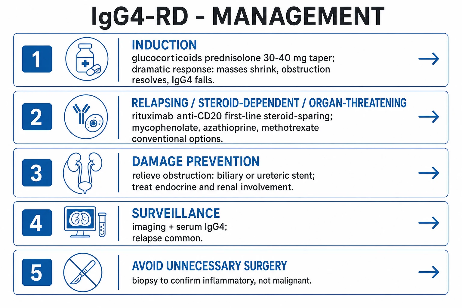
Glucocorticoids (first-line induction). Prednisolone 30 to 40 mg daily (about 0.5 to 0.6 mg/kg) for 2 to 4 weeks, then a gradual taper over 3 to 6 months. A widely taught taper reduces the dose by 2.5 to 5 mg every two weeks once the induction phase is complete, continuing down to a low maintenance dose before withdrawal. The dramatic response — shrinkage of masses, resolution of obstruction, fall in serum IgG4 — typically begins within two weeks and is maximal by four weeks; this response is so characteristic that it is regarded as near-diagnostic. Some Japanese protocols use an induction of 0.6 mg/kg/day for two to four weeks before tapering. A failure to respond within four weeks should prompt re-questioning of the diagnosis (consider malignancy or an alternative), because a genuine IgG4-RD mass that does not shrink with steroids is rare and warns that the lesion may in fact be cancer.[1][4]
Watch-and-wait. Asymptomatic, mild, isolated, histologically quiescent disease — especially uncomplicated lymphadenopathy or a small, non-obstructing lesion — may be monitored rather than treated, reserving therapy for progression or organ threat. [1]
Steroid-sparing agents. Conventional DMARDs (azathioprine, mycophenolate mofetil, methotrexate, ciclosporin) are used as steroid-sparers, but the evidence of efficacy is limited and the relapse rate remains high; they are weaker than rituximab. [1]
Rituximab (anti-CD20, B-cell depletion) — the preferred first-line steroid-sparing agent. It is indicated for relapsing, steroid-dependent, steroid-refractory or organ-threatening disease, and is increasingly used upfront in many centres. Standard dosing is two infusions of 1 gram two weeks apart (or 375 mg per square metre weekly for four doses). It depletes IgG4-expressing plasmablast precursors, reduces fibrotic progression, and achieves remission in the large majority of treated patients. Response is monitored clinically, by imaging and by CD19-positive B-cell depletion (and ideally circulating plasmablast counts).[3]
Other biologics for refractory disease. When rituximab is contraindicated, poorly tolerated, or ineffective, the interleukin-6 receptor antagonist tocilizumab has been used for refractory or relapsing IgG4-RD, exploiting the pathogenic role of B-cell activating cytokines and the acute-phase response; evidence is limited to case series rather than randomised trials, so it remains a third-line option.[2] Novel agents directed against the B-cell axis — including belimumab (anti-BAFF) and ianalumab (anti-BAFF-R) — are under active investigation and reflect the move from blanket immunosuppression toward targeted B-cell biology.
Relapse management. Relapse is common on tapering steroids — up to about half of patients within months of withdrawal. A relapse is managed with a short glucocorticoid re-induction and, more durably, with repeat rituximab (maintenance infusions, often every six to twelve months based on B-cell reconstitution) or a conventional DMARD. Most patients with multi-organ or relapsing disease need long-term maintenance. [1]
Damage prevention, surgery and adjuncts. Relieve obstruction early — biliary or ureteric stenting (ERCP or percutaneous transhepatic drainage for the biliary tree; ureteric stent or nephrostomy for retroperitoneal fibrosis) takes priority over immunosuppression when cholangitis or obstructive uropathy threatens. Surgical intervention is reserved for the complications rather than the disease itself: drainage of a walled-off pancreatic collection, decompression of a threatened aortic aneurysm, or rarely resection of a lesion that proved refractory and could not be distinguished from malignancy even after biopsy. Treat pancreatic endocrine insufficiency (new-onset diabetes with insulin or oral agents) and exocrine insufficiency (pancreatic enzyme replacement for steatorrhoea), and provide renal support where tubulointerstitial nephritis or obstructive uropathy has reduced function. Urology, hepatobiliary, vascular and endocrine input as the organs dictate.[1][4]
Multidisciplinary care and harm prevention. Rheumatology coordinates with gastroenterology/hepatology, nephrology, surgery/vascular, ophthalmology, ENT and endocrinology. Give bone protection (calcium, vitamin D, a bisphosphonate where indicated) and PJP prophylaxis (co-trimoxazole) for prolonged steroids. Vaccinate before rituximab (pneumococcal, influenza, COVID-19, and hepatitis B as indicated) because the antibody response is blunted for months after B-cell depletion; rituximab can also cause infusion reactions, late-onset neutropenia and hypogammaglobulinaemia, so monitor immunoglobulins and full blood count. [1]
Specific Subtypes & Scenarios
- Type 1 (IgG4) autoimmune pancreatitis — the prototypic manifestation; older men, painless jaundice, sausage-shaped pancreas, raised IgG4, dramatic steroid response. Distinguished from type 2 AIP (idiopathic duct-centric pancreatitis): younger patients, equal sex distribution, inflammatory bowel disease association, neutrophilic duct-centric histology with granulocytic epithelial lesions, and IgG4-negative; type 2 AIP is not part of the IgG4-RD spectrum.[1]
- IgG4-related sclerosing cholangitis — biliary strictures mimicking cholangiocarcinoma or PSC; classically a long intra-pancreatic common bile duct stricture; steroid-responsive. Distinguished from PSC by raised IgG4, other-organ involvement and a dramatic steroid response (PSC is steroid-unresponsive and progresses to cirrhosis and cholangiocarcinoma).
- Mikulicz syndrome and head-and-neck disease — bilateral painless lacrimal and salivary gland enlargement (Mikulicz), Kuttner tumour, dacryoadenitis, orbital pseudotumour, chronic sinusitis. Distinguished from Sjogren syndrome by sex distribution, anti-Ro/La negativity and IgG4-rich (rather than lymphocytic focal) sialadenitis.
- Retroperitoneal fibrosis — a periaortic and periureteric fibrous plaque; hydronephrosis and renal failure; relieve obstruction then immunosuppress. Distinguish from malignancy-associated, drug-related (methysergide) and radiation-related retroperitoneal fibrosis by histology and history.
- IgG4-related kidney disease — tubulointerstitial nephritis and renal pseudotumours, often bilateral peripheral lesions; respond to steroids; may coexist with membranous glomerulonephritis (then proteinuria and low complement). Always screen for other organ involvement.[6]
- Vascular IgG4-RD — aortitis, inflammatory abdominal aortic aneurysm (the association of idiopathic retroperitoneal fibrosis with inflammatory AAA is classic), coronary periarteritis.
- Miscellaneous — Riedel thyroiditis (stony-hard goitre mimicking anaplastic cancer), hypertrophic pachymeningitis (headache, cranial-nerve palsies), pulmonary nodules and infiltrates, IgG4-related hypophysitis, prostatic involvement (a hard prostate mimicking prostate cancer), skin pseudotumours and reactive lymphadenopathy.
Complications & Pitfalls
- Irreversible fibrotic organ damage from delayed treatment — persistent biliary obstruction and secondary biliary cirrhosis, chronic kidney disease from obstructive uropathy or tubulointerstitial nephritis, and pancreatic exocrine and endocrine insufficiency (new-onset diabetes and steatorrhoea).[1]
- Inflammatory aortic aneurysm with risk of rupture, and coronary periarteritis with ischaemia — aortic disease may be silent until catastrophic.
- Portal hypertension from periportal fibrosis, hepatic IgG4-RD or secondary biliary cirrhosis, presenting with varices, ascites and splenomegaly alongside the more familiar obstructive jaundice.
- Unnecessary surgery (pancreaticoduodenectomy/Whipple, nephrectomy, gland excision) for a lesion presumed malignant that was actually IgG4-RD — the cardinal preventable harm. Biopsy first.[1]
- Relapse on steroid taper — common (up to about half of patients); mandates a steroid-sparing strategy with rituximab or a conventional DMARD.
- Treatment-related toxicity — glucocorticoid adverse effects (diabetes, osteoporosis, infection, cataracts, hypertension, avascular necrosis); rituximab risks (infusion reactions, hypogammaglobulinaemia, infection, blunted vaccine response, late-onset neutropenia); and opportunistic infection such as PJP with prolonged immunosuppression.
- Pitfalls: relying on serum IgG4 alone (a normal level does not exclude disease; a raised level does not confirm it); failing to biopsy a mass before resection; conflating IgG4-related sclerosing cholangitis with PSC; conflating Mikulicz syndrome with Sjogren syndrome; conflating type 1 with type 2 AIP; and mistaking a hard prostate or thyroid for cancer without histology.
Prognosis & Disposition
The overall prognosis is good with prompt recognition and treatment, because the dramatic steroid response reverses mass lesions and obstruction in most patients.[1][2] Relapse is the rule rather than the exception on steroid withdrawal, so most patients need long-term steroid-sparing maintenance with rituximab or a conventional DMARD. Irreversible fibrotic damage (chronic kidney disease, biliary cirrhosis, pancreatic insufficiency) determines long-term outcome, and early treatment limits it. Mortality is low and usually relates to delayed diagnosis (unnecessary surgery, irreversible organ failure), treatment toxicity, an associated or incipient malignancy, or a catastrophic vascular event (ruptured inflammatory aneurysm). Follow-up is multidisciplinary, with imaging and serum IgG4 surveillance, recognising that IgG4 may flare without symptoms and that symptoms may recur without a rise — clinical vigilance and repeat imaging are the safety net.
Prevention
There is no specific primary prevention for IgG4-RD because the aetiology and triggering antigens remain unknown. Prevention is therefore secondary and harm-directed — early recognition of a mass-like lesion in an at-risk older man, biopsy before resection, prompt glucocorticoid induction once histology is secured, and a planned steroid-sparing strategy to prevent relapse and treatment toxicity.[1][4] Specific preventive actions include: bone protection (calcium, vitamin D and a bisphosphonate where indicated) and PJP prophylaxis with co-trimoxazole for anyone on prolonged glucocorticoids; vaccination before rituximab because the humoral response is blunted for months after B-cell depletion; relapse surveillance with imaging and serum IgG4 during and after the taper, so that recurrence is caught before it inflicts new fibrotic damage; and malignancy surveillance, since a modest excess of pancreatic cancer and lymphoma has been reported. The single most powerful preventive measure is cultural rather than pharmacological — maintaining a biopsy-first mindset so that no patient undergoes a Whipple, nephrectomy or gland excision for what was in fact a steroid-responsive IgG4-RD mass.[2]
Special Populations
- Older men with pancreaticobiliary disease — the classic phenotype; maintain a high index of suspicion and biopsy before resection.
- Pregnancy — rare in this older population; manage with pregnancy-compatible prednisolone and avoid mycophenolate, methotrexate and cyclophosphamide; rituximab is avoided in pregnancy if possible because of neonatal B-cell depletion.
- Renal impairment at presentation (obstructive or tubulointerstitial) — relieve obstruction first, dose-adjust immunosuppression to renal function, and monitor renal function closely; screen for membranous glomerulonephritis (urinalysis, complement) when proteinuria is present.
- Patients with a normal serum IgG4 — a normal level does not exclude IgG4-RD; histology and imaging decide. Up to 30 to 40 percent of biopsy-proven cases have a normal serum IgG4.[2]
- Patients on anticoagulation or with suspected malignancy — biopsy carefully (consider bleeding risk); coordinate with oncology and surgery; never assume a mass is cancer without tissue.
- Younger patients or those with isolated organ disease — confirm histology to exclude type 2 AIP, lymphoma and the histiocytic mimics before committing to long-term immunosuppression.
Evidence, Guidelines & Regional Differences
- Kamisawa T, Zen Y, Pillai S, Stone JH. IgG4-related disease. Lancet 2015 — the landmark comprehensive review that unified the disease concept.[1]
- Wallace ZS et al. International Consensus Guidance Statement on the Management and Treatment of IgG4-Related Disease. Arthritis Rheumatol 2015 — glucocorticoid induction, rituximab for refractory, relapsing or organ-threatening disease, conventional DMARDs as steroid-sparers, and the principle of treating organ-threatening disease while observing mild disease.[4]
- Rituximab prospective open-label trial (Ann Rheum Dis 2015) — B-cell depletion is highly effective in refractory and relapsing disease and depletes IgG4-expressing plasmablasts.[3]
- Umehara H et al. The 2020 Revised Comprehensive Diagnostic Criteria for IgG4-RD. Mod Rheumatol 2021 — the Japanese three-criterion scheme (clinical, serum IgG4, histology) used in parallel with the international criteria.[5]
- 2019 ACR/EULAR Classification Criteria for IgG4-RD — entry and exclusion criteria plus a domain score (histopathology, imaging, serum IgG4, other-organ involvement, steroid response); a score of 20 or more classifies disease.
- Lanzillotta M et al. Advances in the diagnosis and management of IgG4 related disease. BMJ 2020, and Mbengue et al. IgG4-related kidney disease. Clin Nephrol 2021 — contemporary diagnosis-to-management overviews.[2][6]
Regional delta in criteria. The Japanese 2020 revised comprehensive criteria (Umehara) and the international 2019 ACR/EULAR classification criteria are used in parallel worldwide. The ACR/EULAR scheme is weighted toward histopathology, characteristic imaging and exclusion criteria (and includes a glucocorticoid-response domain), whereas the Japanese RCD criteria lean on the serum IgG4 threshold (above 135 mg/dL) and the IgG4-positive/IgG-positive plasma-cell ratio (above 40 percent). Both coexist; many specialists apply both to the same patient, and a clear histology with characteristic imaging will satisfy either framework. For type 1 autoimmune pancreatitis specifically, the older HISORt (Mayo) criteria remain widely taught.
Red Flags
Exam application bank (NEET-PG / INICET)
One-line answer
IgG4-related disease (IgG4-RD) is a fibro-inflammatory immune-mediated disorder characterised by tumefactive (mass-like) lesions, a dense lymphoplasmacytic infiltrate rich in IgG4-positive plasma cells, storiform fibrosis and obliterative phlebitis, and often elevated serum IgG4. It can affect nearly any organ — type 1 autoimmune pancreatitis, IgG4-related sclerosing cholangitis, sialadenitis (Mikulicz syndrome), dacryoadenitis, retroperitoneal fibrosis, tubulointerstitial nephritis, aortitis, Riedel thyroiditis and hypertrophic pachymeningitis. It predominantly affects older men and often presents as painless organ enlargement or a mass that mimics malignancy. Diagnosis rests on histology (the gold standard) plus serum IgG4 and imaging. Treatment gives a dramatic response to glucocorticoids; rituximab is the key steroid-sparing and refractory agent. The cardinal rule: biopsy mass-like l
Worked stems (answer without another resource)
Stem 1 — Classic presentation. Map symptoms to mechanism; name the first investigation and first treatment step with dose/route if drug therapy is standard. [1]
Stem 2 — Unstable / complicated. List red flags that force immediate resuscitation, theatre, ICU, antidote, or reperfusion — and what you do in the first 15 minutes. [1]
Stem 3 — Atypical group. Elderly, pregnancy, child, or immunocompromised: how presentation and thresholds change. [1]
Stem 4 — Differential trap. Name the three closest mimics and one discriminator for each. [1]
Stem 5 — Disposition. Who goes home with safety-netting, who is admitted, who needs HDU/ICU/theatre, and what follow-up is mandatory. [1]
Rapid viva checklist
- Definition + classification
- Pathophysiology chain
- Bedside signs / criteria
- Score with exact components (if any)
- Emergency bundle
- Definitive therapy with doses
- Complications of disease and of treatment
- Special populations
- Guideline/trial name if classic
- Three exam traps
Coverage self-check
If you cannot answer any stem above from this page alone, re-read the matching section — the page is intended to be self-sufficient for final-prof and NEET-PG/INICET questions on IgG4-Related Disease.
Exam Pearls
Self-test: histology trio and criteria
State the three defining histological features of IgG4-RD, the 2020 RCD serum-IgG4 and plasma-cell-ratio thresholds, and the 2019 ACR/EULAR score that classifies disease. Answers: storiform fibrosis, obliterative phlebitis, dense IgG4-positive-plasma-cell-rich infiltrate; serum IgG4 above 135 mg/dL with IgG4-positive/IgG-positive plasma-cell ratio above 40 percent and more than 10 IgG4-positive cells/HPF; and a 2019 ACR/EULAR score of 20 or more.
References
- [1]Kamisawa T, Zen Y, Pillai S, Stone JH. IgG4-related disease Lancet, 2015.PMID 25481618
- [2]Lanzillotta M, Mancuso G, Della-Torre E. Advances in the diagnosis and management of IgG4 related disease BMJ, 2020.PMID 32546500
- [3]Khosroshahi A, Wallace ZS, Crowe JL, et al. Rituximab for IgG4-related disease: a prospective, open-label trial Ann Rheum Dis, 2015.PMID 25667206
- [4]Wallace ZS, Carruthers MN, Khosroshahi A, et al. International Consensus Guidance Statement on the Management and Treatment of IgG4-Related Disease Arthritis Rheumatol, 2015.PMID 25809420
- [5]Umehara H, Okazaki K, Kawano M, et al. The 2020 revised comprehensive diagnostic (RCD) criteria for IgG4-RD Mod Rheumatol, 2021.PMID 33274670
- [6]Mbengue M, Goumri N, Niang A, et al. IgG4-related kidney disease: Pathogenesis, diagnosis, and treatment Clin Nephrol, 2021.PMID 33860756