Rheumatology · General Medicine
Low Back Pain (Approach & Red Flags)
Also known as Low back pain · Lumbago · Mechanical back pain · Sciatica · Lumbar radiculopathy · Cauda equina syndrome
Low back pain is the leading cause of disability worldwide; the vast majority is non-specific (mechanical) and self-limiting (over 90 percent recover within 6 weeks). The central clinical skill is identifying red flags (serious pathology — fracture, infection, malignancy, cauda equina, inflammatory) that mandate imaging and referral, while reassuring and mobilising the remainder. Red flags: significant trauma; age over 50 with new pain; history of cancer; fever/weight loss/night sweats; immunosuppression, IV drug use; night pain or pain unrelieved by rest; recent bacterial infection; corticosteroid use; cauda equina (saddle anaesthesia, urinary retention/incontinence, bilateral neurology, progressive weakness) = surgical emergency. Manage non-specific pain: reassure, stay active, simple analgesia (avoid opioids), exercise + CBT; surgery only for severe persistent radiculopathy or cauda equina.
On this page & tools
Your progress
Saved locally on this device.
Exam tags
Red flags
Overview & Definition
Low back pain is defined as pain, muscle tension or stiffness localised between the lower costal margin and the inferior gluteal folds, with or without referred leg pain (sciatica), lasting typically less than 6 weeks (acute) and often less than 12 weeks (subacute).[1] It is the single biggest cause of years-lived-with-disability worldwide, ahead of ischaemic heart disease, headache and depression, and it is rising: global years-lived-with-disability from low back pain increased by more than 50 percent between 1990 and 2015, driven by ageing and population growth. Yet it remains profoundly over-medicalised — excess imaging, opioid prescribing, spinal injections and surgery consume vast resources and demonstrably cause harm without benefit in the majority of cases.[2]
The central clinical skill is the structured, deliberate search for red flags that separate the dangerous minority — cauda equina syndrome, vertebral fracture, spinal infection, malignancy, and axial spondyloarthritis — from the benign mechanical majority. Cauda equina syndrome is the one true surgical emergency: a delayed or missed diagnosis produces permanent paralysis, bladder and bowel incontinence, and sexual dysfunction. Conversely, unnecessary imaging of non-specific pain produces harm, because incidental disc bulges, disc degeneration and facet arthropathy are common in pain-free adults (about 30 percent of pain-free 20-year-olds and over 80 percent of pain-free 80-year-olds have a disc bulge on MRI) and chasing these findings drives anxiety, opioid use and unnecessary intervention.[4]
The condition is broadly triaged into three categories — non-specific (mechanical, over 90 percent), radicular (nerve root, sciatica), and serious specific pathology (red flags) — and this triage drives every downstream decision (image or not, refer or not, reassure or operate).[5]
Classification
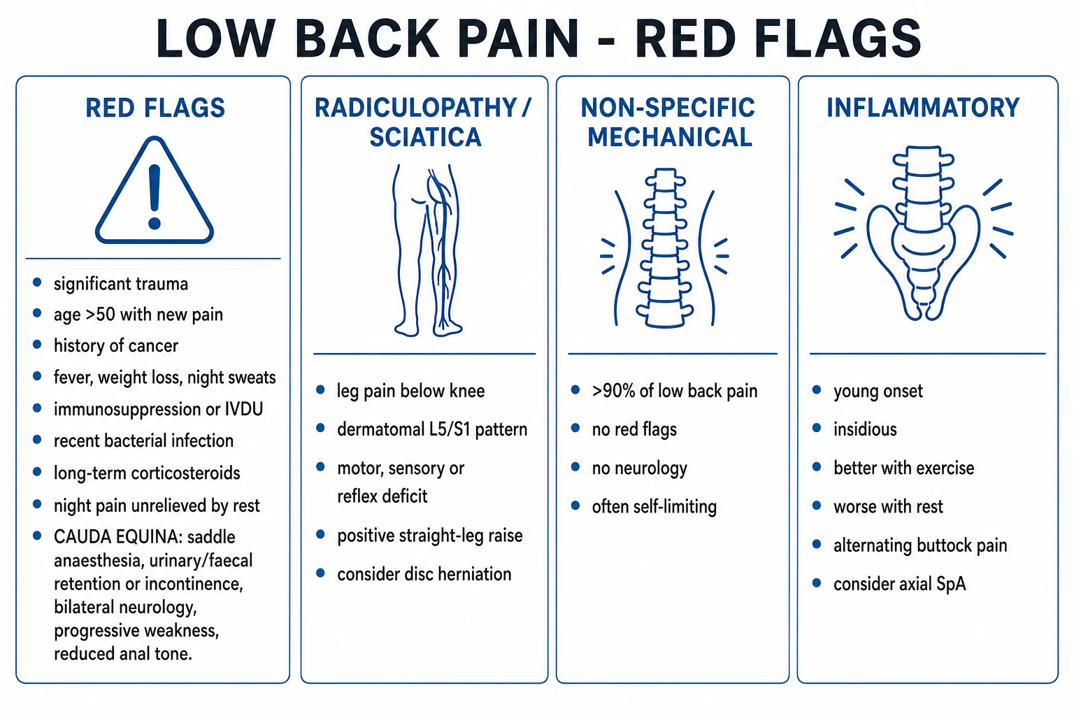
Low back pain is classified by the underlying mechanism — and that classification drives whether you image, refer, reassure, or operate. The triage below is the universal first step at every back-pain encounter, primary or secondary care. [1]
Non-specific (mechanical)
- Over 90 percent of all low back pain presentations
- Muscle/ligament strain, disc degeneration, facet arthropathy, sacroiliac strain
- No red flags, no objective neurological deficit, no systemic features
- Worse with activity and prolonged sitting, better with rest and lying flat
- Self-limiting — about 90 percent recover within 6 weeks
- Management: reassure, stay active, simple analgesia (avoid opioids), exercise; NO routine imaging
Radicular (sciatica)
- Nerve root irritation or compression — disc herniation or foraminal stenosis
- Leg pain radiating below the knee in a dermatomal distribution (L5, S1 commonest)
- Motor, sensory, or reflex deficit; positive straight leg raise
- Conservative first (over 80 percent recover within 6 to 12 weeks)
- MRI if persistent over 6 weeks or progressive neurological deficit
- Surgical microdiscectomy for severe persistent radiculopathy or progressive motor loss
Serious specific (red flags)
- Cauda equina, fracture, infection, malignancy, axial spondyloarthritis
- Saddle anaesthesia, bladder/bowel dysfunction, fever, weight loss, night pain
- History of cancer, immunosuppression, IV drug use, corticosteroids, significant trauma
- Needs urgent targeted imaging (MRI gold standard) and specialist referral
- Cauda equina = surgical emergency, decompression within 24 to 48 hours
Epidemiology & Risk Factors
Low back pain is the leading cause of years-lived-with-disability worldwide according to the Global Burden of Disease studies.[2] The lifetime prevalence is about 80 to 84 percent — almost everyone will have at least one disabling episode — and the point prevalence is about 12 to 33 percent of the adult population at any given moment. The annual incidence in working-age adults is around 15 to 45 percent. Peak onset is between 30 and 50 years, and it is the leading cause of work absence and activity limitation in adults of working age. The prevalence rises again in the elderly, where degenerative spinal stenosis, osteoporotic compression fracture and malignancy account for a larger share.
Low back pain — key numbers
Risk factors operate in two domains. Mechanical/physical risk factors include heavy or repeated manual lifting, whole-body vibration (for example driving tractors, trucks or helicopters), twisting and bending at work, smoking, obesity, and low cardiovascular and muscular fitness. Psychosocial factors — depression, anxiety, fear-avoidance beliefs, pain catastrophising, low job satisfaction, job strain and seeking compensation — are not merely associated with low back pain; they are the strongest predictors of transition from acute to chronic disabling pain (the so-called yellow flags).[1] This biopsychosocial understanding is the basis of modern management: a clinician who addresses only the disc and ignores the patient's mood, work and beliefs will fail to prevent chronicity.
Pathophysiology
The pathophysiology of low back pain differs by category, and understanding it determines which patients recover spontaneously and which need imaging or surgery. [1]
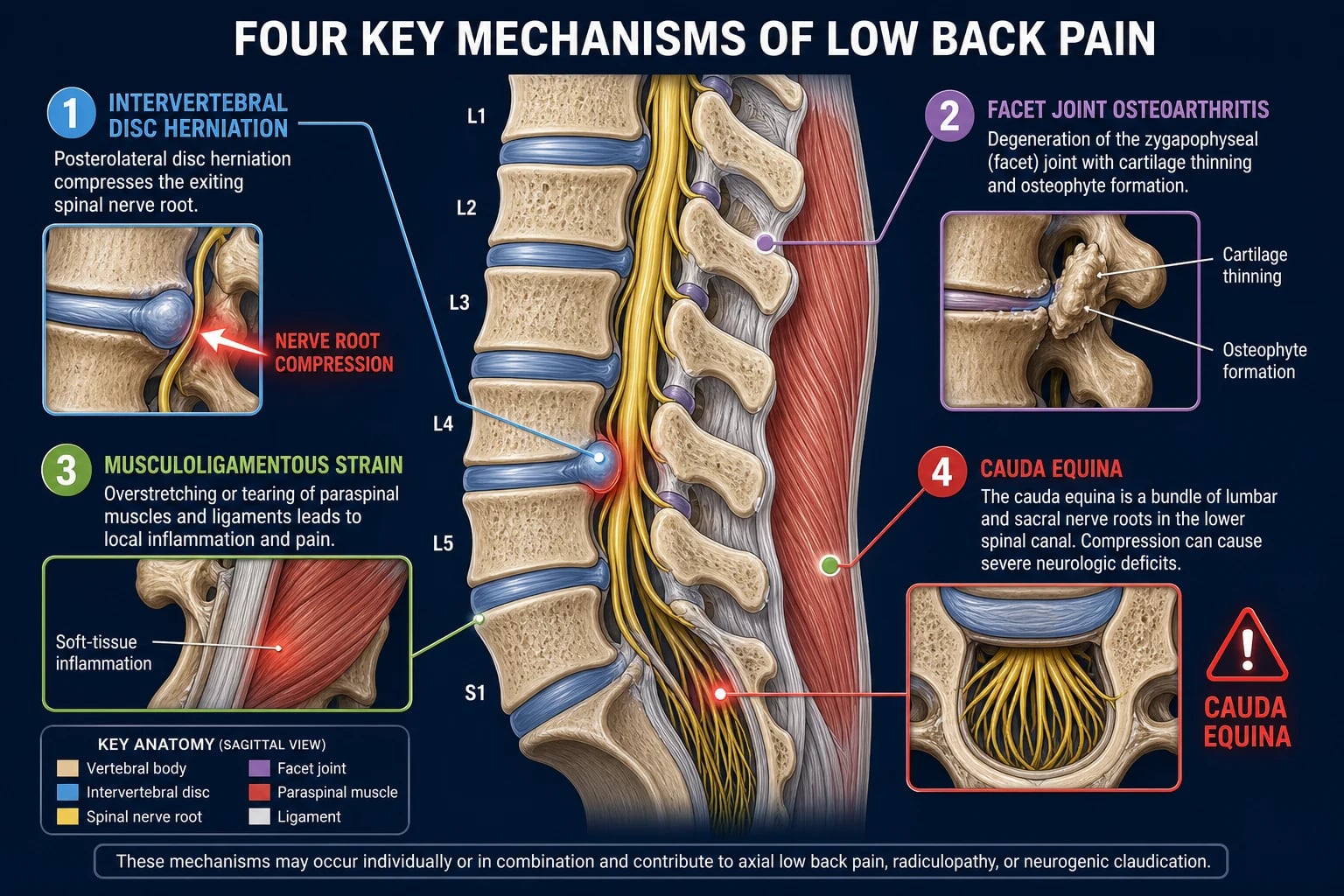
Mechanical (non-specific). Repeated axial loading, bending and twisting over years causes intervertebral disc degeneration: the nucleus pulposus desiccates and loses proteoglycans, the annulus fibrosus develops radial tears, and disc height collapses. The resulting loss of mechanical support overloads the facet joints, which develop osteoarthritis (cartilage thinning, osteophytes, joint effusion) — and the paraspinal muscles, ligaments and thoracolumbar fascia, which strain. These structures are richly innervated by the sinuvertebral nerve (annulus), the medial branch of the dorsal ramus (facet capsule), and segmental sensory nerves (muscles, ligaments); their nociceptors fire in response to mechanical distortion and local inflammation, producing local pain that is typically worse with activity and relieved by rest.[1]
Radicular (sciatica). A disc herniation — classically posterolateral, where the annulus is thinnest and the posterior longitudinal ligament tapers — compresses and inflames a lumbar nerve root. The most commonly affected roots are L4-L5 and L5-S1, because the L4-L5 and L5-S1 segments carry the greatest axial load and have the widest range of motion. The pain of radiculopathy has two components: mechanical compression of the dorsal root ganglion (worse on sitting, coughing, sneezing and straight leg raise, all of which increase tension in the nerve) and inflammatory cytokines (TNF-alpha, IL-1, IL-6) released from the herniated nucleus pulposus, which sensitise the root to otherwise painless mechanical stimuli. This dual mechanism underlies both the surgical (decompression) and the pharmacological (anti-inflammatory, anti-TNF) approaches. [1]
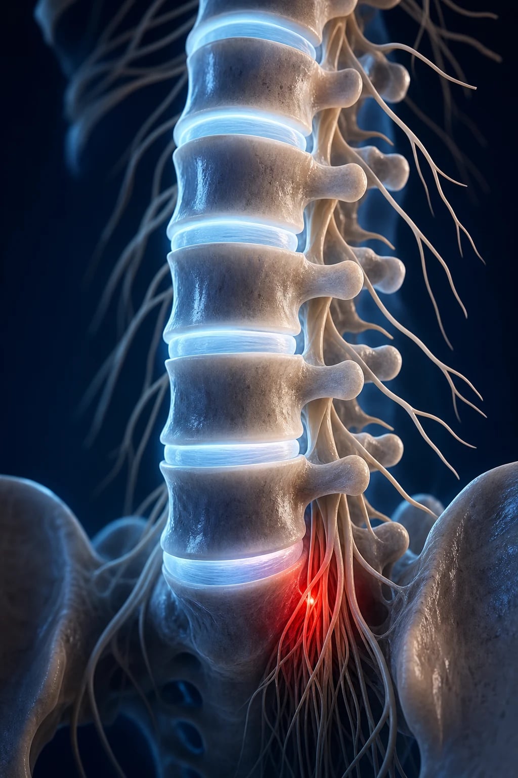
Cauda equina. The spinal cord ends at the L1-L2 conus medullaris; below this, the lumbar and sacral nerve roots travel as a bundle (the cauda equina) within the spinal canal. A large central (or paracentral) disc herniation, or any other mass effect (tumour, abscess, haematoma, fractured bone retropulsion), compresses the sacral nerve roots (S2-S4) that carry the pelvic visceral afferents and efferents — producing the cardinal features of saddle anaesthesia (perianal and genital numbness in the S2-S4 dermatomes), bladder dysfunction (retention or incontinence from loss of detrusor/sphincter coordination), bowel dysfunction, erectile dysfunction, and bilateral leg motor and sensory loss. The internal sphincter tone falls, the bulbocavernosus reflex is abolished, and post-void residual rises dramatically. The window to decompress before permanent injury is narrow — most surgeons aim for surgery within 24 to 48 hours of symptom onset.[2]
Chronic pain. In a minority, acute pain fails to resolve and central sensitisation develops — dorsal horn and brain nociceptive pathways amplify and perpetuate pain signals long after the original tissue injury has healed. Biopsychosocial factors — fear-avoidance beliefs, catastrophising, low mood, social isolation, deconditioning, sleep disturbance, and work/compensation stress — are the chief drivers of this transition and are the primary targets of multidisciplinary rehabilitation.[1]
RED FLAGS — the serious pathology to never miss
RED FLAG
Significant trauma, or minor trauma in an osteoporotic or steroid-treated patient — vertebral fracture
Age over 50 with new pain, or long-term corticosteroids — vertebral fracture or malignancy
History of cancer (spinal metastasis), or IV drug use or immunosuppression — spinal infection
Fever, weight loss, night sweats — spinal infection (vertebral osteomyelitis, epidural abscess, discitis)
Cauda equina — saddle anaesthesia, bladder/bowel retention, bilateral neurology, progressive weakness
Insidious inflammatory pain, better with exercise, worse at rest — axial spondyloarthritis (HLA-B27)
Pain unrelieved by rest or waking the patient at night — malignancy or infection until proven otherwise
Clinical Presentation
The tempo and pattern of pain is the single most discriminating clue, and taking a deliberate, structured history (often remembered as the SOCRATES dimensions, supplemented by an explicit red-flag and cauda equina screen) is what separates mechanical pain from serious pathology. [1]
Mechanical (non-specific). Pain is typically worse with activity and prolonged sitting, better with rest and lying flat, of acute or subacute onset after a recognisable mechanical trigger (lifting, twisting, a fall), and unaccompanied by red flags or objective neurological deficit. It may radiate to the buttock or thigh (sclerotomal pain) but not below the knee. The patient is systemically well. [1]
Inflammatory (axial spondyloarthritis). Suspect this when four of the ASAS inflammatory back pain criteria are met: onset under 40 years, insidious onset, improvement with exercise (not rest), no improvement with rest, pain at night (especially second half of the night) or on rising, plus morning stiffness lasting over 30 minutes. Alternating buttock pain is highly characteristic. Ask also about extra-articular features — uveitis (red painful photophobic eye), psoriasis, inflammatory bowel disease, dactylitis, enthesitis (Achilles, plantar fascia) — and a family history of spondyloarthritis or psoriasis.[5]
Radicular (sciatica). Leg pain radiating below the knee in a dermatomal distribution is the cardinal feature, often more severe than the back pain itself. The L5 root gives weakness of great toe and ankle dorsiflexion (foot drop when severe), sensory loss over the lateral leg and dorsum of the foot, with no reliable reflex loss. The S1 root gives weakness of ankle plantarflexion (cannot tip-toe), loss of the ankle (Achilles) reflex, and sensory loss over the lateral foot and little toe. The L4 root (higher herniation) gives weakness of knee extension (quadriceps), loss of the knee (patellar) reflex, and sensory loss over the medial leg and foot. Pain is typically worse on sitting, coughing, sneezing and straining — all of which raise intra-abdominal pressure and increase tension in the inflamed root. [1]
Cauda equina syndrome. This is the must-not-miss presentation. The classical features are: saddle anaesthesia (perianal, genital, inner thigh numbness — S2-S4 dermatomes); urinary dysfunction — retention (a painless distended bladder is a late, ominous sign), overflow incontinence, or loss of urethral sensation; faecal incontinence or loss of anal tone; bilateral sciatica or bilateral neurological deficit (the bilaterality is the discriminator from unilateral disc herniation); progressive motor weakness in the legs; reduced or absent anal tone and bulbocavernosus reflex; and sexual dysfunction (erectile dysfunction, loss of sensation). The patient may describe "loss of control down below" or numbness when wiping after toileting — those volunteered phrases should trigger emergency action.[2]
Atypical presentations. The elderly may have lumbar spinal stenosis with neurogenic claudication — bilateral leg pain, heaviness, numbness or cramping on walking, relieved within minutes by sitting and forward flexion (the bicycle test — exercise tolerance on a bicycle is preserved because cycling flexes the spine and opens the canal — is positive). The pregnant patient has mechanical and postural pain from altered centre of gravity and the relaxin-mediated ligamentous laxity, with limited imaging options. The immunocompromised or IV drug user may harbour spinal infection with an indolent, fluctuating, febrile course. Vertebral compression fracture in the osteoporotic elderly presents as sudden, severe back pain after minimal or no trauma (a sneeze, a cough, sitting down hard). Spinal metastasis typically produces progressive, unremitting back pain, worse at night and unrelieved by rest — the night-pain feature is the cardinal discriminator from mechanical pain. [1]
Differential Diagnosis
The differential is best organised by the triage categories. Referred (non-spinal) causes must always be considered when the pain is vague, unrelieved by posture, accompanied by systemic or abdominal features, or when the examination of the spine is unremarkable — a particularly dangerous mimic is the expanding or ruptured abdominal aortic aneurysm in the elderly back-pain patient. [1]
- Mechanical (over 90 percent of presentations): muscle or ligament strain, degenerative disc disease, facet joint arthropathy, spondylolysis (pars defect, in athletes), spondylolisthesis (anterior slip of one vertebra on another), sacroiliac joint dysfunction, myofascial pain.
- Radicular: lumbar disc herniation (L4-L5, L5-S1 commonest), lumbar spinal stenosis (degenerative, congenital, or post-surgical), foraminal stenosis, lateral recess stenosis, piriformis syndrome (sciatic nerve compressed by piriformis muscle).
- Inflammatory: axial spondyloarthritis — ankylosing spondylitis, psoriatic arthritis, reactive arthritis, enteropathic (IBD-associated) arthritis, undifferentiated SpA.
- Infective: vertebral osteomyelitis, epidural abscess, discitis (Staphylococcus aureus commonest; Gram-negatives from urinary or gastrointestinal source; Mycobacterium tuberculosis — Pott disease — in endemic areas or immigrants; Brucella in herd-animal regions; fungal in immunocompromised).
- Malignancy: spinal metastasis (the spine is the commonest site of bony metastasis — primary sites are breast, lung, prostate, kidney, thyroid in the classic "BLT-with-Kosher-Pickle" mnemonic), multiple myeloma, lymphoma, and primary bone tumours (chordoma, Ewing sarcoma in children, osteosarcoma).
- Fracture: osteoporotic compression fracture (minimal or no trauma), traumatic fracture (significant mechanism), pathological fracture (through tumour or infection).
- Referred (non-spinal): abdominal aortic aneurysm (a must-not-miss in the elderly — pulsatile abdominal mass, hypotension, tearing pain), pancreatitis, peptic ulcer disease, renal colic, pyelonephritis, pelvic inflammatory disease, endometriosis, retrocaecal appendicitis, prostatitis, hip pathology (osteoarthritis, fracture), sacroiliitis.[1]
A focused abdominal and hip examination is therefore part of a complete back-pain assessment, particularly in elderly patients where referred causes are over-represented. [1]
Clinical & Bedside Assessment
A focused, structured examination identifies neurological deficit, cauda equina syndrome, inflammatory features, and referred causes — and the findings drive the imaging decision. [1]
- Inspection: posture (list to one side), gait (antalgic, or foot-drop indicating L5 lesion), spinal curvature (scoliosis, kyphosis, hyperlordosis), paraspinal muscle spasm, step deformity of the spinous processes (spondylolisthesis), skin over the lower back (sinus, hair tuft, lipoma, naevus suggesting spinal dysraphism), surgical scars.
- Palpation: point tenderness (localised tenderness suggests infection or fracture; widespread non-organic tenderness is non-specific), paraspinal muscle spasm, step deformity, sacroiliac joint tenderness and provocative tests (FABER, Gaenslen), abdominal palpation for pulsatile mass (AAA) and renal angle tenderness.
- Range of motion: flexion, extension, lateral flexion, rotation. Modified Schober test — measure 10 cm above the S2 spinous process, then ask the patient to forward-flex; an increase of less than 5 cm screens for inflammatory spinal restriction (sensitivity moderate, specificity high in established ankylosing spondylitis).[5]
- Neurological — mapped to roots:
- L4 — quadriceps weakness (knee extension), knee (patellar) reflex loss, medial leg and knee numbness.
- L5 — great-toe dorsiflexion (extensor hallucis longus) weakness (and ankle dorsiflexion, foot drop when severe), lateral leg and dorsum of foot numbness; no reliable reflex.
- S1 — ankle plantarflexion weakness (cannot tip-toe), ankle (Achilles) reflex loss, lateral foot and little-toe numbness.
- Provocative manoeuvres:
- Straight leg raise (Lasègue) — reproduction of leg pain below the knee at under 70 degrees of hip flexion indicates L5 or S1 root irritation (sensitivity high, specificity moderate).
- Crossed (contralateral) straight leg raise — raising the asymptomatic leg reproduces the symptomatic leg pain — is more specific (and a positive crossed SLR is highly predictive of a large, central disc herniation).
- Femoral nerve stretch test (prone, knee flexed, hip extended) reproduces anterior thigh pain in L2-L4 root lesion (high lumbar herniation).
- Slump test — sensitises the nerve-root tension test.
- Cauda equina screen (mandatory in any back-pain assessment with red-flag features): perianal (saddle) sensation, anal tone on digital rectal examination, bulbocavernosus reflex, and bladder scan for post-void residual — a residual over 100 to 200 mL suggests retention and supports the diagnosis.
A red flag on history must be matched with the relevant physical sign: fever and vertebral tenderness with infection, a palpable bladder with retention, a pulsatile abdominal mass with AAA, a step deformity with spondylolisthesis, restricted spinal mobility with axial SpA. [1]
Investigations
Most low back pain needs no investigation at all. Imaging is reserved for red flags, progressive neurological deficit, suspected cauda equina, suspected infection or malignancy, suspected inflammatory disease, or persistent radiculopathy despite conservative care — because routine imaging of non-specific pain increases harm without improving any outcome (incidental findings drive anxiety, opioid prescribing, injections, and unnecessary surgery, with no improvement in pain or function).[4]
When to image — and with what
1
Acute non-specific low back pain (under 6 weeks), no red flags, no objective neurological deficit — over 90 percent recover with reassurance and time
2
Suspected fracture from significant trauma; screening for gross destructive lesions (limited sensitivity for early infection/malignancy and zero sensitivity for neural compression)
3
Cauda equina (today), suspected infection (osteomyelitis, epidural abscess, discitis), suspected malignancy (metastasis, myeloma), progressive neurological deficit, persistent radiculopathy over 6 weeks despite conservative care
4
Better for cortical bone detail — fracture characterisation, surgical planning; useful when MRI is contraindicated (pacemaker, some metallic implants)
5
Suspected multifocal bony metastasis; identifying occult infection
6
Differentiating radiculopathy from peripheral neuropathy or plexopathy when the MRI is equivocal or symptoms are atypical; usually not needed in straightforward sciatica
7
FBC, CRP, ESR (infection or malignancy); blood cultures if febrile; PSA, serum electrophoresis and serum free light chains (myeloma); calcium, ALP, LFTs; HLA-B27 and inflammatory markers for suspected axial SpA
Specific investigations by suspected diagnosis. For suspected infection (epidural abscess, vertebral osteomyelitis), the CRP and ESR are usually markedly elevated and blood cultures should be drawn before antibiotics; MRI with gadolinium is the imaging gold standard and defines the abscess extent. For suspected malignancy, MRI defines the cord or cauda compromise and biopsy (image-guided) provides tissue diagnosis; look for the primary (mammogram, prostate exam and PSA, chest CT, myeloma screen). For suspected fracture, plain X-ray is the initial screen; MRI defines marrow oedema (acute fracture) versus healed fracture; DEXA confirms underlying osteoporosis. For suspected axial spondyloarthritis, HLA-B27 plus MRI of the sacroiliac joints looking for bone marrow oedema (active sacroiliitis) is more sensitive than plain films in early disease.[5]
Management — Resuscitation

The single resuscitation scenario in low back pain is cauda equina syndrome — a surgical emergency. Time is nerve: every hour of compression beyond the threshold of injury erodes the chance of neurological recovery. [1]
- Recognise the triad at the bedside: saddle anaesthesia + bladder or bowel dysfunction + bilateral neurology or progressive weakness. Any one of these in a back-pain patient mandates the cauda equina workup, not the whole triad.
- Urgent MRI of the lumbar spine (whole spine if multifocal suspicion, prior malignancy, or high clinical suspicion with negative lumbar MRI). The MRI should be performed today, with the patient remaining nil by mouth.
- Immediate neurosurgical or orthopaedic spinal referral for urgent surgical decompression (laminectomy plus discectomy, plus fusion if instability) — ideally within 24 to 48 hours of symptom onset to maximise neurological recovery. Late decompression (over 48 hours, and especially over 72 hours) is associated with substantially worse bladder, motor and sexual outcomes.
- Urinary catheterisation for retention (catheterise early — a distended bladder worsens the voiding prognosis); protect pressure areas; provide analgesia; keep nil by mouth for theatre.
- Baseline pre-operative bloods: FBC, U&E, coagulation, group and save; document a careful neurological examination (motor power MRC grade by myotome, reflexes, sensory including saddle area, anal tone) before surgery so postoperative recovery can be tracked.
- If infection or malignancy with cord compromise is found instead of a disc herniation, treat the underlying cause (long intravenous antibiotics, oncology referral, urgent radiotherapy) and consider urgent decompression as appropriate.[2]
Management — Definitive & Stepwise
For non-specific low back pain, the modern evidence is unequivocal: less is more.[1] The biggest iatrogenic harms in low back pain are over-imaging, opioid prescribing, spinal injections and surgery — interventions that do not improve outcomes and demonstrably cause harm. The ACP, NICE and the Lancet Low Back Pain Series all converge on the same first-line bundle: education, reassurance, encouragement to stay active and return to work, simple analgesia, and exercise.[2][3]
1. Acute non-specific back pain (under 6 weeks) — first-line, non-pharmacological:
- Education and reassurance — explain that the pain is mechanical, non-serious and very likely to improve over weeks; advise the patient to stay active and to return to work as soon as possible; avoid bed rest (prolonged bed rest worsens pain, deconditions and prolongs disability).
- Simple analgesia — first-line non-steroidal anti-inflammatory drugs (e.g. ibuprofen 400 mg orally three times daily, or naproxen 250 to 500 mg orally twice daily, with a proton-pump inhibitor if there is GI risk), at the lowest effective dose for the shortest time. Paracetamol is a second choice — the PACE trial demonstrated that paracetamol (regular or as-needed, 4 g/day) was no better than placebo for acute low back pain, challenging its traditional first-line status.[9]
- Heat (hot packs, hot baths) and short-term manual therapy or spinal manipulation (e.g. up to 6 to 8 sessions of osteopathic or physiotherapy manipulation) for short-term symptom relief.
- AVOID opioids (dependence, overdose, constipation, falls — no long-term benefit and significant harm), AVOID routine imaging in the absence of red flags, AVOID injections.
2. Persistent or subacute pain (over 6 weeks, under 12 weeks):
- Supervised exercise programme (group or individual; any exercise modality the patient will adhere to — Pilates, core strengthening, aerobic, yoga).
- Cognitive behavioural therapy (CBT) combined with exercise — addresses fear-avoidance, catastrophising, and low mood.
- Manual therapy (up to 12 weeks) alongside exercise.
- Address yellow flags (psychosocial risk factors for chronicity) — these predict chronic disabling pain more powerfully than any imaging finding. [1]
3. Chronic disabling pain (over 12 weeks):
- Multidisciplinary biopsychosocial rehabilitation combining physical, psychological and occupational components — the only intervention with consistent evidence for improving pain, function and work outcomes in chronic disabling low back pain.
- AVOID opioids, repeat imaging, and further invasive interventions unless there is a new red flag. [1]
4. Sciatica (lumbar radiculopathy):
- Conservative management first — the natural history is favourable, with over 80 percent recovering within 6 to 12 weeks. The Peul group's NEJM trial showed early surgery for sciatica achieved faster relief of leg pain in the first year but no advantage at one- and two-year follow-up compared with prolonged conservative care — patients who tolerate conservative care can be reassured they will not be harmed by waiting.[7]
- MRI if persistent over 6 weeks despite conservative care, or if there is a progressive neurological deficit.
- Surgical microdiscectomy for severe persistent radiculopathy (over 6 to 12 weeks despite conservative care and confirmed disc herniation on MRI) or progressive motor deficit. The SPORT randomised trial (Weinstein et al, JAMA 2006) confirmed that patients treated surgically for lumbar disc herniation recovered better than those treated non-operatively, although the treatment effect was attenuated by crossover.[8]
- Cauda equina is operated urgently (see Resuscitation).
5. Lumbar spinal stenosis:
- Flexion-based exercise (cycling, swimming, walking uphill — flexion opens the canal and reduces claudication), simple analgesia.
- Epidural steroid injection for short-term relief (weeks to a few months) in selected patients; not a cure and not for long-term use.
- Decompressive laminectomy (with or without fusion for instability or deformity) for refractory neurogenic claudication limiting quality of life. [1]
6. Axial spondyloarthritis: continuous NSAIDs (e.g. naproxen 1000 mg/day) plus exercise and physiotherapy; biologic anti-TNF (e.g. adalimumab 40 mg subcutaneously every 2 weeks, or etanercept 50 mg weekly) for patients with high disease activity despite NSAIDs. Refer to the ankylosing-spondylitis topic for the full ladder. [1]
7. Specific pathology: treat the cause — antibiotics for spinal infection (long intravenous course, e.g. 6 weeks of flucloxacillin 2 g IV every 6 hours with rifampicin, then oral continuation, tailored to organism and sensitivity); vertebroplasty or kyphoplasty for painful osteoporotic compression fractures unresponsive to conservative care; oncology referral for spinal metastasis (radiotherapy, surgery for cord compression, systemic therapy); antiresorptive therapy (bisphosphonates, denosumab) plus calcium and vitamin D for underlying osteoporosis.[5]
Specific Subtypes & Scenarios
- Acute non-specific (under 6 weeks): reassure, mobilise, simple analgesia; over 90 percent recover within 6 weeks. Recurrence is common (about 60 percent within a year) and patients should be warned.
- Chronic disabling (over 12 weeks): biopsychosocial approach — supervised exercise plus CBT plus multidisciplinary rehabilitation; treat yellow flags; avoid opioids and repeat imaging.
- Sciatica (lumbar radiculopathy): L5 and S1 roots commonest (from L4-L5 and L5-S1 disc herniations respectively). Conservative first; surgery (microdiscectomy) for severe or persistent radiculopathy (over 6 to 12 weeks) or progressive motor loss. Cauda equina is operated urgently.
- Lumbar spinal stenosis: neurogenic claudication — bilateral leg pain or heaviness on walking, relieved within minutes by sitting and forward flexion (bicycle test positive because cycling flexes the spine and opens the canal). Distinguish from vascular claudication (which is relieved simply by stopping, requires no sitting or flexion, and is associated with absent pulses and skin signs of ischaemia). Flexion exercise, epidural steroid injection for short-term relief, decompressive laminectomy for refractory cases.
- Cauda equina syndrome: large central disc (or tumour, abscess, fractured bone) compressing the sacral roots; emergency decompression within 24 to 48 hours.
- Axial spondyloarthritis: young patient (under 40), insidious onset, exercise-relieved back pain, morning stiffness over 30 minutes, alternating buttock pain; HLA-B27, MRI of sacroiliac joints for bone marrow oedema; treat with NSAIDs plus exercise, anti-TNF if resistant.
- Vertebral compression fracture (osteoporotic): sudden severe back pain after minimal or no trauma in an elderly patient (or any patient on long-term corticosteroids, or with risk factors for osteoporosis). Plain films screen; MRI confirms acuity (marrow oedema). Conservative with analgesia, bracing, early mobilisation; vertebroplasty or kyphoplasty for unresponsive severe pain. Treat underlying osteoporosis (DEXA, bisphosphonate or denosumab, calcium, vitamin D, fall prevention).
- Spinal infection (vertebral osteomyelitis, epidural abscess, discitis): fever, raised inflammatory markers, severe localised tenderness, possibly neurological deficit; Staphylococcus aureus commonest; MRI with gadolinium is gold standard; long intravenous antibiotics (typically 6 weeks) with surgical decompression if there is cord compromise, neurological deficit, or failure of medical therapy. [1]
Complications & Pitfalls
- Chronic pain and disability — the biggest complication of acute non-specific low back pain. Persistent pain produces work loss, depression, anxiety, relationship breakdown, sleep disturbance, and physical deconditioning.
- Opioid dependence and overdose — driven by over-prescription for a condition that is overwhelmingly self-limiting. Constipation, falls, fractures, hypogonadism, overdose and death are downstream harms; avoid opioids for non-specific low back pain.
- Pitfall — over-investigation: incidental disc bulges and degeneration are common in pain-free adults; imaging non-specific pain leads to overdiagnosis, anxiety, opioid prescribing and unnecessary surgery with no benefit.[4]
- Pitfall — missed cauda equina: permanent paralysis, bladder and bowel incontinence, and sexual dysfunction. Always ask about saddle numbness and bladder function, and bladder-scan for retention; never attribute urinary symptoms to "mechanical" pain without ruling out cauda equina.
- Pitfall — missed infection or malignancy: irreversible neurological damage, sepsis, death. Image whenever red flags are present, especially fever, weight loss, night pain, history of cancer, or immunosuppression.
- Pitfall — missing a non-spinal mimic: an elderly back-pain patient with hypotension may have a ruptured abdominal aortic aneurysm — a fatal miss. Always examine the abdomen and feel the pulses.
- Pitfall — over-medicalising a self-limiting condition: driving fear, dependence, and disability through inappropriate referrals, scans, injections and surgery.
- Surgical complications: dural tear, nerve root injury, infection, recurrent disc herniation, failed back surgery syndrome (persistent post-surgical pain).
Prognosis & Disposition
The prognosis is generally favourable — about 90 percent of acute non-specific low back pain recovers within 6 weeks, and most of the remainder improves by 12 weeks. However, recurrence is common (about 60 percent within a year), and a minority progress to chronic disabling pain. [1]
The strongest predictors of poor outcome — the yellow flags — are not imaging findings but psychosocial:[1]
- High baseline disability and pain intensity.
- Psychological distress — depression, anxiety, somatisation.
- Fear-avoidance beliefs ("if it hurts I must be damaging my back") and pain catastrophising.
- Low job satisfaction, job strain, poor social support, heavy manual work, seeking compensation.
- Widespread pain (suggests central sensitisation), and prior prolonged episodes of back pain. [1]
Addressing these yellow flags early — through education, reassurance, CBT, graded exercise and return-to-work support — is the single most effective strategy to prevent chronic disabling low back pain. [1]
Disposition. Most patients are managed in the community or primary care. Emergency disposition is reserved for cauda equina syndrome (urgent admission, MRI, surgical referral, decompression within 24 to 48 hours) and other red-flag pathology (suspected infection or malignancy, fracture with neurological compromise). Patients with severe or persistent radiculopathy (over 6 to 12 weeks) are referred for specialist assessment and possible surgical opinion. [1]
Special Populations
- Elderly: higher risk of lumbar spinal stenosis, osteoporotic compression fracture (sudden severe pain after minimal or no trauma), vertebral malignancy, and referred pain from abdominal aortic aneurysm. Maintain a lower threshold to image. Polymyalgia rheumatica and giant cell arteritis may present with back and shoulder girdle pain and must not be missed (ESR/CRP markedly raised).
- Pregnancy: mechanical and postural pain (altered centre of gravity, relaxin-mediated ligamentous laxity, weight gain). Avoid imaging unless red flags are present (and then weigh radiation risk — MRI without gadolinium is preferred). Simple analgesia (paracetamol; avoid NSAIDs in the third trimester — premature closure of the ductus arteriosus and oligohydramnios), exercise, posture, and supportive belts help.
- Children and adolescents: consider spondylolysis and spondylolisthesis in young athletes (gymnasts, fast bowlers, dancers), infection (discitis — young child refusing to walk, back pain, limp), tumour (Ewing sarcoma, osteoid osteoma, aneurysmal bone cyst), and leukaemia. Persistent or night pain in a child warrants imaging — do not dismiss it as growing pains.
- Immunocompromised and IV drug users: high risk of spinal infection with atypical organisms (Staphylococcus aureus commonest, but Gram-negatives, Mycobacterium tuberculosis, fungi, and Candida must be considered). Maintain a low threshold for MRI, blood cultures and inflammatory markers.[5]
- Patients on long-term corticosteroids: high risk of osteoporotic compression fracture and avascular necrosis (the latter at the hip rather than the spine, but may refer pain to the back).
- Anticoagulated patients: any sudden severe back pain with hypotension or anaemia may be a spinal epidural haematoma — urgent MRI, neurosurgical referral.
Evidence, Guidelines & Regional Differences
The evidence base for low back pain management has shifted dramatically over the last two decades, away from invasive intervention and toward conservative, biopsychosocial care. The pivotal publications: [1]
- Lancet Low Back Pain Series (2018) — Foster, Buchbinder and colleagues issued a global call to action against over-medicalisation, championing first-contact conservative care, stay-active messaging, and avoidance of opioids and routine imaging. They emphasised the biopsychosocial model, the social determinants of chronicity, and the special challenges of low- and middle-income countries.[1][2]
- American College of Physicians (ACP) Guideline, Qaseem et al 2017 — recommends non-pharmacologic care first for acute, subacute and chronic low back pain (heat, massage, acupuncture, spinal manipulation); pharmacologic options only if non-pharmacologic fails, with NSAIDs or skeletal muscle relaxants as first-line (paracetamol downgraded on the strength of the PACE trial); for chronic low back pain, exercise, multidisciplinary rehabilitation, acupuncture, CBT, spinal manipulation, tai chi, yoga, motor control exercise, progressive relaxation. Recommendation against opioids except in refractory severe cases, against imaging without red flags, against injections and surgery in most cases.[3][4][5]
- PACE trial (Williams et al, Lancet 2014) — the first placebo-controlled trial of paracetamol for acute low back pain showed no benefit of paracetamol (regular or as-needed, 4 g/day) over placebo on pain, disability, function, symptom resolution or quality of life. This finding reshaped first-line pharmacotherapy globally.[9]
- Sport and sciatica surgery trials — the Peul group's NEJM 2007 trial showed early microdiscectomy for sciatica gave faster short-term leg pain relief but no advantage at one- and two-year follow-up over prolonged conservative care; the SPORT trial (Weinstein et al, JAMA 2006) confirmed surgical superiority for lumbar disc herniation despite crossover.[7][8]
- Diagnostic imaging for low back pain (Chou et al, 2011) — ACP high-value care advice confirming that routine imaging does not improve outcomes and causes harm.[4]
The Lancet Series (2018) highlights the global divide — high-income countries struggle with over-imaging, opioid over-prescription and unnecessary surgery, while low- and middle-income countries face limited access to MRI, surgery and rehabilitation, reinforcing the value of a conservative, primary-care-first approach. WHO and the Global Spine Care Initiative promote task-shifting to community health workers, exercise and education as scalable solutions.
Controversies. The role of paracetamol has been contested since PACE; some guidelines (NICE, ACP) now place NSAIDs above paracetamol. The role of spinal manipulation is supported for short-term relief but not long-term. The role of epidural steroid injections for spinal stenosis is contested (short-term benefit, no long-term benefit, repeated injections carry harm). Vertebroplasty and kyphoplasty for osteoporotic compression fracture have mixed evidence (two sham-controlled trials were negative; subsequent trials and meta-analyses suggest benefit in selected acute severe fractures). Surgery for axial back pain (so-called "fusion for discogenic back pain") has poor evidence and is generally not recommended. [1]
Exam Pearls
- Over 90 percent of low back pain is non-specific and self-limiting — recovers within 6 weeks. Over-investigation, opioids, injections and surgery cause harm.[1]
- Screen red flags at every assessment: cauda equina, fracture, infection, malignancy, axial spondyloarthritis. The systematic red-flag screen is the most examinable skill in this topic.
- Cauda equina = saddle anaesthesia + bladder or bowel dysfunction + bilateral neurology (with or without progressive weakness). This is a surgical emergency: urgent MRI today, decompression within 24 to 48 hours, urinary catheterisation.
- Reserve MRI for red flags, progressive neurology, or persistent radiculopathy over 6 weeks — not for routine non-specific pain.[4]
- Non-specific pain: reassure, stay active, NSAIDs first (avoid opioids), exercise and CBT for persistent pain. PACE showed paracetamol is no better than placebo for acute pain.[3][9]
- Sciatica is L5 or S1 in the vast majority (from L4-L5 or L5-S1 disc herniations). L5 — great toe dorsiflexion weakness, no reflex; S1 — ankle reflex loss, plantarflexion weakness. Crossed straight leg raise is more specific.
- Inflammatory back pain (ASAS criteria) — onset under 40, insidious, better with exercise, morning stiffness over 30 min, alternating buttock — points to axial spondyloarthritis. Send HLA-B27 and MRI of sacroiliac joints.
- Spinal stenosis pseudoclaudication — worse walking and downhill, better with flexion and sitting (bicycle test positive). Distinguish from vascular claudication (no flexion requirement, absent pulses).
- Sciatica surgery: Peul 2007 NEJM showed early surgery gives faster leg pain relief but no advantage at one year vs prolonged conservative care; SPORT confirmed surgical superiority for disc herniation despite crossover.[7][8]
- Don't miss the AAA in the elderly back-pain patient — palpate the abdomen for a pulsatile mass.
- Pregnancy back pain — avoid imaging unless red flags; avoid NSAIDs in third trimester.
Exam application bank (NEET-PG / INICET)
One-line answer
Low back pain is the leading cause of disability worldwide; the vast majority is non-specific (mechanical) and self-limiting (over 90 percent recover within 6 weeks). The central clinical skill is identifying red flags (serious pathology — fracture, infection, malignancy, cauda equina, inflammatory) that mandate imaging and referral, while reassuring and mobilising the remainder. Red flags: significant trauma; age over 50 with new pain; history of cancer; fever/weight loss/night sweats; immunosuppression, IV drug use; night pain or pain unrelieved by rest; recent bacterial infection; corticosteroid use; cauda equina (saddle anaesthesia, urinary retention/incontinence, bilateral neurology, progressive weakness) = surgical emergency. Manage non-specific pain: reassure, stay active, simple analgesia (avoid opioids), exercise + CBT; surgery only for severe persistent radiculopathy or cauda e
Worked stems (answer without another resource)
Stem 1 — Classic presentation. Map symptoms to mechanism; name the first investigation and first treatment step with dose/route if drug therapy is standard. [1]
Stem 2 — Unstable / complicated. List red flags that force immediate resuscitation, theatre, ICU, antidote, or reperfusion — and what you do in the first 15 minutes. [1]
Stem 3 — Atypical group. Elderly, pregnancy, child, or immunocompromised: how presentation and thresholds change. [1]
Stem 4 — Differential trap. Name the three closest mimics and one discriminator for each. [1]
Stem 5 — Disposition. Who goes home with safety-netting, who is admitted, who needs HDU/ICU/theatre, and what follow-up is mandatory. [1]
Rapid viva checklist
- Definition + classification
- Pathophysiology chain
- Bedside signs / criteria
- Score with exact components (if any)
- Emergency bundle
- Definitive therapy with doses
- Complications of disease and of treatment
- Special populations
- Guideline/trial name if classic
- Three exam traps
Coverage self-check
If you cannot answer any stem above from this page alone, re-read the matching section — the page is intended to be self-sufficient for final-prof and NEET-PG/INICET questions on Low Back Pain (Approach & Red Flags).
References
- [1]Foster NE, Anema JR, Cherkin D, Chou R, Cohen SP, Gross DP, et al. Prevention and treatment of low back pain: evidence, challenges, and promising directions Lancet, 2018.PMID 29573872
- [2]Buchbinder R, van Tulder M, Öberg B, Costa LM, Woolf A, Schoene M, Croft P, et al. Low back pain: a call for action Lancet, 2018.PMID 29573871
- [3]Qaseem A, Wilt TJ, McLean RM, Forciea MA, Clinical Guidelines Committee of the American College of Physicians. Noninvasive Treatments for Acute, Subacute, and Chronic Low Back Pain: A Clinical Practice Guideline From the American College of Physicians Ann Intern Med, 2017.PMID 28192789
- [4]Chou R, Deyo RA, Friedly J, Fu R, Bougatsos C, Dana T, Sullivan SD, et al. Diagnostic imaging for low back pain: advice for high-value health care from the American College of Physicians Ann Intern Med, 2011.PMID 21282698
- [5]Chou R, Qaseem A, Snow V, Casey D, Cross JT Jr, Shekelle P, Owens DK, et al. Diagnosis and treatment of low back pain: a joint clinical practice guideline from the American College of Physicians and the American Pain Society Ann Intern Med, 2007.PMID 17909209
- [6]Peul WC, van Houwelingen HC, van den Hout WB, Brand R, Eekhof JA, Tans JT, Thomeer RT, et al. Magnetic resonance imaging in follow-up assessment of sciatica N Engl J Med, 2013.PMID 23484826
- [7]Peul WC, van den Hout WB, Brand R, Thomeer RTWM, Koes BW, Leiden-The Hague Spine Intervention Prognostic Study Group. Surgery versus prolonged conservative treatment for sciatica N Engl J Med, 2007.PMID 17538084
- [8]Weinstein JN, Tosteson TD, Lurie JD, Tosteson ANA, Hanscom B, Skinner JS, Abdu WA, et al. Surgical vs nonoperative treatment for lumbar disk herniation: the Spine Patient Outcomes Research Trial (SPORT): a randomized trial JAMA, 2006.PMID 17119140
- [9]Williams CM, Maher CG, Latimer J, McLachlan AJ, Hancock MJ, Day RO, Lin CCW, et al. Efficacy of paracetamol for acute low-back pain: a double-blind, randomised controlled trial Lancet, 2014.PMID 25064594