Rheumatology · General Medicine
Osteoarthritis
Also known as Osteoarthritis · OA · Degenerative joint disease · Osteoarthrosis
Osteoarthritis (OA) is the commonest joint disorder — a degenerative, mechanically driven arthritis of articular (hyaline) cartilage with bony remodelling (osteophytes, subchondral sclerosis and cysts), meniscal damage and low-grade synovial inflammation. It predominantly affects the knees, hips, hands (DIP — Heberden nodes, PIP — Bouchard nodes, first CMC) and spine. Risk factors are age, obesity, female sex, prior joint injury, repetitive occupational stress and genetics. It presents with use-related joint pain (worse with activity, better with rest, brief morning stiffness under 30 minutes), functional limitation, crepitus and bony swelling, with late deformity. Diagnosis is clinical, supported by X-ray (LOSS — joint-space narrowing, osteophytes, subchondral sclerosis and cysts), though imaging severity correlates poorly with symptoms. Management is built on education, exercise and weight loss (core, disease-modifying), plus topical NSAIDs, short-course oral NSAIDs, intra-articular steroid for flares and joint replacement for end-stage disease.
On this page & tools
Your progress
Saved locally on this device.
Practise this topic
Exam tags
Red flags
Overview & Definition
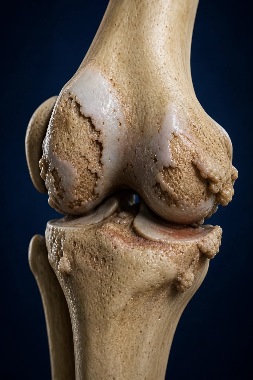
Osteoarthritis (OA) — historically called degenerative joint disease or osteoarthrosis — is a whole-organ disorder of the synovial joint in which progressive structural failure of articular cartilage is accompanied by bony remodelling (osteophytes, subchondral sclerosis and subchondral cysts), meniscal degeneration, capsular thickening and low-grade synovial inflammation.[1] It is not, as once taught, a passive process of "wear and tear": it is an active, cell-mediated repair response of joint tissues that becomes maladaptive under sustained mechanical overload, ageing, metabolic stress and genetic predisposition. The clinical syndrome is the combination of use-related joint pain, brief morning stiffness, functional limitation, crepitus and bony swelling in a characteristic joint distribution.
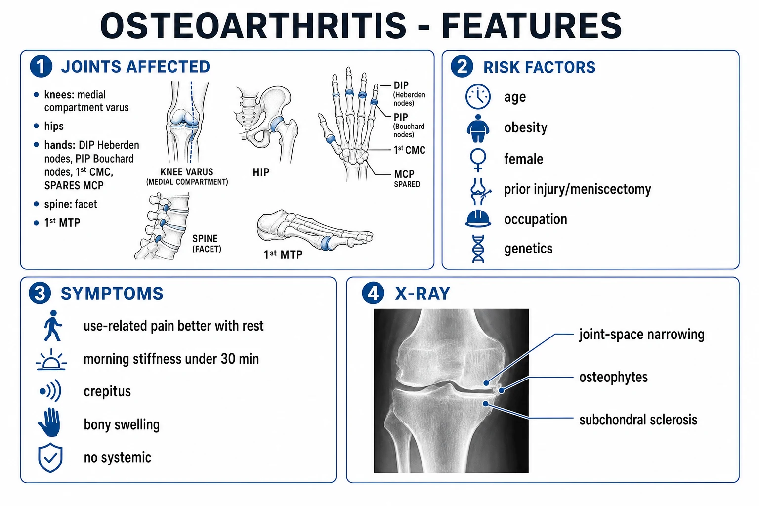
OA is the commonest form of arthritis worldwide, the leading cause of disability in older adults and the single largest contributor to the global burden of musculoskeletal disease, with an estimated 250 million people affected globally.[1] The knee, hip, hand (DIP, PIP, first CMC) and spine (facet joints) are the principal targets; in the hand the metacarpophalangeal (MCP) joints are characteristically spared, a fact that does more diagnostic work than any single investigation.
The clinical reasoning around OA turns on three insights. First, mechanical versus inflammatory: OA pain is use-related with short stiffness and normal inflammatory markers, whereas rheumatoid arthritis is inflammatory with prolonged stiffness, symmetric small-joint involvement and DIP sparing. Second, non-pharmacological core measures — exercise and weight loss — are disease-modifying and first-line, not adjuncts; they slow progression of knee OA and reduce symptoms more durably than any drug. Third, imaging severity does not dictate treatment — symptoms and function do, because radiographic severity correlates poorly with pain. A patient with alarming X-ray changes may have mild symptoms, and a patient with near-normal films may be disabled.[1][2]
Classification
OA is classified by cause into primary (idiopathic) and secondary forms, and by joint distribution into localised and generalised patterns. The distinction matters because secondary OA demands a search for the underlying driver — treat the cause, and you may slow or localise the disease.[1]
Primary (idiopathic) OA has no identifiable precipitant and is the overwhelmingly common form in older adults. It is further subdivided by distribution: generalised nodal OA (the classic Heberden phenotype, with symmetric DIP, PIP and first CMC involvement in post-menopausal women, strongly familial); isolated knee, hip or first CMC OA; and erosive (inflammatory) OA, an aggressive subset of hand OA marked by central erosions in the DIP/PIP ("gull-wing" deformity) with marked inflammatory overlay — distinguished from RA by DIP predominance, sparing of MCP/wrists and a self-limiting course.
[1]Secondary OA has an identifiable cause. The categories worth memorising are: post-traumatic (meniscectomy, ACL tear, intra-articular fracture — the commonest secondary cause in younger patients); developmental/structural (developmental dysplasia of the hip, slipped capital femoral epiphysis, femoroacetabular impingement, Legg-Calvé-Perthes, genu varum/valgum); prior inflammatory arthritis (burned-out RA, gout, septic arthritis); metabolic/endocrine (haemochromatosis classically affects the second and third MCP; Wilson disease; acromegaly; hyperparathyroidism; ochronosis/alkaptonuria; Paget disease of bone); neuropathic (Charcot) arthropathy; and crystal deposition (CPPD, BCP). When OA appears in an atypical joint (MCP, wrist, shoulder, ankle) or at a young age, the diagnosis is not primary OA until a secondary cause has been excluded.[1][3]
Primary (idiopathic) OA
- No identifiable cause — diagnosis of exclusion in older adults
- Subtypes: generalised nodal (Heberden), isolated knee/hip/CMC, erosive inflammatory hand OA
- Classic distribution: knee, hip, DIP/PIP/1st CMC, spine facets, first MTP
- Strong familial clustering (heritability 40-65% for hand/knee OA)
Secondary OA
- Post-traumatic: meniscectomy, ACL tear, intra-articular fracture (commonest in the young)
- Developmental: DDH, FAI, SCFE, Perthes, malalignment
- Metabolic: haemochromatosis (2nd/3rd MCP), acromegaly, hyperparathyroidism, ochronosis
- Inflammatory/burned-out: RA, gout, sepsis, CPPD, Charcot neuropathic
Epidemiology & Risk Factors
OA is the commonest form of arthritis and a major cause of disability and lost productivity. The Global Burden of Disease 2019 study estimated roughly 250 million people with knee OA worldwide and a prevalence that rises steeply with age — radiographic knee OA is found in about 30% of those over 65, although only around half of these are symptomatic.[1] Symptomatic hip OA is less common than knee OA; hand OA (especially nodal) is the commonest form in women beyond the menopause.
Osteoarthritis — key numbers
The risk factors divide neatly into modifiable and non-modifiable. The non-modifiable cluster — age (the strongest risk factor; cartilage loses proteoglycan and chondrocyte repair capacity with time), female sex (women overtake men after the menopause, especially for hand and knee OA — implicating oestrogen loss), and genetics (heritability estimates 40-65% for hand and knee OA; candidate genes include GDF5, DVWA, ASTN2) — set the substrate. The modifiable cluster is where prevention and treatment live: obesity (the single strongest modifiable risk for knee OA, with a hazard ratio around 2-3 — and a smaller but real effect for hand OA via adipokine-mediated inflammation, not loading), prior joint injury (meniscectomy and ACL tear multiply the risk of later knee OA several-fold), repetitive occupational stress (kneeling, squatting, lifting, farming — knee OA; prolonged standing or farming — hip OA), muscle weakness (especially quadriceps), and joint malalignment (varus or valgus concentrating load on one compartment).
[1]Two relationships reward careful explanation because examiners probe them. Why does obesity cause OA of the hand? Because adipose tissue is not inert fat but an endocrine organ secreting adipokines (leptin, interleukin-6, adiponectin) that drive low-grade systemic inflammation and chondrocyte catabolism at non-weight-bearing joints; the mechanical load story is necessary but not sufficient. Why do farmers and floor-sitters (squatting/cross-legged postures common in India and East Asia) get hip and knee OA? Because the deep-flexed postures concentrate contact stress on the posteromedial tibial plateau and the anterosuperior femoral head, accelerating cartilage failure in compartments already vulnerable to malalignment.[1]
Pathophysiology
OA is not simple 'wear and tear' but an active, cell-mediated process of cartilage destruction and aberrant repair affecting the whole joint organ. The driver is excessive or abnormal mechanical load acting on a chondrocyte already rendered vulnerable by ageing, genetics and metabolic stress. The stressed chondrocyte switches from a matrix-maintaining phenotype (synthesising type II collagen and aggrecan) to a catabolic, pro-inflammatory phenotype: it up-regulates matrix metalloproteinases (MMP-1, MMP-3, MMP-13) and the aggrecanases (ADAMTS-4, ADAMTS-5) that together cleave type II collagen fibrils and degrade aggrecan, the major cartilage proteoglycan responsible for water retention and compressive resilience.[1]
Once the collagen network is disrupted, the cartilage loses its aggrecan-bound water, fibrillates, thins and ulcerates, progressing from superficial fraying to full-thickness erosions that expose the underlying subchondral bone. The subchondral bone does not sit passively: it remodels in response to the altered load, becoming denser and stiffer (subchondral sclerosis) and developing subchondral cysts (geodes) — fluid-filled cavities formed by synovial pressurisation of microfractures. At the joint margins, osteophytes (new bone outgrowths) form as a reparative response — and are the most specific radiographic feature of OA.
[1]Cartilage debris and calcium pyrophosphate / basic calcium phosphate crystals released into the joint space drive a low-grade synovitis, distinct from the high-grade pannus of RA. Inflammatory mediators (IL-1β, TNF-α, IL-6) feed back to amplify chondrocyte catabolism. Periarticular muscle weakness (especially quadriceps inhibition) and proprioceptive impairment further destabilise the joint, completing a vicious cycle. Meniscal degeneration and extrusion in knee OA narrow the tibiofemoral contact area and increase peak stress. Nerve ingrowth into the subchondral bone and osteophytes, plus central sensitisation, explains why some patients have severe pain out of proportion to structural change.[1]
The molecular hierarchy is worth fixing: type II collagen is the tensile scaffold (analogous to the steel reinforcement in concrete); aggrecan is the water-binding proteoglycan that resists compression (the concrete). ADAMTS attacks aggrecan early (reversible water loss, swelling on MRI), MMP-13 attacks type II collagen late (irrevable structural failure). This is why cartilage loss on X-ray is a late finding — by the time joint-space narrowing is visible, much of the matrix is already gone.
[1]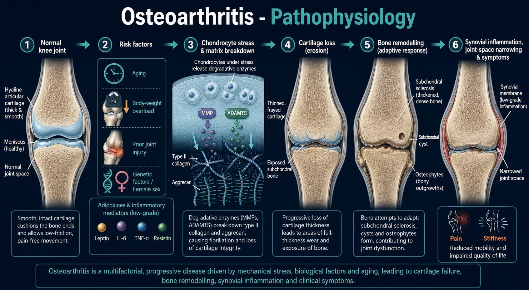
LOSS — the X-ray hallmarks of osteoarthritis
LOSS
Narrowing from cartilage thinning (unlike inflammatory arthritis where narrowing may be periarticular osteopenia-driven)
New bone at the joint margins — the most specific radiographic feature of OA
Thickened, dense bone immediately beneath the worn cartilage
Fluid-filled cavities in the subchondral bone (geodes)
Clinical Presentation
The clinical signature of OA is a characteristic pain pattern and joint distribution, and these two features do most of the diagnostic work. The pain is use-related — it worsens with activity and weight-bearing (walking, climbing stairs, gripping) and improves with rest. Patients describe an end-of-day aching and stiffness after inactivity ("gelling") that resolves within 30 minutes of movement. Crucially, there are no systemic features — no fatigue, fever, weight loss or morning systemics — and inflammatory markers (ESR, CRP) are normal.[1]
The joint distribution is asymmetric and patterned:
[1]- Knee — the medial tibiofemoral compartment is most often affected first, producing varus (bow-leg) malalignment in advanced disease. Patellofemoral OA causes anterior knee pain on stairs and rising from a chair.
- Hip — typically groin pain (worse on weight-bearing and internal rotation) that may refer to the knee via the obturator nerve, the source of much diagnostic confusion. Internal rotation is the first movement lost.
- Hand — DIP (Heberden nodes), PIP (Bouchard nodes) and first CMC (giving a squared hand base); the MCP joints are characteristically spared. Nodal OA evolves in a stuttering, episodic fashion with periods of warmth and redness as new nodes form.
- Spine — facet-joint OA causes mechanical back pain worse on extension and rotation, with referred pain to the buttock and thigh (rarely below the knee) and possible spinal stenosis with neurogenic claudication.
- First MTP — hallux rigidus (stiff big toe with dorsal osteophyte), distinct from hallux valgus.
Examination shows bony swelling (hard, cool, non-tender nodes; compare with the soft, warm, boggy synovitis of RA), crepitus (a palpable/audible grating on active movement), restricted range of motion, joint-line tenderness, muscle wasting (quadriceps in knee OA), varus/valgus deformity and functional limitation. Cool effusions may occur; a hot, tense effusion should prompt aspiration to exclude sepsis or crystals.[1]
Differential Diagnosis
The pivotal decision at the bedside is mechanical versus inflammatory, and the first action in any acute hot joint is aspiration to exclude sepsis or crystals. OA is distinguished from inflammatory and crystal arthropathies by its pain pattern, joint distribution and normal inflammatory markers.[1]
Osteoarthritis
- Use-related pain, morning stiffness under 30 min, better with rest
- Asymmetric: knee, hip, hands (DIP Heberden, PIP Bouchard, first CMC); MCP SPARED
- Bony swelling, crepitus, late deformity; no systemic features
- Normal ESR/CRP. X-ray = LOSS (joint-space narrowing, osteophytes, subchondral sclerosis, cysts)
- First-line: exercise + weight loss + physio (disease-modifying); topical NSAIDs; joint replacement end-stage
Rheumatoid arthritis
- Symmetric small joints: MCP, PIP, wrists, ankles; DIP SPARED
- Prolonged morning stiffness over 1 hour, synovial (soft-tissue) swelling
- Systemic fatigue; raised ESR/CRP; RF and anti-CCP positive
- X-ray: periarticular osteopenia, marginal erosions early
- Disease-modifying antirheumatic drugs (methotrexate first) — needs early rheumatology
Gout
- Acute monoarthritis, rapid onset, extremely painful red-hot joint; first MTP (podagra)
- Tophi (pinna, elbows, Achilles) in chronic disease; raised serum urate
- Negatively birefringent NEEDLE-shaped monosodium urate crystals in synovial fluid
- Acute: NSAIDs, colchicine, steroids; chronic: allopurinol (urate-lowering)
Calcium pyrophosphate (CPPD / pseudogout)
- Acute or chronic; classically knee and wrist; often accompanies established OA (especially knee)
- Positively birefringent RHOMBOID crystals; chondrocalcinosis on X-ray (knee meniscus, triangular fibrocartilage of wrist, symphysis pubis)
- Acute: NSAIDs, colchicine, intra-articular steroid; chronic urate-lowering does NOT apply
Septic arthritis
- Single hot, swollen, very tender joint; severe pain on movement, fever, rigors
- Synovial WBC over 50,000, neutrophilic; POSITIVE Gram stain and culture
- Staphylococcus aureus commonest; urgent washout plus IV antibiotics — emergency
- Aspirate ANY acute hot joint first to exclude it
Psoriatic arthritis
- Asymmetric oligoarthritis with psoriasis and nail dystrophy (pitting, onycholysis)
- DIP involvement, dactylitis (sausage digit), enthesitis
- Younger onset, often HLA-B27; no preceding infection
- DMARDs and biologics early
Two mimics deserve special mention. Avascular necrosis (osteonecrosis) of the femoral head or knee produces sudden-onset severe pain in a previously stable OA joint, often with night pain and a history of steroid use, alcohol excess, sickle cell disease or trauma; the X-ray may be normal early and MRI is diagnostic. Haemochromatosis should be considered whenever "OA" involves the second and third MCP joints (a site primary OA spares) — check iron studies (transferrin saturation over 45%, raised ferritin) and treat by venesection to prevent progression and end-organ damage (cirrhosis, cardiomyopathy, diabetes).[1]
Clinical & Bedside Assessment
The focused joint examination in suspected OA confirms the mechanical, asymmetric, bony pattern and excludes inflammatory and septic mimics. Inspect for bony swelling (Heberden at the DIP, Bouchard at the PIP), first CMC squaring, varus/valgus deformity at the knee, muscle wasting (quadriceps vastus medialis obliquus, interossei in hand OA), gait (antalgic, Trendelenburg in hip OA). Palpate for bony enlargement (hard, cool, in contrast to the soft, warm, boggy synovitis of RA), joint-line tenderness, a cool effusion (a patellar tap or fluid thrill), and warmth — a genuinely hot joint should prompt aspiration. Move the joint actively and passively: crepitus (a palpable crunch on flexion-extension) is typical; restriction of range (especially internal rotation at the hip, flexion and extension at the knee) reflects structural damage and capsular fibrosis.[1]
Named signs worth knowing: the squaring sign at the first CMC (a squared-off radial border of the hand base from osteophyte and ligamentous laxity); varus thrust gait in medial-compartment knee OA; Faber (Patrick) test reproducing hip pain in hip OA. Functional impact is best quantified with a validated patient-reported tool — the WOMAC (Western Ontario and McMaster Universities Osteoarthritis Index, 24 items across pain, stiffness and function) or the KOOS (Knee injury and Osteoarthritis Outcome Score) for knee OA, and the AUSCAN for hand OA. A gait assessment and a balance/falls screen (get-up-and-go) are essential in the elderly, in whom OA is a major contributor to falls.[1]
Investigations & Criteria
OA is a clinical diagnosis, and investigations serve to exclude inflammatory and septic mimics and to seek secondary causes in atypical cases — they do not make the diagnosis in isolation. Blood tests are used to rule out other arthritides: ESR and CRP are normal, full blood count is normal (anaemia of chronic disease or leukocytosis argue against simple OA), rheumatoid factor and anti-CCP are negative, urate may be normal or high but does not diagnose gout in a chronic joint. In atypical "OA" (young age, MCP/wrist/shoulder involvement, rapid onset, family history) add iron studies (haemochromatosis — second/third MCP), calcium, phosphate, parathyroid hormone and vitamin D (hyperparathyroidism), copper/caeruloplasmin (Wilson, in the young), and a fasting glucose and lipids (metabolic syndrome clustering).[1]
X-ray is the imaging modality of choice and the four cardinal features are encoded in the LOSS mnemonic — Loss of joint space (cartilage thinning), Osteophytes (new bone at the joint margins — the most specific sign), Subchondral sclerosis (thickened dense bone beneath the worn cartilage) and Subchondral cysts (geodes). X-ray severity correlates poorly with symptoms — a film showing bone-on-bone change may belong to a patient with mild discomfort, and a near-normal film may belong to a patient in severe pain — so treatment is guided by the patient, not the film.[1]
MRI is reserved for red flags or diagnostic dilemmas — suspected avascular necrosis, stress fracture, early inflammatory arthritis with normal X-ray, or meniscal/root tear — and shows cartilage thinning, bone marrow lesions (BMLs), synovitis, effusion and meniscal pathology not visible on plain film. Ultrasound can guide intra-articular injections and detect effusion, synovitis and osteophytes, but is operator-dependent. CT has a limited role (surgical planning, complex anatomy). Synovial fluid analysis is mandatory in any acutely hot, swollen joint to exclude sepsis and crystals: OA fluid is clear, viscous (high hyaluronan), non-inflammatory with WBC under 2,000 cells/μL (predominantly mononuclear); gout shows negatively birefringent needle-shaped monosodium urate crystals; CPPD shows weakly positively birefringent rhomboid crystals; septic arthritis shows a cloudy, low-viscosity fluid with WBC over 50,000 cells/μL, neutrophil predominance, and a positive Gram stain and culture.[1][2]
The ACR clinical classification criteria are designed for research and clinical trial inclusion, but they codify the pattern that examiners test. For hand OA (Altman 1990): hand pain, aching or stiffness plus 3 of the following 4 features — hard tissue enlargement of 2 or more of 10 selected joints (the 2nd and 3rd DIP, 2nd and 3rd PIP, and 1st CMC of both hands), fewer than 3 swollen MCP joints, hard tissue enlargement of 2 or more DIP joints, and deformity of at least 1 of the 10 selected joints — gives sensitivity 94% and specificity 87%.[3] For knee OA (ACR 1986): knee pain plus at least 3 of 6 — age over 50, crepitus on active motion, morning stiffness 30 minutes or less, bony tenderness, bony enlargement, and no palpable warmth — gives sensitivity 91%, specificity 86%. For hip OA (ACR 1991): hip pain plus at least 2 of 3 — ESR under 20 mm/h, femoral or acetabular osteophytes, and joint-space narrowing (superior, axial, or medial) — gives sensitivity 91%, specificity 89%. The unifying principle: OA is a clinical diagnosis, and the criteria operationalise that clinical pattern; imaging is confirmatory, not primary.[2][3]
Management — Acute Flare
The immediate priority in OA is to exclude the dangerous mimics before attributing worsening to a flare. Any sudden change in a chronic OA joint — new night pain, rest pain, severe acute pain, systemic features, a hot and tense effusion — mandates reassessment for fracture (insufficiency or stress), avascular necrosis, septic arthritis, or an acute crystal attack superimposed on chronic OA. The single safe rule: aspirate any acute hot joint before attributing it to an OA flare. Send synovial fluid for cell count, Gram stain, culture, and crystal microscopy.[1]
Once sepsis and crystals are excluded, an OA flare is managed with rest, ice, simple analgesia, a short course of oral NSAID (e.g. naproxen 500 mg twice daily for 7 to 14 days with a proton-pump inhibitor), and — for a clearly monoarticular flare, especially of the knee or hip — a single intra-articular corticosteroid injection (e.g. triamcinolone acetonide 40 mg for a large joint such as the knee, methylprednisolone acetate 40 mg is an equivalent alternative) with aseptic technique; benefit typically peaks at 1 to 3 weeks and lasts 4 to 8 weeks. Injections should be limited to 3 to 4 per joint per year to limit theoretical risk of cartilage and skin atrophy, and should be avoided if there is any suspicion of sepsis or a prosthetic joint.[1][2]
Management — Definitive & Stepwise
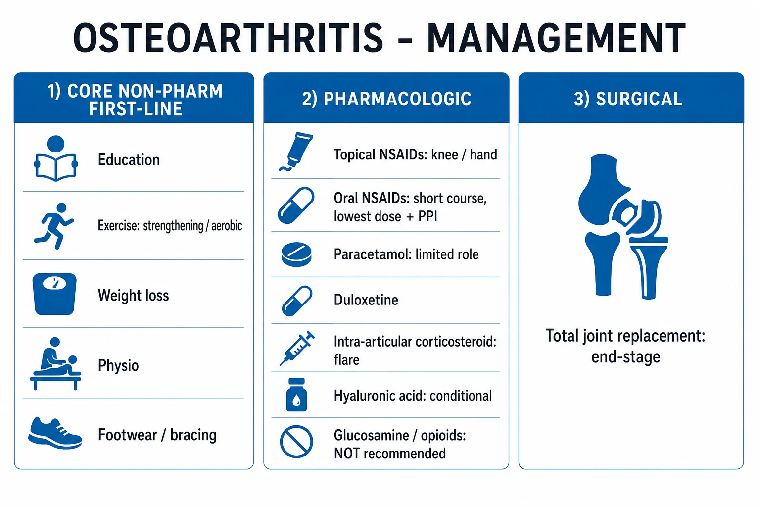
Management of OA is staged, multidisciplinary, individualised, and — critically — built on the core non-pharmacological measures that are first-line AND disease-modifying, not adjuncts. Every patient, regardless of severity, begins here. The four core measures are: education and self-management, aerobic and strengthening exercise (especially quadriceps strengthening for knee OA — strongly evidence-based), weight loss (targetting 5 kg in obese patients, which reduces knee-OA symptom burden by roughly 50%), and biomechanical aids (appropriate footwear, lateral-wedge insoles for medial compartment OA, bracing, walking aids). Physiotherapy delivers structured exercise, manual therapy, taping and modalities; occupational therapy delivers joint protection, pacing, and aids for hand OA (splinting the first CMC).[1][2]
Stepwise management of knee OA
**Step 1 — Core non-pharmacological (every patient)**: education, aerobic and strengthening exercise (especially quadriceps), weight loss, biomechanical aids (footwear, lateral-wedge insole), self-management. Disease-modifying.
**Step 2 — Topical pharmacological**: topical NSAID (e.g. diclofenac 1% gel applied four times daily) is first-line for knee and hand OA — effective with the best safety profile.
**Step 3 — Oral pharmacological (shortest time, lowest dose)**: oral NSAID at lowest effective dose with a PPI (e.g. naproxen 500 mg twice daily + omeprazole 20 mg once daily, or celecoxib 200 mg twice daily). Duloxetine 30 to 60 mg once daily for chronic musculoskeletal pain. Capsaicin 0.025% cream for hand/knee OA.
**Step 4 — Intra-articular therapy**: corticosteroid (triamcinolone 40 mg) for a clearly defined flare; benefit weeks. Hyaluronic acid is NOT recommended (NICE/ACR against).
**Step 5 — Surgery**: total joint arthroplasty for end-stage refractory pain and functional loss; osteotomy for unicompartmental knee disease in younger active patients. Arthroscopy has NO role in uncomplicated OA.
The pharmacological ladder, with agent, dose, route, timing and rationale:[2]
- Topical NSAIDs — e.g. diclofenac 1% gel (2 to 4 g applied four times daily) or ketoprofen 2.5% gel — are strongly recommended first-line for knee and hand OA because they deliver similar symptomatic benefit to oral NSAIDs with a fraction of the systemic exposure and a far better gastrointestinal, renal and cardiovascular safety profile.[2]
- Oral NSAIDs — e.g. naproxen 500 mg twice daily, ibuprofen 400 to 600 mg three times daily, celecoxib 200 mg twice daily — are the next step, used at the lowest effective dose for the shortest time, always co-prescribed with a proton-pump inhibitor (e.g. omeprazole 20 mg once daily) for patients over 45 or with prior GI history. The cardiovascular risk hierarchy is roughly: naproxen (lowest), then ibuprofen, then diclofenac and the COX-2 inhibitors (highest); celecoxib at low dose has the best GI profile but a CV risk similar to diclofenac. Avoid in CKD (eGFR under 30), heart failure, active peptic ulcer, and bleeding diathesis.[2][4]
- Capsaicin 0.025% to 0.075% cream applied four times daily is conditionally recommended by the ACR for hand and knee OA — useful in patients who cannot take NSAIDs, though local burning limits adherence.[2]
- Duloxetine 30 to 60 mg once daily (a serotonin–noradrenaline reuptake inhibitor) is conditionally recommended by the ACR for chronic musculoskeletal pain of OA, particularly in patients with central sensitisation, coexistent depression, or those who cannot take NSAIDs.[2]
- Intra-articular corticosteroid — e.g. triamcinolone acetonide 40 mg or methylprednisolone acetate 40 mg for a large joint — gives short-term (4 to 8 week) benefit, especially for a clear flare with effusion; limited to 3 to 4 injections per joint per year.[1][2]
- Paracetamol (acetaminophen) — historically first-line — is no longer recommended as first-line by NICE (2022) and is conditionally recommended against by the ACR 2019 because the Roberts 2016 systematic review demonstrated minimal efficacy and a dose-dependent increase in mortality, gastrointestinal bleeding, and cardiovascular events at chronic high doses (3 g/day or more). It retains a limited role as short-term add-on in patients who cannot take NSAIDs.[5][2]
- Tramadol is conditionally recommended by the ACR for refractory pain; strong opioids, oxycodone, fentanyl and morphine should be avoided (recommendation against) because the risks (dependence, falls, fracture, constipation, respiratory depression in the elderly) outweigh benefit.[2]
Naproxen / Ibuprofen / Celecoxib
Triamcinolone acetonide / Methylprednisolone acetate
Agents recommended AGAINST (do not use in OA): paracetamol as first-line (NICE 2022, ACR 2019), intra-articular hyaluronic acid (NICE and ACR recommend against — modern meta-analyses show effect indistinguishable from placebo and an inflated placebo response from the injection itself), glucosamine and chondroitin (no benefit over placebo in large RCTs; ACR recommends against), strong opioids (dependence, falls, mortality), stem cell therapy, and platelet-rich plasma (insufficient evidence, do not use outside a trial).[2][4][5]
Surgical management is reserved for end-stage OA with refractory pain and functional loss despite optimal conservative management. Total hip arthroplasty (THA) and total knee arthroplasty (TKA) are among the most cost-effective interventions in medicine, transforming quality of life in appropriately selected patients; implant survival exceeds 90% at 10 to 15 years for both. High tibial osteotomy has a role in younger, active patients with isolated medial compartment knee OA and varus malalignment (shifting the mechanical axis), delaying arthroplasty. Unicompartmental knee replacement is appropriate for single-compartment disease. Arthroscopic lavage and debridement have NO role in uncomplicated OA — multiple RCTs (including the Moseley sham-controlled trial) show no benefit over placebo; arthroscopy is reserved for mechanical locking from a clearly displaced meniscal tear in an otherwise preserved joint. Joint fusion (arthrodesis) is a salvage option for the ankle, first MTP, and wrist in younger high-demand patients. Treat the patient (symptoms and function), not the X-ray.[1][2]
Specific Subtypes & Scenarios
Generalised nodal OA (the Heberden phenotype) is the classic presentation of hand OA in post-menopausal women, with symmetric Heberden nodes at the DIP, Bouchard nodes at the PIP, and first CMC involvement, strongly familial. It evolves in episodic flares (warm, tender, red nodes) that settle to leave hard, bony, often painless swellings; functional impairment is usually modest once the nodes mature. Erosive (inflammatory) OA is an aggressive subset marked by central erosions in the DIP/PIP ("gull-wing" deformity on X-ray), more pronounced inflammatory symptoms, and a self-limiting course over years; it can mimic RA but the MCP and wrists are spared and inflammatory markers are normal. Management mirrors nodal OA with topical NSAIDs and joint protection.[1]
Post-traumatic OA is the commonest form of secondary OA in younger patients — a prior meniscectomy, ACL tear or intra-articular fracture accelerates cartilage failure by altering joint mechanics. It is often unicompartmental (e.g. the lateral compartment after a lateral meniscectomy) and may benefit from osteotomy to offload the damaged compartment. Developmental hip dysplasia (DDH) and femoroacetabular impingement (FAI) cause anterior/groin hip OA in young and middle-aged adults, often with a positive impingement test (Faber, Fadir); a young patient with hip OA mandates hip imaging (hip dysplasia is markedly more common in women and in breech presentations). Slipped capital femoral epiphysis and Legg-Calvé-Perthes in childhood leave a malformed femoral head that develops OA in middle age.[1]
Haemochromatosis arthropathy deserves separate emphasis because it is a treatable cause of "OA" that is frequently missed: it classically affects the second and third MCP joints (a site primary OA spares) and may involve the ankles and wrists, with chondrocalcinosis. A young or middle-aged man with MCP OA, fatigue, bronze skin, diabetes or liver disease should have iron studies checked (transferrin saturation over 45%, raised ferritin), and treatment by venesection can prevent progression to cirrhosis, cardiomyopathy and diabetes — though the arthritis, once established, is largely irreversible.[1]
Charcot (neuropathic) arthropathy — diabetes, tabes dorsalis, syringomyelia, leprosy — produces a destructive, relatively painless arthropathy with preserved sensation to pain, joint effusion, bony debris and disorganisation on X-ray; management is offloading and bracing, not arthroplasty (high failure rate). CPPD-deposition disease (pseudogout) frequently co-exists with OA, especially of the knee and wrist, and presents with acute attacks superimposed on chronic structural change.[1]
Complications & Pitfalls
The complications of OA and its management fall into three groups. Disease-related: progressive deformity (varus/valgus, fixed flexion, squaring), loss of range and mobility, muscle wasting and weakness, falls and fracture risk in the elderly (knee and hip OA impair balance), depression and social isolation from chronic pain and lost independence, and spinal stenosis from facet OA and ligamentous hypertrophy. Drug-related: oral NSAID toxicity — gastrointestinal (peptic ulcer, bleeding and perforation — the commonest serious NSAID adverse effect, dose and age dependent), renal (sodium and water retention, acute kidney injury, hyperkalaemia, acceleration of chronic kidney disease, especially with ACE inhibitors and diuretics), cardiovascular (hypertension, heart failure exacerbation, increased risk of myocardial infarction and stroke, especially with diclofenac and high-dose COX-2 inhibitors), and hepatic (transaminase rise). Opioid-related: falls, fracture, constipation, respiratory depression, dependence and overdose — the reason strong opioids are recommended against. Surgical: periprosthetic infection (1 to 2%), deep vein thrombosis and pulmonary embolism (mechanical and pharmacological prophylaxis required), dislocation (hip), aseptic loosening and osteolysis (the long-term failure mode requiring revision), limb-length discrepancy, stiffness and arthrofibrosis, and fracture at surgery.[1][2]
The classic diagnostic errors worth naming: (1) falsely attributing an acute hot swollen knee to "an OA flare" without aspirating — the missed septic joint is the catastrophic error; (2) missing avascular necrosis in a patient with sudden new hip/knee pain and a steroid or alcohol history (MRI is diagnostic when X-ray is normal); (3) missing haemochromatosis when "OA" involves the second and third MCP; (4) over-treating based on the X-ray rather than the patient (operating on a film, not a symptom); (5) prescribing long-term oral NSAIDs in the elderly without a PPI, renal monitoring or review. The recurring safety lesson — aspirate any acute hot joint — deserves repeating.[1]
Prognosis & Disposition
OA is progressive but not life-threatening — it does not shorten life, but it is a major cause of years lived with disability, especially in the elderly and the obese. The course is highly variable: some patients progress rapidly to bone-on-bone change over a few years, while others remain stable for decades with the same radiographic grade. Predictors of progression at the knee include obesity, malalignment (varus/valgus concentrating load), quadriceps weakness, presence of bone marrow lesions on MRI, and large effusions. Predictors of functional decline include pain, depression, comorbidity, and social isolation.[1]
Total hip and total knee arthroplasty transform outcomes in appropriately selected patients: 90% or more of patients report substantial pain relief and functional improvement, and implant survival is over 90% at 10 to 15 years; revision rates rise with younger age, obesity, and high-impact activity. The disposition after a clinic visit is GP and physiotherapy-led conservative management for most patients, with rheumatology referral for diagnostic uncertainty or suspected inflammatory/secondary OA, and orthopaedic referral for end-stage refractory pain and functional loss. The safety-net for every patient is the warning to return urgently with any sudden change — new night or rest pain, a hot swollen joint, systemic features, or neurological symptoms — that should prompt reassessment for fracture, AVN, sepsis or crystal arthritis.[1][2]
Special Populations
The elderly with comorbidity are the largest OA population. Prefer topical NSAIDs (knee and hand) over oral to avoid gastrointestinal, cardiovascular and renal harm; minimise polypharmacy; screen for falls and frailty; treat depression and insomnia that amplify pain. Duloxetine is useful when oral NSAIDs are contraindicated. Joint replacement is well tolerated even into the ninth decade in medically fit patients — age alone is not a contraindication.[2]
Obesity and knee OA — the single largest preventable driver of knee OA. Weight loss of 5 kg reduces knee-OA symptom burden by roughly 50%, and bariatric surgery in severe obesity produces sustained weight loss and substantial improvements in pain and function. Quadriceps strengthening is the most evidence-based exercise. The message for the patient is that weight loss and exercise are disease-modifying, not merely symptom relief — this reframing drives adherence.[1]
Younger patients with OA should prompt a search for a secondary cause: post-traumatic (meniscectomy, ACL), developmental (DDH, FAI, SCFE), metabolic (haemochromatosis, Wilson), neuropathic (Charcot), or crystal (CPPD). In the young, joint-preserving surgery (osteotomy, hip arthroscopy for FAI) is preferred to delay arthroplasty, because revision rates of arthroplasty are higher in younger, high-demand patients.[1]
Pregnancy — OA is uncommon in women of reproductive age, but when symptomatic, prefer topical NSAIDs (avoid oral NSAIDs in the third trimester — premature closure of the ductus arteriosus and oligohydramnios; paracetamol remains acceptable short-term despite the Roberts review, which was based on chronic high-dose use). Intra-articular corticosteroid (single injection) is acceptable in pregnancy for a disabling flare. Avoid duloxetine and opioids where possible.[2][5]
Anticoagulated patients — joint injection carries a small bleeding risk; do not stop warfarin if INR is in range (under 3.0) per international consensus; direct oral anticoagulants can be paused for one dose for high-risk injections. Aspiration of a hot joint in an anticoagulated patient should still be done — sepsis risk outweighs bleeding risk.[1]
Evidence, Guidelines & Regional Differences
OA is a global disease, but its drugs, thresholds and ladder are regional — name the guideline you quote. The three dominant contemporary guidelines are NICE NG226 (2022) — Osteoarthritis in over 16s, the 2019 American College of Rheumatology/Arthritis Foundation Guideline (Kolasinski) for hand, hip and knee OA, and the OARSI 2019 Non-surgical Management Guideline (Bannuru).[2][4]
NICE NG226 (2022)
- Core non-pharm first-line for ALL: education, exercise, weight loss
- Topical NSAIDs first-line for knee and hand OA
- Paracetamol NOT recommended (weak analgesic, harms at chronic high dose)
- Strongly AGAINST hyaluronic acid, glucosamine, chondroitin, stem cell, PRP
- Oral NSAIDs at lowest dose, shortest time, with PPI; consider duloxetine
ACR/AF 2019 (Kolasinski)
- Strongly FOR: exercise, weight loss, self-efficacy, tai chi, cane, tibiofemoral bracing
- Strongly FOR topical NSAIDs for knee; conditionally FOR capsaicin, oral NSAIDs, intra-articular steroid
- Conditionally FOR duloxetine and tramadol; conditionally AGAINST hyaluronic acid, PRP
- Strongly AGAINST glucosamine, chondroitin, fish oil, strong opioids; conditionally AGAINST paracetamol
- Conditional recommendations reflect weak evidence base, individualise
OARSI 2019 (Bannuru)
- Core treatments (exercise, weight loss) strongly recommended for all joints
- Topical NSAIDs strongly recommended for knee OA
- Conditionally recommends intra-articular steroid for knee/hip; conditional on hyaluronic acid
- Conditionally against paracetamol and glucosamine for knee
- Most permissive on hyaluronic acid among the three guidelines
Where they agree: the four core non-pharmacological measures are first-line for every patient everywhere; topical NSAIDs are first-line pharmacological therapy for knee and hand OA; paracetamol is no longer first-line (the Roberts 2016 review changed practice worldwide by showing minimal efficacy and dose-dependent harm — mortality, GI bleeding, CV events at 3 g/day or more); hyaluronic acid is not recommended (NICE strongly against, ACR conditionally against, OARSI most permissive but still conditional); glucosamine, chondroitin, fish oil, strong opioids, stem cell and PRP are recommended against.[2][4][5]
The NICE (UK), ACR (US) and OARSI (international) guidelines converge on the core-plus-topical-NSAID algorithm and on no role for paracetamol first-line or for hyaluronic acid. The largest divergence is on hyaluronic acid (OARSI conditional for, ACR and NICE against) and on the precise strength of duloxetine and tramadol (ACR conditional for). Indian practice emphasises knee OA and squatting/cross-legged posture (a cultural driver of medial compartment OA), late presentation with severe deformity, and limited access to arthroplasty — hence a relatively heavier reliance on core measures and intra-articular steroid.[1]
Landmark trials that shaped modern OA management: the Moseley 2002 sham-controlled RCT showed arthroscopic lavage and debridement were no better than placebo for uncomplicated knee OA — ending the routine use of arthroscopy. The Roberts 2016 systematic review (PMID 25732175) changed paracetamol from first-line to conditionally against, demonstrating dose-dependent mortality, GI and CV harm at chronic high doses. The GAIT trial (Clegg 2006) showed glucosamine and chondroitin were no better than placebo for overall knee OA pain. The 2018 BMJ individual patient data meta-analysis (Hunter cohort) confirmed topical NSAIDs match oral NSAIDs for knee and hand OA with a fraction of the systemic risk.[2][4][5]
Exam Pearls & High-Yield Minutiae
- OA = mechanically driven cartilage degeneration. Use-related pain, morning stiffness under 30 minutes, crepitus, bony swelling. No systemic features, normal ESR/CRP.[1]
- Joints: knee, hip, hands (DIP Heberden, PIP Bouchard, first CMC; MCP SPARED), spine, first MTP. Asymmetric.
- Risks: age, obesity, female sex, prior injury, occupation, genetics, malalignment, developmental dysplasia.
- X-ray = LOSS: Loss of joint space, Osteophytes, Subchondral sclerosis, Subchondral cysts. Osteophytes are the most specific sign.
- Pathophysiology: stressed chondrocytes release MMP and ADAMTS → degrade type II collagen and aggrecan → cartilage fibrillation → subchondral sclerosis, cysts, osteophytes + low-grade synovitis.
- Distinguish from RA: OA is mechanical, brief stiffness, DIP involved, MCP spared, normal ESR/CRP.
- Core first-line: exercise (especially quadriceps) and weight loss (target 5 kg) are disease-modifying — not just symptom relief.
- Pharmacologic: topical NSAIDs (knee/hand) first; oral NSAIDs short with a PPI (naproxen 500 mg BD lowest CV risk); intra-articular steroid (triamcinolone 40 mg) for a flare; duloxetine 30 to 60 mg OD for chronic pain; joint replacement for end-stage.
- Paracetamol is NO LONGER first-line (NICE 2022, ACR 2019) — minimal efficacy, dose-dependent harm at 3 g/day or more.
- Avoid: hyaluronic acid, glucosamine/chondroitin, strong opioids, stem cell, PRP. No arthroscopy in uncomplicated OA.
- X-ray severity does NOT correlate with symptoms — treat the patient, not the film.
- New night/rest pain in an OA joint — consider fracture, avascular necrosis, or septic/crystal arthritis; aspirate a hot joint.
- Haemochromatosis causes OA of the second and third MCP — check iron studies in atypical hand OA; venesection prevents systemic disease.
- Synovial fluid: OA WBC under 2,000 (non-inflammatory); gout under 50,000 with needle crystals; sepsis over 50,000 with positive Gram stain.
- Synovial fluid crystal microscopy: gout = negatively birefringent needle; CPPD = positively birefringent rhomboid.
Quick check — the joint that is characteristically SPARED in hand OA
The metacarpophalangeal (MCP) joints are spared in OA. The DIP (Heberden), PIP (Bouchard) and first CMC are involved. MCP involvement in "OA" should prompt a search for haemochromatosis (especially second/third MCP), rheumatoid arthritis, or CPPD.[1]
Quick check — the single most specific radiographic feature of OA
Osteophytes (new bone at the joint margins) are the most specific radiographic feature of OA — more specific than joint-space narrowing or subchondral sclerosis. The full radiographic quartet is encoded in the LOSS mnemonic.[1]
Red Flags
Exam application bank (NEET-PG / INICET)
One-line answer
Osteoarthritis (OA) is the commonest joint disorder — a degenerative, mechanically driven arthritis of articular (hyaline) cartilage with bony remodelling (osteophytes, subchondral sclerosis and cysts), meniscal damage and low-grade synovial inflammation. It predominantly affects the knees, hips, hands (DIP — Heberden nodes, PIP — Bouchard nodes, first CMC) and spine. Risk factors are age, obesity, female sex, prior joint injury, repetitive occupational stress and genetics. It presents with use-related joint pain (worse with activity, better with rest, brief morning stiffness under 30 minutes), functional limitation, crepitus and bony swelling, with late deformity. Diagnosis is clinical, supported by X-ray (LOSS — joint-space narrowing, osteophytes, subchondral sclerosis and cysts), though imaging severity correlates poorly with symptoms. Management is built on education, exercise and we
Worked stems (answer without another resource)
Stem 1 — Classic presentation. Map symptoms to mechanism; name the first investigation and first treatment step with dose/route if drug therapy is standard. [1]
Stem 2 — Unstable / complicated. List red flags that force immediate resuscitation, theatre, ICU, antidote, or reperfusion — and what you do in the first 15 minutes. [1]
Stem 3 — Atypical group. Elderly, pregnancy, child, or immunocompromised: how presentation and thresholds change. [1]
Stem 4 — Differential trap. Name the three closest mimics and one discriminator for each. [1]
Stem 5 — Disposition. Who goes home with safety-netting, who is admitted, who needs HDU/ICU/theatre, and what follow-up is mandatory. [1]
Rapid viva checklist
- Definition + classification
- Pathophysiology chain
- Bedside signs / criteria
- Score with exact components (if any)
- Emergency bundle
- Definitive therapy with doses
- Complications of disease and of treatment
- Special populations
- Guideline/trial name if classic
- Three exam traps
Coverage self-check
If you cannot answer any stem above from this page alone, re-read the matching section — the page is intended to be self-sufficient for final-prof and NEET-PG/INICET questions on Osteoarthritis.
References
- [1]Hunter DJ, Bierma-Zeinstra S. Osteoarthritis Lancet, 2019.PMID 31034380
- [2]Kolasinski SL, Neogi T, Hochberg MC, et al. 2019 American College of Rheumatology/Arthritis Foundation Guideline for the Management of Osteoarthritis of the Hand, Hip, and Knee Arthritis Care Res (Hoboken), 2020.PMID 31908149
- [3]Altman R, Alarcon G, Appelrouth D, et al. The American College of Rheumatology criteria for the classification and reporting of osteoarthritis of the hand Arthritis Rheum, 1990.PMID 2242058
- [4]Bannuru RR, Osani MC, Vaysbrot EE, et al. OARSI guidelines for the non-surgical management of knee, hip, and polyarticular osteoarthritis Osteoarthritis Cartilage, 2019.PMID 31278997
- [5]Roberts E, Delgado Nunes V, Buckner S, et al. Paracetamol: not as safe as we thought? A systematic literature review of observational studies Ann Rheum Dis, 2016.PMID 25732175