Rheumatology · General Medicine
Osteoporosis
Also known as Osteoporosis · Osteopaenia · Low bone mineral density · Fragility fracture · Postmenopausal bone loss
Osteoporosis is a progressive systemic skeletal disease of low bone mass and microarchitectural deterioration of bone tissue, leading to increased bone fragility and a consequent increase in fracture risk. Operationally (WHO) it is defined by a DEXA T-score of minus 2.5 or less at the femoral neck, total hip or lumbar spine; a low-trauma (fragility) fracture establishes the diagnosis regardless of the T-score. It is the commonest metabolic bone disease of older adults and the underlying cause of most fragility fractures (low-trauma — fall from standing height or less) of the vertebra, hip, distal forearm (Colles) and proximal humerus. Commonest in postmenopausal women and older adults; secondary causes include glucocorticoids, hypogonadism, hyperthyroidism, hyperparathyroidism, CKD, malabsorption, chronic liver disease, smoking, alcohol, low BMI and immobility. Often silent until a fragility fracture. Diagnosis: DEXA T-score plus FRAX 10-year fracture probability. Treat: lifestyle (weight-bearing exercise, calcium 1000 to 1200 mg, vitamin D 800 to 1000 IU, smoking cessation, fall prevention) + bisphosphonates first-line (alendronate weekly, zoledronate IV annually), denosumab SC 6-monthly; anabolic agents (teriparatide, romosozumab) for very-high-risk disease; drug holiday after 3 to 5 years.
On this page & tools
Your progress
Saved locally on this device.
Exam tags
Red flags
Overview & Definition
Osteoporosis literally means "porous bone". The 1993 and 2001 Consensus Development Conference definitions, still current, describe it as "a progressive systemic skeletal disease characterised by low bone mass and microarchitectural deterioration of bone tissue, leading to increased bone fragility and a consequent increase in fracture risk." Bone strength — the property that resists fracture — reflects two integrated features: (a) bone mineral density (the amount of mineral per unit volume) and (b) bone quality (the architecture, turnover, damage accumulation, and mineralisation). Bone mineral density (BMD) is the easiest to measure and explains roughly 70 percent of bone strength; the rest depends on microarchitecture, which DEXA cannot directly assess.[1]
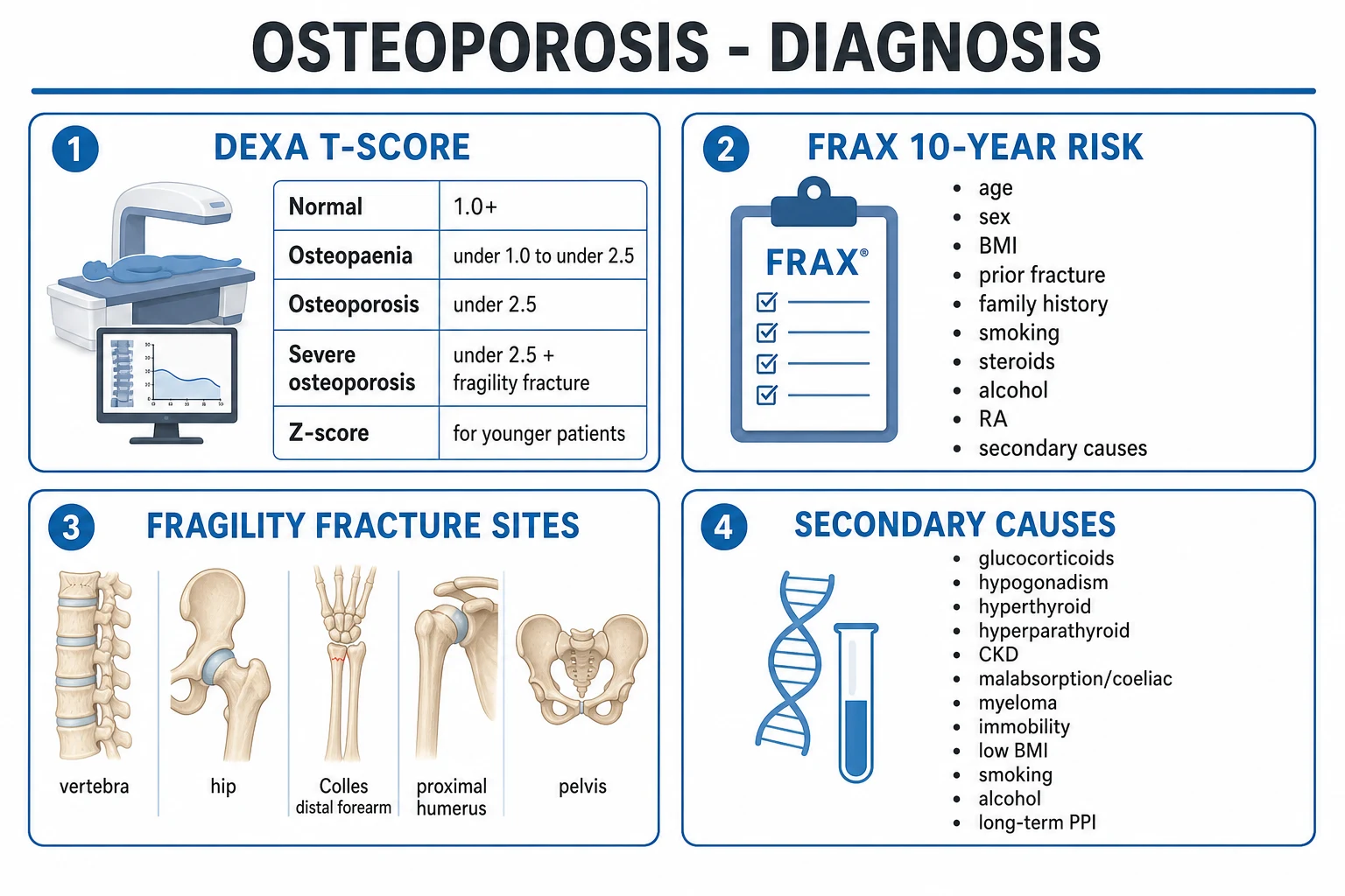
The operational WHO definition, used worldwide and at NEET-PG/INICET, is DEXA-based: in a postmenopausal woman or a man aged 50 years or older, the diagnosis is established by a T-score of minus 2.5 or less at the femoral neck, total hip or lumbar spine. The T-score is the number of standard deviations the patient's BMD lies above or below the young-adult healthy female reference mean. A T-score of minus 2.5 means the BMD is 2.5 standard deviations below that mean.[1][2]
The clinical skill in osteoporosis is prevention and case-finding: identify and treat at-risk people before the first fracture, because a prior fragility fracture is the strongest single predictor of the next one. The disease is largely silent until fracture, so screening and risk-stratification with DEXA and FRAX are the workhorses. Bisphosphonates are first-line therapy for the great majority; anabolic agents (teriparatide, romosozumab) are reserved for very-high-risk disease (multiple fractures or a very low T-score).[2]
Classification
Osteoporosis is classified by (a) the underlying cause (primary versus secondary) and by (b) the DEXA T-score, which also distinguishes normal, osteopaenia, osteoporosis, and severe (established) osteoporosis (T-score of minus 2.5 or less plus a fragility fracture).[1]
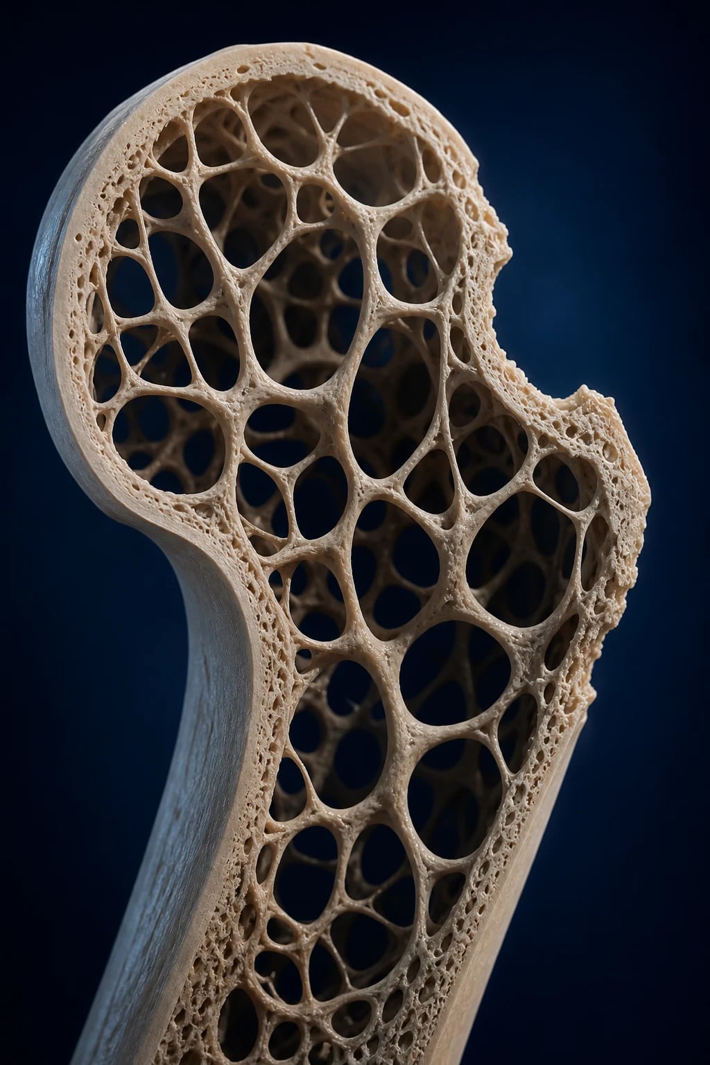
PRIMARY OSTEOPOROSIS — the two classical types
RIGS
Oestrogen-deficiency driven; accelerated trabecular bone loss; women aged 50 to 70; vertebral and Colles fractures predominate; high turnover
Age-related; men and women over 70; cortical and trabecular loss; low turnover; hip and pelvic fractures predominate
Highest secondary risk; mixed mechanism; needs prophylaxis from steroid outset
Hypogonadism, hyperthyroid, hyperparathyroid, CKD, malabsorption/coeliac, myeloma, RA, low BMI, smoking, immobility, anti-epileptics, PPIs
The classical Riggs and Melton clinical classification, although superseded in mechanistic thinking, is still useful at the viva: [1]
- Primary Type 1 (postmenopausal) osteoporosis — driven by oestrogen deficiency after the menopause, producing accelerated, high-turnover trabecular bone loss. Predominantly affects women aged 50 to 70. Classical fractures: vertebral wedge/compression and Colles (distal radial).
- Primary Type 2 (senile) osteoporosis — age-related, arising from declining osteoblast formation and osteocyte viability, producing cortical porosity and low-turnover loss in both men and women after about 70. Classical fractures: hip (femoral neck and intertrochanteric) and pelvic. [1]
The mechanistic distinction is useful because it predicts both the fracture pattern and the targeted therapy: high-turnover postmenopausal bone loss responds briskly to antiresorptives (bisphosphonates, denosumab), while very low T-score or multiple fractures favour an anabolic-first sequence.[1]
Epidemiology & Risk Factors
Osteoporosis is the commonest metabolic bone disease, affecting an estimated 200 million people worldwide. Approximately one in three women and one in five men aged over 50 years will sustain a fragility fracture in their remaining lifetime. The female:male ratio for vertebral and hip fracture is roughly 2:1, reflecting the rapid loss of trabecular bone that follows the menopause, but men have a worse post-hip-fracture mortality. The global burden is rising rapidly as the population ages; the annual number of hip fractures globally is projected to rise from about 1.6 million in 1990 to over 6 million by 2050.[1]
Osteoporosis — high-yield numbers
Risk factors cluster into BMD-dependent (factors that lower bone density) and BMD-independent (factors that increase fracture risk at any given BMD — age, prior fracture, fall risk): [1]
- Unmodifiable — age (the strongest), female sex, Caucasian or Asian ethnicity, family history of hip fracture (parental), premature menopause (under 45), short stature, slender frame (low peak bone mass).
- Hormonal — postmenopausal oestrogen deficiency, hypogonadism in men (low testosterone), hyperthyroidism, primary or secondary hyperparathyroidism, hypercortisolism (Cushing), hyperprolactinaemia, pregnancy and lactation (transient).
- Drug-induced — glucocorticoids (the dominant cause; above 5 mg prednisolone daily for over 3 months), aromatase inhibitors (breast cancer), androgen-deprivation therapy (prostate cancer), excess levothyroxine, antiepileptics (phenytoin, carbamazepine, valproate — vitamin D metabolism), proton-pump inhibitors (chronic — reduced calcium absorption), heparin (long term), loop diuretics, SSRIs, thiazolidinediones, calcineurin inhibitors, transplant immunosuppression.
- Nutritional and lifestyle — low calcium and vitamin D intake, malabsorption (coeliac disease, inflammatory bowel disease, post-bariatric surgery, chronic pancreatitis), low BMI (under 21), smoking (reduces osteoblast activity and oestrogen), excess alcohol (over 3 units/day; direct osteoblast toxicity, falls), high salt/protein (calciuria), excessive caffeine.
- Chronic disease — CKD (renal osteodystrophy; low active vitamin D), chronic liver disease, rheumatoid arthritis and other inflammatory arthritides (cytokine-driven bone loss plus glucocorticoid exposure), diabetes mellitus (especially type 1; type 2 paradoxically despite higher BMI), COPD, chronic heart failure, organ transplant, HIV, anorexia nervosa, haemophilia, sickle cell, beta-thalassaemia, mastocytosis, glycogen storage diseases.
- Mechanical — immobilisation (paraplegia, stroke, prolonged bed rest, spaceflight), low physical activity, regional (complex regional pain syndrome, transient regional osteoporosis). [1]
Pathophysiology
Bone is a dynamic tissue that is continually remodelled throughout life in tightly coupled basic multicellular units (BMUs). Three cells cooperate: [1]
- Osteoclasts — multinucleated, bone-resorbing cells of monocyte/macrophage lineage.
- Osteoblasts — bone-forming cells of mesenchymal origin that lay down osteoid and mineralise it.
- Osteocytes — former osteoblasts entombed within lacunae in the mineralised matrix; the mechanosensors of bone that detect loading and microdamage and orchestrate remodelling. [1]
Coupled remodelling proceeds: osteocyte apoptosis at sites of microdamage triggers osteoclast recruitment and resorption of a packet of old bone, which is then reversal, followed by osteoblast recruitment and bone formation to refill the cavity. In health the volume of bone formed equals the volume resorbed (zero net balance). [1]
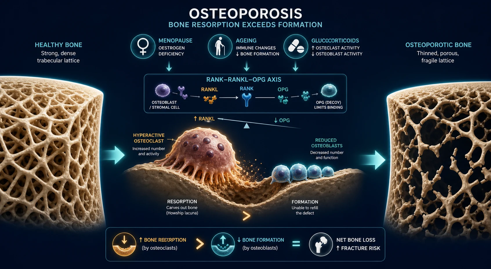
The RANK-RANKL-OPG axis is the master regulator of osteoclastogenesis and the target of modern antiresorptive therapy: [1]
- RANK (Receptor Activator of NF-kB) is expressed on the surface of osteoclast precursors and mature osteoclasts.
- RANKL (RANK Ligand) is a membrane-bound and soluble protein produced mainly by osteoblasts and osteocytes (and by activated T cells in inflammation). Binding of RANKL to RANK drives osteoclast differentiation, activation and survival.
- OPG (osteoprotegerin) is a soluble decoy receptor produced by osteoblasts and other cells that binds RANKL and prevents it activating RANK, thereby suppressing osteoclastogenesis.
- Oestrogen promotes OPG and suppresses RANKL; glucocorticoids suppress OPG and promote RANKL; PTH intermittently stimulates osteoblasts but continuously upregulates RANKL (hence teriparatide anabolic, hyperparathyroidism catabolic).
- Denosumab is a fully human monoclonal antibody against RANKL — given 60 mg subcutaneously every 6 months, it produces rapid, deep, reversible suppression of osteoclast activity.[4]
The Wnt-LRP5/6-sclerostin axis governs osteoblast activity and bone formation, and is the target of the newer anabolic agents: [1]
- Wnt signalling through LRP5/6 co-receptors on osteoblast-lineage cells drives their proliferation, differentiation and bone formation.
- Sclerostin, a glycoprotein produced almost exclusively by mature osteocytes, binds LRP5/6 and inhibits Wnt signalling, suppressing bone formation.
- Romosozumab is a humanised monoclonal antibody against sclerostin — given monthly for 12 months, it produces rapid, large increases in bone formation (anabolic) while modestly reducing resorption — a dual effect.[5][6]
Pathophysiology by type: [1]
- Postmenopausal (Type 1, high turnover) — oestrogen withdrawal removes its tonic restraint on osteoclastogenesis. RANKL rises, OPG falls; osteoclasts are recruited, activated and live longer, producing deep resorption lacunae that perforate trabeculae. Trabecular plates lose struts, become rods, and the connections ("nodes") that confer strength are lost — this is why the vertebra (predominantly trabecular) and distal radius fail first.
- Senile (Type 2, low turnover) — ageing is associated with a decline in osteoblast number and function (mesenchymal stem cells preferentially differentiate into adipocytes rather than osteoblasts), sclerostin accumulation, and accumulation of apoptotic osteocytes. Remodelling becomes inefficient at repairing microdamage. Cortical bone becomes porous, the femoral neck thins, and hip fractures predominate.
- Glucocorticoid-induced — glucocorticoids cause direct osteoblast and osteocyte apoptosis, decrease osteoid formation, increase osteoclast survival via RANKL/OPG tilt, reduce intestinal calcium absorption and increase renal calcium loss (secondary hyperparathyroidism), and suppress gonadotropins (hypogonadism). Fracture risk rises rapidly, within 3 to 6 months of starting therapy and at higher BMD than for postmenopausal osteoporosis. [1]
Secondary hyperparathyroidism from chronic vitamin D deficiency (common in the elderly and housebound) drives chronic low-grade PTH-mediated resorption and contributes to both cortical and trabecular loss. [1]
Clinical Presentation
Osteoporosis is silent until it complicates — the first manifestation is usually a fragility fracture. Case-finding depends on screening and risk stratification, not waiting for symptoms.[1]
Vertebral compression fracture — the commonest osteoporotic fracture and often asymptomatic (only about one in three comes to clinical attention). When symptomatic it presents with: [1]
- Acute, severe thoracic or lumbar back pain after minimal trauma (lifting, bending, coughing), occasionally radicular.
- Loss of height (over 4 cm prompts vertebral imaging), progressive thoracic kyphosis ("dowager hump"), protuberant abdomen (from loss of lumbar lordosis and abdominal muscle tone).
- Late complications: restrictive respiratory impairment (reduced vital capacity from kyphosis), early satiety and weight loss (gastric compression), reduced exercise tolerance and depression.
- Most heal within 6 to 8 weeks; chronic pain suggests multifracture disease. [1]
Hip fracture — the most serious osteoporotic fracture. Typically a low-trauma fall from standing height or less in an older adult, with: [1]
- Pain in the groin or lateral hip, inability to weight-bear, shortened and externally rotated leg (in displaced femoral-neck and intertrochanteric fractures).
- Surgical emergency — orthogeriatric co-management, prompt internal fixation or arthroplasty.
- One-year mortality 20 to 30 percent; only about 40 percent of survivors regain their pre-fracture level of independence; many require institutional care. [1]
Distal radial (Colles) fracture — typically in younger postmenopausal women (under 65) who fall on an outstretched hand. Produces the dinner-fork deformity (dorsal displacement), pain, swelling and limited wrist movement. Treated by closed reduction and below-elbow cast, occasionally K-wires or volar locking plate. [1]
Proximal humeral fracture — fall on outstretched hand or directly onto the shoulder; usually impacted or surgical-neck fractures. Most treated conservatively in a sling. [1]
Other fragility fractures — pelvis, proximal femur (subtrochanteric — bisphosphonate-related atypical femoral fracture (AFF) has a distinct prodrome and radiographic pattern), distal femur, ribs, sacral insufficiency. [1]
Atypical presentations: [1]
- Younger woman (under 50) — consider pregnancy-associated osteoporosis, anorexia nervosa with the female-athlete triad, premature ovarian failure, coeliac disease, Cushing, mastocytosis, connective-tissue disease, prolonged depot-medroxyprogesterone or GnRH analogues, anti-epileptic drugs, glucocorticoids.
- Man of any age — always search a secondary cause: hypogonadism (measure serum testosterone), hyperthyroidism, hyperparathyroidism, myeloma, glucocorticoids, alcoholism, haemochromatosis, hypogonadotropic hypogonadism.
- Elderly housebound patient — vitamin D deficiency with secondary hyperparathyroidism, immobility, multiple comorbidities and polypharmacy, falls risk. [1]
Differential Diagnosis
The pivotal step is to distinguish generalised osteoporosis from other metabolic bone diseases that share low BMD, and to identify secondary causes that change management. The key discriminator is that in uncomplicated osteoporosis the serum calcium, phosphate and alkaline phosphatase are normal.[1]
Osteoporosis
- Older postmenopausal woman, man over 70, or chronic glucocorticoid user; asymptomatic until fragility fracture
- DEXA: T-score of minus 2.5 or less; vertebra/hip/Colles/humerus fractures
- Serum calcium, phosphate and ALP all NORMAL; vitamin D often low
- FRAX elevated; bone turnover markers may be high (postmenopausal) or low (senile)
- Treat: bisphosphonate first-line, denosumab, anabolic for very-high-risk
Osteomalacia
- Vitamin D deficiency, coeliac, antiepileptics, CKD, X-linked hypophosphataemia, tumour-induced osteomalacia
- Generalised bone pain and tenderness (especially long bones, ribs, pelvis), proximal muscle weakness (waddling gait, difficulty rising from chair)
- LOW calcium, LOW phosphate, HIGH ALP, HIGH PTH (secondary hyperparathyroidism), LOW 25-hydroxy-vitamin D
- Looser zones (pseudofractures) on X-ray; osteoid seams on biopsy; low BMD at DEXA
- Treat the cause: cholecalciferol or calcitriol; oral phosphate in XLH/TIO
Multiple myeloma
- Same age group (over 60); bone pain, fatigue, recurrent infections; CRAB (hypercalcaemia, renal failure, anaemia, lytic bone lesions)
- Punched-out lytic lesions and diffuse osteopaenia; vertebral fractures; not a true BMD problem
- Monoclonal band on serum/urine protein electrophoresis, raised free light chains, serum and urine immunofixation
- Normocytic anaemia, raised creatinine, hypercalcaemia; bone marrow plasma cells over 10 percent
- Treat with chemotherapy, bisphosphonate (or denosumab) for myeloma bone disease
Metastatic bone disease
- History of breast, prostate, lung, thyroid or renal cancer; focal or multifocal bone pain worse at night
- Lytic, sclerotic or mixed lesions (breast and lung lytic; prostate and breast sclerotic); pathologic fractures; spine, pelvis, ribs
- Bone scan hot at metastatic sites; MRI/CT/PET for staging; biopsy to confirm primary
- Hypercalcaemia of malignancy (PTHrP); tumour markers as indicated
- Treat primary; radiotherapy, orthopaedic fixation, denosumab or zoledronate for skeletal events
Primary hyperparathyroidism
- Postmenopausal woman; 'stones, bones, abdominal groans, psychic moans'; nephrolithiasis, peptic ulcer disease, fatigue, depression
- Bone pain; subperiosteal resorption (radial aspect of middle phalanges); salt-and-pepper skull; brown tumours
- HIGH calcium, LOW phosphate, HIGH ALP, HIGH intact PTH; nephrolithiasis
- DEXA shows preferential cortical (distal 1/3 radius) bone loss; osteitis fibrosa cystica in severe disease
- Parathyroidectomy if symptomatic or meeting criteria; cinacalcet if unfit
Paget disease of bone
- Older adult; asymptomatic incidental finding or bone pain, deformity, hearing loss (skull base), increased hat size, chalk-stick fractures
- Markedly RAISED ALP with normal calcium and phosphate; pelvic, spine, skull, long-bone involvement
- X-ray: mixed lytic-sclerotic, expanded, thickened cortex with coarse trabeculae; flame-shaped lytic edge in long bones
- Bone scan markedly hot at pagetic sites; normal PTH and calcium
- Treat symptomatic: zoledronic acid 5 mg IV once; bisphosphonates are first-line
Other considerations: transient regional (migratory) osteoporosis (focal, self-limiting, often hip), reflex sympathetic dystrophy / complex regional pain syndrome type 1 (focal periarticular osteopaenia after minor injury), regional migratory osteoporosis, post-traumatic disuse osteopaenia, and idiopathic juvenile osteoporosis (prepubertal, self-limiting). [1]
Clinical & Bedside Assessment
A focused structural and functional assessment for osteoporosis covers four domains: [1]
-
Anthropometry and skeletal signs:
- Measure height annually using a stadiometer; height loss of 4 cm or more (or any acute loss) prompts vertebral imaging (lateral thoracolumbar spine X-ray or DEXA-based vertebral fracture assessment, VFA).
- Occiput-to-wall distance over 5 cm suggests thoracic kyphosis from vertebral collapse; rib-to-pelvis distance reduced.
- Thoracic kyphosis ("dowager hump"), protuberant abdomen, reduced intercostal spaces. [1]
-
Fall risk assessment — every patient with low BMD should be assessed for fall risk:
- Get-up-and-go test (over 12 seconds indicates increased fall risk), TUG, Romberg, tandem gait, functional reach.
- Identify intrinsic fall-risk factors: visual impairment (cataracts, multifocal glasses), cognitive impairment, depression, orthostatic hypotension, vestibular dysfunction, neuropathy (diabetes, alcohol), muscle weakness (sarcopenia), urinary urgency, syncope (carotid sinus hypersensitivity, vasovagal, arrhythmia).
- Extrinsic factors: polypharmacy (especially sedatives, hypnotics, antipsychotics, anticholinergics, opioids, antihypertensives, vasodilators), environmental hazards (loose rugs, poor lighting, no grab-rails, ill-fitting footwear), pets underfoot. [1]
-
Identify secondary causes:
- Drug history: glucocorticoids, aromatase inhibitors, GnRH agonists/antagonists, antiepileptics, PPIs, loop diuretics, thyroxine excess, anticoagulants (heparin).
- Past medical: rheumatoid arthritis and other inflammatory arthritides, CKD, chronic liver disease, coeliac disease, diabetes, hyperthyroidism, hyperparathyroidism, Cushing syndrome, anorexia nervosa, organ transplant, HIV, mastocytosis, haematological disease.
- Lifestyle: smoking, alcohol (over 14 units/week), low calcium intake (under 700 mg/day), low sunlight exposure, immobilisation.
- In men — always ask about libido, erectile dysfunction (hypogonadism) and check testosterone, LH, FSH, prolactin.
- In younger women — menstrual history (premature menopause, anorexia, exercise-induced amenorrhoea, hyperprolactinaemia). [1]
-
Examine for complications and secondary clues:
- Focal bony tenderness, deformity, kyphosis, rib crowding.
- Proximal muscle weakness (osteomalacia), proximal myopathy in vitamin D deficiency.
- Cushingoid features, goitre, hyperreflexia (hyperthyroid), proximal weakness (osteomalacia, Cushing), striae, buffalo hump (Cushing).
- Signs of underlying cause: anaemia (myeloma), hepatosplenomegaly (mastocytosis, myeloma), skin pigmentation (haemochromatosis). [1]
Investigations
The aims of investigation are: (1) confirm the diagnosis and quantify BMD (DEXA); (2) detect occult vertebral fractures (VFA/lateral spine X-ray); (3) stratify fracture risk (FRAX); (4) identify secondary causes (targeted blood and urine tests); and (5) monitor therapy (DEXA, bone turnover markers). [1]
1. Dual-energy X-ray absorptiometry (DEXA)
DEXA is the gold-standard investigation for diagnosis and monitoring. Two X-ray beams of different energy are passed through the patient; software subtracts soft tissue and reports the BMD (g/cm squared) at the lumbar spine (L1 to L4), femoral neck, total hip and (sometimes) distal one-third radius. DEXA is quick (under 10 minutes), low-radiation (under 10 microSv), precise and cheap. [1]
[1]2. Vertebral fracture assessment (VFA) and lateral spine X-ray
About two-thirds of vertebral fractures are silent. VFA (performed on the DEXA machine in lateral projection, very low dose) or a lateral thoracolumbar spine X-ray finds occult vertebral fractures. Indications include: height loss over 4 cm, historical height loss over 6 cm, kyphosis, age over 70 (women) or 80 (men), recent or ongoing glucocorticoid therapy, T-score below minus 1.0. A vertebral fracture of at least 20 to 25 percent height reduction (semi-quantitative grade 1 to 3) is diagnostic of osteoporosis regardless of the T-score and independently raises future fracture risk. [1]
3. FRAX — fracture risk assessment
FRAX (the WHO Fracture Risk Assessment Tool) integrates clinical risk factors (with or without femoral-neck BMD) to estimate the 10-year probability of hip fracture and of major osteoporotic fracture (forearm, hip, spine, proximal humerus — a composite of the four classical sites). It is freely available at sheffield.ac.uk/FRAX and embedded in many guidelines.[7]
The FRAX clinical risk factors (each contributes additively): [1]
- Age (rises exponentially)
- Sex (female higher)
- BMI (under 21)
- Prior fragility fracture (the strongest single predictor)
- Parental history of hip fracture
- Current tobacco smoking
- Glucocorticoids (over 5 mg prednisolone daily for over 3 months; FRAX auto-adjusts upward)
- Rheumatoid arthritis (clinically confirmed)
- Secondary osteoporosis (type 1 diabetes, untreated hyperthyroidism, hypogonadism, premature menopause under 45, chronic malabsorption, chronic liver disease)
- Alcohol intake (3 or more units/day) [1]
FRAX risk factors — what the algorithm asks for
BAG-PIPSA
Body mass index under 21 kg per metre squared
Risk rises with age; female higher
Prednisolone over 5 mg daily for over 3 months
Strong family-history predictor
Type 1 diabetes, untreated hyperthyroidism, hypogonadism, premature menopause, malabsorption, chronic liver disease
Strongest single predictor — doubles future risk
Current tobacco use; alcohol 3 or more units per day
Confirmed rheumatoid arthritis (not OA)
Treatment thresholds vary by country (cost-effectiveness based): [1]
- US (National Osteoporosis Foundation / AACE 2020): treat when hip-fracture probability is at least 3 percent OR major-osteoporotic-fracture probability is at least 20 percent on FRAX, or in any patient with a T-score of minus 2.5 or below, or with a fragility fracture of the hip or spine.[2]
- UK (NOGG, NICE NG211/2024): treatment thresholds are age-dependent — a fixed 10-year probability cut-off is not used; the patient's FRAX score is plotted against age on an "age-intervention threshold" chart, treating when the score exceeds the equivalent risk of a same-aged person with a prior fracture.
4. Secondary-cause workup
Routine bloods in every newly-diagnosed osteoporosis patient: [1]
- Serum calcium, phosphate, albumin, alkaline phosphatase, 25-hydroxy-vitamin D, creatinine (eGFR), liver function, full blood count, TSH.
- In men add serum total testosterone (morning), LH, FSH, prolactin (and sex-hormone-binding globulin to calculate free testosterone). [1]
Conditional tests (when the history or basic workup suggest): [1]
- Intact PTH — if calcium is high or low-normal, or suspected hyperparathyroidism.
- 24-hour urinary calcium — to detect hypercalciuria, hypocalciuria, idiopathic versus secondary disease.
- Serum and urine protein electrophoresis, serum free light chains, immunofixation — if myeloma is suspected (anaemia, raised ESR, lytic lesions, renal impairment).
- Coeliac serology (anti-tissue transglutaminase IgA, total IgA) — especially in iron-deficiency, weight loss, diarrhoea, family history, type 1 diabetes, autoimmune thyroid disease, Down syndrome, dermatitis herpetiformis.
- Cortisol (24-hour urinary free cortisol, dexamethasone suppression, midnight salivary cortisol) — if Cushing suspected.
- Serum tryptase — mastocytosis.
- Bone marrow biopsy — if myeloma/mastocytosis suspected.
- IL-23, genetic testing — rare monogenic causes. [1]
5. Bone turnover markers (BTMs)
- Serum CTX (C-terminal telopeptide of type I collagen) — marker of bone resorption.
- Serum P1NP (propeptide of type I procollagen) — marker of bone formation.
- Used optionally to monitor adherence and treatment response (CTX falls within weeks of starting an antiresorptive; P1NP rises within weeks of starting an anabolic). Not used for diagnosis.
- Sample in the fasting morning state (CTX has a diurnal rhythm). [1]
6. Other imaging
- Plain X-ray — for symptomatic fracture; lateral spine for suspected vertebral compression; shows wedge, endplate or crush deformity; may show intravertebral gas (Kummell), sclerotic end-plates in healing. AP and lateral of long bones for suspected atypical femoral fracture (lateral cortical thickening, "beaking", transverse subtrochanteric or diaphyseal fracture).
- MRI spine — acute versus old vertebral fracture (oedema in acute), exclude malignancy or infection.
- CT — fracture detail, lytic/sclerotic lesion characterisation, vertebral augmentation planning.
- Bone scan / PET-CT — multifocal metastatic disease.
- Quantitative CT (QCT) — research, and in obese patients where DEXA artefact is problematic.
- Trabecular bone score (TBS) — texture analysis of DEXA image; augments FRAX.
- HR-pQCT — research tool assessing microarchitecture. [1]
Management — Resuscitation
Osteoporosis itself rarely presents as an acute emergency; the resuscitation phase applies to the acute fragility fracture, particularly the hip:[1][2]
-
Acute hip fracture is a surgical and orthogeriatric emergency:
- Prompt analgesia (opioid-sparing: paracetamol, low-dose opioid, regional/fascia-iliaca block), oxygen, IV fluids, optimisation of comorbidities (heart failure, AF, diabetes, anticoagulation).
- Surgery within 36 hours (NICE/AAOS) — internal fixation (cannulated screws, dynamic hip screw, intramedullary nail) or arthroplasty (hemiarthroplasty or total hip replacement for displaced femoral-neck fractures in cognitively intact, ambulant patients).
- Co-managed orthogeriatric care (Rapid-recovery / Hip-fracture pathway) reduces mortality and length of stay.
- VTE prophylaxis (LMWH, fondaparinux) until mobile.
- Pressure-area care, delirium prevention, early mobilisation, physiotherapy, nutritional support, falls review. [1]
-
Acute painful vertebral fracture:
- Stepwise analgesia: paracetamol ± NSAID (with caution in elderly/CKD); short-course opioid if severe; calcitonin 200 IU intranasally daily for up to 4 weeks has evidence for acute vertebral-fracture pain (likely via central analgesia).
- Brief bed rest (1 to 3 days), then early mobilisation; thoracolumbar orthosis (brace) for severe pain.
- Vertebroplasty or kyphoplasty for persistent severe pain unresponsive to 4 to 6 weeks of conservative care — injects polymethylmethacrylate cement into the collapsed vertebra (kyphoplasty uses a balloon to restore height first). Risks include cement leak (pulmonary, neural), adjacent-level fracture.
- Start specific osteoporosis therapy immediately (oral or IV bisphosphonate; anabolic agent first is increasingly favoured for very-high-risk acute vertebral-fracture patients). [1]
-
Prevent complications of immobility: VTE prophylaxis, pressure-area care, pneumonia prevention, falls reduction. [1]
-
Initiate osteoporosis-specific therapy as soon as the acute fracture is managed. IV zoledronate 5 mg annually, started within 90 days of hip-fracture fixation, reduced subsequent clinical fractures and all-cause mortality in the HORIZON-Recurrent fracture trial.[8]
Management — Definitive & Stepwise
Definitive management of osteoporosis has two pillars: (A) universal lifestyle and nutritional measures for all patients, and (B) drug therapy in those who meet treatment thresholds.[1][2]
A. Universal baseline (all patients)
- Calcium 1000 to 1200 mg per day (dietary first — dairy, leafy greens, fortified foods; supplement only the shortfall). Excessive supplementation (over 1500 mg/day) is associated with nephrolithiasis and possibly cardiovascular events.
- Vitamin D 800 to 1000 IU per day; aim for serum 25-hydroxy-vitamin D over 50 nmol/L (20 ng/mL). Higher doses (up to 4000 IU) may be needed in obesity, malabsorption, hepatic or renal disease.
- Weight-bearing and resistance exercise: walking, dancing, jogging, stair-climbing, weights or resistance bands — 30 minutes, three times weekly. High-impact exercise avoided in established vertebral fracture.
- Balance and fall-prevention exercise (Tai Chi, supervised balance training) for high fall risk.
- Smoking cessation; alcohol moderation (under 14 units/week); caffeine in moderation.
- Fall prevention: home hazard assessment, adequate lighting, grab-rails, non-slip footwear, removing loose rugs and clutter; review and reduce sedating medications.
- Hip protectors for high-risk institutionalised patients. [1]
B. Drug therapy — when to treat
Treat with pharmacotherapy when any of the following holds:[2]
- A fragility fracture of the hip or spine (regardless of BMD).
- A T-score of minus 2.5 or less at the femoral neck, total hip or spine.
- A T-score between minus 1.0 and minus 2.5 (osteopaenia) AND a 10-year FRAX probability of at least 3 percent for hip fracture or at least 20 percent for major osteoporotic fracture.
- Patients on chronic glucocorticoids at high risk (see Glucocorticoid-induced osteoporosis below). [1]
C. Antiresorptive therapy — bisphosphonates first-line
Bisphosphonates are synthetic analogues of pyrophosphate that bind to hydroxyapatite at sites of active resorption and induce osteoclast apoptosis, suppressing bone resorption. They are first-line for the great majority of patients.[3]
- Alendronate 70 mg orally once weekly (or 10 mg daily) — first-line; reduces vertebral, hip and non-vertebral fractures. The Fracture Intervention Trial (FIT) showed alendronate halved vertebral fractures and reduced hip fracture by over 50 percent in women with existing vertebral fractures.[3]
- Risedronate 35 mg orally once weekly (or 5 mg daily) — equivalent to alendronate; reduces vertebral and non-vertebral fractures.
- Zoledronic acid 5 mg IV over 15 minutes once yearly — for those intolerant to oral bisphosphonates (oesophagitis, dyspepsia), unable to comply with oral dosing, or post-hip-fracture. The HORIZON-Recurrent fracture trial showed zoledronate given within 90 days of hip-fracture fixation reduced subsequent clinical fractures by 35 percent and all-cause mortality by 28 percent.[8]
- Ibandronate 150 mg orally once monthly (or 3 mg IV every 3 months) — reduces vertebral but not hip fractures, so is not preferred for high hip-fracture risk.
Oral bisphosphonate dosing rules — must be taken on an empty stomach, first thing in the morning, with a full glass of water, 30 minutes before any food, drink or other medication, and the patient must sit upright for at least 30 minutes and not lie down (to prevent oesophageal irritation). Contraindicated in active oesophageal motility disorders, achalasia, severe reflux, inability to sit upright, and in eGFR under 30 to 35 mL/min (acute tubular necrosis). Common side effects: oesophagitis, gastritis, musculoskeletal pain; rare: osteonecrosis of the jaw (ONJ), atypical femoral fracture (AFF), acute-phase reaction (IV zoledronate — flu-like illness, transient). [1]
D. Denosumab — alternative antiresorptive
Denosumab 60 mg subcutaneously every 6 months is a fully human monoclonal antibody against RANKL. It produces rapid, deep suppression of bone resorption and is not nephrotoxic (no renal excretion), so it is preferred in CKD (eGFR under 30 to 35). The FREEDOM trial showed denosumab reduced vertebral fractures by 68 percent and hip fractures by 40 percent over 3 years in postmenopausal women.[4]
Critical safety rule: never stop denosumab without a transition bisphosphonate — discontinuation produces rapid rebound bone resorption and a cluster of multiple vertebral fractures within months; transition with zoledronate or oral bisphosphonate at the next scheduled dose. [1]
E. Anabolic therapy — for very-high-risk disease
Anabolic agents build new bone (rather than preserving existing bone) and are used in patients with very high fracture risk: multiple or severe (especially vertebral) fractures while on antiresorptive therapy, very low T-scores (below minus 3.5), or fractures while on therapy. They are always followed by an antiresorptive to consolidate the BMD gain. [1]
- Teriparatide (PTH 1-34) 20 micrograms subcutaneously daily for up to 24 months — intermittent PTH is paradoxically anabolic (versus the catabolic effect of continuous endogenous PTH in hyperparathyroidism). The Neer trial showed teriparatide reduced vertebral fractures by 65 percent and non-vertebral fractures by 53 percent over a median of 21 months in postmenopausal women with prior vertebral fractures.[9] Side effects: orthostatic hypotension, hypercalcaemia, leg cramps; avoid in prior skeletal radiation, bone metastases or history of skeletal malignancy (theoretical osteosarcoma risk; the black-box warning has been relaxed in 2020 — the human risk appears minimal).
- Romosozumab 210 mg subcutaneously monthly for 12 months — humanised anti-sclerostin monoclonal antibody with a dual action: rapidly increases bone formation while reducing resorption. The FRAME trial showed romosozumab reduced vertebral fractures by 73 percent versus placebo over 12 months; the ARCH trial showed romosozumab followed by alendronate was superior to alendronate alone for reducing vertebral, clinical and hip fractures in severe osteoporosis. The FDA and EMA carry a cardiovascular warning — avoid in patients with recent (within 1 year) myocardial infarction or stroke.[5][6]
- Abaloparatide (PTHrP analogue) 80 micrograms SC daily — similar to teriparatide, available in some markets.
F. Other antiresorptive agents
- Raloxifene — a selective oestrogen receptor modulator (SERM) that acts as an oestrogen agonist on bone and liver (lowers LDL) but an antagonist on breast and uterus. 60 mg orally daily; reduces vertebral (not hip) fractures; reduces oestrogen-receptor-positive breast cancer risk. Side effects: hot flushes, venous thromboembolism (similar to tamoxifen), leg cramps. Useful in younger postmenopausal women with breast-cancer risk.
- Hormone replacement therapy (HRT) — oestrogen (with progestogen if uterus intact) is effective for preventing postmenopausal bone loss and fractures, but long-term use is no longer first-line for osteoporosis because of increased risk of breast cancer, VTE, stroke and cardiovascular events (Women's Health Initiative). Reserved for early-menopausal women with vasomotor symptoms and short-term use.
- Tibolone — a STEAR (selective tissue oestrogenic activity regulator); useful in some postmenopausal women.
- Calcitonin — modest antiresorptive; mainly used as nasal spray for acute painful vertebral fracture.
- Strontium ranelate — modest efficacy; largely withdrawn due to cardiovascular concerns. [1]
G. Drug holiday
After 3 to 5 years of oral bisphosphonate therapy (or 3 years of IV zoledronate), reassess. In a stable, lower-risk patient (T-score above minus 2.5, no recent fracture, no new risk factors), consider a planned drug holiday of 1 to 2 years, exploiting the prolonged skeletal retention of bisphosphonates. Reassess with DEXA at 1 to 2 years; resume therapy if T-score falls significantly (typically by 5 percent or more), or a fracture occurs, or fracture risk rises. Higher-risk patients (very low T-score, prior hip fracture, multiple fractures) should continue without a holiday. [1]
H. Monitoring
- DEXA every 1 to 3 years (longer intervals in stable patients) to confirm treatment response (T-score stable or rising).
- Reinforce adherence at every visit — non-adherence is the single biggest real-world barrier.
- Bone turnover markers (CTX, P1NP) — optional; a fall in CTX within 3 to 6 months of starting an antiresorptive, or a rise in P1NP within 3 months of starting an anabolic, confirms biological response.
- Annual height measurement; VFA/lateral spine X-ray if height loss over 4 cm or back pain.
- Dental review before bisphosphonate initiation (theoretical ONJ risk). [1]
Specific Subtypes & Scenarios
- Postmenopausal osteoporosis (primary Type 1) — highest prevalence; high-turnover trabecular bone loss driven by oestrogen deficiency. Screen with DEXA at age 65 (or earlier if risk factors); treat with bisphosphonate first-line; review at 3 to 5 years for a possible drug holiday.
- Senile osteoporosis (primary Type 2) — age-related cortical and trabecular bone loss in men and women over 70; falls risk is the dominant cofactor. Hip-fracture prevention with zoledronate (also reduces mortality post-hip-fracture) is preferred when oral adherence is poor.
- Glucocorticoid-induced osteoporosis (GIOP) — the commonest iatrogenic cause of secondary osteoporosis; fracture risk rises within 3 to 6 months of starting steroids, and at higher BMD than postmenopausal osteoporosis. Prophylax from the outset of any course of over 5 mg prednisolone daily for over 3 months: calcium and vitamin D for all, plus an oral bisphosphonate in medium- to high-risk patients (prior fracture, age over 50, FRAX-adjusted risk high). Teriparatide is superior to alendronate in GIOP for vertebral-fracture prevention.
- Male osteoporosis — under-recognised; always measure serum testosterone (hypogonadism is common and reversible). Treat identically to women: bisphosphonates first-line (alendronate, risedronate, zoledronate all approved in men), denosumab, or teriparatide. Testosterone replacement in hypogonadal men (with BMD benefit) — but counsel on fertility, prostate and cardiovascular considerations.
- Juvenile idiopathic osteoporosis — rare, prepubertal-onset, self-limiting within 3 to 5 years; presents with vertebral compression, metaphyseal fractures and pain. Treat supportively (analgesia, physiotherapy, bracing); bisphosphonates reserved for severe progressive disease under specialist supervision.
- Pregnancy- and lactation-associated osteoporosis — rare; presents with vertebral fractures in the third trimester or postpartum. Conservative management (analgesia, bracing, calcium and vitamin D); usually resolves after weaning. Avoid bisphosphonates (long skeletal half-life, theoretical teratogenicity). Teriparatide has been used in severe cases.
- Disuse / immobilisation osteoporosis — after stroke, paraplegia, prolonged bed rest, spaceflight; rapidly progressive bone loss. Treat with bisphosphonates and early mobilisation, standing frames, functional electrical stimulation.
- Transplantation osteoporosis — rapid bone loss in the first 6 to 12 months post-solid-organ transplant from glucocorticoids and calcineurin inhibitors; prophylactic bisphosphonate from the outset.
- Diabetes mellitus — both type 1 (low BMD) and type 2 (paradoxically normal/high BMD but fragile bone) increase fracture risk; manage by maintaining reasonable glycaemic control without hypoglycaemia (which causes falls), preventing falls, and using bisphosphonates when thresholds are met. Avoid thiazolidinediones in high-risk patients.
- Anorexia nervosa / female athlete triad — low BMI, hypo-oestrogenism, low IGF-1; address nutrition and menstrual function first; bisphosphonates only in severe disease and after failure of weight restoration. [1]
Complications & Pitfalls
- Fragility fractures — the central complication. Hip (20 to 30 percent one-year mortality, loss of independence, institutionalisation, postoperative pneumonia, VTE, pressure ulcers, infection); vertebral (acute pain, chronic pain, kyphosis, restrictive respiratory impairment, reduced vital capacity and FEV1, early satiety, depression, 2-fold to 9-fold risk of a further vertebral fracture within a year); Colles (pain, deformity, malunion, complex regional pain syndrome, median nerve injury); proximal humeral (malunion, frozen shoulder).
- Kyphosis with reduced vital capacity and early satiety from vertebral body collapse.
- Loss of independence and institutionalisation after hip fracture.
- Long-term bisphosphonate: atypical femoral fracture (AFF) — stress-type fracture in the subtrochanteric or diaphyseal femur in patients on long-term (over 3 to 5 years) potent bisphosphonates; presents with prodromal thigh or groin pain, often bilateral; characteristic radiographic features: lateral cortical thickening ("beaking"), transverse fracture line, minimal trauma. Manage with discontinuation of bisphosphonate, orthopaedic fixation, calcium and vitamin D, and switch to an alternative (e.g. teriparatide may speed healing).
- Osteonecrosis of the jaw (ONJ) — exposed necrotic mandibular or maxillary bone for over 8 weeks in a patient on a bisphosphonate (or denosumab) without local malignancy or radiation therapy; rare in osteoporosis doses (about 1 in 10,000 to 1 in 100,000), more common in oncology high-dose use. Risk reduced by good dental hygiene, completing dental work before starting therapy.
- Denosumab discontinuation — rebound multiple vertebral fractures — a non-negotiable pitfall: always transition to a bisphosphonate on stopping.
- Teriparatide — avoid in prior skeletal radiation or malignancy (theoretical osteosarcoma risk; absolute risk in humans appears very small — the FDA relaxed the boxed warning in 2020).
- Romosozumab — cardiovascular risk — avoid in patients with recent (within 1 year) myocardial infarction or stroke (per ARCH/FDA label).
- Atrial fibrillation (rare, mostly with IV zoledronate) and acute-phase reaction (fever, myalgia, arthralgia for 24 to 72 hours after IV bisphosphonate).
- Hypocalcaemia — risk with denosumab and zoledronate, especially in CKD or severe vitamin D deficiency; correct vitamin D and calcium before antiresorptive therapy.
- Treatment failure — defined as 2 or more fractures or significant BMD decline despite 12 months of adherent therapy; reassess for secondary causes, adherence, malabsorption, vitamin D deficiency, and consider switching to an anabolic agent. [1]
Prognosis & Disposition
Osteoporosis is preventable and treatable but not cured. With effective therapy, vertebral-fracture risk falls by 40 to 70 percent and hip-fracture risk by 25 to 50 percent over 3 years. A prior fragility fracture roughly doubles the future fracture risk; the strongest single predictor of a future fracture is a recent one, so the time after a fracture is a high-risk "fragility fracture gateway" that demands prompt assessment and treatment. [1]
Hip fractures carry a one-year all-cause mortality of 20 to 30 percent (worse in men, with comorbidity and cognitive impairment), and a major loss of independence — only about 40 percent of survivors regain their pre-fracture level of mobility, and 20 to 30 percent require long-term institutional care. [1]
Vertebral fractures carry an 8-fold increased risk of further vertebral fracture within 1 year and an age-adjusted mortality excess — the more severe and numerous the fractures, the worse the prognosis. [1]
Early detection (DEXA, FRAX), good adherence to therapy, fall prevention and prompt management of fragility fractures transform the outlook. Non-adherence is the single biggest real-world barrier: oral bisphosphonate persistence at 1 year is only 30 to 50 percent in routine practice. [1]
Disposition: most osteoporosis care is outpatient / primary care, with rheumatology or endocrinology referral for young or male patients, treatment failure, complex secondary causes, consideration of anabolic therapy, or severe disease. Acute hip fracture is a surgical emergency with orthogeriatric co-management. Acute painful vertebral fracture may need inpatient analgesia or vertebroplasty/kyphoplasty. [1]
Special Populations
- Glucocorticoid users — fracture risk rises rapidly (within 3 months) and at higher BMD than postmenopausal disease. Prophylax from the outset: calcium and vitamin D for all; add an oral bisphosphonate (alendronate, risedronate) for medium- to high-risk patients (FRAX-adjusted). Teriparatide is superior to alendronate in GIOP for vertebral fracture prevention.
- Chronic kidney disease — bisphosphonates are contraindicated below eGFR 30 to 35 (acute tubular necrosis). Prefer denosumab (non-renal clearance, but watch for hypocalcaemia, especially in CKD stage 4 to 5D — replete vitamin D and calcium first), or teriparatide (no renal dose adjustment). Dialysis patients need CKD-MBD assessment first (PTH, calcium, phosphate) — many have low-turnover (adynamic) bone disease in which antiresorptives can harm.
- Pregnancy and lactation — pregnancy- and lactation-associated osteoporosis is rare, presents with vertebral fractures in late pregnancy or postpartum; conservative management (analgesia, bracing, calcium and vitamin D); avoid bisphosphonates (long skeletal half-life). Teriparatide has been used in severe cases (limited data).
- Elderly / frail — falls risk, polypharmacy, malnutrition and sarcopenia dominate the picture. Multifactorial fall-prevention programmes, vitamin D supplementation (prevents falls in deficient elderly), hip protectors, calcium, and IV zoledronate (also reduces mortality post-hip-fracture) are preferred over oral bisphosphonates when adherence is poor.
- Immunocompromised / transplant recipients — high fracture risk; prophylactic bisphosphonate from pre-transplant through the first 12 months.
- Men — always measure testosterone; hypogonadism is common and reversible. Bisphosphonates (alendronate, risedronate, zoledronate), denosumab and teriparatide are all approved in men.
- Children / adolescents — use Z-score (not T-score); a Z-score below minus 2.0 with a fragility fracture warrants investigation for secondary causes (glucocorticoids, immobility, hypogonadism, anorexia, cystic fibrosis, inflammatory bowel disease, osteogenesis imperfecta, idiopathic juvenile osteoporosis). Treat the underlying cause; bisphosphonates only under specialist supervision in severe progressive disease. [1]
Evidence, Guidelines & Regional Differences
The landmark trials that established modern osteoporosis therapy:[1][2]
- FIT (Fracture Intervention Trial), Black et al. Lancet 1996 — alendronate in 2027 postmenopausal women with existing vertebral fractures: 47 percent reduction in new vertebral fractures, 51 percent reduction in hip fracture, 28 percent reduction in clinical fractures over 3 years. Established alendronate as first-line antiresorptive.[3]
- FREEDOM, Cummings et al. NEJM 2009 — denosumab 60 mg SC 6-monthly vs placebo in 7868 postmenopausal women: 68 percent reduction in vertebral fracture, 40 percent reduction in hip fracture, 20 percent reduction in non-vertebral fracture over 3 years. Established denosumab as an alternative antiresorptive.[4]
- FRAME, Cosman et al. NEJM 2016 — romosozumab vs placebo for 12 months: 73 percent reduction in vertebral fracture at 12 months, 36 percent reduction in clinical fracture. Established romosozumab as a dual anabolic-antiresorptive.[5]
- ARCH, Saag et al. NEJM 2017 — romosozumab then alendronate vs alendronate alone in 4093 postmenopausal women with severe osteoporosis: 48 percent lower vertebral fracture, 19 percent lower clinical fracture, 38 percent lower hip fracture at 33 months. An imbalanced cardiovascular safety signal (more serious cardiovascular events with romosozumab) led to an FDA boxed warning; romosozumab is avoided in patients with recent MI or stroke.[6]
- HORIZON-Recurrent, Lyles et al. NEJM 2007 — zoledronic acid 5 mg IV within 90 days of hip-fracture fixation: 35 percent reduction in new clinical fracture, 28 percent reduction in all-cause mortality over median 1.9 years. Established IV zoledronate as a key therapy post-hip-fracture.[8]
- Neer et al. NEJM 2001 — teriparatide (PTH 1-34) 20 micrograms SC daily in postmenopausal women with prior vertebral fracture: 65 percent reduction in vertebral fracture, 53 percent reduction in non-vertebral fracture. Established PTH as an anabolic.[9]
- Kanis et al. Osteoporos Int 2008 — FRAX development and validation — built and validated the FRAX algorithm on 9 international cohorts, providing the 10-year fracture-probability tool now embedded in guidelines worldwide.[7]
Exam Pearls
OSTEOPOROSIS — the exam-critical pearls
BONEDEF
WHO definition; minus 1.0 to minus 2.5 = osteopaenia; plus fracture = severe
Lifetime fragility-fracture risk — exam-favoured epidemiology
Distinguishes osteoporosis from osteomalacia, myeloma, hyperparathyroidism, Paget
RANKL up, OPG down; trabecular loss; vertebra and Colles; bisphosphonate first-line
FRAX: 10-year hip (treat at 3 percent) and major-osteoporotic (treat at 20 percent) probability
Denosumab 60 mg SC every 6 months; never stop without transition bisphosphonate (rebound vertebral fractures)
Drug holiday at 3 to 5 years; rare ONJ and atypical femoral fracture; zoledronate 5 mg IV yearly post-hip-fracture
Exam application bank (NEET-PG / INICET)
One-line answer
Osteoporosis is a progressive systemic skeletal disease of low bone mass and microarchitectural deterioration of bone tissue, leading to increased bone fragility and a consequent increase in fracture risk. Operationally (WHO) it is defined by a DEXA T-score of minus 2.5 or less at the femoral neck, total hip or lumbar spine; a low-trauma (fragility) fracture establishes the diagnosis regardless of the T-score. It is the commonest metabolic bone disease of older adults and the underlying cause of most fragility fractures (low-trauma — fall from standing height or less) of the vertebra, hip, distal forearm (Colles) and proximal humerus. Commonest in postmenopausal women and older adults; secondary causes include glucocorticoids, hypogonadism, hyperthyroidism, hyperparathyroidism, CKD, malabsorption, chronic liver disease, smoking, alcohol, low BMI and immobility. Often silent until a fragi
Worked stems (answer without another resource)
Stem 1 — Classic presentation. Map symptoms to mechanism; name the first investigation and first treatment step with dose/route if drug therapy is standard. [1]
Stem 2 — Unstable / complicated. List red flags that force immediate resuscitation, theatre, ICU, antidote, or reperfusion — and what you do in the first 15 minutes. [1]
Stem 3 — Atypical group. Elderly, pregnancy, child, or immunocompromised: how presentation and thresholds change. [1]
Stem 4 — Differential trap. Name the three closest mimics and one discriminator for each. [1]
Stem 5 — Disposition. Who goes home with safety-netting, who is admitted, who needs HDU/ICU/theatre, and what follow-up is mandatory. [1]
Rapid viva checklist
- Definition + classification
- Pathophysiology chain
- Bedside signs / criteria
- Score with exact components (if any)
- Emergency bundle
- Definitive therapy with doses
- Complications of disease and of treatment
- Special populations
- Guideline/trial name if classic
- Three exam traps
Coverage self-check
If you cannot answer any stem above from this page alone, re-read the matching section — the page is intended to be self-sufficient for final-prof and NEET-PG/INICET questions on Osteoporosis.
References
- [1]Compston JE, McClung MR, Leslie WD. Osteoporosis Lancet, 2019.PMID 30696576
- [2]Camacho PM, Petak SM, Binkley N, et al. AMERICAN ASSOCIATION OF CLINICAL ENDOCRINOLOGISTS/AMERICAN COLLEGE OF ENDOCRINOLOGY CLINICAL PRACTICE GUIDELINES FOR THE DIAGNOSIS AND TREATMENT OF POSTMENOPAUSAL OSTEOPOROSIS-2020 UPDATE Endocr Pract, 2020.PMID 32427503
- [3]Black DM, Cummings SR, Karpf DB, et al. Randomised trial of effect of alendronate on risk of fracture in women with existing vertebral fractures. Fracture Intervention Trial Research Group Lancet, 1996.PMID 8950879
- [4]Cummings SR, Martin JS, McClung MR, et al. Denosumab for prevention of fractures in postmenopausal women with osteoporosis N Engl J Med, 2009.PMID 19671655
- [5]Cosman F, Crittenden DB, Adachi JD, et al. Romosozumab Treatment in Postmenopausal Women with Osteoporosis N Engl J Med, 2016.PMID 27641143
- [6]Saag KG, Petersen J, Brandi ML, et al. Romosozumab or Alendronate for Fracture Prevention in Women with Osteoporosis N Engl J Med, 2017.PMID 28892457
- [7]Kanis JA, Oden A, Johansson H, Borgstrom F, Strom O, McCloskey E. Case finding for the management of osteoporosis with FRAX--assessment and intervention thresholds for the UK Osteoporos Int, 2008.PMID 18751937
- [8]Lyles KW, Colon-Emeric CS, Magaziner JS, et al. Zoledronic acid and clinical fractures and mortality after hip fracture N Engl J Med, 2007.PMID 17878149
- [9]Neer RM, Arnaud CD, Zanchetta JR, et al. Effect of parathyroid hormone (1-34) on fractures and bone mineral density in postmenopausal women with osteoporosis N Engl J Med, 2001.PMID 11346808