Rheumatology · General Medicine
Polyarteritis Nodosa (PAN)
Also known as Polyarteritis nodosa · PAN · Periarteritis nodosa · Necrotising medium-vessel vasculitis
Polyarteritis nodosa (PAN) is a necrotising vasculitis of medium-sized muscular arteries characterised by transmural inflammation, fibrinoid necrosis, disruption of the internal elastic lamina and microaneurysm formation. It is defined by what it is NOT: it does not involve glomeruli or capillaries, does not involve the lungs (no pulmonary capillaritis), and is not ANCA-associated — the three findings that distinguish it from the ANCA-associated vasculitides (especially microscopic polyangiitis). Presentation: constitutional symptoms, mononeuritis multiplex (wrist/foot drop from nerve infarction), mesenteric ischaemia (abdominal angina), renovascular hypertension with bland urinalysis, livedo reticularis, tender subcutaneous nodules, ulcers, testicular pain. Diagnosis: mesenteric/renal angiography (microaneurysms and beading — the 'rosary sign'), tissue biopsy (medium-vessel transmural fibrinoid necrosis), ANCA negative, bland urinalysis, HBV serology mandatory. Forms: idiopathic and hepatitis B-associated. Treatment: idiopathic — high-dose corticosteroids (+ cyclophosphamide for severe); HBV-associated — antivirals (entecavir/tenofovir) + plasma exchange + short steroids (goal is HBV clearance, not long-term immunosuppression). Five-year survival is around 80 percent treated (versus 13 percent untreated).
On this page & tools
Your progress
Saved locally on this device.
Exam tags
Red flags
Overview & Definition
Polyarteritis nodosa (PAN) is the archetype of the medium-vessel vasculitides and one of the oldest recognised vasculitic syndromes — Kussmaul and Maier coined the name periarteritis nodosa in 1866 after autopsy on a young tailor with widespread nodular arterial lesions. The 2012 Revised International Chapel Hill Consensus Conference (CHCC) Nomenclature defines PAN as a necrotising arteritis of medium or small muscular arteries without glomerulonephritis and without ANCA — a deliberately negative definition, because those two exclusions are exactly what separate PAN from the ANCA-associated vasculitides.[3]
The clinical skill in PAN is three simultaneous recognitions. First, recognise the medium-vessel pattern — ischaemic end-organ damage from arterial stenosis, thrombosis or aneurysm (nerve infarction, gut ischaemia, renovascular hypertension, skin infarction) rather than the small-vessel pattern of capillaritis (glomerulonephritis, pulmonary haemorrhage, palpable purpura). Second, recognise the two defining negatives — a bland urinalysis and a negative ANCA — which, taken together with medium-vessel features, make the diagnosis. Third, recognise the HBV-PAN subtype, because it is treated completely differently from idiopathic PAN: antivirals and plasma exchange to clear circulating HBV-antigen immune complexes, not long-term cyclophosphamide.[1][2]
Untreated PAN was once uniformly fatal — five-year survival in the pre-steroid era was approximately 13 percent. With modern treatment (high-dose corticosteroids plus cyclophosphamide for severe disease, or antiviral-based regimens for HBV-PAN), five-year survival now exceeds 80 percent.[2] Yet it remains a rare and easily missed diagnosis, because its presentation overlaps with infection, malignancy and the more common ANCA-associated vasculitides, and its hallmark — microaneurysms — is invisible on routine imaging unless specifically sought by visceral angiography.
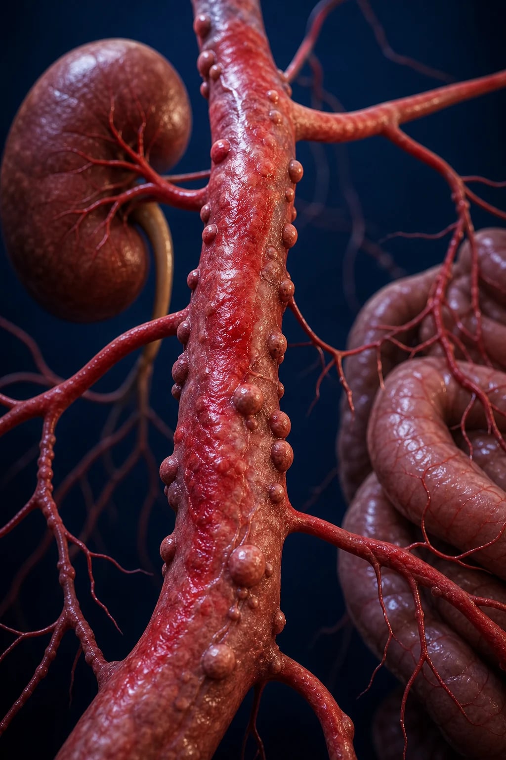
Classification
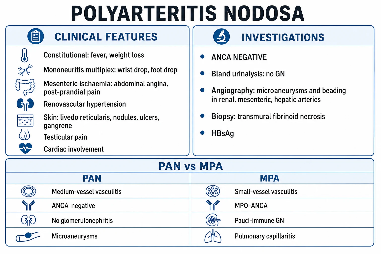
PAN is classified on two axes: the dominant vessel size (which defines it against the other vasculitides) and the presence of an associated trigger (which dictates treatment).[1][3]
Idiopathic (primary) PAN
- Necrotising vasculitis of medium muscular arteries with no identifiable cause; the common form in modern practice
- Treated with high-dose corticosteroids plus cyclophosphamide for severe/organ-threatening disease
- Can relapse; requires maintenance immunosuppression
- Definition requires exclusion of HBV, HCV, HIV and DADA2
HBV-associated PAN
- Secondary (immune-complex) PAN triggered by circulating HBV-antigen immune complexes; usually within 6 to 12 months of HBV infection
- Often with low complement and cryoglobulins; classic 'panarteritis' presentation with mononeuritis multiplex, abdominal angina, hypertension
- Treated with antivirals (entecavir/tenofovir) + plasma exchange + SHORT steroids — goal is HBV clearance, not chronic immunosuppression
- Usually monophasic; rarely relapses once HBV is cleared; now uncommon in vaccinated populations
Cutaneous-limited PAN
- Skin-only disease: livedo reticularis, tender subcutaneous nodules, ulcers, usually on the lower limbs
- No systemic involvement; benign course but often relapsing
- Biopsy confirms medium-vessel vasculitis in subcutis
- Treated with steroids +/- colchicine, dapsone or methotrexate; surveillance for systemic spread
DADA2 (PAN-mimic)
- Autosomal recessive loss-of-function mutations in ADA2 (CECR1); monogenic vasculopathy/vasculitis
- Presents in childhood with livedoid rash, PAN-like vasculitis, recurrent ischaemic and haemorrhagic strokes, immunodeficiency, pure red cell aplasia
- Treated with anti-TNF (etanercept) — NOT steroids; haematopoietic stem cell transplant for severe haematological disease
- Must be excluded in any child, adolescent or young adult with PAN-like disease or unexplained stroke
The 2012 Chapel Hill classification places PAN within the medium-vessel vasculitis (MVV) group, alongside Kawasaki disease. Crucially, CHCC stripped PAN of all small-vessel disease: a patient with glomerulonephritis, pulmonary capillaritis, or an active ANCA does not have PAN and should be reclassified as microscopic polyangiitis or another AAV.[3] The older 1990 American College of Rheumatology (ACR) classification criteria for PAN predate the separation of microscopic polyangiitis and are no longer diagnostically reliable; the 2021 ACR/Vasculitis Foundation (VF) criteria recognise PAN as a distinct medium-vessel entity with weight for visceral angiographic findings and against ANCA positivity and glomerulonephritis.[1]
Epidemiology & Risk Factors
PAN is rare. The annual incidence is approximately 2 to 9 per million population per year, and the prevalence is approximately 30 per million; incidence has fallen over the past three decades as the proportion of HBV-associated cases has declined with universal HBV vaccination, leaving the idiopathic form dominant in modern practice.[1][2]
The peak age of onset is 40 to 60 years, and there is a male predominance of roughly 2 to 1. There is no strong racial or geographic predilection, though historical cohorts over-represented HBV-endemic regions.[1]
PAN — the epidemiological numbers
Recognised triggers and associations drive the secondary workup:[1][6]
- Hepatitis B virus (HBV) — the classical association. Historically up to 30 to 50 percent of PAN cases were HBV-associated (immune-complex disease classically within 6 to 12 months of acute HBV infection); with vaccination, this has fallen to under 10 percent in modern series.
- Hepatitis C virus (HCV) — rare cause of true PAN; HCV more typically causes mixed cryoglobulinaemic vasculitis (small-vessel, low C4, GN), which is a separate entity.
- Hairy cell leukaemia — the most cited haematological association; an underlying malignancy should be considered in atypical or refractory PAN.
- Other associations — minimal-change nephropathy, hairy cell leukaemia, myelodysplasia, and rarely drugs; DADA2 (genetic) is now a distinct differential rather than 'idiopathic' PAN.[1][6]
Pathophysiology
PAN is driven by immune-complex deposition in the walls of medium muscular arteries. The cascade proceeds in four stages:[1][6]
- Immune-complex deposition and complement activation. Circulating immune complexes (idiopathic antigen unknown; in HBV-PAN they are HBV-antigen/anti-HBV complexes, often with mixed cryoglobulins) deposit in the vessel wall and activate complement — classically via the classical pathway, producing low serum complement in HBV-PAN.
- Neutrophilic infiltration and transmural inflammation. Complement-derived chemotaxins (C5a, C3a) recruit neutrophils and macrophages into the vessel wall. The infiltrate is transmural — involving intima, media and adventitia — and produces fibrinoid necrosis of the full arterial wall.
- Disruption of the internal elastic lamina and microaneurysm formation. Neutrophil-derived proteases and reactive oxygen species fragment the internal elastic lamina and weaken the muscular media, producing the microaneurysms (the 'nodosa' — nodular aneurysms palpable along an artery) alternating with segmental stenotic lesions. On angiography this gives the 'beaded' or 'rosary sign' appearance — alternating dilatations and narrowings.
- Thrombosis, rupture, and healing with fibrosis. The damaged vessel may thrombose (downstream infarction), rupture (haemorrhage — renal, hepatic, mesenteric), or heal by fibrosis — producing permanent segmental narrowing and the chronic ischaemic phenotype.[1][6]
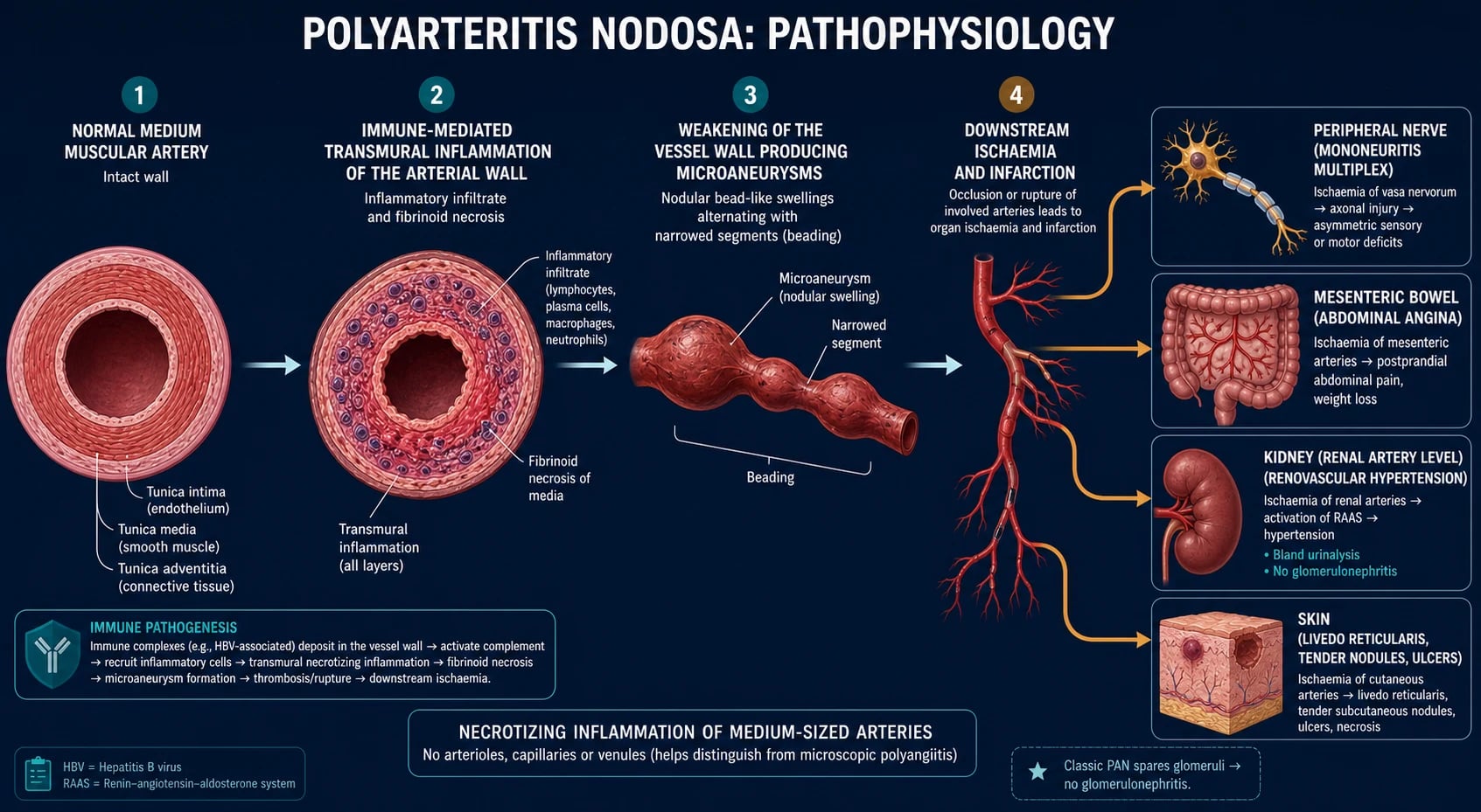
Two defining negatives follow directly from the medium-vessel distribution. Because PAN involves arteries — not arterioles, capillaries or venules — it spares the glomerulus (so the urinalysis is bland, with no proteinuria, haematuria or red-cell casts) and it spares the lungs (no pulmonary capillaritis, so no alveolar haemorrhage). Both are contrasts with the ANCA-associated vasculitides. In HBV-associated PAN the antigen is known (HBV surface, e and core antigens), which is why the goal of therapy is clearance of HBV by antivirals and removal of circulating complexes by plasma exchange, rather than indefinite immunosuppression.[1][6]
Clinical Presentation
PAN is a multi-system disease with a subacute onset over weeks to months. Symptoms cluster into constitutional and organ-specific patterns, and the combination — rather than any single feature — makes the diagnosis. There is no single pathognomonic clinical feature; the diagnostic skill is recognising the medium-vessel pattern across multiple territories.[1]
Constitutional features
Fever, malaise, fatigue and weight loss are present in most patients and are often the first and most misleading feature — mimicking infection, malignancy or endocarditis. Weight loss may be profound (10 percent body weight) and is often compounded by post-prandial abdominal pain that the patient avoids eating (sitophobia).[1]
Organ-specific features
Neurological (50–75%)
- Mononeuritis multiplex — the HALLMARK: painful, asymmetric, sensorimotor neuropathy from nerve infarction (peroneal nerve causing foot drop; ulnar/median causing wrist drop; evolving over days to weeks)
- Less commonly a symmetric sensorimotor polyneuropathy from overlapping mononeuropathies
- CNS involvement in 2 to 10 percent, usually late: stroke, seizures, encephalopathy
- Cranial neuropathy rare
Gastrointestinal (30–50%)
- Mesenteric ischaemia — post-prandial central abdominal pain (abdominal angina), weight loss, diarrhoea, bleeding
- May progress to gut infarction, perforation or ischaemic pancreatitis — surgical emergencies
- Pain out of proportion to examination is typical early; peritonism indicates infarction/perforation
Renal (30–60%)
- Renovascular hypertension and renal impairment with BLAND urinalysis (no proteinuria/haematuria/casts)
- Renal infarction, microaneurysm rupture with perinephric haemorrhage
- NOT glomerulonephritis — bland urine distinguishes PAN from MPA/AAV
Skin (25–50%)
- Livedo reticularis (the most characteristic skin sign — net-like, on the lower limbs)
- Tender subcutaneous nodules along arteries (the 'nodosa')
- Ulcers (usually lower limb), digital gangrene, occasionally palpable purpura
Cardiac
- Coronary arteritis (especially in HBV-PAN) causing angina, myocardial infarction
- Hypertension-driven cardiac failure and left ventricular hypertrophy
- Pericarditis, arrhythmia less common
Genitourinary
- Testicular pain and epididymal tenderness — CHARACTERISTIC of PAN
- Ovarian pain less commonly
- A frequently-tested distinguishing feature from other vasculitides
Musculoskeletal
- Myalgia, arthralgia (non-erosive, migratory)
- Rarely frank myositis with elevated CK
SPARED organs
- Lungs — NO pulmonary capillaritis, NO alveolar haemorrhage, NO asthma
- Glomerulus — NO glomerulonephritis (bland urine)
- Both are the cardinal distinctions from AAV/MPA
Atypical presentations
- Cutaneous PAN: skin-only disease with livedo, nodules and ulcers — no systemic features; benign but relapsing; biopsy decisive.
- Mononeuritis multiplex as the sole feature: a painful asymmetric sensorimotor neuropathy may precede systemic features by weeks — every mononeuritis multiplex workup must include vasculitis screen (HBV, ANCA, cryoglobulins, ESR/CRP).
- HBV-PAN: often a severe, explosive panarteritis with mononeuritis multiplex, abdominal angina, hypertension, orchitis and gastrointestinal bleeding occurring within 6 to 12 months of acute HBV infection.
- Paediatric/young-adult PAN or recurrent stroke: consider DADA2 (ADA2 deficiency) — a monogenic PAN-mimic requiring anti-TNF therapy, not steroids.[7][8]
- Elderly: more renal and mesenteric disease, poorer tolerance of cyclophosphamide — favour steroid-sparing strategies.
Differential Diagnosis
The pivotal differentials are (1) the ANCA-associated vasculitides (especially microscopic polyangiitis), which also cause neuropathy and renal disease but are small-vessel, ANCA-positive and have glomerulonephritis; (2) the other medium-vessel vasculitides (Kawasaki, DADA2); (3) immune-complex small-vessel vasculitides (IgA vasculitis, cryoglobulinaemia); and (4) non-vasculitic mimics (infection, emboli, antiphospholipid syndrome, malignancy).[1][2]
Microscopic polyangiitis (MPA)
- SMALL-vessel, MPO-ANCA (p-ANCA) positive in 40 to 80 percent
- Pauci-immune rapidly progressive glomerulonephritis — ACTIVE urinary sediment (proteinuria, dysmorphic RBCs, red-cell casts)
- Pulmonary capillaritis / alveolar haemorrhage
- NO granulomas, NO destructive ENT disease
- Treated with steroids + rituximab or cyclophosphamide; PLEX if severe renal
GPA (Wegener)
- Small-vessel + granulomatous; PR3-ANCA (c-ANCA) positive
- ENT (saddle nose, epistaxis, sinusitis, subglottic stenosis) + lung nodules/cavitation + pauci-immune GN
- ANCA positive, active urine — both distinguish from PAN
EGPA (Churg-Strauss)
- Adult-onset ASTHMA + eosinophilia + nasal polyposis then vasculitic phase
- Mononeuritis multiplex (common), eosinophilic cardiomyopathy (leading cause of death)
- MPO-ANCA positive in about 40 percent; eosinophilia over 10 percent; eosinophilic tissue infiltrate on biopsy
Kawasaki disease
- Medium-vessel vasculitis of YOUNG CHILDREN
- Fever over 5 days plus conjunctivitis, oral changes, rash, cervical lymphadenopathy, extremity changes
- Coronary artery aneurysms the feared complication; treated with IVIG + aspirin
IgA vasculitis (Henoch-Schonlein)
- Small-vessel IgA immune deposits; children; post-URI
- Palpable purpura (lower limbs), abdominal pain, arthritis, IgA nephritis (glomerulonephritis)
- Mesangial IgA on biopsy; normal complement
Cryoglobulinaemic vasculitis
- Mixed cryoglobulins (Types II/III); hepatitis C association
- Palpable purpura, arthralgia, weakness, peripheral neuropathy, glomerulonephritis
- Low C4; immune-complex deposition on biopsy
DADA2 (ADA2 deficiency)
- Autosomal recessive mutations in ADA2 (CECR1) gene; monogenic vasculopathy/vasculitis
- Childhood onset; livedoid rash, PAN-like vasculitis, recurrent ischaemic and haemorrhagic STROKES, immunodeficiency, pure red cell aplasia
- Treated with anti-TNF (etanercept) and HSCT for severe haematological disease — NOT steroids
Non-vasculitic mimics
- Infective endocarditis, disseminated infection — culture, echo
- Cholesterol emboli (post-catheter, eosinophilia, livedo, AKI), atrial myxoma
- Antiphospholipid syndrome, cocaine/levamisole vasculopathy
- Malignancy (lymphoma, including hairy cell leukaemia)
Clinical & Bedside Assessment
A focused multi-system examination is mandatory at first contact and at every follow-up, because PAN's territory is arterial and the organs map the vessel distribution.[1][5]
General and vital signs. Confirm blood pressure in both arms (renovascular hypertension is common and may be the first clue); temperature, weight (loss), and a general search for cachexia, fever patterns.[1]
Skin. Examine the lower limbs and trunk in daylight for livedo reticularis (net-like, blanchable network), tender subcutaneous nodules along arteries (the 'nodosa' — palpate the course of the sural, posterior tibial, radial and ulnar arteries), ulcers, digital infarcts and gangrene. Nodules are best felt along the lower-limb arteries and may be biopsied.[1]
Neurological. This is the most important bedside domain. Test ankle and foot dorsiflexion (peroneal nerve — foot drop), wrist and finger extension (radial/ulnar/median — wrist drop), and sensation in the territories of named peripheral nerves. Mononeuritis multiplex — an asymmetric, painful, sensorimotor neuropathy in the distribution of two or more individual nerves — is the single most characteristic neurological finding and reflects nerve infarction. Also perform a cranial nerve and cerebellar examination and screen for stroke (CNS involvement is uncommon but recognised).[5]
Abdomen. Examine for tenderness out of proportion to findings (early mesenteric ischaemia), distension, and signs of peritonism (perforation/infarction — a surgical emergency). Auscultate for bruits (renal, abdominal aortic, mesenteric). Perform a testicular examination for epididymal tenderness and testicular pain — characteristic of PAN and often forgotten.[1][5]
Cardiovascular. Heart sounds (pericarditis, failure), murmurs (endocarditis is a key mimic), peripheral pulses, bruits, and signs of cardiac failure (hypertensive or ischaemic).[1]
Eye and fundoscopy. Hypertensive retinopathy (the kidney is renovascular in PAN); rarely retinal vasculitis.[1]
PAN clinical features — NODULES
NODULES
wrist/foot drop from nerve infarction — the hallmark
abdominal angina, renovascular hypertension, bland urine
medium-vessel skin infarction
the two negatives that distinguish PAN from MPA/AAV
unlike the ANCA-associated vasculitides
characteristic of PAN
treatment by form
Investigations
A staged workup confirms the vasculitis, defines the vessel size and organ involvement, excludes mimics, screens for HBV and other secondary causes, and informs the treatment intensity. The most important principles are that ANCA is negative, the urinalysis is bland, and visceral angiography and tissue biopsy are the two confirmatory tests.[1]
First-line bloods. FBC (normocytic anaemia of chronic disease, leucocytosis, thrombocytosis as acute-phase responses — eosinophilia points AWAY from PAN toward EGPA); ESR and CRP (markedly elevated but non-specific); U&E and creatinine (renal impairment); LFTs (hepatitic pattern in HBV-PAN, low albumin in active inflammation); CK (myositis); glucose and lipids (cardiovascular risk stratification).[1]
Urinalysis and urine microscopy. The single most important bedside test. In PAN the urine is bland — no proteinuria, no haematuria, no red-cell casts. An active sediment mandates reconsideration of the diagnosis (MPA, anti-GBM, IgA nephritis, lupus). Send a urine protein-to-creatinine ratio; significant proteinuria in suspected PAN again points to a glomerular disease.[1][3]
Immunology — the discriminating panel. ANCA is NEGATIVE — both indirect immunofluorescence (IIF) pattern and PR3/MPO ELISA. If ANCA is positive, reconsider MPA, GPA or EGPA. ANA, rheumatoid factor and anti-dsDNA to exclude lupus/overlap. Complement (C3, C4) — normal or LOW in HBV-PAN (immune-complex consumption; C4 may be selectively low if cryoglobulins are present). Cryoglobulins — send if HBV/HCV suspected (sample must be kept warm). Anti-GBM if pulmonary-renal overlap considered. Serum immunoglobulins and serum protein electrophoresis to screen for paraproteinaemia and hairy cell leukaemia association.[1]
Infection screen — mandatory in every case. HBV serology (HBsAg, anti-HBc, anti-HBs) is mandatory — it changes treatment. Also send HCV antibodies, HIV, and consider hepatitis B DNA viral load if HBsAg positive. HBV-PAN is treated with antivirals and plasma exchange, not cyclophosphamide; missing HBV commits the patient to the wrong regimen.[1]
Visceral angiography — the radiological hallmark. CT, MR or conventional mesenteric and renal angiography reveals the pathognomonic findings: multiple microaneurysms and a 'beaded' (rosary sign) irregular outline of the renal, mesenteric and hepatic arteries. The microaneurysms are typically 1 to 5 mm, saccular, and distributed at branch points. Conventional digital subtraction angiography has the highest spatial resolution and is preferred when CT/MR is equivocal. Renal biopsy is generally unhelpful in PAN because the disease involves the arteries at the hilum, not the glomerulus sampled by a standard biopsy.[1][6]
Tissue biopsy — the gold standard. Biopsy an involved organ with the highest yield and lowest risk: skin nodule (in cutaneous disease), sural nerve plus adjacent muscle (when neuropathy is the lead feature — combined nerve-muscle biopsy increases yield), testis (when epididymal/orchitis is present), gut (resected at surgery for perforation/infarction), or kidney (targeting perirenal tissue rather than the glomerulus). Histology shows transmural medium-vessel vasculitis with fibrinoid necrosis, disruption of the internal elastic lamina and a mixed inflammatory infiltrate at the acute stage, healing with fibrosis and intimal hyperplasia in older lesions. Skip lesions are common — a negative biopsy never excludes PAN, and angiography then becomes decisive.[1][6]
Nerve conduction studies and EMG. Localise mononeuritis multiplex (axonal, asymmetric, in named-nerve distribution), confirm the pattern, and guide the choice of nerve for biopsy (typically biopsy a clinically affected nerve with absent sensory action potential, plus adjacent muscle).[5]
Other imaging. Chest X-ray (lungs should be clear — infiltrates or haemorrhage point away from PAN). CT abdomen/pelvis for organomegaly, infarcts, haemorrhage. Echocardiogram for cardiac involvement and to exclude endocarditis (a key mimic).[1]
Management — Resuscitation
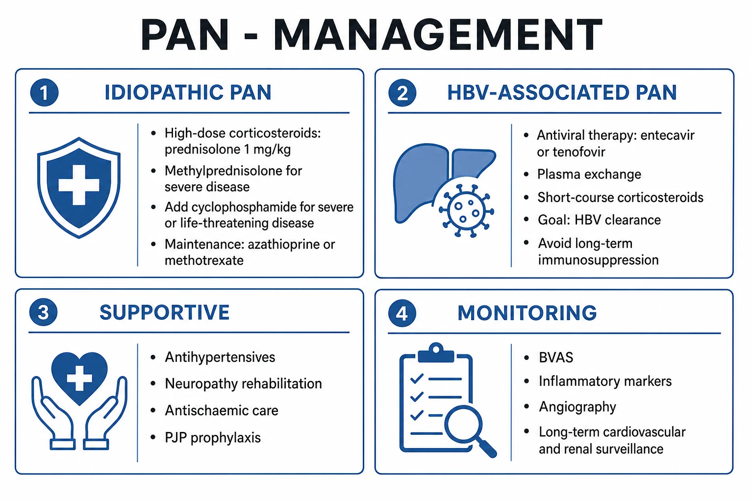
PAN rarely presents with the seconds-count emergency of AAV pulmonary-renal syndrome (it spares the lungs and the glomerulus), but several life- or organ-threatening presentations need immediate inpatient care:[1][6]
- Mesenteric ischaemia with infarction or perforation — acute severe abdominal pain with peritonism, lactic acidosis, pneumatosis on imaging. Urgent surgical review, broad-spectrum antibiotics, fluid resuscitation, and laparotomy/laparoscopy for resection; immunosuppression is withheld or reduced peri-operatively.
- Malignant hypertension / hypertensive emergency (renovascular) — IV labetalol or nicardipine infusion to lower mean arterial pressure by no more than 25 percent in the first hour, then gradual control; monitor for end-organ damage.
- Severe mononeuritis multiplex with rapid functional loss (bilateral foot/wrist drop, falling function) — begin urgent high-dose corticosteroids (IV methylprednisolone pulses) to abort further nerve infarction.
- Microaneurysm rupture with renal, hepatic or mesenteric haemorrhage — resuscitation, transfusion, and interventional radiology for selective arterial embolisation.
- HBV-PAN presenting fulminantly — expedite the diagnosis and begin antiviral therapy plus plasma exchange without delay.[1][6]
A supportive bundle applies across all of these: analgesia, fluid and electrolyte correction, gastric protection, Pneumocystis jirovecii prophylaxis (cotrimoxazole 480 mg or 960 mg daily, or alternative if allergic) when high-dose steroids or cyclophosphamide are started, bone protection (calcium, vitamin D, bisphosphonate where indicated), and vaccination (influenza, pneumococcal, COVID-19, hepatitis B if non-immune — given ideally before immunosuppression; avoid live vaccines during immunosuppression).[1]
Management — Definitive & Stepwise
Treatment is stratified by HBV status and severity. The pivotal fork — and a favourite examiner question — is between idiopathic and HBV-associated PAN: the former is treated with corticosteroids plus cyclophosphamide (for severe disease); the latter is treated with antivirals and plasma exchange to clear HBV, NOT long-term immunosuppression.[1][2][6]
[1] [6]Idiopathic PAN
Corticosteroids are the cornerstone. Oral prednisolone 1 mg/kg/day (maximum 60 to 80 mg) is started immediately for confirmed PAN; for severe or organ-threatening disease (severe mononeuritis multiplex, cardiac, gut, renal), begin with IV methylprednisolone 500 to 1000 mg daily for 3 days then convert to oral prednisolone 1 mg/kg/day. A rapid clinical and biochemical response (falling ESR/CRP, stabilising renal function, improving neuropathy) is characteristic.[1]
Add cyclophosphamide for severe disease. The French Vasculitis Study Group approach uses cyclophosphamide 15 mg/kg IV every 2 weeks for 6 doses (then every 3 weeks) or oral 2 mg/kg/day (maximum 200 mg/day) for 3 to 6 months, with dose reduction for age over 65 and for renal impairment. Indications: severe neuropathy, gut, cardiac, renal or CNS involvement, or steroid-refractory disease. After induction, switch to a safer maintenance agent.[1]
Maintenance therapy (after cyclophosphamide induction): azathioprine 2 mg/kg/day or methotrexate 20 to 25 mg once weekly (with folate) for 18 to 24 months. Rituximab (375 mg per square metre weekly for 4 doses, or 1 g two weeks apart) is used in refractory, relapsing or cyclophosphamide-intolerant PAN; evidence is extrapolated from AAV, but B-cell depletion is rational given the immune-complex pathogenesis.[1]
Steroid taper. Once remission is achieved (typically 4 to 8 weeks), taper prednisolone slowly — reduce by approximately 10 percent every 1 to 2 weeks to 20 mg, then by 2.5 mg every 2 to 4 weeks to 10 mg, then by 1 mg every 1 to 2 months — guided by symptoms, ESR/CRP and organ function. The tapering window is typically 12 to 24 months.[1]
HBV-associated PAN
The principle is clearance of HBV with antivirals and plasma exchange, plus a short course of corticosteroids to control the early inflammatory phase — NOT long-term immunosuppression. The regimen, refined by Guillevin and the French Vasculitis Study Group, is:[1][5]
- Antiviral therapy — entecavir 0.5 to 1.0 mg orally once daily (1 mg in decompensated liver disease or lamivudine resistance) or tenofovir disoproxil fumarate 300 mg once daily (or tenofovir alafenamide 25 mg once daily). These potent nucleos(t)ide analogues suppress HBV replication and clear circulating HBV-antigen immune complexes that drive the vasculitis. Treat in collaboration with hepatology.
- Plasma exchange (PLEX) — typically 3 exchanges per week for the first 2 to 3 weeks, then progressively less frequent (1 to 2 per week for several weeks), exchanging one plasma volume per session, replaced with 4 to 5 percent human albumin (fresh-frozen plasma for bleeding risk or pre-procedure). PLEX removes circulating HBV-antigen-antibody complexes and cryoglobulins.
- Short-course corticosteroids — prednisolone 1 mg/kg/day for 1 to 2 weeks to control early inflammation, then rapidly tapered and stopped as the antiviral and PLEX take effect. The aim is to avoid prolonged immunosuppression, which impairs HBV clearance.[1][5]
HBV-PAN is usually monophasic and rarely relapses once HBV is cleared. HBeAg seroconversion correlates with vasculitis remission. Successful HBV vaccination programmes have made HBV-PAN uncommon in countries with universal infant vaccination.[1]
Cutaneous-limited PAN
Skin-only PAN (livedo, nodules, ulcers, no systemic involvement) is treated with prednisolone 0.5 to 1 mg/kg/day tapered over weeks to months, with colchicine 0.6 mg twice daily, dapsone 50 to 100 mg/day (check G6PD), or methotrexate as steroid-sparing agents for relapsing disease. Local wound care for ulcers and surveillance for systemic spread (regular clinical review, ESR/CRP, urinalysis) are essential.[1]
Supportive care and monitoring
- Antihypertensives for renovascular hypertension — ACE inhibitor/ARB (monitor renal function and potassium), calcium-channel blocker; aim for tight control.
- Neuropathy rehabilitation — physiotherapy, ankle-foot orthosis for foot drop, occupational therapy, pain management (neuropathic agents: amitriptyline, gabapentin, pregabalin).
- PJP prophylaxis — cotrimoxazole 480 mg or 960 mg daily throughout high-dose steroids or cyclophosphamide.
- Bone protection — calcium 1000 mg/day, vitamin D 800 to 1000 IU/day, and a bisphosphonate (e.g. alendronate 70 mg weekly) for glucocorticoid-induced osteoporosis risk; DEXA scan baseline.
- Gastric protection — proton-pump inhibitor with high-dose steroids or concurrent NSAIDs/anticoagulation.
- Vaccination — pneumococcal, influenza, COVID-19, hepatitis B (if non-immune); avoid live vaccines during immunosuppression.
- Monitoring — Birmingham Vasculitis Activity Score (BVAS) for disease activity, ESR/CRP, renal function, blood pressure, and follow-up angiography in selected cases.[1]
Specific Subtypes & Scenarios
- Idiopathic PAN — the common form in modern practice; treated with steroids plus cyclophosphamide for severe disease, then azathioprine or methotrexate maintenance; can relapse.
- HBV-associated PAN — secondary immune-complex PAN; antivirals + plasma exchange + short steroids; usually monophasic.
- Cutaneous-limited PAN — skin-only; steroids plus colchicine/dapsone/methotrexate; surveillance for systemic spread.
- DADA2 (ADA2 deficiency) — a monogenic PAN-mimic, autosomal recessive, loss-of-function mutations in ADA2 (CECR1) on chromosome 22q11. Presents in childhood with livedoid rash, PAN-like medium-vessel vasculitis, recurrent ischaemic and haemorrhagic strokes, immunodeficiency (recurrent infections), haematological disease (pure red cell aplasia, neutropenia), and hepatosplenomegaly. Treated with anti-TNF (etanercept) — NOT corticosteroids — and haematopoietic stem cell transplant for severe haematological disease. Must be excluded in any child, adolescent or young adult presenting with PAN-like disease or unexplained stroke.[7][8]
- Hairy cell leukaemia-associated PAN — the classic haematological association; an underlying malignancy should be considered in atypical or refractory PAN. Treatment of the leukaemia (cladribine) may resolve the vasculitis.
- Paediatric PAN — rare; DADA2 must be excluded; weight-based dosing of steroids and cyclophosphamide; paediatric rheumatology involvement.
- Pregnancy — plan conception during remission (at least 6 months); avoid cyclophosphamide, methotrexate, mycophenolate (teratogenic); use azathioprine maintenance; prednisolone at lowest effective dose; multidisciplinary care.
Complications & Pitfalls
PAN's complications arise from end-organ ischaemia (the disease itself) and from treatment toxicity (the drugs).[1][6] Disease complications:
- Mononeuritis multiplex with residual disability — persistent foot drop, wrist drop, sensory deficit and chronic neuropathic pain; rehabilitation and orthoses.
- Mesenteric ischaemia, gut infarction and perforation — surgical emergencies; the leading cause of mortality in severe PAN.
- Ischaemic pancreatitis — from mesenteric/pancreatic arteritis.
- Renovascular hypertension and chronic kidney disease (NOT glomerulonephritis) — long-term antihypertensive and nephrology follow-up.
- Coronary arteritis, myocardial infarction, cardiac failure — especially in HBV-PAN.
- Cerebral vasculitis, stroke and microaneurysm rupture with haemorrhage (renal, hepatic, mesenteric).
- Digital gangrene requiring amputation.[1][6]
Treatment complications:
- Glucocorticoids — diabetes, osteoporosis, infection, hypertension, weight gain, cataracts, adrenal suppression, mood disturbance.
- Cyclophosphamide — infection, haemorrhagic cystitis (maintain hydration; consider mesna), bone marrow suppression, infertility (offer fertility preservation — sperm/egg banking), late malignancy (bladder, myelodysplasia).
- Plasma exchange — catheter-related infection, hypotension, bleeding (citrate-induced hypocalcaemia; replace with FFP for bleeding risk), venous access complications.
- Rituximab — infusion reactions, late-onset neutropenia, hypogammaglobulinaemia, progressive multifocal leukoencephalopathy (very rare), HBV reactivation (screen and antiviral-prophylax).[1]
Pitfalls:
- Mislabelling MPA as PAN (or vice versa) — the cardinal error. ANCA positivity or an active urinary sediment excludes PAN and mandates a small-vessel diagnosis.
- Treating HBV-PAN with cyclophosphamide — the wrong regimen; it impairs HBV clearance. Always screen HBsAg before committing to cyclophosphamide.
- Performing a standard renal biopsy in suspected PAN — it samples the glomerulus, which PAN spares, and is usually unhelpful. Biopsy an involved organ or perform angiography.
- Forgetting DADA2 in a child or young adult with PAN-like disease or recurrent stroke — anti-TNF therapy is life-changing, and steroids do not work.
- Missing a surgical abdomen in mesenteric PAN — pain may be out of proportion early, with peritonism indicating established infarction/perforation.[1][7]
Prognosis & Disposition
Five-year survival is approximately 80 percent with modern treatment, compared with 13 percent untreated. Outcome is stratified by the Five-Factor Score (FFS, 2009 revision — Guillevin, Medicine 2011, PMID 21200183), the prognostic instrument for the systemic necrotising vasculitides (PAN, MPA, GPA, EGPA):[4]
HBV-associated PAN is usually monophasic and rarely relapses once HBV is cleared, with HBeAg seroconversion correlating with vasculitis remission.[1] Idiopathic PAN can relapse and requires maintenance immunosuppression with long-term cardiovascular and renal surveillance — residual renovascular hypertension is common and an independent cardiovascular risk factor. Cutaneous-limited PAN has an excellent prognosis but relapses frequently.
Disposition: admit for organ-threatening or suspected surgical disease (mesenteric ischaemia/perforation, severe neuropathy, malignant hypertension, microaneurysm rupture, fulminant HBV-PAN); outpatient management for stable disease under combined rheumatology, nephrology, neurology and hepatology (HBV-PAN) care. Long-term follow-up is lifelong for cardiovascular risk, renal surveillance and relapse detection.[1][4]
PAN — prognostic numbers
Special Populations
- HBV-associated PAN — treat with antivirals + plasma exchange + short corticosteroids to clear HBV; engage hepatology early; now uncommon due to universal HBV vaccination.
- DADA2 (paediatric/young-adult PAN) — anti-TNF (etanercept), NOT steroids; HSCT for severe haematological disease; genetic counselling (autosomal recessive); screen siblings.[7][8]
- Hairy cell leukaemia-associated PAN — treat the underlying malignancy (cladribine); the vasculitis often resolves with haematological remission.
- Pregnancy — plan conception during remission; avoid cyclophosphamide, methotrexate, mycophenolate (teratogenic); azathioprine maintenance; prednisolone at lowest effective dose; multidisciplinary maternal-foetal medicine and rheumatology care.
- Children (juvenile PAN) — rare; weight-based dosing; DADA2 must be excluded; growth, fertility and bone concerns with chronic steroids — minimise exposure.
- Elderly — more renal and mesenteric disease; poorer tolerance of cyclophosphamide and high-dose steroids; favour steroid-sparing strategies and lower thresholds for plasma exchange; age over 65 is an FFS point.
- Renal impairment — dose-adjust cyclophosphamide; rituximab dosing unaffected by renal function; consider plasma exchange for severe disease.
- Immunosuppressed patients — screen for latent TB, HBV, HCV, HIV before induction; antiviral prophylaxis for HBsAg-positive or anti-HBc-positive patients throughout therapy and for 12 months after rituximab; PJP prophylaxis; vaccinate (avoid live vaccines).
Evidence, Guidelines & Regional Differences
Landmarks that shaped modern PAN classification and management
Guidelines that govern PAN management. The 2022 EULAR recommendations for the management of primary medium- and small-vessel vasculitis and the 2021 ACR/VF classification criteria are the contemporary reference standards. The French Vasculitis Study Group algorithms underpin the stratification by FFS and the HBV-PAN regimen. HBV vaccination is the single most effective public-health measure to reduce HBV-PAN incidence — the prevalence of HBV-PAN has fallen dramatically in countries with universal infant HBV vaccination.[1][2]
Exam Pearls
- PAN = necrotising vasculitis of MEDIUM muscular arteries; MICROANEURYSMS on angiography (rosary sign); NO glomerulonephritis; NO ANCA; lungs spared.[1]
- Hallmark presentation: mononeuritis multiplex (wrist/foot drop from nerve infarction) + constitutional symptoms + livedo + nodules.[5]
- Abdominal angina (post-prandial pain) + weight loss + new hypertension = mesenteric + renal PAN.
- Bland urinalysis with renal impairment + hypertension = renovascular PAN (artery, NOT glomerulus).
- Testicular pain/epididymitis is characteristic of PAN — frequently tested.
- ALWAYS screen HBsAg — HBV-PAN is treated with antivirals + plasma exchange + short steroids, NOT cyclophosphamide.
- Distinguish from MPA: small-vessel, MPO-ANCA positive, pauci-immune glomerulonephritis, pulmonary capillaritis.
- PAN spares the lungs AND the glomerulus — the two cardinal negatives.
- Idiopathic PAN: high-dose steroids (prednisolone 1 mg/kg/day; methylprednisolone 500 to 1000 mg IV for severe) + cyclophosphamide 15 mg/kg for severe; maintenance azathioprine (2 mg/kg/day) or methotrexate (20 to 25 mg/week).
- Microaneurysms + beading of renal, mesenteric and hepatic arteries on angiography is the radiological hallmark.
- Child or young adult with PAN-like disease or recurrent stroke — exclude DADA2, treated with anti-TNF (etanercept), not steroids.[7][8]
- FFS: age over 65, cardiac, GI, renal insufficiency, no ENT — each scores 1 point; 5-year mortality 9 percent (FFS 0), 21 percent (FFS 1), 40 percent (FFS 2 or more).[4]
PAN — the diagnostic negatives
NONE
medium-vessel disease; an active sediment points to MPA/AAV
transmural fibrinoid necrosis and microaneurysms (rosary sign)
negative ANCA distinguishes PAN from the ANCA-associated vasculitides
lungs spared — no pulmonary capillaritis, no alveolar haemorrhage
Exam application bank (NEET-PG / INICET)
One-line answer
Polyarteritis nodosa (PAN) is a necrotising vasculitis of medium-sized muscular arteries characterised by transmural inflammation, fibrinoid necrosis, disruption of the internal elastic lamina and microaneurysm formation. It is defined by what it is NOT: it does not involve glomeruli or capillaries, does not involve the lungs (no pulmonary capillaritis), and is not ANCA-associated — the three findings that distinguish it from the ANCA-associated vasculitides (especially microscopic polyangiitis). Presentation: constitutional symptoms, mononeuritis multiplex (wrist/foot drop from nerve infarction), mesenteric ischaemia (abdominal angina), renovascular hypertension with bland urinalysis, livedo reticularis, tender subcutaneous nodules, ulcers, testicular pain. Diagnosis: mesenteric/renal angiography (microaneurysms and beading — the 'rosary sign'), tissue biopsy (medium-vessel transmural f
Worked stems (answer without another resource)
Stem 1 — Classic presentation. Map symptoms to mechanism; name the first investigation and first treatment step with dose/route if drug therapy is standard. [1]
Stem 2 — Unstable / complicated. List red flags that force immediate resuscitation, theatre, ICU, antidote, or reperfusion — and what you do in the first 15 minutes. [1]
Stem 3 — Atypical group. Elderly, pregnancy, child, or immunocompromised: how presentation and thresholds change. [1]
Stem 4 — Differential trap. Name the three closest mimics and one discriminator for each. [1]
Stem 5 — Disposition. Who goes home with safety-netting, who is admitted, who needs HDU/ICU/theatre, and what follow-up is mandatory. [1]
Rapid viva checklist
- Definition + classification
- Pathophysiology chain
- Bedside signs / criteria
- Score with exact components (if any)
- Emergency bundle
- Definitive therapy with doses
- Complications of disease and of treatment
- Special populations
- Guideline/trial name if classic
- Three exam traps
Coverage self-check
If you cannot answer any stem above from this page alone, re-read the matching section — the page is intended to be self-sufficient for final-prof and NEET-PG/INICET questions on Polyarteritis Nodosa (PAN).
One-liner — what is PAN and how is it treated?
PAN is a necrotising vasculitis of medium muscular arteries with transmural fibrinoid necrosis and microaneurysms — distinguished from MPA by bland urinalysis, negative ANCA and spared lungs. Diagnose with angiography (rosary sign) and tissue biopsy; screen HBsAg. Treat idiopathic PAN with high-dose steroids (+ cyclophosphamide for severe) and HBV-associated PAN with antivirals + plasma exchange + short steroids. Five-year survival is approximately 80 percent treated.[1][2]
References
- [1]Hernández-Rodríguez J, Alba MA, Prieto-González S, Cid MC. Diagnosis and classification of polyarteritis nodosa J Autoimmun, 2014.PMID 24485157
- [2]Springer JM, Byram K, Dey ID, Warrington KJ. Polyarteritis nodosa: an evolving primary systemic vasculitis Postgrad Med, 2023.PMID 35709399
- [3]Jennette JC, Falk RJ, Bacon PA, Basu N, Cid MC, Ferrario F, Flores-Suarez LF, Gross WL, Guillevin L, Hagen EC, Hoffman GS, Jayne DR, Kallenberg CG, Lamprecht P, Langford CA, Luqmani RA, Mahr AD, Matteson EL, Merkel PA, Ozen S, Pusey CD, Rasmussen N, Rees AJ, Scott DG, Specks U, Stone JH, Takahashi K, Watts RA. 2012 revised International Chapel Hill Consensus Conference Nomenclature of Vasculitides Arthritis Rheum, 2013.PMID 23045170
- [4]Guillevin L, Pagnoux C, Seror R, Mahr A, Mouthon L, Toumelin PL; French Vasculitis Study Group. The Five-Factor Score revisited: assessment of prognoses of systemic necrotizing vasculitides based on the French Vasculitis Study Group (FVSG) cohort Medicine (Baltimore), 2011.PMID 21200183
- [5]de Boysson H, Guillevin L. Polyarteritis Nodosa Neurologic Manifestations Neurol Clin, 2019.PMID 30952413
- [6]Colmegna I, Maldonado-Cocco JA. Polyarteritis nodosa revisited Curr Rheumatol Rep, 2005.PMID 16045832
- [7]Meyts I, Aksentijevich I. Deficiency of Adenosine Deaminase 2 (DADA2): Updates on the Phenotype, Genetics, Pathogenesis, and Treatment J Clin Immunol, 2018.PMID 29951947
- [8]Sharma V, Suri D, Rawat A, Verma Ghosh R, Minz RW, Singh S. Deficiency of adenosine deaminase 2 (DADA2): Review Best Pract Res Clin Rheumatol, 2023.PMID 37328410