Rheumatology · General Medicine
Psoriatic Arthritis
Also known as Psoriatic arthritis · PsA · Psoriatic arthropathy · Arthropathy of psoriasis · Arthritis psoriatica · Arthritis mutilans
Psoriatic arthritis (PsA) is an inflammatory arthritis associated with psoriasis, usually rheumatoid-factor negative, classified within the seronegative spondyloarthropathies. Diagnosis is clinical using the CASPAR criteria (established inflammatory articular disease plus at least 3 points from current psoriasis, personal or family history of psoriasis, current or historical dactylitis, juxta-articular new bone formation, rheumatoid-factor negativity, and nail dystrophy). The five Moll and Wright patterns are asymmetric oligoarthritis (commonest), symmetric polyarthritis, DIP-predominant, arthritis mutilans, and predominant spondylitis or sacroiliitis. Hallmarks: dactylitis (sausage digit), enthesitis (Achilles, plantar fascia), nail disease (pitting, onycholysis, hyperkeratosis), and DIP involvement. Pathophysiology is genetic (HLA-Cw6 for skin, HLA-B27 for axial) plus environmental triggers (Koebner phenomenon, infection, obesity, drugs) driving the IL-23 / Th17 / IL-17 axis and TNF, producing the unique paradoxical bone remodelling in which erosions and pathological new bone formation coexist. Treat-to-target aims for minimal disease activity or remission: NSAIDs first-line, conventional DMARDs (methotrexate) for moderate peripheral disease, TNF inhibitors first-line biologic, then IL-17 and IL-23 inhibitors for refractory or skin-predominant disease, with JAK inhibitors as oral alternatives.
On this page & tools
Your progress
Saved locally on this device.
Exam tags
Red flags
Overview & Definition
Psoriatic arthritis (PsA) is the inflammatory arthritis that accompanies psoriasis and is the prototype of a seronegative spondyloarthropathy with prominent peripheral and entheseal disease. The term seronegative denotes that both rheumatoid factor (RF) and anti-citrullinated protein antibodies (ACPA) are absent, in contrast to rheumatoid arthritis. The core pathological process is enthesitis — inflammation at the insertion of tendon, ligament or joint capsule into bone — which the synovitis and the psoriatic skin lesion then mirror, giving PsA its multi-domain character (peripheral joints, axial skeleton, entheses, skin, nails).[1]
The clinical skill is recognising the pattern — psoriasis or nail dystrophy plus dactylitis, enthesitis, DIP involvement or asymmetry in an RF-negative patient — and then distinguishing it from rheumatoid arthritis (symmetric, small joints, RF and ACPA positive, DIP spared, no enthesitis or dactylitis) and osteoarthritis (non-inflammatory, Heberden and Bouchard nodes, brief stiffness, normal CRP). Once recognised, the goal is treat-to-target: escalating from NSAIDs through conventional DMARDs to biologics to prevent the destructive, paradoxical bone remodelling that is unique to this disease — simultaneous erosions and pathological new bone formation in the same joint.[1][4]
A defining feature that makes PsA distinct from the other spondyloarthropathies is its multi-domain phenotype: one patient may have a few swollen toes and nail pitting, another destructive arthritis mutilans with telescoping digits, and a third an ankylosing-spondylitis-like spine with uveitis. The same patient may shift between patterns over years, which is why the CASPAR classification criteria (rather than any single test) anchor the diagnosis and why domain-driven treatment — addressing peripheral joints, axial disease, enthesitis, dactylitis, and skin and nail disease in parallel — is the organising principle of modern care.[1][3]
Classification
The seronegative spondyloarthropathy family
PsA belongs to the seronegative spondyloarthropathies (SpA), a family unified by overlapping genetics (HLA-B27), clinical pattern (inflammatory back pain, asymmetric oligoarthritis, enthesitis, dactylitis) and pathology (enthesitis with paradoxical bone remodelling):[1]
- Ankylosing spondylitis — axial prototype.
- Psoriatic arthritis — this topic.
- Reactive arthritis — one to four weeks after GU or GI infection (the classic triad of arthritis, urethritis, conjunctivitis).
- Enteropathic arthritis — arthritis associated with inflammatory bowel disease (Crohn disease, ulcerative colitis).
- Undifferentiated spondyloarthropathy — features of SpA that do not meet a specific subset.[1]
The shared (unifying) features of the SpA family are the high-yield list: inflammatory back pain; sacroiliitis (often asymmetric in PsA, reactive and IBD disease, in contrast to the bilateral symmetric pattern of ankylosing spondylitis); asymmetric oligoarthritis of the lower limbs; enthesitis (especially Achilles tendon and plantar fascia); dactylitis (sausage digit); RF and ACPA negative; HLA-B27 association (strongest in ankylosing spondylitis, weaker in PsA); extra-articular features (uveitis, psoriasis, IBD); and the characteristic paradoxical bone remodelling on imaging.[1]
CASPAR criteria (reproduced verbatim) — the classification cornerstone
PsA is a clinical diagnosis; the CASPAR (ClASsification criteria for Psoriatic ARthritis) criteria were developed from a large international study and are the standard tool for classification.[2]
The entry requirement is that the patient must have established inflammatory articular disease — inflammatory articular disease of the joints (peripheral or axial) or entheseal disease. At least 3 points are then required from the following categories:[2]
| Category | Feature | Points |
|---|---|---|
| Current psoriasis | Psoriatic skin or scalp disease confirmed today | 2 |
| Personal history of psoriasis | History of psoriasis (only if current psoriasis absent) | 1 |
| Family history of psoriasis | History in a first- or second-degree relative (if current and personal absent) | 1 |
| Current dactylitis | Swelling of an entire digit | 1 |
| History of dactylitis | History recorded by a rheumatologist | 1 |
| Juxta-articular new bone formation | Ill-defined ossification near joint margins on hand or foot X-ray | 1 |
| Rheumatoid factor negativity | RF negative by any method (other than latex) | 1 |
| Nail dystrophy | Onycholysis, pitting, hyperkeratosis on examination | 1 |
| CASPAR reproduced verbatim from Taylor 2006; threshold at least 3 points; specificity 98.7 percent, sensitivity 91.4 percent.[2] |
The clinical implication: a patient with current psoriasis alone earns 2 points, so just one further feature (nail dystrophy, RF negativity, dactylitis or new bone formation) completes the criteria. This is why examining the nails and skin of any patient with inflammatory arthritis is mandatory — a single overlooked plaque of psoriasis or one field of nail pitting can change the diagnosis.[2]
The five Moll and Wright patterns
Moll and Wright (1973) described five clinical patterns, still taught because each behaves differently and shapes treatment:[1]
- Asymmetric oligoarthritis — the commonest pattern (about half of patients; up to 70 percent in some series including overlap). Affects fewer than five joints, often of the lower limbs (knees, ankles, feet), frequently with DIP involvement, dactylitis and enthesitis. Often the presenting pattern; many evolve into polyarthritis.
- Symmetric polyarthritis — about 25 percent. Clinically resembles rheumatoid arthritis but is RF and ACPA negative, with more DIP involvement, nail dystrophy and a generally less erosive course.
- DIP-predominant arthritis — about 5 percent. Predominantly affects the DIP joints and is strongly associated with nail disease. The pattern most easily confused with osteoarthritis — distinguish by inflammation, nail changes and psoriasis.
- Arthritis mutilans — the most destructive form, about 1 to 5 percent. Marked osteolysis producing the opera-glass hand with telescoping digits; function is devastated. Often combined with spondylitis.
- Predominant spondylitis or sacroiliitis (axial disease) — about 5 to 20 percent, frequently coexisting with peripheral disease. Produces inflammatory back pain, sacroiliitis (often asymmetric) and syndesmophytes (typically bulky, asymmetric, non-marginal — the parasyndesmophyte — in contrast to the delicate symmetric marginal syndesmophytes of ankylosing spondylitis). HLA-B27 is more frequent here.[1]
Self-test: which Moll and Wright pattern?
A 42-year-old man with psoriasis has an asymmetric swollen left knee, a sausage right second toe and pitting of several fingernails. Pattern: asymmetric oligoarthritis (the commonest) with dactylitis and nail dystrophy. He earns 2 (current psoriasis) plus 1 (dactylitis) plus 1 (nail dystrophy) = 4 points on CASPAR.[1]
Epidemiology & Risk Factors
Psoriasis affects about 2 to 3 percent of the general population, and psoriatic arthritis develops in roughly 15 to 30 percent of patients with psoriasis — every patient with psoriasis should therefore be asked proactively about joint symptoms, and screened with a tool such as the PEST (Psoriasis Epidemiology Screening Tool). Population prevalence of PsA is about 0.3 to 1 percent, making it about half as common as rheumatoid arthritis but still one of the commonest inflammatory arthritides.[1]
Peak onset is between 30 and 50 years (slightly later than ankylosing spondylitis, earlier than rheumatoid arthritis) and the sex ratio is roughly equal (male equals female), in contrast to the female predominance of rheumatoid arthritis and the male predominance of ankylosing spondylitis. Juvenile psoriatic arthritis accounts for a distinct paediatric subgroup.[1]
Psoriasis precedes the arthritis by months to years in about 70 percent; the arthritis and psoriasis appear together in about 15 percent; and the arthritis precedes the psoriasis in 10 to 15 percent — a clinical trap, because the patient has inflammatory arthritis but no visible skin disease, and only a careful family history, dactylitis, enthesitis, nail changes and RF negativity point to PsA.[1]
The dominant genetic associations are HLA-Cw6 (strongly linked to the skin disease and to early-onset, erosive PsA), HLA-B27 (with axial disease), and HLA-B38, HLA-B39, HLA-DR4 and HLA-B27 in peripheral and erosive disease. ERAP1 and IL23R polymorphisms reinforce the IL-23 / Th17 pathogenesis (ERAP1 risk is amplified specifically in HLA-Cw6-positive individuals — a classic gene-environment interaction).[1]
Environmental triggers include skin micro-trauma (Koebner phenomenon) — psoriatic plaques appear at sites of injury; streptococcal infection (especially guttate psoriasis); stress, obesity and smoking; and certain drugs (lithium, beta-blockers, antimalarials, NSAID withdrawal, rapid systemic corticosteroid taper). Obesity is both a risk factor for developing PsA in patients with psoriasis and a predictor of poor treatment response, including reduced biologic efficacy; weight loss improves drug response.[1]
Psoriatic arthritis — the numbers that decide an answer
Pathophysiology
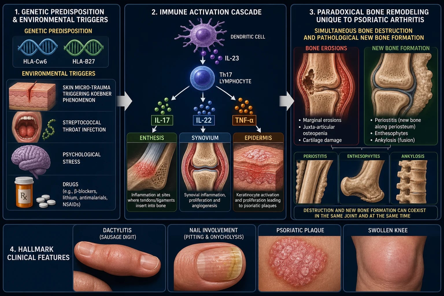
PsA arises from the interaction of genetic susceptibility, environmental triggers and immune dysregulation, converging on the enthesis as the unifying lesion of the spondyloarthropathies.[1]
The enthesis is the insertion of tendon, ligament or joint capsule into bone — a mechanically stressed transition zone (fibrocartilage over mineralised fibrocartilage over subchondral bone). In PsA, mechanical stress and microdamage in a genetically susceptible host trigger a sterile inflammatory cascade at the enthesis that then secondarily involves the adjacent joint and the overlying skin. This enthesis-centred model explains why enthesitis, dactylitis (a flexor-tendon entheseal and synovial phenomenon) and synovitis coexist, and why enthesitis is the unifying lesion of the entire SpA family.[1]
Activated dendritic cells in skin, gut and enthesis release IL-23 (and TNF). IL-23 drives Th17 cells and type-3 innate lymphoid cells to secrete IL-17A, IL-17F, IL-22 and IL-23 itself, generating a self-amplifying inflammatory loop alongside TNF-alpha. These cytokines act simultaneously at three target tissues — the enthesis, the synovium and the epidermis — explaining the multi-domain phenotype of PsA. This cytokine biology is directly therapeutic: TNF inhibitors were the first biologics (and remain first-line), then IL-17 inhibitors (secukinumab, ixekizumab, bimekizumab) and IL-23 inhibitors (guselkumab, risankizumab) and IL-12/23 inhibitors (ustekinumab) followed, each targeting a node of this cascade.[1][3]
HLA-Cw6 is strongly linked to the skin disease (psoriasis) and to early-onset, more erosive PsA; HLA-B27 is associated with axial (spinal) involvement but is found in only about 25 to 40 percent of axial-PsA patients — much lower than the 90 percent seen in ankylosing spondylitis. Additional loci include ERAP1 (whose risk effect is amplified in HLA-Cw6 carriers), IL23R, TNFAIP3, RUNX3 and others, with over 100 loci identified by genome-wide association.[1]
Koebner phenomenon (isomorphic response) is the appearance of psoriatic plaques at sites of skin trauma — a scratch, a surgical scar, a tattoo, sunburn. The same mechanical-stress principle operates at the enthesis: microdamage drives local inflammation. Other triggers include streptococcal pharyngitis (guttate psoriasis), stress, obesity, smoking, and drugs (lithium, beta-blockers, antimalarials, systemic corticosteroid withdrawal, NSAID withdrawal).[1]
The defining and unique feature of PsA is paradoxical bone remodelling: the same disease causes both erosive bone destruction and pathological new bone formation in the same joint. Look for the pencil-in-cup deformity at DIP joints, fluffy periostitis and enthesophytes near joint margins, bony ankylosis, and bulky asymmetric non-marginal syndesmophytes (parasyndesmophytes) in the spine. This duality reflects the IL-23 / Th17-driven enthesitis that first erodes the bone and then drives pathological repair via the Wnt and beta-catenin osteoproliferation pathway (modulated by DKK-1 and sclerostin); as inflammation resolves under treatment, the brake on osteoblasts is released, permitting new bone — which explains why anti-inflammatory therapy alone does not reliably halt radiographic progression.[1]
PSORIATIC — the disease at a glance
PSORIATIC
Inflammatory arthritis associated with psoriasis; skin disease usually precedes joints
RF and ACPA negative; a spondyloarthropathy alongside AS, reactive, IBD-associated
Asymmetric oligoarthritis is the commonest pattern; lower limbs and DIPs favoured
Simultaneous erosions AND new bone formation on X-ray — unique to PsA
IL-23 drives Th17 IL-17/IL-22 plus TNF; Koebner phenomenon; HLA-Cw6, HLA-B27
Severe patterns: ankylosis, spondylitis, opera-glass hand (arthritis mutilans)
NSAIDs, then methotrexate / DMARDs, then TNFi / IL-17 / IL-23 biologics to remission
CASPAR criteria classify PsA; X-ray shows pencil-in-cup, fluffy periostitis
Cardiovascular and metabolic disease, obesity, depression; screen for TB before biologics
Clinical Presentation
PsA is clinically heterogeneous — the same disease may present as a few swollen toes with nail pitting, a destructive arthritis mutilans, or an ankylosing-spondylitis-like spine. The articular, entheseal, skin, nail and systemic manifestations are each examinable.[1]
The articular presentation follows one of the five Moll and Wright patterns described above. Regardless of pattern, the arthritis is inflammatory: pain and stiffness that are worse in the morning and with rest, improve with activity, and are accompanied by morning stiffness of more than 30 minutes. Key distinguishing features from rheumatoid arthritis: asymmetry, lower-limb predominance, DIP involvement, dactylitis, enthesitis and RF and ACPA negativity.[1]
Dactylitis is the diffuse swelling of an entire digit (finger or toe) — the "sausage digit" — produced by simultaneous synovitis and enthesitis of the flexor tendon sheath. It is one of the most specific features of the spondyloarthropathies and contributes a point to the CASPAR criteria (current or historical). Dactylitis is painful, persistent, and a marker of more severe disease; on imaging it shows flexor-tendon tenosynovitis, joint synovitis and soft-tissue swelling.[1]
Enthesitis — inflammation at tendon or ligament insertions — is the unifying lesion of the spondyloarthropathies. The classic sites are the Achilles tendon insertion at the calcaneus and the plantar fascia origin (also at the calcaneus), producing heel pain that is worse on first standing in the morning. Other sites include the patellar tendon at the tibial tuberosity, the quadriceps insertion, the iliac crests, the ischial tuberosities and the costosternal junctions. Enthesitis is often the first symptom of PsA and may precede overt arthritis by years.[1]
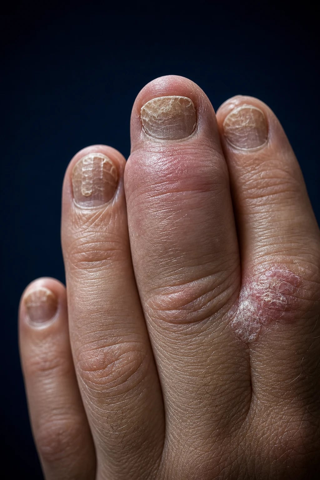
Nail disease is present in up to 80 percent of patients with PsA (versus about 30 percent of patients with psoriasis alone) and is the feature most strongly associated with joint disease. Examine every nail in any patient with psoriasis. The findings are pitting (small depressions from parakeratosis of the proximal nail matrix), onycholysis (separation of the nail plate from the bed), subungual hyperkeratosis (thick keratinous debris), discolouration (oil-drop sign, salmon patch), and onychorrhexis (longitudinal ridging) or nail crumbling in severe disease.[1]
Look for psoriasis at the classic sites: extensor surfaces of elbows and knees, the scalp (especially the retro-auricular margin), the umbilicus, gluteal cleft (natal cleft), intergluteal fold, genitals and the perianal skin — these hidden sites are easily missed and high-yield for diagnosis. Patterns include chronic plaque psoriasis (commonest), guttate psoriasis (small teardrop plaques, often after streptococcal pharyngitis), palmoplantar pustular psoriasis, erythrodermic psoriasis (medical emergency), and inverse (flexural) psoriasis. The Koebner phenomenon may produce linear plaques in surgical or scratch scars.[1]
Every system is examinable for the extra-articular features. Ocular: uveitis — in PsA classically more chronic, bilateral and posterior than the acute anterior uveitis of ankylosing spondylitis, though acute anterior uveitis also occurs; any red eye with visual change needs urgent ophthalmology. Gastrointestinal: clinical or subclinical inflammatory bowel disease (Crohn disease or ulcerative colitis) overlaps with PsA. Cardiovascular and metabolic: PsA carries excess cardiovascular risk driven by chronic inflammation — a cardiometabolic cluster of obesity, hypertension, dyslipidaemia, insulin resistance and type 2 diabetes, non-alcoholic fatty liver disease and metabolic syndrome. Psychological: depression and anxiety are common and disabling. Constitutional: fatigue is often the most disabling symptom; low-grade fever and weight loss may occur in severe disease.[1][3]
Atypical presentations must be recognised. Arthritis preceding psoriasis (10 to 15 percent) has no visible skin disease at presentation — diagnose on family history, dactylitis, enthesitis, RF negativity, nail changes and juxta-articular new bone. Juvenile psoriatic arthritis is an asymmetric large-joint oligoarthritis with nail pitting, dactylitis or a family history of psoriasis. Acute monoarthritis mimicking gout or sepsis — a hot, swollen joint in a patient with psoriasis — must be aspirated first to exclude septic arthritis, which can coexist. Predominant axial disease presenting as inflammatory back pain in a patient known to have psoriasis warrants MRI of the sacroiliac joints (asymmetric sacroiliitis) even if the plain film is normal.[1]
Differential Diagnosis
The pivotal differential is rheumatoid arthritis versus psoriatic arthritis versus osteoarthritis, with gout, reactive arthritis and ankylosing spondylitis completing the set.[1]
Psoriatic arthritis
- Asymmetric, oligoarticular, DIP involved; enthesitis, dactylitis (sausage digit), nail dystrophy, psoriasis
- RF and ACPA NEGATIVE; HLA-Cw6 (skin), HLA-B27 (axial); IL-23 / Th17 axis
- X-ray: erosions WITH new bone formation; pencil-in-cup, fluffy periostitis, enthesophytes, parasyndesmophytes
- NSAIDs, methotrexate / DMARDs; TNFi first-line biologic; IL-17 / IL-23 inhibitors
Rheumatoid arthritis
- SYMMETRIC, small joints of hands and feet; DIP SPARED; prolonged morning stiffness over 1 hour
- RF and ACPA POSITIVE; HLA-DR4; CD4 T-cell and B-cell driven
- X-ray: marginal erosions, periarticular osteopenia, symmetric joint-space narrowing; NO new bone
- Methotrexate first DMARD; add TNFi / IL-6 / JAK if refractory; treat-to-target (DAS28)
Osteoarthritis
- Non-inflammatory; DIP (Heberden) and PIP (Bouchard) nodes, first CMC, knees, hips; brief stiffness
- RF / ACPA negative; age and mechanical load driven; CRP normal
- X-ray: osteophytes, subchondral sclerosis, asymmetric joint-space narrowing, subchondral cysts
- Analgesia, weight loss, physiotherapy, joint replacement
Ankylosing spondylitis
- Inflammatory back pain (young male); bilateral symmetric sacroiliitis, bamboo spine; uveitis; HLA-B27 about 90%
- RF / ACPA negative; enthesitis, dactylitis; overlaps with PsA axial pattern
- X-ray / MRI: bilateral sacroiliitis, delicate marginal syndesmophytes, bamboo spine, shiny corners
- NSAIDs first-line; TNFi or IL-17i for refractory; physiotherapy
Gout
- Acute monoarthritis (first MTP, podagra); tophi; hyperuricaemia; triggered by alcohol, purines, diuretics
- Negatively birefringent monosodium urate crystals in synovial fluid; RF / ACPA negative
- X-ray: punched-out erosions with overhanging edges (rat-bite); preserved joint space until late
- NSAIDs / colchicine for acute attack; allopurinol / febuxostat for urate lowering
Reactive arthritis
- 1 to 4 weeks after GU (Chlamydia) or GI (Salmonella, Shigella, Campylobacter, Yersinia) infection
- Asymmetric lower-limb oligoarthritis, enthesitis, dactylitis; RF / ACPA negative
- Often self-limiting; sacroiliitis may be asymmetric; fluffy periostitis overlaps with PsA
- NSAIDs; antibiotics for the trigger infection if persistent; TNFi if severe and refractory
The single most examinable comparison is PsA versus RA: PsA is RF and ACPA negative, asymmetric, lower-limb and DIP predominant, with enthesitis, dactylitis and nail and skin disease, and shows erosions with new bone on X-ray. RA is RF and ACPA positive, symmetric, upper-limb and small-joint predominant, with DIP spared, no enthesitis or dactylitis, and shows purely erosive change (marginal erosions, periarticular osteopenia, no new bone). If a patient with apparent RA has DIP involvement, dactylitis, enthesitis or psoriasis, think PsA — and check RF and ACPA.[1]
The can't-miss mimics are septic arthritis (hot joint, systemically unwell — aspirate and culture), gout and CPPD (crystals on synovial microscopy, hyperuricaemia for gout), and HIV-associated arthritis (an explosive, treatment-resistant SpA-like picture in advanced HIV — check HIV serology in the right clinical context before starting immunosuppression).[1]
Clinical & Bedside Assessment
The diagnosis rests on pattern recognition at the bedside. A focused examination looks for skin, nails, joints, entheses, dactylitis, spine, eyes and comorbidities.[1]
Examine the skin at the extensor elbows and knees, scalp (including retro-auricular), umbilicus, gluteal and natal cleft, genitals, perianal skin and flexures — the easily missed sites where a single plaque of psoriasis earns 2 CASPAR points. Examine every nail for pitting, onycholysis, hyperkeratosis, discolouration and ridging — nail disease is the feature most strongly associated with PsA and contributes a CASPAR point. Perform a full 68/66 joint count (tender and swollen); look specifically for DIP involvement (versus RA, which spares the DIP), asymmetry and lower-limb predominance; squeeze across the metacarpophalangeal and metatarsophalangeal joints for tenderness.[1]
Palpate the entheses — Achilles tendon insertion, plantar fascia origin, patellar tendon insertion, tibial tuberosity and costochondral junctions — for tenderness (quantify with the Leeds Enthesitis Index or SPARCC). Look for the sausage digit of dactylitis (quantify with the Leeds Dactylitis Index). If axial disease is suspected, assess for inflammatory back pain (worse at rest, better with exercise, morning stiffness, alternating buttock pain) and measure modified Schober, occiput-to-wall distance and chest expansion. Examine the eyes for redness, photophobia and visual acuity — uveitis needs urgent ophthalmology.[1]
Measure BMI, blood pressure, lipids, HbA1c and liver function, screen for depression, and ask about smoking and alcohol — PsA carries excess cardiovascular risk, and these are part of routine care, not an afterthought. The PEST (Psoriasis Epidemiology Screening Tool) — five self-report questions — screens dermatology patients for PsA; a score of 3 or more triggers rheumatology referral.[3]
For disease activity, use the DAPSA (Disease Activity Score for Psoriatic Arthritis) — tender joint count (68) plus swollen joint count (66) plus patient pain (0 to 10) plus patient global (0 to 10) plus CRP (mg per dL); remission is under 4, low activity 4 to 14, moderate 14 to 28, high over 28. The treat-to-target endpoint is Minimal Disease Activity (MDA), which requires 5 of 7 of: tender joint count under 1, swollen joint count under 1, PASI under 1 (or BSA under 3), patient pain under 15 mm, patient global under 20 mm, HAQ under 0.5, tender entheseal points under 1. The PASI (Psoriasis Area and Severity Index) is the standard skin severity score.[3][4]
Investigations
PsA is a clinical diagnosis — apply the CASPAR criteria; no single test confirms it. Investigations support the diagnosis, exclude mimics, stage disease and prepare for treatment.[1]
Rheumatoid factor (RF) is negative (the key discriminator from RA) and contributes a CASPAR point; note that up to 13 percent of PsA patients are RF positive in low titre — a positive RF does not exclude PsA if other features dominate. ACPA (anti-CCP) is negative and more specific than RF for RA, so a negative result is reassuring. CRP and ESR are often raised in active disease (but may be normal); a baseline and on-treatment value track response. HLA-B27 may be requested when axial disease is prominent. Baseline full blood count, urea and electrolytes, liver function are required before DMARDs and biologics; hepatitis B and C and HIV are screened before biologics (and HIV serology in a treatment-resistant explosive SpA picture).[1]
If a hot joint is aspirated to exclude sepsis, PsA synovial fluid is inflammatory (white cell count 2,000 to 50,000 per microlitre, predominantly neutrophilic), sterile and crystal-negative. Check serum urate to exclude gout (though hyperuricaemia and PsA can coexist).[1]
The radiographic hallmarks of established PsA on plain X-ray of the hands, feet and pelvis are the pencil-in-cup deformity at DIP joints (the eroded, narrowed base of the middle phalanx sits in the widened, cup-shaped head of the distal phalanx — classic and highly characteristic), fluffy periostitis (ill-defined new bone near joint margins — the CASPAR "juxta-articular new bone formation"), marginal erosions that coexist with new bone (the paradoxical remodelling), bony ankylosis of small joints, sausage-digit soft-tissue swelling with acro-osteolysis, asymmetric sacroiliitis (versus the bilateral symmetric pattern of ankylosing spondylitis), bulky asymmetric non-marginal syndesmophytes (parasyndesmophytes) in the spine, and the ivory phalanx (sclerosis of a distal phalanx).[1]
MRI of the sacroiliac joints and peripheral joints is the cornerstone for early disease: STIR or T2 fat-saturated sequences show bone-marrow oedema (osteitis) at entheses and sacroiliac joints — the earliest lesion, preceding plain X-ray by years — and reveal synovitis, tenosynovitis (flexor tendon sheath in dactylitis), enthesitis and bone erosion. Whole-body MRI can map multi-site enthesitis and dactylitis for research and difficult cases. Musculoskeletal ultrasound at the bedside detects synovitis, tenosynovitis, enthesitis (with power Doppler) and nail disease, and guides injections. DEXA screens for the osteoporosis of chronic inflammation and immobility.[1]
Management — Resuscitation
PsA is not usually a resuscitation diagnosis, but three situations are time-critical and must be recognised first.[1]
Acute hot monoarthritis must be aspirated before treating, to exclude septic arthritis (which can coexist with PsA and is easily missed). Send synovial fluid for cell count, Gram stain, culture and crystals. If septic arthritis cannot be excluded, treat as septic (joint washout and intravenous antibiotics) until culture is negative. Acute uveitis — a red eye with photophobia and visual change — is uveitis, not conjunctivitis; refer urgently to ophthalmology within 24 hours. Treatment is topical corticosteroid (prednisolone acetate 1 percent drops, hourly for severe attacks) plus a cycloplegic (cyclopentolate 1 percent or homatropine 2 percent) to relieve ciliary spasm and prevent posterior synechiae. If uveitis is recurrent, favour a TNF inhibitor monoclonal antibody (adalimumab, infliximab, certolizumab) over etanercept, which is less effective for uveitis. New axial pain with bowel or bladder dysfunction, saddle anaesthesia or progressive leg weakness must be imaged urgently to exclude spinal instability or cauda equina syndrome.[1]
Management — Definitive & Stepwise
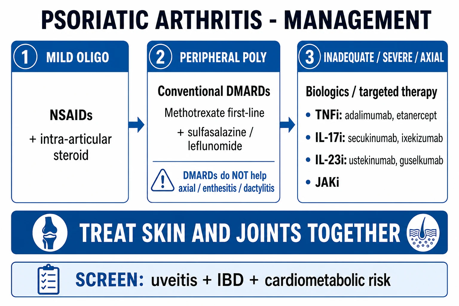
Modern PsA management is domain-driven and treat-to-target, coordinated between rheumatology and dermatology (and gastroenterology, ophthalmology and psychology as needed). The target is minimal disease activity or remission across all active domains — peripheral joints, axial skeleton, entheses, dactylitis, skin and nails.[1][3][4]
Treat-to-target (GRAPPA 2021 and EULAR 2023)
Both the GRAPPA 2021 update and the EULAR 2023 update endorse a treat-to-target approach aiming for Minimal Disease Activity (MDA) or remission, with regular assessment and escalation if the target is not met. The choice of therapy should address every active domain in that patient (e.g. a patient with peripheral arthritis plus severe skin disease and nail dystrophy benefits from an agent effective across joints and skin, such as a TNF inhibitor, IL-17 inhibitor or IL-23 inhibitor).[3][4]
Step 1 — NSAIDs (first-line for mild disease)
NSAIDs at full anti-inflammatory dose are first-line for mild peripheral disease and mild axial symptoms. EULAR 2023 specifies NSAIDs in monotherapy only for mild PsA and short-term. Try at least two different NSAIDs over 2 to 4 weeks before declaring failure.[3][4]
| NSAID | Dose and route | Frequency |
|---|---|---|
| Naproxen | 500 mg orally | Twice daily |
| Diclofenac | 50 to 75 mg orally | Two to three times daily |
| Ibuprofen | 400 to 800 mg orally | Three to four times daily |
| Indometacin | 25 to 50 mg orally | Two to three times daily (effective but more CNS and GI side-effects) |
| Celecoxib | 100 to 200 mg orally | Once to twice daily (COX-2 selective, GI-sparing) |
| Doses are oral adult doses per GRAPPA 2021 and EULAR 2023.[3][4] |
Add a proton-pump inhibitor (omeprazole 20 mg once daily) for patients with GI risk. Avoid NSAIDs in significant renal impairment, severe heart failure, active peptic ulcer, anticoagulation, and late pregnancy.[3]
Step 2 — Local glucocorticoid injections
Intra-articular or intra-lesional corticosteroid injection (e.g. triamcinolone acetonide 40 mg into a swollen joint or enthesis) is useful for focal inflammation, including a stubbornly inflamed DIP or a single enthesis. Oral glucocorticoids are NOT recommended by EULAR 2023 — they are ineffective for axial disease, carry long-term toxicity, and rapid withdrawal can flare psoriasis.[4]
Step 3 — Conventional synthetic DMARDs (csDMARDs)
For moderate-to-severe peripheral arthritis (multiple swollen joints, elevated CRP, or failure of NSAIDs), start a conventional synthetic DMARD. EULAR 2023 recommends rapid initiation of a csDMARD and prefers methotrexate. Note that csDMARDs are effective for peripheral arthritis but NOT for axial disease or enthesitis — axial disease needs an NSAID or a biologic.[4]
| csDMARD | Dose and route | Notes |
|---|---|---|
| Methotrexate | 10 to 25 mg once weekly orally or subcutaneously (start 7.5 to 10 mg, titrate up); plus folic acid 5 mg once weekly | First-line; also improves skin psoriasis; monitor CBC and LFT (monthly for 3 months then 3-monthly); screen hepatitis and latent TB; teratogenic — stop 3 months before conception in both sexes |
| Sulfasalazine | 2 to 3 g per day orally in divided doses (start 500 mg once daily, titrate up) | Peripheral arthritis and IBD overlap; monitor CBC and LFT; sulpha allergy; haemolysis in G6PD deficiency (screen); reversible male infertility |
| Leflunomide | Loading 100 mg once daily for 3 days then 10 to 20 mg once daily orally | Alternative or add-on; monitor LFT and blood pressure; long half-life; teratogenic — cholestyramine washout before conception |
| Doses per GRAPPA 2021 and EULAR 2023; methotrexate is preferred because it also improves skin disease.[3][4] |
Step 4 — Biologic DMARDs (bDMARDs)
If the target is not met with csDMARDs, or if disease is axial, refractory, or severely impacting quality of life, advance to a biologic DMARD. EULAR 2023 recommends initiating a bDMARD without preference among modes of action (TNFi, IL-17i, IL-23i) for peripheral disease, but skin-predominant disease should orient towards IL-23p40, IL-23p19 or IL-17A / IL-17AF inhibitors, and uveitis or IBD should orient towards a monoclonal TNF inhibitor.[4]
TNF inhibitors are the most extensively used biologic class in PsA, effective across peripheral arthritis, axial disease, enthesitis, dactylitis, skin and nails. The pivotal ADEPT trial of adalimumab demonstrated significant improvement in joint and skin manifestations, inhibition of radiographic progression, improved function and quality of life in moderate-to-severe PsA.[5]
| TNF inhibitor | Dose and route | Notes |
|---|---|---|
| Adalimumab | 40 mg subcutaneously every 2 weeks | Fully human monoclonal anti-TNF; effective for uveitis; low placental transfer |
| Etanercept | 50 mg subcutaneously weekly | Fusion protein (TNF receptor-Fc); effective for joints and skin but LESS effective for uveitis — avoid in recurrent uveitis |
| Infliximab | 5 mg/kg intravenously at weeks 0, 2, 6, then every 8 weeks | Chimeric monoclonal; infusion reactions; effective for uveitis |
| Golimumab | 50 mg subcutaneously monthly | Once-monthly dosing; convenient |
| Certolizumab pegol | 400 mg at weeks 0, 2, 4, then 200 mg every 2 weeks (or 400 mg every 4 weeks) | PEGylated; absent placental transfer and minimal breast-milk transfer — preferred in pregnancy |
| Doses per GRAPPA 2021 and EULAR 2023; ADEPT (Mease 2005) established adalimumab efficacy and inhibition of radiographic progression.[3][4][5] |
IL-17 inhibitors are highly effective for peripheral arthritis, axial disease, enthesitis, dactylitis and (especially) skin and nails. Secukinumab is given as 150 mg subcutaneously at weeks 0, 1, 2, 3, 4, then 150 to 300 mg monthly; ixekizumab as a 160 mg loading dose, then 80 mg every 2 weeks for 12 weeks, then every 4 weeks; bimekizumab (dual IL-17A and IL-17F blockade) as a 320 mg loading dose then 320 mg every 8 weeks (every 4 weeks if inadequate response). Caution — IL-17 inhibitors may worsen or trigger inflammatory bowel disease, so avoid them in IBD.[3][4]
IL-23 and IL-12/23 inhibitors are excellent for peripheral arthritis and for skin-predominant disease, and effective for enthesitis and dactylitis, but are not effective for axial PsA (a key gap). Ustekinumab (IL-12/23) is given weight-based: 45 mg (under 100 kg) or 90 mg (over 100 kg) subcutaneously at weeks 0, 4, then every 12 weeks. Guselkumab (IL-23) is 100 mg at weeks 0, 4, then every 8 weeks. Risankizumab (IL-23) is 150 mg at weeks 0, 4, then every 12 weeks, with very high skin clearance.[3][4]
EULAR 2023 positions JAK inhibitors primarily after a bDMARD has failed, or when a bDMARD is not appropriate, taking risk factors into account (venous thromboembolism, malignancy, cardiovascular disease, age over 65). Tofacitinib is given as 5 mg orally twice daily; upadacitinib as 15 mg orally once daily (JAK1-selective); filgotinib as 200 mg orally once daily (not available in the USA; available in Europe and Japan). All share class cautions of VTE, herpes zoster and lipid and LFT monitoring. The PDE4 inhibitor apremilast — 30 mg orally twice daily (titrate up from 10 mg to minimise GI intolerance) — is effective for oral ulcers, mild skin disease and mild peripheral arthritis, with no requirement for TB screening or blood monitoring; common side-effects are nausea, diarrhoea and weight loss.[4]
Pre-biologic screen and safety
Before any biologic or JAK inhibitor, screen for latent tuberculosis with IGRA (QuantiFERON-TB Gold) and chest X-ray; treat latent TB (e.g. isoniazid 300 mg once daily for 6 to 9 months or rifampicin plus isoniazid for 3 to 4 months) before starting a TNF inhibitor. Screen for hepatitis B and C (HBsAg, anti-HBc, anti-HBs, anti-HCV) and treat or refer before starting; check HIV in appropriate risk groups; record CBC, LFT, renal function, lipids, blood pressure, pregnancy test. Vaccinate before immunosuppression (pneumococcal, influenza, hepatitis B, COVID-19, HPV, recombinant herpes zoster) and avoid live vaccines during biologic therapy. Coordinate with dermatology for skin assessment and gastroenterology if IBD coexists (favour monoclonal TNFi; avoid IL-17i in IBD); counsel on contraception (many agents are teratogenic).[3][4]
EULAR 2023 recommends: NSAIDs monotherapy only for mild PsA, short term; no oral glucocorticoids; rapid csDMARD (methotrexate preferred) for peripheral arthritis; bDMARD without preference among modes of action if the target is not met; skin disease should orient towards IL-23p40 / IL-23p19 / IL-17A / IL-17AF; uveitis or IBD should orient towards a monoclonal TNFi; JAK inhibitors mainly after bDMARD failure taking risk factors into account.[4]
Adjuncts and lifestyle
Physiotherapy and exercise preserve joint mobility and muscle strength — non-negotiable, especially in axial disease. Smoking cessation improves both skin and joint disease and biologic response (smokers have more severe disease). Weight loss improves biologic efficacy and reduces cardiovascular risk. Cardiovascular risk modification (statins, antihypertensives, glycaemic control) is essential — PsA carries excess cardiovascular mortality. Occupational therapy, splints for dactylitis, and psychological support for depression and anxiety complete the package.[1][3]
Specific Subtypes & Scenarios
Asymmetric oligoarthritis (the commonest pattern) is managed with NSAIDs plus local corticosteroid injection for symptomatic joints, escalating to a csDMARD (methotrexate) if recurrent or persistent, and a biologic if refractory; prognosis is excellent with early treatment. Symmetric polyarthritis (RA-like) is treated with methotrexate first-line, treat-to-target with DAPSA, escalating to biologics as in RA but with the option of IL-17 and IL-23 inhibitors not available in RA. DIP-predominant arthritis, strongly linked to nail disease, is managed with NSAIDs, methotrexate, or a biologic (TNFi or IL-17i — IL-17i particularly effective for nail disease); intra-articular steroid to a DIP is technically difficult but useful.[1][3]
Arthritis mutilans (opera-glass hand) is destructive osteolysis with telescoping digits — it needs aggressive early biologic therapy to halt destruction and surgical reconstruction (arthrodesis, arthroplasty) for salvage; it is often combined with spondylitis. Predominant axial disease (spondylitis, sacroiliitis) is treated with NSAIDs first-line and a biologic if active (TNFi or IL-17i — both effective for axial PsA; IL-23 inhibitors are NOT effective for axial disease and must not be chosen for isolated axial PsA); csDMARDs do not work for axial disease.[4]
Psoriasis without joint symptoms should be screened proactively with PEST; up to a third of psoriasis patients have undiagnosed PsA, and a score of 3 or more triggers rheumatology referral. Arthritis preceding psoriasis is diagnosed on family history, dactylitis, enthesitis, RF negativity, nail changes and juxta-articular new bone; CASPAR still applies (family history, dactylitis, RF negative, nail dystrophy and new bone formation can complete 3 points even without current psoriasis).[2]
Juvenile psoriatic arthritis is an asymmetric large-joint oligoarthritis with nail pitting, dactylitis, or psoriasis in the child or a first-degree relative; treat as juvenile idiopathic arthritis with NSAIDs and intra-articular steroids first, then methotrexate and biologics. Screen for uveitis (slit-lamp every 6 to 12 months). Severe or refractory disease in adults is escalated biologic-naive to TNFi then IL-17i or IL-23i; consider switching class if the first biologic fails at 12 to 16 weeks (about 40 to 50 percent respond to the second agent); coordinate rheumatology, dermatology and where needed gastroenterology and ophthalmology.[1][3]
Complications & Pitfalls
The principal musculoskeletal complication is joint destruction, deformity, arthritis mutilans with telescoping digits and functional impairment — the outcome of untreated disease. Uveitis is a visual threat; untreated attacks cause posterior synechiae, glaucoma and cataract. Inflammatory bowel disease overlaps (Crohn disease, ulcerative colitis) — coordinate with gastroenterology. Cardiovascular disease is the leading driver of excess mortality, amplified by the cardiometabolic cluster (obesity, diabetes, hypertension, dyslipidaemia, fatty liver). Metabolic syndrome, non-alcoholic fatty liver disease, depression, anxiety and fatigue are common; osteoporosis results from chronic inflammation and immobility. Drug-related complications include NSAID gastropathy and renal effects, methotrexate hepatotoxicity and pneumonitis, biologic infections (TB reactivation, serious bacterial infections), and JAK inhibitor VTE, herpes zoster and malignancy signals.[1][4]
The common pitfalls are missing PsA because the psoriasis is hidden (always examine the scalp, umbilicus, natal cleft, genitals and nails); mislabelling PsA as RA because RF happened to be low-positive (re-examine for DIP involvement, dactylitis, enthesitis and nail disease, and check ACPA); aspirating a hot joint as "a flare of PsA" without excluding sepsis (always culture); using systemic corticosteroids for axial disease (ineffective and toxic; rapid withdrawal flares psoriasis); forgetting pre-biologic TB and hepatitis screening (reactivation TB is a preventable disaster); choosing an IL-17 inhibitor in IBD (may worsen bowel disease — choose a monoclonal TNFi); choosing an IL-23 inhibitor for isolated axial disease (not effective); choosing etanercept for recurrent uveitis (less effective — choose adalimumab, infliximab or certolizumab); neglecting cardiovascular and metabolic comorbidity (the leading driver of excess mortality); and ignoring the skin and nails (domain-driven care means treating skin and nails — often IL-17i or IL-23i are best for nails — alongside the joints).[1][4]
Prognosis & Disposition
The natural history is highly variable — some patients have mild disease controlled on NSAIDs for decades; others develop destructive arthritis mutilans, an ankylosing-spondylitis-like spine and systemic complications within years. Early diagnosis and treat-to-target have transformed outcomes, reducing radiographic progression and disability.[1][3]
The poor prognostic factors are polyarticular or axial disease at presentation; elevated acute-phase reactants (CRP, ESR) at baseline; HLA-B27 positivity (especially with axial disease), HLA-DR4 and HLA-Cw6 (erosive disease); erosions on baseline X-ray (predicts further radiographic progression); dactylitis (a marker of more severe disease and future erosions); young age at onset, family history, obesity and smoking; and delay in starting DMARD or biologic therapy.[1]
Mortality is modestly increased (standardised mortality ratio about 1.3 to 1.6), driven by cardiovascular disease (chronic inflammation and the cardiometabolic cluster), complications of severe disease, and the side-effects of long-term immunosuppression. Aggressive cardiovascular risk-factor modification is part of routine care. Untreated, up to 20 to 40 percent of patients develop significant functional impairment; modern treat-to-target and biologics substantially reduce this, and workplace modifications and occupational therapy remain important.[1]
All patients with suspected PsA should be referred to rheumatology for confirmation and treatment; coordinate with dermatology for skin disease, ophthalmology for uveitis, gastroenterology for IBD, and cardiology for cardiovascular risk. A safety-net of regular monitoring of disease activity, drug toxicity and comorbidity (at least every 3 to 6 months) underpins long-term care.[3]
Special Populations
Pregnancy and lactation
NSAIDs are safe in the first and second trimester but should be avoided after 28 to 30 weeks (premature closure of the ductus arteriosus and oligohydramnios); ibuprofen and diclofenac are low-risk in lactation. Sulfasalazine is safe throughout pregnancy and lactation; supplement with folic acid 5 mg once daily. Low-dose corticosteroids (prednisolone under 10 mg once daily) are acceptable; avoid in the first trimester where possible (oral cleft concern) and in late pregnancy (gestational diabetes, premature rupture of membranes).[3]
Among the biologics, certolizumab is preferred because of absent placental transfer (no Fc region); adalimumab and infliximab are generally continued through the first and second trimesters and stopped in the third to avoid placental transfer; etanercept is low-risk. Methotrexate and leflunomide are TERATOGENIC and CONTRAINDICATED — stop methotrexate at least 3 months before conception (both men and women) and use cholestyramine washout for leflunomab. JAK inhibitors and IL-17 / IL-23 inhibitors should be avoided in pregnancy and lactation (limited data).[3][4]
Children (juvenile psoriatic arthritis)
Juvenile PsA is an asymmetric large-joint oligoarthritis with nail pitting, dactylitis, or psoriasis in the child or a first-degree relative; treat as juvenile idiopathic arthritis with NSAIDs and intra-articular steroids first, then methotrexate and biologics (etanercept, adalimumab). Screen for uveitis (slit-lamp every 6 to 12 months).[1]
Elderly
Late-onset PsA may present with more metabolic comorbidity and is easily confused with osteoarthritis or polymyalgia rheumatica; NSAID and methotrexate toxicity is higher — adjust doses for renal function and polypharmacy. Biologics are effective and generally well tolerated in selected elderly patients.[3]
Immunocompromised or on-biologic host
Vaccinate before starting the biologic (pneumococcal, influenza, hepatitis B, COVID-19, recombinant herpes zoster); avoid live vaccines during biologic therapy; monitor for infection (TB, opportunistic) with a low threshold for culture; coordinate with infectious diseases if latent TB is treated, hepatitis B is reactivated, or HIV coexists.[3][4]
Evidence, Guidelines & Regional Differences
GRAPPA 2021 treatment recommendations (international)
The Group for Research and Assessment of Psoriasis and Psoriatic Arthritis issued its third update in 2021 (Coates et al, 2022), with domain-focused recommendations for peripheral arthritis, axial disease, enthesitis, dactylitis, and skin and nail psoriasis, plus PsA-related conditions (uveitis, IBD) and comorbidities. Overarching principles emphasise shared decision-making, treating all active domains, and screening for and managing comorbidities. The recommendations strongly endorse NSAIDs, csDMARDs (methotrexate first), TNF inhibitors, IL-17 inhibitors, IL-12/23 and IL-23 inhibitors, JAK inhibitors and PDE4 inhibitors, with the choice tailored to the dominant domain and the patient's comorbidities. The 2021 update added principles on biosimilars and tapering of therapy in sustained remission.[3]
EULAR 2023 treatment recommendations (international)
The 2023 update (Gossec, Smolen et al, 2024) comprises 7 overarching principles and 11 recommendations. Key points: NSAIDs in monotherapy only for mild PsA, short term; oral glucocorticoids NOT recommended; rapid csDMARD (methotrexate preferred) for peripheral arthritis; bDMARD without preference among modes of action if the target is not met; skin psoriasis should orient towards IL-23p40, IL-23p19, IL-17A and IL-17AF inhibitors; predominant entheseal or axial disease has its own algorithm; JAK inhibitors mainly after bDMARD failure taking risk factors into account; uveitis or IBD should orient towards a monoclonal TNFi; drug switches and tapering in sustained remission are addressed.[4]
ACR / National Psoriasis Foundation 2018 guidance (USA) and NICE NG65 (UK)
The ACR / NPF 2018 guidance endorses TNFi, IL-17i and IL-12/23i as first-line biologics (no strict ordering), PDE4 and JAK inhibitors as oral alternatives, and permits biologic as first-line for severe disease; it does not mandate a csDMARD trial for axial or severe skin disease. NICE NG65 (UK, 2017 with updates) mandates a DMARD-first approach for peripheral arthritis before a biologic; favours the cheapest biologic within a class (biosimilars); and prefers certolizumab in pregnancy.[3]
Landmark trials — what they changed
ADEPT (Mease 2005) showed adalimumab significantly improved joint and skin manifestations, inhibited radiographic progression, improved function and quality of life in moderate-to-severe PsA — establishing TNF inhibition as a cornerstone. FUTURE 1 and 2 (secukinumab) showed IL-17A inhibition effective across peripheral arthritis, axial disease, enthesitis, dactylitis and skin, establishing IL-17 blockade. PSUMMIT 1 and 2 (ustekinumab) showed IL-12/23 blockade effective for peripheral arthritis and skin. DISCOVER 1 and 2 (guselkumab) showed selective IL-23 blockade effective for peripheral arthritis and very high skin clearance but not for axial disease — a key evidence gap shaping domain-driven prescribing. SELECT-PsA 1 and 2 (tofacitinib) showed JAK inhibition effective for peripheral arthritis after csDMARD or bDMARD failure.[5][3][4]
Controversies and open questions
Treat-to-target with MDA improves outcomes but the optimal target and frequency of assessment are still refined. IL-23 inhibitors work for peripheral but not axial PsA — a biologically puzzling dissociation that shapes domain-driven prescribing. Tapering of biologics in sustained remission is feasible for many but risks flare; EULAR and GRAPPA differ in emphasis. Obesity reduces biologic efficacy — weight loss as a therapeutic strategy is increasingly recognised. Cardiometabolic comorbidity is under-treated in PsA; whether aggressive systemic inflammation control reduces cardiovascular events is under study.[3][4]
Exam Pearls
- Psoriatic arthritis = inflammatory arthritis plus psoriasis plus negative RF; a seronegative spondyloarthropathy.[1]
- Genetics: HLA-Cw6 (skin), HLA-B27 (axial); pathophysiology is IL-23 / Th17 / IL-17 plus TNF; Koebner phenomenon.[1]
- Five Moll and Wright patterns; asymmetric oligoarthritis is the commonest (about half of patients).[1]
- Hallmarks: dactylitis (sausage digit), enthesitis (Achilles, plantar fascia), nail disease (pitting, onycholysis, hyperkeratosis), DIP involvement.[1]
- CASPAR criteria classify PsA clinically — current psoriasis 2 points, personal or family history, dactylitis, juxta-articular new bone formation, RF negative, nail dystrophy — 1 point each; at least 3 points.[2]
- X-ray: pencil-in-cup deformity, fluffy periostitis, erosions with new bone formation (paradoxical remodelling); asymmetric sacroiliitis, bulky parasyndesmophytes.[1]
- NSAIDs first-line (mild disease, short term); methotrexate for moderate peripheral; TNF inhibitors first-line biologic class; IL-17 and IL-23 inhibitors for refractory or skin-predominant disease; JAK inhibitors after bDMARD failure.[3][4]
- Screen for latent TB before biologics; manage cardiovascular and metabolic comorbidity.[3]
- Distinguish from RA — PsA is RF and ACPA negative, DIP involved, asymmetric, enthesitis and dactylitis, psoriasis present.[1]
- Nail disease is the feature most strongly associated with PsA — examine every nail.[1]
- Always aspirate a hot joint to exclude sepsis; refer uveitis urgently to ophthalmology.[1]
- Avoid IL-17 inhibitors in IBD; choose a monoclonal TNFi for uveitis or IBD; IL-23 inhibitors do NOT work for axial disease.[4]
- Certolizumab is the preferred biologic in pregnancy (no placental transfer); methotrexate and leflunomide are teratogenic and contraindicated.[3]
CASPAR criteria — the 6-item mnemonic
CASPAR
current psoriasis = 2 points (the heaviest item)
juxta-articular new bone formation on X-ray = 1 point
current or historical dactylitis = 1 point
personal or family history of psoriasis = 1 point (if no current psoriasis)
RF negative = 1 point
onycholysis, pitting, hyperkeratosis = 1 point
Self-test: CASPAR scoring
A 38-year-old woman has inflammatory arthritis of three fingers, current plaque psoriasis on the elbows, onycholysis of two fingernails, and RF negative. Score: current psoriasis (2) plus nail dystrophy (1) plus RF negative (1) = 4 points — CASPAR positive (threshold 3).[2]
Self-test: biologic choice by domain
A 45-year-old man with PsA has recurrent uveitis and mild IBD. Best biologic class? A monoclonal TNF inhibitor (adalimumab, infliximab or certolizumab) — effective for uveitis and IBD. Avoid etanercept (worse for uveitis) and IL-17 inhibitors (may worsen IBD). IL-23 inhibitors help IBD but not uveitis.[4]
Exam application bank (NEET-PG / INICET)
One-line answer
Psoriatic arthritis (PsA) is an inflammatory arthritis associated with psoriasis, usually rheumatoid-factor negative, classified within the seronegative spondyloarthropathies. Diagnosis is clinical using the CASPAR criteria (established inflammatory articular disease plus at least 3 points from current psoriasis, personal or family history of psoriasis, current or historical dactylitis, juxta-articular new bone formation, rheumatoid-factor negativity, and nail dystrophy). The five Moll and Wright patterns are asymmetric oligoarthritis (commonest), symmetric polyarthritis, DIP-predominant, arthritis mutilans, and predominant spondylitis or sacroiliitis. Hallmarks: dactylitis (sausage digit), enthesitis (Achilles, plantar fascia), nail disease (pitting, onycholysis, hyperkeratosis), and DIP involvement. Pathophysiology is genetic (HLA-Cw6 for skin, HLA-B27 for axial) plus environmental tri
Worked stems (answer without another resource)
Stem 1 — Classic presentation. Map symptoms to mechanism; name the first investigation and first treatment step with dose/route if drug therapy is standard. [1]
Stem 2 — Unstable / complicated. List red flags that force immediate resuscitation, theatre, ICU, antidote, or reperfusion — and what you do in the first 15 minutes. [1]
Stem 3 — Atypical group. Elderly, pregnancy, child, or immunocompromised: how presentation and thresholds change. [1]
Stem 4 — Differential trap. Name the three closest mimics and one discriminator for each. [1]
Stem 5 — Disposition. Who goes home with safety-netting, who is admitted, who needs HDU/ICU/theatre, and what follow-up is mandatory. [1]
Rapid viva checklist
- Definition + classification
- Pathophysiology chain
- Bedside signs / criteria
- Score with exact components (if any)
- Emergency bundle
- Definitive therapy with doses
- Complications of disease and of treatment
- Special populations
- Guideline/trial name if classic
- Three exam traps
Coverage self-check
If you cannot answer any stem above from this page alone, re-read the matching section — the page is intended to be self-sufficient for final-prof and NEET-PG/INICET questions on Psoriatic Arthritis.
References
- [1]Ritchlin CT, Colbert RA, Gladman DD. Psoriatic Arthritis N Engl J Med, 2017.PMID 28273019
- [2]Taylor W, Gladman D, Helliwell P, Marchesoni A, Mease P, Mielants H; CASPAR Study Group. Classification criteria for psoriatic arthritis: development of new criteria from a large international study Arthritis Rheum, 2006.PMID 16871531
- [3]Coates LC, Soriano ER, Corp N, et al. Group for Research and Assessment of Psoriasis and Psoriatic Arthritis (GRAPPA): updated treatment recommendations for psoriatic arthritis 2021 Nat Rev Rheumatol, 2022.PMID 35761070
- [4]Gossec L, Kerschbaumer A, Ferreira RJO, et al. EULAR recommendations for the management of psoriatic arthritis with pharmacological therapies: 2023 update Ann Rheum Dis, 2024.PMID 38499325
- [5]Mease PJ, Gladman DD, Ritchlin CT, et al. Adalimumab for the treatment of patients with moderately to severely active psoriatic arthritis: results of a double-blind, randomized, placebo-controlled trial Arthritis Rheum, 2005.PMID 16200601