Rheumatology · General Medicine
Raynaud Phenomenon
Also known as Raynaud phenomenon · Raynaud's phenomenon · Raynaud syndrome · Primary Raynaud · Secondary Raynaud
Raynaud phenomenon is episodic reversible vasospasm of the digital arteries and cutaneous arterioles, classically producing the triphasic colour change: white (pallor / ischaemia), blue (cyanosis) and red (reperfusion hyperaemia) of the fingers and toes on cold exposure or emotional stress. Two forms: primary (benign, idiopathic, young women, symmetric, no tissue loss, normal nailfold capillaries, negative autoantibodies) and secondary (associated with connective tissue disease — systemic sclerosis above all, plus SLE, MCTD; occupational vibration; drugs such as beta-blockers, bleomycin, cisplatin, ergotamine; thromboembolic and hyperviscosity states; older onset, asymmetric, abnormal nailfold capillaroscopy with dilated tortuous loops and avascular dropout, positive ANA, risk of digital ulcers and critical ischaemia). Diagnosis is clinical, supported by nailfold capillaroscopy and an autoantibody screen. Management: cold avoidance, smoking cessation, hand and whole-body warming + calcium-channel blockers (nifedipine 10 to 40 mg OD, amlodipine 5 to 10 mg OD) first-line; in secondary Raynaud add PDE-5 inhibitors (sildenafil, tadalafil), IV prostacyclin (iloprost) for severe disease, and bosentan to prevent new digital ulcers; critical digital ischaemia is a rheumatological emergency.
On this page & tools
Your progress
Saved locally on this device.
Exam tags
Red flags
Overview & Definition
Raynaud phenomenon is an episodic, fully reversible vasospasm of the digital arteries and cutaneous arterioles of the fingers (and less often the toes, ears, nose, tongue and nipples), triggered by cold exposure or emotional stress. The defining clinical sign is the triphasic colour change: the digit first turns white (pallor) as the digital arteries close and blood flow ceases, then blue (cyanosis) as the trapped blood deoxygenates, and finally red (hyperaemia) on rewarming as the vessels reopen and perfusion returns.[1][2]
The phenomenon is extremely common, affecting roughly 3 to 5 per cent of the population with a strong female predominance (about 9 to 1 in primary disease) and a higher prevalence in cold climates. For the great majority it is a benign, functional exaggeration of the normal cold-induced vasoconstriction. The clinical task, however, is never to stop at the diagnosis of "Raynaud" alone: the phenomenon exists in two forms whose management and prognosis diverge sharply. Primary Raynaud (also called Raynaud disease) is idiopathic and benign, occurring in young women with structurally normal vessels. Secondary Raynaud (Raynaud phenomenon proper) reflects structural vessel disease or an underlying disorder — systemic sclerosis is the single most important and the commonest cause, and Raynaud is so often its first manifestation that any patient presenting with new Raynaud over the age of 30, with asymmetric attacks, abnormal nailfold capillaries or digital ischaemia, must be screened for a connective tissue disease.[1]
The pathophysiology, differential diagnosis and treatment ladder all flow from this single distinction, so the examiner's question almost always reduces to: "Is this primary or secondary, and what will you do about it?"[1]

Classification — Primary vs Secondary
The single most important clinical decision in Raynaud is to separate primary from secondary. Primary Raynaud is a benign, functional disorder needing only lifestyle measures and occasional vasodilators; secondary Raynaud demands a connective tissue disease screen, aggressive vasodilation and surveillance for organ disease. The distinction rests on age of onset, symmetry, the presence of tissue ischaemia, nailfold capillaroscopy and autoantibodies.[1]
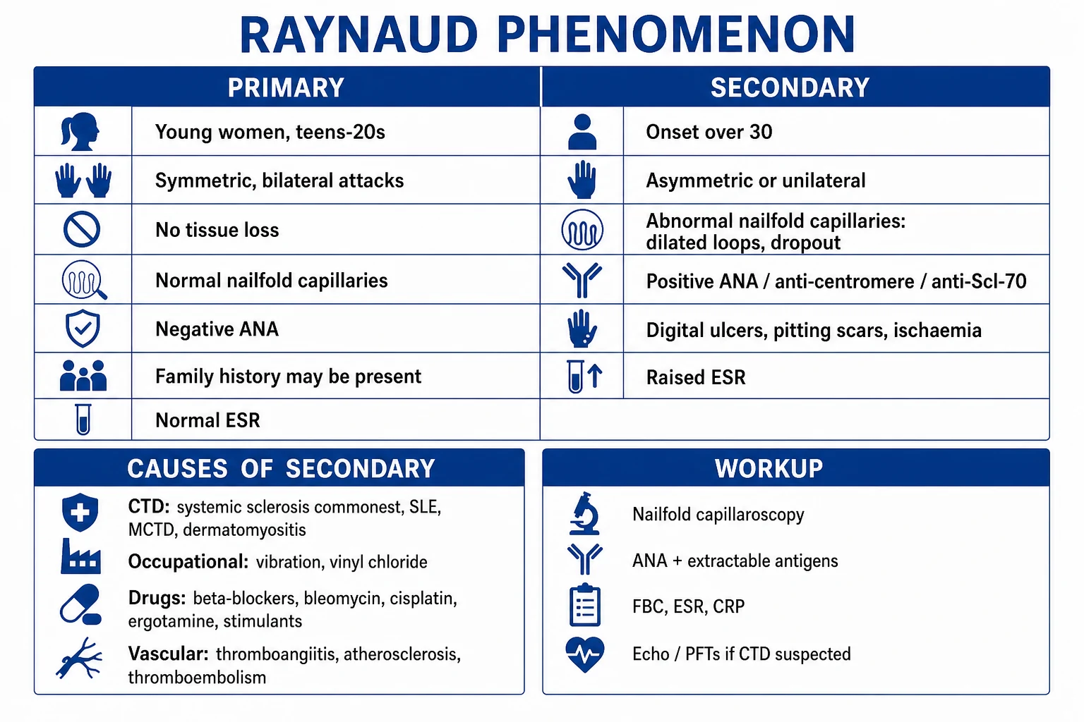
Primary Raynaud (Raynaud disease)
benign, functional, idiopathic
- Onset UNDER age 30 (teens/20s); strong female predominance (~9:1)
- Bilateral, SYMMETRIC, all fingers; toes/nose/ears less often
- NO tissue loss — no ulcers, no pitting scars, no gangrene
- NORMAL nailfold capillaroscopy; NEGATIVE ANA; normal ESR
- Family history common; benign course; reassurance + lifestyle (± CCB)
Secondary Raynaud (Raynaud phenomenon)
structural vessel disease / underlying disorder
- Onset OVER age 30; either sex; systemic sclerosis commonest cause
- ASYMMETRIC or unilateral; may spare thumbs
- Tissue loss — digital ulcers, pitting 'ice-pick' scars, critical ischaemia
- ABNORMAL nailfold capillaroscopy (dilated tortuous loops, megacapillaries, avascular dropout); POSITIVE ANA; raised ESR
- Treat the underlying disease + aggressive vasodilation (CCB, PDE-5i, IV iloprost, bosentan)
Chilblains (perniosis)
cold-induced inflammatory skin lesion — a key mimic
- Pruritic or painful erythematous-violaceous swellings hours after cold exposure
- NO triphasic colour change and NO true reversible vasospasm
- Usually on toes, fingers, ears; resolves over days to weeks
- Warming, topical corticosteroid; oral nifedipine for recurrent/severe disease
Raynaud phenomenon — the numbers that matter
Epidemiology & Risk Factors
Raynaud phenomenon is one of the commonest vascular disorders seen in general practice. Population prevalence is around 3 to 5 per cent, but this figure climbs steeply in cold climates and in women. Risk factors cluster into demographic, environmental and disease-related groups.[1]
Demographic and constitutional factors. Primary Raynaud is far commoner in women (ratio about 9 to 1), begins in the teens or twenties, and is reported in up to 20 per cent of women in some series living in temperate regions. Family history is positive in roughly a quarter of first-degree relatives, suggesting a heritable component, and oestrogen is thought to modulate vascular tone. Age of onset over 30 is itself a red flag: it shifts the prior probability toward secondary Raynaud and mandates investigation.[1]
Environmental and occupational triggers. Cold exposure is the universal trigger — the threshold is lower in Raynaud patients because their digital vessels are hypersensitive. Vibrating tools (chain saws, pneumatic drills, grinders, mining equipment) cause hand-arm vibration syndrome (HAVS, vibration white finger), the commonest occupational secondary Raynaud; risk scales with cumulative vibration dose. Vinyl chloride, epoxy resin and frostbite are rarer occupational causes.[1]
Drug-induced Raynaud. The classic offenders are the non-selective beta-blockers (propranolol) — unopposed alpha-mediated vasoconstriction worsens attacks — followed by ergotamine, methysergide, clonidine, the triptan antimigraine drugs, sympathomimetics and stimulants (amphetamine, cocaine, caffeine in excess). Chemotherapy combinations are an important iatrogenic cause: bleomycin (often with cisplatin and vinblastine, as in germ-cell tumour regimens) is the classic, and interferon, ciclosporin and bromocriptine have been implicated.[1]
Underlying disease (secondary Raynaud). Systemic sclerosis is the commonest and most important cause — over 95 per cent of systemic sclerosis patients develop Raynaud, and it is the presenting feature in the majority. Other connective tissue diseases (SLE, mixed connective tissue disease, Sjogren, dermatomyositis/polymyositis, rheumatoid arthritis), Buerger disease (thromboangiitis obliterans) in young male smokers, atherosclerosis and thoracic outlet syndrome, antiphospholipid syndrome, cryoglobulinaemia and hyperviscosity states (polycythaemia, macroglobulinaemia) make up the rest of the differential.[1]
Pathophysiology
A Raynaud attack is the end-result of an exaggerated vasoconstrictor response of the digital arteries and arterioles to cold or emotional stress. The mechanism is multifactorial, and the relative weight of each mechanism differs between primary and secondary disease — which is precisely why the two forms behave so differently.[1]
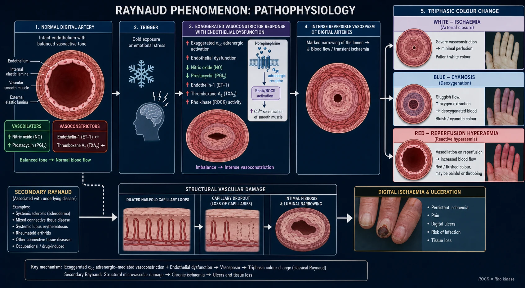
Neural mechanism — the cold-sensitive alpha-2C-adrenoceptor. Vascular smooth muscle in the digital arteries carries the alpha-2C-adrenoceptor, which at normal body temperature is held inside the cell. On cooling — even by a few degrees — the receptor translocates to the cell surface, dramatically increasing the contractile response to sympathetic noradrenaline. In Raynaud patients this response is exaggerated. Rho-kinase activity is increased, sensitising the myosin light chain to calcium and so prolonging the spasm once it starts. This neural hypersensitivity is the dominant mechanism in primary Raynaud, where the vessel wall is otherwise structurally normal.[1][2]
Endothelial dysfunction. In secondary Raynaud the endothelium tips the balance firmly toward vasoconstriction: there is reduced production of the vasodilators nitric oxide (NO) and prostacyclin (PGI2), and increased production of the vasoconstrictors endothelin-1 and thromboxane A2. Endothelin-1 is a potent, long-acting vasoconstrictor whose levels are raised in systemic sclerosis and which drives both acute spasm and chronic structural narrowing. Oxidative stress, reactive oxygen species from ischaemia-reperfusion, and intravascular platelet activation with microthrombi further amplify the vasospasm and contribute to digital ulceration.[2]
Impaired neurogenic vasodilation. Sensory afferent nerve fibres that release calcitonin gene-related peptide (CGRP) are powerful vasodilators. In systemic sclerosis the release of CGRP is impaired, removing a key brake on vasoconstriction.[1]
Structural vessel disease — the hallmark of secondary Raynaud. Repeated vasospasm and endothelial injury lead to intimal fibrosis, luminal narrowing, microvascular rarefaction and obliterative vasculopathy, best seen at the nailfold where the capillaries become dilated, tortuous, bushy (megacapillaries) and ultimately drop out (avascular areas). This structural narrowing explains why secondary Raynaud causes ulcers, pitting scars, critical ischaemia and gangrene while primary Raynaud does not — and why nailfold capillaroscopy is the single best discriminator between the two.[2][7]
Intravascular mechanisms. In the secondary forms driven by hyperviscosity, cryoglobulinaemia or antiphospholipid syndrome, red-cell or platelet aggregates, cryoproteins and microthrombi add a rheological component to the ischaemia, independent of vasospasm.[1]
The colour sequence mapped to physiology. White (pallor) = arterial closure, blood flow ceases, the digit is ischaemic. Blue (cyanosis) = the small volume of trapped blood in the capillaries deoxygenates. Red (hyperaemia) = reflex reopening of the arteriovenous anastomoses on rewarming, producing reactive hyperaemia, often with throbbing pain and paraesthesiae. The whole sequence is fully reversible — that reversibility is what defines Raynaud and distinguishes it from fixed arterial occlusion.[1]
Clinical Presentation
The attack is the clinical unit of Raynaud. Cold exposure (often a minor provocation such as reaching into a freezer, or a fall in ambient temperature) or emotional stress triggers bilateral, symmetric blanching (pallor) of the fingers, followed by cyanosis (blue) and then redness (hyperaemia) on rewarming. Attacks last minutes to a few hours, and the patient describes numbness, paraesthesiae and then pain on reperfusion (a throbbing, burning quality). The demarcation between the ischaemic white fingertip and the pink proximal finger is characteristically sharp.[1]
Primary Raynaud. The patient is typically a woman in her teens or twenties, with bilateral, symmetric attacks affecting all fingers (the thumbs are often spared), sometimes the toes, and occasionally the ears, nose, tongue or nipples. Attacks are provoked by cold or stress, are fully reversible, last under an hour, and cause no tissue damage — no ulcers, no scars, no gangrene. Family history is common. Nailfold capillaroscopy is normal and ANA is negative.[7]
Secondary Raynaud. The patient is typically older (onset over 30), of either sex, with asymmetric or unilateral attacks that may spare some fingers. Critically, there is tissue damage: digital pulp ulcers, pitting 'ice-pick' scars, dystrophic or lost nails, and in severe disease critical ischaemia and gangrene. The history and examination must hunt for the underlying disease — especially systemic sclerosis, where the hand examination may reveal sclerodactyly (tight, shiny, thickened skin of the fingers), telangiectasia, calcinosis cutis and digital pitting scars (the components of CREST — Calcinosis, Raynaud, Esophageal dysmotility, Sclerodactyly, Telangiectasia, the limited cutaneous subset that is typically anti-centromere positive). Beyond the hands, ask about dysphagia and heartburn (oesophageal dysmotility), dry cough and exertional dyspnoea (interstitial lung disease), palpitations and syncope (pulmonary arterial hypertension), arthritis, dry eyes/mouth, rashes (malar, Gottron) and Raynaud preceding skin thickening by years.[1][2]
Atypical andoften-missed presentations. Localised Raynaud affecting a single digit suggests secondary disease or a large-vessel cause (thoracic outlet, subclavian stenosis, hypothenar hammer syndrome). Raynaud with absent pulses, arm claudication or a subclavian bruit is not true Raynaud — it points to large-vessel occlusive disease (atherosclerosis, Takayasu, Buerger, subclavian stenosis) and the vessels must be imaged, not dismissed as vasospasm. Raynaud of the nipples in a breastfeeding woman exposed to cold is a recognised, often-missed cause of painful lactation. Paediatric Raynaud is nearly always primary, but any secondary features warrant the same screen as in adults.[1]
Differential Diagnosis
The differential falls into three groups: the connective tissue diseases that cause secondary Raynaud, the non-CTD causes of secondary Raynaud, and the cold-induced acrosyndromes that mimic Raynaud but are not true vasospasm.[1]
Systemic sclerosis (scleroderma)
the commonest cause of secondary Raynaud
- Over 95% have Raynaud; often the FIRST manifestation by years
- Screen: nailfold capillaroscopy, ANA, anti-centromere (limited/CREST), anti-Scl-70/topoisomerase I (diffuse), anti-RNA polymerase III (renal crisis)
- Look for sclerodactyly, pitting scars, telangiectasia, ILD, PAH, dysphagia, renal crisis
- ACR/EULAR 2013 criteria weight abnormal nailfold capillaries, RP, SSc-autoantibodies and pitting scars
Other connective tissue diseases
SLE, MCTD, Sjogren, dermatomyositis, RA
- MCTD (anti-U1-RNP) — Raynaud is almost universal, with overlapping SLE/scleroderma/polymyositis features
- SLE — screen ANA, anti-dsDNA; malar rash, arthritis, nephritis
- Dermatomyositis — Gottron papules, heliotrope rash, proximal myopathy, raised CK
- Rheumatoid arthritis and Sjogren less often cause severe digital ischaemia
Buerger disease (thromboangiitis obliterans)
young male smokers, small/medium vessels
- Young male, heavy smoker; distal ischaemia, claudication of instep, superficial thrombophlebitis migrans
- Small arteries of digits affected — Raynaud, ulcers, gangrene
- Smoking cessation is the ONLY effective treatment; amputation common if continued
Chilblains (perniosis) / acrocyanosis
cold acrosyndromes that MIMIC Raynaud
- Chilblains: pruritic/painful erythematous-violaceous swellings HOURS after cold; no triphasic change
- Acrocyanosis: persistent painless blue-purple discoloration of hands/feet, no paroxysmal pallor
- Distinguish from true Raynaud by the absence of the reversible white phase
- Treat with warming; nifedipine for recurrent chilblains
Don't miss the large-vessel mimic. Asymmetric digital ischaemia with an absent radial/ulnar pulse, arm claudication, a supraclavicular or subclavian bruit, or blood-pressure asymmetry between arms is large-vessel occlusive disease (atherosclerosis, Takayasu arteritis, subclavian stenosis, thoracic outlet obstruction, hypothenar hammer syndrome). These patients need arterial Doppler, CT or MR angiography — nailfold capillaroscopy alone will miss the proximal lesion, and treating them only with vasodilators wastes the window for revascularisation.[1]
Clinical & Bedside Assessment
The clinical assessment has one purpose: to decide whether this is primary or secondary Raynaud, and if secondary, to find the cause. The diagnosis of Raynaud itself is clinical — the triphasic colour change is elicited from the history, and provocation testing (cold challenge) is rarely needed.[1]
History. Establish the age of onset (under 30 favours primary; over 30 favours secondary), the symmetry (bilateral and symmetric in primary; asymmetric in secondary), the triggers (cold, stress, vibration, drugs), the attack duration and frequency, and the presence of tissue damage — ulcers, pitting scars, gangrene. Take a careful occupational and drug history (vibrating tools, vinyl chloride; beta-blockers, ergotamine, chemotherapy, stimulants). Ask for features of connective tissue disease: skin thickening, dysphagia or heartburn, dry cough and exertional dyspnoea, palpitations, arthritis, rashes, dry eyes/mouth, and family history of autoimmune disease.[1]
Examination. Inspect the hands for sclerodactyly, digital pulp pitting scars, active ulcers, telangiectasia, calcinosis and nailfold abnormalities. Palpate the peripheral pulses (radial, ulnar, brachial, subclavian) and listen for a subclavian or carotid bruit; check blood pressure in both arms (asymmetry suggests subclavian/Takayasu). Examine the skin globally (malar rash of SLE, Gottron papules of dermatomyositis, morphea plaques), the chest (basal crackles of ILD, loud P2 and right ventricular heave of PAH), the joints (inflammatory arthritis) and the abdomen.[7]
Nailfold capillaroscopy at the bedside. Place a drop of immersion oil on the proximal nailfold of the fourth finger and examine with a dermatoscope, ophthalmoscope (using the +40 diopter setting) or a USB microscope at roughly 10 to 200 times magnification. Normal nailfolds show neat, parallel, hairpin-shaped capillary loops of uniform size and density. Secondary Raynaud shows the characteristic scleroderma pattern: dilated, tortuous and giant (mega) capillaries, bushy capillaries and avascular dropout areas with microhaemorrhages. A normal nailfold in a young woman with symmetric attacks supports primary Raynaud; an abnormal nailfold mandates an autoantibody screen and surveillance for systemic sclerosis.[1][2][7]
Investigations
The diagnosis of Raynaud is clinical. Investigations serve two purposes: to distinguish primary from secondary and, if secondary, to identify and stage the underlying disease. A young patient with classical primary features (symmetric, no tissue loss, normal nailfold, negative ANA) needs little more than reassurance; any feature of secondary disease triggers the panel below.[1]
The core screen (every suspected secondary case).[1]
- Full blood count, ESR and CRP — anaemia of chronic disease, raised inflammatory markers (a raised ESR argues against uncomplicated primary Raynaud and for inflammation).
- Urea, electrolytes, creatinine, LFTs — baseline organ function and to allow later disease-modifying or vasodilator drugs.
- Creatine kinase — myositis overlap.
- Thyroid function — autoimmune thyroid disease coexists with autoimmune Raynaud.
- Immunoglobulins, complement, rheumatoid factor, cryoglobulins and antiphospholipid antibodies — where the history suggests hyperviscosity, cryoglobulinaemia or antiphospholipid syndrome.
- Hepatitis B, C and HIV serology — when PAN-like vasculitis, cryoglobulinaemia or HBV-associated disease is suspected. [1]
Autoantibodies. ANA is the screening test; a positive ANA with an abnormal nailfold capillaroscopy is the strongest predictor of evolution to systemic sclerosis. If ANA is positive, request the scleroderma-specific autoantibodies: anti-centromere (limited cutaneous SSc / CREST), anti-Scl-70 / anti-topoisomerase I (diffuse SSc and ILD), anti-RNA polymerase III (renal crisis and cancer-associated SSc) and anti-U1-RNP (mixed connective tissue disease). Depending on the clinical picture, add anti-dsDNA and anti-Smith (SLE), anti-Ro/La (Sjogren, neonatal lupus), and anti-Jo-1 and other anti-synthetase antibodies (inflammatory myopathy).[1][2]
Nailfold capillaroscopy — the single best discriminator. Beyond the bedside dermoscope, nailfold video-capillaroscopy is the gold standard and is reproducible enough to monitor progression. The Cutolo classification describes three sequential scleroderma patterns that track with disease duration and severity:[7]
- "Early" pattern — few enlarged/giant capillaries, preserved capillary architecture, no major dropout; corresponds to early systemic sclerosis and the window where Raynaud may be the only feature.
- "Active" pattern — frequent giant capillaries and megacapillaries, microhaemorrhages, moderate loss of capillaries, mildly disordered architecture; the most recognisable pattern at diagnosis.
- "Late" pattern — severe avascular dropout, loss of normal architecture, bushy (ramified) capillaries, few giant capillaries; corresponds to late, severe disease and predicts digital ulcers. [1]
Abnormal nailfold capillaroscopy plus positive ANA is the combination that predicts progression from isolated Raynaud to definite systemic sclerosis, and is now embedded in the ACR/EULAR 2013 classification criteria for systemic sclerosis (where "abnormal nailfold capillaries" and "Raynaud phenomenon" each score points, alongside pitting scars, telangiectasia, PAH/ILD and SSc-autoantibodies).[5][7]
Organ screen in suspected systemic sclerosis. Once secondary Raynaud is confirmed, stage the underlying disease: pulmonary function tests (a fall in DLCO is the earliest sign of ILD or PAH), high-resolution CT chest (interstitial lung disease), echocardiogram (pulmonary arterial hypertension, pericardial effusion), ECG, right heart catheterisation if PAH is suspected, renal function and blood pressure monitoring (scleroderma renal crisis), and gastrointestinal studies (manometry, endoscopy for dysmotility and GORD).[1]
Vascular imaging and functional tests — for the atypical or asymmetric case. Digital pulse volume recording / photoplethysmography confirms whether arterial flow is present; arterial Doppler ultrasound, CT or MR angiography excludes proximal large-vessel disease (subclavian stenosis, thoracic outlet, Buerger, embolus, Takayasu). Cold challenge and thermography are research tools and rarely needed clinically.[1]
Self-test: which two test results best predict progression from isolated Raynaud to systemic sclerosis?
Management — Resuscitation (Critical Digital Ischaemia)
Raynaud is rarely an emergency except when it causes critical digital ischaemia — severe resting pain, tissue loss, absent capillary refill or incipient gangrene, almost always in secondary Raynaud (especially systemic sclerosis). This is a rheumatological / vascular emergency requiring admission.[1][2]
For the acute severe attack that is not yet critical, the immediate measures are whole-body warming (a warm bath or shower, heated clothing), simple analgesia and a prompt dose of a short-acting calcium-channel blocker (e.g. nifedipine 10 mg), while the patient is worked up for secondary causes.[1]
Management — Definitive & Stepwise
Management is stratified by primary versus secondary disease and by severity. The ladder moves from conservative measures through oral vasodilators to intravenous and surgical options, with explicit escalation triggers.[1]
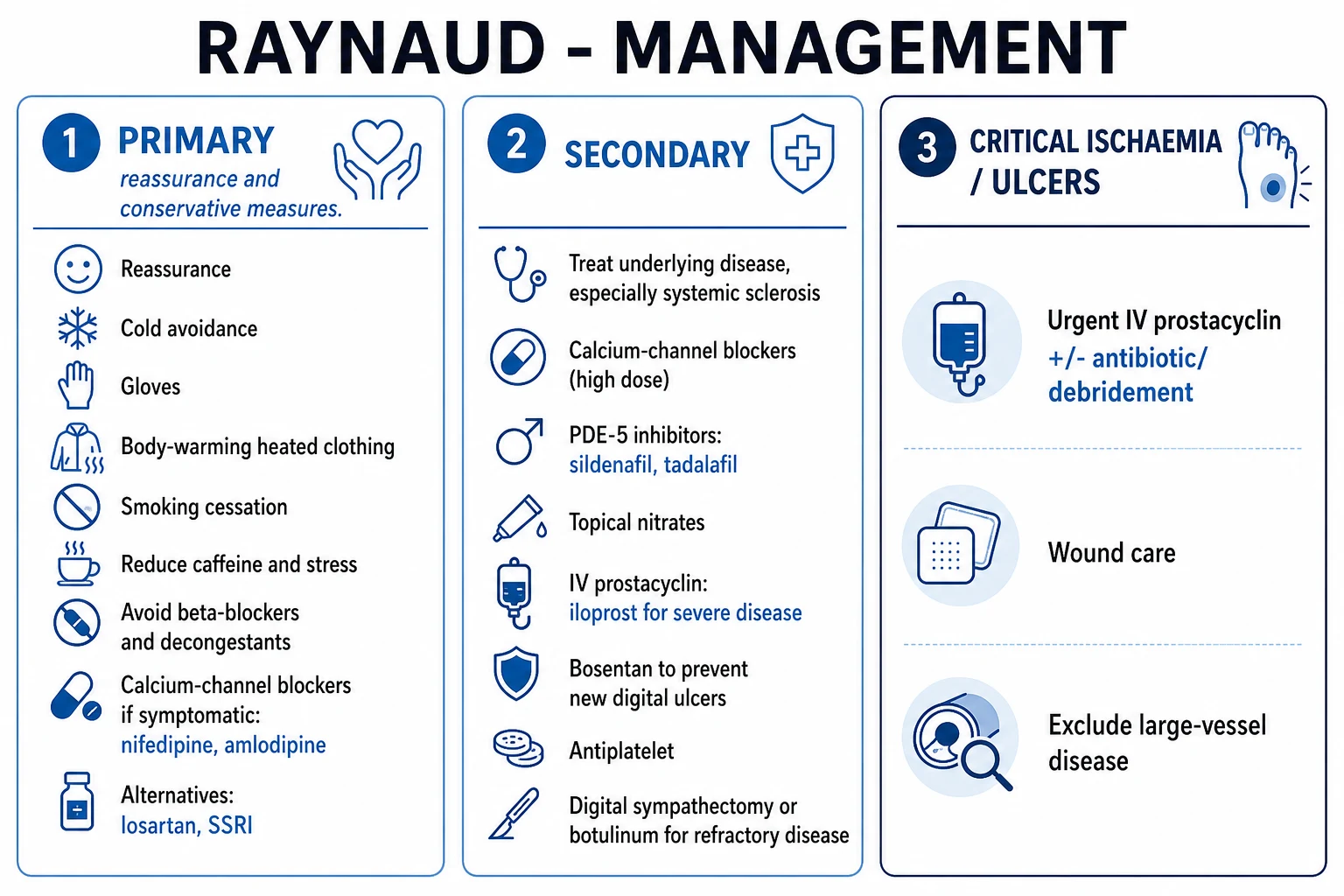
Step 1 — General measures (all patients)
The foundation is non-pharmacological and is often sufficient for primary Raynaud. Avoid cold by dressing warmly before going out (layered clothing, hat, gloves, thick socks), use heated gloves, hand warmers and battery-heated clothing, and keep the whole body warm (not just the hands). Stop smoking — nicotine is a vasoconstrictor and reduces the efficacy of drug therapy. Reduce caffeine and manage stress. Critically, avoid drugs that provoke Raynaud: beta-blockers (switch to a rate-limiting alternative or an ACE inhibitor), sympathomimetic decongestants (pseudoephedrine), ergotamine, clonidine, triptans where possible, and the offending chemotherapy combination where feasible. Vibration avoidance and job modification are central to hand-arm vibration syndrome.[1]
Step 2 — First-line pharmacotherapy: calcium-channel blockers
The dihydropyridine calcium-channel blockers are first-line in both primary and secondary Raynaud. They relax vascular smooth muscle and reduce the frequency and severity of attacks by roughly two to three attacks per week.[1][2][6]
- Nifedipine (modified-release) 10 mg OD, titrated to 30 to 40 mg OD (maximum 120 mg/day in divided doses) — start low to minimise headache, flushing, ankle oedema and reflex tachycardia; the modified-release formulation improves tolerability.[6]
- Amlodipine 5 mg OD, titrated to 10 mg OD — a well-tolerated alternative with a longer half-life and fewer side effects.[6]
These are backed by the Cochrane review of vasodilators for primary Raynaud, which confirms a modest but real benefit of calcium-channel blockers (the dihydropyridines are the only drug class with consistent benefit in primary disease).[6]
Step 3 — Second-line and add-on oral agents (secondary or refractory primary)
When a CCB is insufficient, or not tolerated, the following agents are added or substituted, chosen by comorbidity and tolerability:[1]
- Losartan (an angiotensin-receptor blocker) 50 mg OD (up to 100 mg OD) — modestly effective, well tolerated, useful when CCBs cause oedema.[1]
- Prazosin (alpha-1-blocker) 1 mg TDS, titrated to 2 to 5 mg TDS — directly opposes the alpha-adrenergic vasoconstriction; first-dose hypotension is the main caution.[1]
- Fluoxetine (SSRI) 20 mg OD — a SSRI that reduces platelet serotonin and improves symptoms; recommended by EULAR 2017 for systemic-sclerosis-related Raynaud.[3]
- Statins (e.g. atorvastatin 40 mg OD) — improve endothelial function and reduce vascular events; an adjunct rather than primary therapy.[1]
- Topical nitrates — glyceryl trinitrate 0.4% topical (MQX-503 metered-dose) applied to the fingers; headache and tachyphylaxis limit use. Best as a spot treatment for a single intractable digit.
Step 4 — Severe and refractory secondary Raynaud
For digital ulcers, recurrent severe attacks or critical ischaemia in secondary Raynaud, escalate to:[1]
- PDE-5 inhibitors — sildenafil 25 to 50 mg TDS, or tadalafil 20 mg alternate days or three times weekly. These raise cyclic GMP and potentiate nitric-oxide-mediated vasodilation; the EULAR 2017 update specifically recommends PDE-5 inhibitors for systemic-sclerosis-related Raynaud and digital ulcers. They are among the most effective oral agents for secondary disease.[3]
- Intravenous prostacyclin (iloprost) — 0.5 to 2 ng/kg/min by continuous IV infusion, titrated to tolerance (headache, nausea, hypotension, flushing), over 6 hours daily for 3 to 5 days; courses are repeated every few weeks to months for severe disease. Iloprost is the most potent acute vasodilator and also inhibits platelet aggregation and promotes ulcer healing.[1][2]
- Endothelin-receptor antagonist — bosentan 62.5 mg BD for 4 weeks, then 125 mg BD. Bosentan is reserved for the prevention of new digital ulcers in systemic sclerosis patients with multiple ulcers despite CCB and PDE-5i therapy — it does not heal existing ulcers. This distinction is critical: the RAPIDS-2 trial (188 patients) showed a 30 per cent reduction in the number of new digital ulcers with bosentan but no difference in healing of the cardinal ulcer.[4] Bosentan requires monthly liver function monitoring (transaminase elevation) and is teratogenic, so contraception is mandatory.
Step 5 — Surgical and local interventions
- Botulinum toxin A injection — 25 to 100 units per hand injected around the digital arteries or into the palmar skin; reduces sympathetic tone and is useful for refractory pain and ulcer healing, though repeated injections are needed.[2]
- Digital (peri-arterial) sympathectomy — surgical stripping of the sympathetic fibres around the digital arteries; reserved for a single critically ischaemic digit not responding to medical therapy.
- Cervical or lumbar sympathectomy — largely abandoned for the upper limb because benefit is transient, but still occasionally used for lower-limb disease.
- Amputation — only for irreversible necrosis/auto-amputation.
- Fat grafting (lipofilling) — an emerging surgical adjunct to promote ulcer healing and soften sclerotic tissue.[2]
Specific Subtypes & Scenarios
Primary Raynaud (Raynaud disease). Idiopathic, benign, young women, symmetric, normal nailfold, negative ANA, no tissue loss. Diagnostic criteria: symptoms for over 2 years with no progression and no features of secondary disease. Management is reassurance and lifestyle, with a short course of a CCB if attacks are troublesome. Reassess periodically for evolution.[1]
Systemic-sclerosis-associated Raynaud. The most severe and the archetype of secondary Raynaud — structural obliterative vasculopathy drives recurrent ulcers and critical ischaemia. Treat the underlying disease (immunosuppression for skin/lung disease, ACE inhibitors for renal crisis, PAH-specific therapy) plus aggressive vasodilation: titrated CCB, add PDE-5i, escalate to IV iloprost for severe disease, and bosentan to prevent new ulcers. The EULAR 2017 recommendations formalise this ladder.[3]
Hand-arm vibration syndrome (HAVS, vibration white finger). A compensable occupational disease in miners, forestry workers, fitters and grinders. The history of cumulative vibration exposure is diagnostic. Vibration avoidance and job modification are central; vasodilators (CCB) help symptoms but cannot reverse the structural damage. Coexistent sensorineural hearing loss and carpal tunnel syndrome are common.[1]
Drug-induced Raynaud. Withdraw the offending agent (beta-blocker, ergotamine, clonidine, stimulant, triptan) and substitute as needed; chemotherapy-related Raynaud (bleomycin-cisplatin-vinblastine) may partially resolve after the regimen ends.[1]
Buerger disease (thromboangiitis obliterans). Young male heavy smokers with distal ischaemia, Raynaud, superficial thrombophlebitis migrans and claudication of the instep. Complete smoking cessation is the only effective treatment — vasodilators and sympathectomy are disappointing, and amputation is common if smoking continues.[1]
Paediatric Raynaud. Almost always primary and benign, but any feature of secondary disease (asymmetry, ulcers, abnormal nailfold, positive ANA) mandates the full adult work-up, as systemic sclerosis and SLE can present in adolescence.[1]
Complications & Pitfalls
Digital ulcers are the commonest complication of secondary Raynaud — painful, slow to heal, prone to infection, and a major source of disability in systemic sclerosis. Pitting 'ice-pick' scars of the digital pulp are markers of prior microinfarction and are themselves a diagnostic clue to systemic sclerosis. Critical digital ischaemia, gangrene and auto-amputation or surgical amputation occur in severe secondary disease. Sclerodactyly, flexion contractures and loss of hand function compound the disability. Calcinosis cutis produces painful hard deposits that can ulcerate through the skin. Treatment-related adverse effects — headache, peripheral oedema, flushing and hypotension with CCBs and PDE-5 inhibitors; tachyphylaxis and headache with nitrates; transaminitis, peripheral oedema and teratogenicity with bosentan; hypotension, nausea and flushing with IV iloprost — are common reasons for dose limitation or discontinuation.[1][2]
Classic pitfalls. (1) Labelling secondary Raynaud as primary and discharging the patient, missing early systemic sclerosis — defend with nailfold capillaroscopy and ANA at baseline and re-review. (2) Prescribing a beta-blocker for comorbid hypertension or migraine in a Raynaud patient — switch to an ACE inhibitor, losartan or a rate-limiting agent. (3) Treating the vasospasm while ignoring a proximal large-vessel lesion — image any asymmetric, pulseless or unilateral case. (4) Expecting bosentan to heal an existing ulcer — it prevents new ulcers only. (5) Forgetting contraception and liver monitoring on bosentan.[4]
Prognosis & Disposition
Primary Raynaud is benign — attacks tend to lessen with age and warming, life expectancy is normal, and no organ disease develops. However, approximately 5 to 20 per cent of patients initially labelled 'primary' later evolve into a connective tissue disease, most often systemic sclerosis within 2 to 5 years. The combination of abnormal nailfold capillaroscopy and a positive ANA identifies this at-risk subgroup, who need periodic reassessment (nailfold, autoantibodies, PFTs, echocardiogram) so that organ disease is detected before it declares itself.[1][5][7]
Secondary Raynaud's prognosis is driven by the underlying disease. In systemic sclerosis, interstitial lung disease and pulmonary arterial hypertension are the leading causes of death, while recurrent digital ulcers and critical ischaemia threaten the digits. Early detection (baseline and serial nailfold capillaroscopy, PFTs with DLCO, echocardiogram, renal monitoring) and aggressive vasodilation improve digital outcomes and quality of life, even though they do not change the underlying disease trajectory.[7]
Disposition. Primary Raynaud is managed in primary care with lifestyle advice and, if needed, a CCB. Suspected secondary Raynaud is referred to rheumatology for nailfold capillaroscopy, autoantibody profiling and organ surveillance. Critical digital ischaemia is admitted under rheumatology and vascular surgery for IV iloprost and wound care.[3]
Special Populations
Pregnancy. Primary Raynaud often improves in pregnancy (vasodilated state). Most drugs (CCBs, losartan) need review — losartan is teratogenic (FDA category D) and must be stopped pre-conception; nifedipine is relatively safe. Bosentan is absolutely contraindicated in pregnancy (teratogenic) — enforce reliable contraception in women of childbearing potential.[4]
Elderly. New-onset Raynaud after 60 is almost always secondary — to atherosclerosis, drug effect, or a paraneoplastic/CTD process — and warrants aggressive investigation rather than reassurance.[1]
Systemic sclerosis with renal crisis. Avoid high-dose steroids (above 15 mg/day prednisolone) in systemic sclerosis — they precipitate scleroderma renal crisis; manage Raynaud with CCBs (which also protect the kidney) and PDE-5 inhibitors.[3]
Anticoagulated / prothrombotic patients. In antiphospholipid syndrome with digital ischaemia, combine vasodilators with anticoagulation; aspirin 75 mg OD is standard antiplatelet prophylaxis in secondary Raynaud with ulcers.[1]
Evidence, Guidelines & Regional Differences
The management of Raynaud, particularly systemic-sclerosis-associated disease, is built on a small set of landmark trials and guidelines.[1]
EULAR 2017 — treatment of systemic sclerosis (Kowal-Bielecka et al.)
Population: 16 recommendations across RP, digital ulcers, PAH, skin, lung, renal crisis, GI
Key finding
Recommends dihydropyridine CCBs first-line for SSc-RP; PDE-5 inhibitors and IV iloprost for SSc-RP and digital ulcers; fluoxetine for SSc-RP; bosentan for prevention of new digital ulcers in SSc
RAPIDS-2 (Matucci-Cerinic et al., 2011)
Population: 188 SSc patients with at least one active digital ulcer
Key finding
30 per cent reduction in the number of NEW digital ulcers (1.9 vs 2.7; p=0.04); NO difference in healing of the cardinal ulcer; peripheral oedema and raised aminotransferases with bosentan
Cochrane 2021 — vasodilators for primary Raynaud (Su et al.)
Population: 15 RCTs, 635 participants with primary Raynaud
Key finding
Evidence for vasodilators other than CCBs is of low to very low certainty; PDE-5 inhibitors showed no clear benefit in PRIMARY Raynaud (in contrast to secondary)
ACR/EULAR 2013 — classification criteria for systemic sclerosis (van den Hoogen et al.)
Population: SSc cases vs scleroderma-like controls
Key finding
Sensitivity 0.91 and specificity 0.92 (vs 0.75/0.72 for 1980 ACR criteria)
Regional deltas. EULAR (Europe) sets the international reference ladder for systemic-sclerosis-related Raynaud, endorsing CCB, PDE-5i, IV iloprost, fluoxetine and bosentan.[3] NICE (UK) recommends nifedipine as first-line, with referral for specialist assessment when secondary Raynaud is suspected and admission for critical ischaemia. ACR (US) practice is concordant with EULAR; the FDA-approved topical formulation for Raynaud is nitrates (glyceryl trinitrate / nitroglycerin MQX-503), while bosentan is licensed in Europe for SSc with multiple digital ulcers but its availability and reimbursement differ by country. In low-resource settings (including much of India and South Asia), where nifedipine and amlodipine are cheap and universally available, they remain the backbone; PDE-5 inhibitors, IV iloprost and bosentan are expensive, often available only at tertiary centres, and reserved for severe secondary disease. The diagnostic nailfold capillaroscopy is under-utilised worldwide but can be performed with an inexpensive dermatoscope or USB microscope at any clinic.
Exam Pearls
Causes of SECONDARY Raynaud — COLDER
COLDER
systemic sclerosis (commonest), SLE, MCTD (anti-U1-RNP), dermatomyositis, Sjogren
vibration white finger (HAVS), vinyl chloride disease, frostbite, hypothenar hammer syndrome
beta-blockers, bleomycin/cisplatin/vinblastine, ergotamine, clonidine, triptans, stimulants, interferon
atherosclerosis, thromboembolism, Buerger disease, subclavian stenosis, thoracic outlet
cryoglobulinaemia, hyperviscosity (polycythaemia, macroglobulinaemia), antiphospholipid syndrome
Exam application bank (NEET-PG / INICET)
One-line answer
Raynaud phenomenon is episodic reversible vasospasm of the digital arteries and cutaneous arterioles, classically producing the triphasic colour change: white (pallor / ischaemia), blue (cyanosis) and red (reperfusion hyperaemia) of the fingers and toes on cold exposure or emotional stress. Two forms: primary (benign, idiopathic, young women, symmetric, no tissue loss, normal nailfold capillaries, negative autoantibodies) and secondary (associated with connective tissue disease — systemic sclerosis above all, plus SLE, MCTD; occupational vibration; drugs such as beta-blockers, bleomycin, cisplatin, ergotamine; thromboembolic and hyperviscosity states; older onset, asymmetric, abnormal nailfold capillaroscopy with dilated tortuous loops and avascular dropout, positive ANA, risk of digital ulcers and critical ischaemia). Diagnosis is clinical, supported by nailfold capillaroscopy and an au
Worked stems (answer without another resource)
Stem 1 — Classic presentation. Map symptoms to mechanism; name the first investigation and first treatment step with dose/route if drug therapy is standard. [1]
Stem 2 — Unstable / complicated. List red flags that force immediate resuscitation, theatre, ICU, antidote, or reperfusion — and what you do in the first 15 minutes. [1]
Stem 3 — Atypical group. Elderly, pregnancy, child, or immunocompromised: how presentation and thresholds change. [1]
Stem 4 — Differential trap. Name the three closest mimics and one discriminator for each. [1]
Stem 5 — Disposition. Who goes home with safety-netting, who is admitted, who needs HDU/ICU/theatre, and what follow-up is mandatory. [1]
Rapid viva checklist
- Definition + classification
- Pathophysiology chain
- Bedside signs / criteria
- Score with exact components (if any)
- Emergency bundle
- Definitive therapy with doses
- Complications of disease and of treatment
- Special populations
- Guideline/trial name if classic
- Three exam traps
Coverage self-check
If you cannot answer any stem above from this page alone, re-read the matching section — the page is intended to be self-sufficient for final-prof and NEET-PG/INICET questions on Raynaud Phenomenon.
References
- [1]Herrick AL, Wigley FM. Raynaud's phenomenon Best Pract Res Clin Rheumatol, 2020.PMID 32007400
- [2]Ture HY, Lee NY, Kim NR, Nam EJ. Raynaud's Phenomenon: A Current Update on Pathogenesis, Diagnostic Workup, and Treatment Vasc Specialist Int, 2024.PMID 39040029
- [3]Kowal-Bielecka O, Fransen J, Avouac J, et al.; EUSTAR Coauthors. Update of EULAR recommendations for the treatment of systemic sclerosis Ann Rheum Dis, 2017.PMID 27941129
- [4]Matucci-Cerinic M, Denton CP, Furst DE, et al. Bosentan treatment of digital ulcers related to systemic sclerosis: results from the RAPIDS-2 randomised, double-blind, placebo-controlled trial Ann Rheum Dis, 2011.PMID 20805294
- [5]van den Hoogen F, Khanna D, Fransen J, et al. 2013 classification criteria for systemic sclerosis: an American College of Rheumatology/European League against Rheumatism collaborative initiative Arthritis Rheum, 2013.PMID 24122180
- [6]Su KY, Sharma M, Kim HJ, Kaganov E, Hughes I, Abdeen MH, Ng JHK. Vasodilators for primary Raynaud's phenomenon Cochrane Database Syst Rev, 2021.PMID 33998674
- [7]Ruaro B, Pizzorni C, Paolino S, Smith V, Ghio M, Casabella A, Alessandri E, Patane M, Sulli A, Cutolo M. Correlations between nailfold microvascular damage and skin involvement in systemic sclerosis patients Microvasc Res, 2019.PMID 30974112