Rheumatology · Rheumatology
Rheumatoid Arthritis
Also known as Rheumatoid arthritis · RA · Seropositive arthritis · Atrophic arthritis
Rheumatoid arthritis (RA) is a chronic, symmetric, erosive, autoimmune polyarthritis of unknown cause that preferentially targets the synovial small joints (MCP, PIP, wrists, MTP), spares the DIP, and produces morning stiffness lasting more than 1 hour. Diagnosis is clinical plus anti-CCP antibody (highly specific) and characteristic X-ray erosions; disease activity is graded with the DAS28. Management is treat-to-target to remission or low disease activity, beginning with methotrexate (weekly + folic acid) and escalating through csDMARDs, bDMARDs (anti-TNF, anti-IL-6, anti-CD20, abatacept) and tsDMARDs/JAK inhibitors. Untreated disease causes deformity, disability, atlantoaxial subluxation and premature cardiovascular death.
On this page & tools
Your progress
Saved locally on this device.
Practise this topic
Exam tags
Red flags
Overview & Definition
Rheumatoid arthritis (RA) is a chronic, systemic, autoimmune inflammatory disease of unknown cause whose hallmark is a symmetric, erosive polyarthritis of the diarthrodial (synovial) joints. Persistent synovial inflammation produces a hypertrophic, invasive pannus that destroys cartilage and bone, generating the deformities, disability and excess mortality that define untreated disease. Although joints are the dominant target, RA is systemic: it carries substantial extra-articular manifestations and a cardiovascular mortality that remains the leading cause of premature death.[7][8]
The clinical art in RA is not the diagnosis alone — it is recognising disease early (within the first 12 weeks, the "window of opportunity"), classifying it correctly with the 2010 ACR/EULAR criteria, grading activity objectively with the DAS28 (or CDAI/SDAI), and driving a treat-to-target strategy to remission or low disease activity using a conventional synthetic DMARD (csDMARD, almost always methotrexate) before escalating to biologics or JAK inhibitors. Every step of that chain has evidence of improved outcomes; missing any step — late DMARD, no target, indefinite steroids — costs the patient joint destruction and years of life.[2][4]
Classification
The classification of RA serves two purposes: distinguishing it from the mimic polyarthritides (SLE, psoriatic, viral, OA, gout) and stratifying prognosis and therapy within the disease. [1]
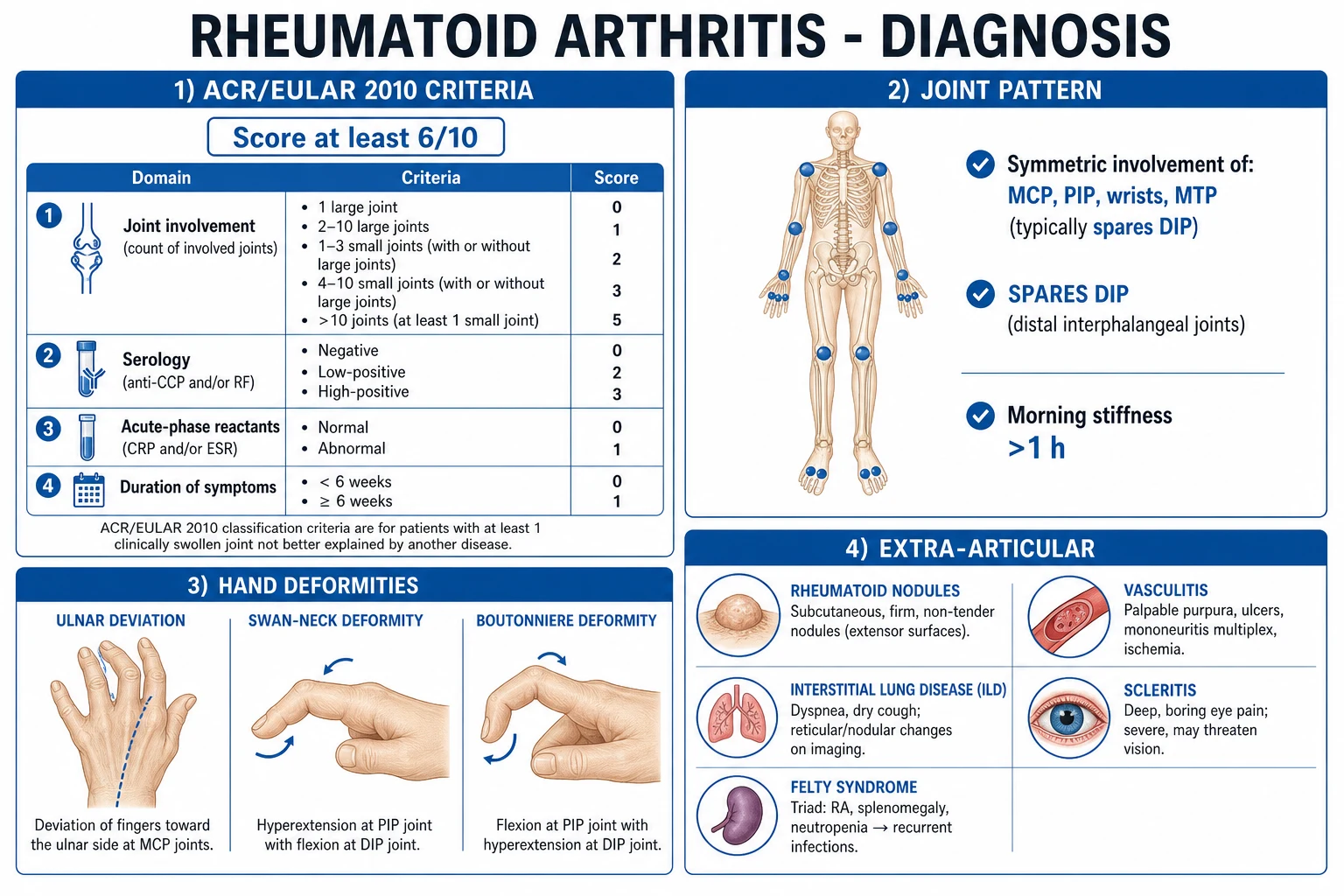
By 2010 ACR/EULAR classification criteria (the score used to formally "label" RA, replacing the 1987 criteria). A patient is classified as having definite RA if the score is 6 or more out of 10, provided synovitis is not better explained by another disease, AND the criteria are applied to someone with at least one swollen joint on examination. The score adds four domains:[1]
2010 ACR/EULAR RA classification — scoring table
Seropositive RA
- ACPA (anti-CCP) and/or RF positive
- More aggressive, erosive course
- Classical nodules and extra-articular disease
- Better response to rituximab and abatacept
- ~70–80% of patients
Seronegative RA
- Both ACPA and RF negative
- Often milder, less erosive
- Differential must exclude psoriatic, reactive, viral and crystal arthropathies
- Treat target identical; biologics used if csDMARDs fail
Elderly-onset RA (EORA)
- Onset after age 60–65
- More abrupt, oligo-articular, large joints
- Prominent systemic symptoms, very high ESR
- Higher PMR/SLE overlap; differentially from polymyalgia rheumatica
- Often RF/ACPA negative
Palindromic RA
- Episodic 1–3 day attacks of mono-/oligo-arthritis
- Complete resolution between attacks
- ~30–50% evolve into classic RA over years
- Often ACPA-positive
- Treat flares with NSAIDs/short steroid bursts
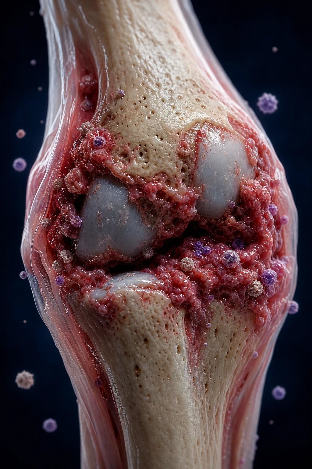
Symmetric vs asymmetric, large vs small joint, and "surgical hand" distinctions are the examiner's favourite "spot-the-arthritis" tool. RA is symmetric, small-joint, and spares the DIP; OA is asymmetric, affects the DIP (Heberden) and PIP (Bouchard) and the first CMC; psoriatic arthritis is asymmetric, involves the DIP and carries nail dystrophy; gout targets the first MTP and tophi; SLE causes a non-erosive, reducible Jaccoud arthropathy with deforming but not destructive X-rays. Holding these five pictures simultaneously is the single most exam-rewarded skill in rheumatology. [1]
Epidemiology & Risk Factors
RA is the commonest chronic inflammatory arthritis in adults, with a global prevalence of about 0.5–1% (higher in some indigenous populations, lower in rural Africa and Asia). The female-to-male ratio is about 3:1; peak onset is between 30 and 50 years, although a second peak occurs in the elderly (EORA).[7]
RA — key numbers
Predisposing factors combine genetic susceptibility, environmental triggers and loss of immune tolerance. The strongest genetic association is HLA-DRB1 alleles carrying the "shared epitope" (a conserved amino-acid motif at positions 70–74 of the DRβ chain) — they account for about one-third of the genetic risk and predict erosive, anti-CCP positive disease. Other genes include PTPN22 (T-cell receptor signalling), PAD14 (peptidylarginine deiminase 4, crucial for citrullination in Asian populations) and STAT4.[7]
Environmental triggers: cigarette smoking is the single most important modifiable risk factor — it triggers pulmonary citrullination, breaks immune tolerance to citrullinated proteins and converts genetic susceptibility (especially HLA-DRB1 shared epitope) into ACPA-positive disease with a multiplicative effect. Other contributors include silica and mineral dust exposure, obesity, nulliparity, postpartum hormonal shifts, periodontitis (Porphyromonas gingivalis expresses its own PAD and citrullinates host proteins) and microbiome dysbiosis (gut and lung). The classic "1st pregnancy improves, postpartum flares" pattern reflects immunological and hormonal modulation.[7]
Pathophysiology
RA is driven by a break in immune tolerance to citrullinated self-proteins, amplified by T-cell, B-cell and innate (macrophage, fibroblast) effector mechanisms, that culminates in a self-sustaining synovial inflammation (pannus) that destroys cartilage and bone. The cascade proceeds in five linked stages.[7]
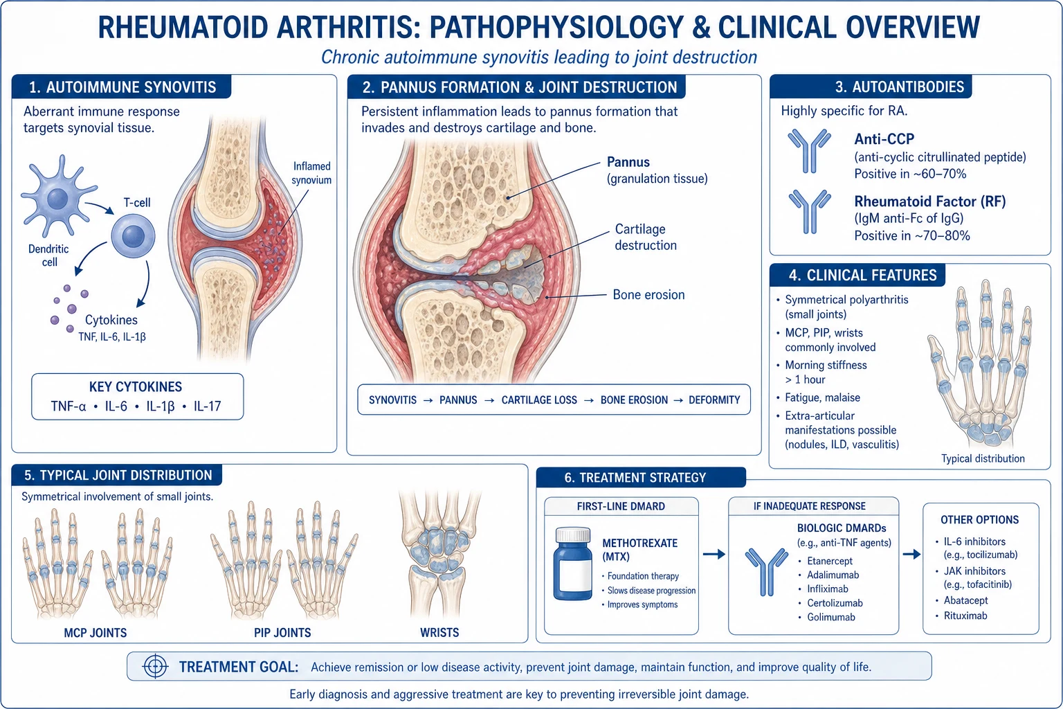
1. Citrullination and loss of tolerance. Peptidylarginine deiminases (PAD enzymes, especially PAD2 and PAD4) convert arginine residues in self-proteins (vimentin, fibrin, fibronectin, type II collagen, alpha-enolase) to citrulline. Citrullinated proteins are normally inert, but in susceptible individuals they are presented by HLA-DRB1 shared-epitope molecules and recognised by autoreactive CD4+ T cells, which provide help to B cells that produce ACPA (anti-citrullinated protein antibodies — what the clinical "anti-CCP" assay detects) and IgM rheumatoid factor (an antibody against the Fc portion of IgG).[3][7]
2. Synovial inflammation and pannus formation. ACPA-IgG immune complexes deposit in the joint, fix complement (classical pathway), activate macrophages and mast cells, and trigger release of TNF-alpha, IL-1, IL-6, IL-17 and granulocyte-macrophage colony-stimulating factor (GM-CSF). These cytokines drive: (a) synovial capillary proliferation and oedema; (b) massive infiltration by CD4+ T cells, B cells, plasma cells, macrophages and neutrophils into the synovial fluid; and (c) transformation of fibroblast-like synoviocytes (FLS) into a tumour-like, semi-autonomous population that no longer respects the joint boundary. The swollen, hyperplastic synovium covered by these cells is the pannus — the destructive tissue of RA.[7]
3. Cartilage and bone destruction. Two parallel effector pathways execute the damage that distinguishes RA from non-erosive arthropathies. Cartilage: FLS and macrophages secrete matrix metalloproteinases (MMP-1, MMP-3, MMP-13) and aggrecanases that digest the collagen and proteoglycan matrix; the pannus grows directly into the cartilage at the bare area. Bone: activated T cells and FLS express RANKL, which binds RANK on osteoclast precursors and drives their differentiation into mature, bone-resorbing osteoclasts — the cellular basis of the marginal erosion. The pro-inflammatory milieu simultaneously suppresses osteoblasts, so erosions do not heal.[7]
4. Why it persists. Three mechanisms keep RA self-sustaining: ectopic lymphoid neogenesis (the synovium organises into follicle-like structures with germinal centres producing high-affinity ACPA); epigenetic reprogramming of FLS to an aggressive phenotype that survives even when inflammation is dampened (a "imprinted stromal memory"); and defective regulatory T cells with impaired IL-2 signalling. This is why disease-modifying therapy must be started early — once FLS imprint and erosions form, structural damage becomes irreversible.[7]
5. Systemic consequences. The same cytokines (TNF, IL-6) that drive synovitis produce the anaemia of chronic disease (hepcidin-mediated iron sequestration), the thrombocytosis, the fatigue, anorexia and weight loss, the hypergammaglobulinaemia, the accelerated atherosclerosis (vascular inflammation, dyslipidaemia paradoxically improved by inflammation, endothelial dysfunction) and the osteoporosis seen in active disease. The CVD risk is approximately 1.5–2 times population baseline and is the single biggest cause of premature death.[8]
Clinical Presentation
RA presents most often with an insidious (weeks–months), symmetric polyarthritis of the small joints of the hands and feet with morning stiffness lasting more than one hour that improves with activity. An acute, explosive onset (over days) is less common but does occur, especially in elderly-onset disease and in palindromic patterns.[12]
Articular symptoms. Patients describe pain, swelling, warmth and stiffness in the MCP, PIP, wrist, elbow, shoulder, knee, ankle, MTP and cervical spine joints. The DIP joints are characteristically spared (a hallmark distinguishing RA from OA and psoriatic arthritis); the thoracic, lumbar and sacroiliac joints are also spared (distinguishing RA from axial spondyloarthritis). Morning stiffness improving with use is the opposite of OA, which worsens with use and is shorter-lived (under 30 minutes). The metacarpophalangeal "squeeze" test (pain on compression of the MCP joints) is a sensitive early sign.[1]
Classical hand deformities — high-yield for OSCEs and image stems: [1]
Ulnar deviation
- Fingers drift ulnarward at the MCP
- Due to capsular weakening and extensor imbalance
- Most visible MCP feature of chronic RA
Swan-neck deformity
- PIP hyperextension + DIP flexion
- Due to volar plate laxity and FDS tightening
- Lateral bands sublux dorsally
Boutonniere deformity
- PIP flexion + DIP hyperextension
- Due to central slip rupture with lateral band volar subluxation
- PIP pokes through like a button-hole
Z-thumb
- MCP flexion + IP hyperextension
- Characteristic 'Z' shape of thumb
Hitch-hiker's toe
- Hallux valgus with great toe overriding
- Plus clawing of lateral toes and subluxed MTP heads (cock-up toes)
Foot involvement — subluxed MTP heads feel like "walking on marbles"; the lateral toes develop claw/hammer deformities. Cervical spine involvement — atlantoaxial subluxation (anterior C1 on C2 from transverse ligament attenuation) produces neck pain radiating to the occiput and can cause cord compression.[10]
Extra-articular and systemic features (especially in seropositive, longstanding disease): [1]
- Rheumatoid nodules — firm, non-tender, subcutaneous nodules on extensor surfaces (olecranon, proximal ulna, Achilles, occiput), on pressure points and in tendon sheaths; histology shows central fibrinoid necrosis surrounded by palisading histiocytes.
- Sjögren syndrome and sicca symptoms (dry eyes, dry mouth) — most common overlap.
- Pulmonary — interstitial lung disease (UIP pattern most common), pleural effusion (low glucose, high LDH exudate), rheumatoid nodules in the lung (Caplan syndrome if pneumoconiosis coexists), bronchiectasis, obliterative bronchiolitis.
- Cardiovascular — accelerated atherosclerosis and ischaemic heart disease (the leading cause of premature death), pericarditis, myocarditis, coronary and aortic vasculitis.[8]
- Ocular — episcleritis, scleritis (scleromalacia perforans is sight-threatening), keratoconjunctivitis sicca, scleromalacia, peripheral ulcerative keratitis.
- Neurological — carpal tunnel syndrome, cervical myelopathy, mononeuritis multiplex (rheumatoid vasculitis), entrapment neuropathies.
- Haematological — anaemia of chronic disease, thrombocytosis, and Felty syndrome (the triad of longstanding seropositive RA + splenomegaly + neutropenia, with recurrent infections and leg ulcers; associated with large granular lymphocyte syndrome).
- Vascular — rheumatoid vasculitis (digital infarcts, leg ulcers, palpable purpura, mononeuritis multiplex) in severe seropositive disease.
- Renal — secondary (AA) amyloidosis presenting as nephrotic-range proteinuria after many years of uncontrolled inflammation.
- Constitutional — low-grade fever, fatigue, weight loss, anorexia, morning "gelling".
Atypical presentations that examiners deliberately test: [1]
- Elderly — onset after 60 with oligo-articular, large-joint, very systemic picture mimicking polymyalgia rheumatica; high ESR/CRP, often seronegative; co-consider giant-cell arteritis.
- Pregnancy — RA often improves in pregnancy (immune shift to Th2) but flares postpartum; remember methotrexate is teratogenic and must be stopped 3 months before conception.
- Diabetic / immunocompromised — septic arthritis can mimic a flare; always aspirate a hot monoarticular joint in these patients before assuming flare.
- Children — chronic inflammatory polyarthritis in a child under 16 is juvenile idiopathic arthritis (JIA), a related but distinct entity with different subtypes (oligo-articular, systemic, polyarticular, etc.). [1]
Differential Diagnosis
A symmetric small-joint polyarthritis with morning stiffness is not always RA. The dangerous omissions are missing septic arthritis in a hot monoarticular presentation, missing a treatable mimic (parvovirus, HBV/HCV, sarcoid, gout), and mislabelling OA or SLE as RA.[12]
Osteoarthritis
- Asymmetric; affects DIP (Heberden), PIP (Bouchard), 1st CMC, knees, spine
- Brief morning stiffness under 30 min, worse with use
- Crepitus, bony swelling, normal ESR/CRP, RF/ACPA negative
- X-ray: joint-space narrowing, osteophytes, subchondral sclerosis, NO erosions
Psoriatic arthritis
- Asymmetric; DIP involvement; enthesitis; dactylitis (sausage digit)
- Nail changes: pitting, onycholysis, hyperkeratosis
- Negative RF and ACPA in most cases
- X-ray: pencil-in-cup, fluffy periostitis, ankylosis, juxta-articular new bone
Systemic lupus erythematosus (Jaccoud)
- Symmetric, small joints — can be deforming but NON-erosive and REDUCIBLE
- Malar rash, photosensitivity, oral ulcers, renal/haematological disease
- ANA strongly positive; dsDNA, anti-Sm; low complement
- X-ray: normal — no erosions despite deformity
Gout
- Asymmetric; first MTP (podagra); tophi; episodic at first
- Hyperuricemia; MSU (negatively birefringent needles) or CPPD crystals in joint fluid
- X-ray: punched-out erosions with overhanging edges; preserved joint space
- Often male, metabolic syndrome, alcohol, diuretics
Viral arthritis (parvovirus B19, HBV, HCV, rubella, chikungunya)
- Acute onset; symmetric polyarthritis that can mimic RA exactly
- Self-limiting in weeks (parvovirus) to months (HCV); rash, fever, lymphadenopathy
- Parvovirus: slapped-cheek rash, IgM anti-parvovirus; HCV: positive HCV RNA ± cryoglobulins
- Usually non-erosive; resolves without DMARDs
Ankylosing spondylitis / axial SpA
- Young male; inflammatory back pain; asymmetric large-joint or lower-limb oligoarthritis
- HLA-B27, sacroiliitis on MRI; enthesitis, uveitis, psoriasis family history
- Negative RF and ACPA
- X-ray/MRI: sacroiliitis, syndesmophytes, bamboo spine
Polymyalgia rheumatica
- Age over 50; shoulder and hip girdle pain and stiffness
- Very high ESR/CRP; no synovitis or only mild peripheral arthritis
- Dramatic response to low-dose prednisolone (15 mg)
- Co-consider giant-cell arteritis; labelled elderly-onset RA if ACPA positive
Reactive arthritis
- Asymmetric oligoarthritis 1–4 weeks after GU/GI infection (Chlamydia, Salmonella, Shigella, Campylobacter, Yersinia)
- Triad: arthritis + urethritis + conjunctivitis (Reiter)
- Enthesitis, dactylitis, keratoderma blennorrhagicum, circinate balanitis
- Self-limiting in most; HLA-B27 associated
The single safest bedside rule for a hot, swollen joint is: any acutely inflamed monoarticular joint must be aspirated to exclude septic arthritis and crystals before NSAIDs or steroids are given. [1]
Clinical & Bedside Assessment
General — assess posture, gait, and functional independence (shoe-lacing, buttoning, grip). Look for rheumatoid nodules on extensor surfaces, psoriasis (scalp, umbilicus, natal cleft, nails), Sjögren features, eye signs (scleritis, episcleritis), leg ulcers and digital infarcts. [1]
Hand examination (the long-case centrepiece): [1]
- Inspection — symmetrical swelling of MCPs and PIPs, spared DIP, ulnar deviation, swan-neck, boutonniere and Z-thumb deformities, muscle wasting (thenar, interossei from disuse/entrapment), rheumatoid nodules, surgical scars (carpal tunnel release, joint replacement).
- Palpation — synovial boggy thickening (the pathognomonic feel of RA), warmth, tender and swollen joint count, squeeze test across MCPs and MTPs (positive if painful compression), restricted range, crepitus.
- Functional tests — grip strength, key pinch, button-fastening, writing, and a validated HAQ (Health Assessment Questionnaire) score. [1]
Other joints — elbows, shoulders, knees (effusion, Baker cyst — rupture mimics DVT), ankles, MTPs ("walking on marbles", claw toes), temporomandibular, and the cervical spine (assess for neck pain, Lhermitte's sign, and signs of myelopathy; check flexion-extension views before any anaesthesia/intubation).[10]
Systemic examination — chest (ILD, pleural effusion), cardiovascular (pericardial rub, ischaemic signs, valve lesions), eyes (sicca, scleritis), abdominal (splenomegaly of Felty), neurological (carpal tunnel, mononeuritis multiplex) and skin (vasculitic ulcers, nodulosis). [1]
Joints assessed in the DAS28 (28-joint count)
DAS 28 JOINTS
10 metacarpophalangeal joints ( bilateral 2nd–5th)
10 proximal interphalangeal joints (bilateral 2nd–5th)
2 wrists
2 elbows
2 shoulders
2 knees
DIP, the first CMC and first MTP are excluded — they are OA sites. The 28-joint set also omits ankles and the hips. [1]
Investigations
Investigations serve three purposes: to confirm inflammation, to classify RA (anti-CCP/RF, X-ray) and to monitor activity (DAS28) and therapy (drug toxicity). No single test diagnoses RA — the diagnosis is clinical, supported by serology and imaging. [1]
Acute-phase and routine bloods: ESR and CRP are raised in active disease and track with response to therapy (although up to 40% of patients have normal inflammatory markers at presentation). Full blood count shows anaemia of chronic disease (normocytic, normochromic, low ferritin relative to iron stores, high hepcidin), thrombocytosis and sometimes neutropenia (Felty). U&E, LFTs and a baseline chest X-ray are needed before methotrexate and other DMARDs.[2]
Serology (the diagnostic core): [1]
- Anti-cyclic citrullinated peptide (anti-CCP / ACPA) — high specificity (~95–98%), moderate sensitivity (~70%), positive early (often years before symptoms), predicts erosive disease. The single best serology.[3]
- Rheumatoid factor (IgM anti-Fc) — sensitivity ~70%, specificity ~70–80%; also positive in Sjögren, SLE, chronic infections (HBV, HCV, endocarditis, TB) and in ~5% of healthy elderly.
- ANA — to exclude SLE; ANA positivity with low/absent dsDNA and absent anti-Smith in RA does not change management.
- Viral and infection screens — parvovirus B19 IgM, HBsAg/HCV RNA, HIV when an acute symmetric polyarthritis could be viral; chikungunya in endemic areas.
Synovial fluid — sterile, inflammatory (WBC 2,000–50,000/μL, predominantly neutrophilic), negative culture, negative crystals. Always aspirate a hot, monoarticular joint to exclude septic arthritis (WBC typically over 50,000/μL, positive culture) and crystal arthropathy.[12]
Imaging: [1]
- Plain X-rays of hands, wrists and feet are the workhorse. Early changes are periarticular osteopenia and soft-tissue swelling; the pathognomonic feature is the marginal erosion at the bare area; later joint-space narrowing (uniform), subchondral cysts, subluxation and ankylosis. Foot X-rays (especially the 5th MTP head) often show erosions earlier than the hands.
- Ultrasound and power Doppler — detect subclinical synovitis, erosions and active hyperaemia; more sensitive than X-ray and useful for joint injection.
- MRI — most sensitive for early erosions, synovitis and bone marrow oedema (a precursor of erosion).
- Cervical spine lateral flexion/extension views — when neck pain or pre-operative: an atlantodens interval (ADI) over 3 mm (over 5 mm in children) suggests atlantoaxial subluxation.[10]
- CT/high-resolution CT chest — when ILD is suspected (RA-ILD carries significant mortality).
Disease activity scores — reproduced verbatim
The DAS28 (Disease Activity Score based on 28 joints) is the most widely used activity score.[5] It is calculated from tender joint count (TJC28), swollen joint count (SJC28), ESR (or CRP) and patient global health (0–100 mm VAS):
DAS28 (ESR) — interpretation
The EULAR response criteria combine current DAS28 with the change in DAS28 from baseline — a good responder improves by more than 1.2 and reaches low activity/remission.[5]
The ACR/EULAR 2022 Boolean remission criteria define true remission as TJC28 ≤1 AND SJC28 ≤1 AND CRP ≤1 mg/dL AND patient global assessment (0–10) ≤1 (the 2022 revision permits a patient global up to 2 in "Boolean 2.0").[6] The simpler CDAI (Clinical Disease Activity Index) and SDAI are also used in clinic and require no lab values (CDAI) or only CRP (SDAI).
The 2010 ACR/EULAR classification criteria — reproduced
To classify a patient as having RA, apply the scoring table reproduced in the Classification section above: at least one swollen joint, no better alternative, and a total score of 6 or more out of 10. The criteria were designed to identify early RA (unlike the 1987 criteria, which described late, deforming disease) and to start DMARD therapy early.[1]
Management — Resuscitation
RA is not the classic ABCDE-resuscitation disease, but several acute, time-critical scenarios demand immediate action. The two most important: rule out septic arthritis in any hot monoarticular joint, and exclude or stabilise cervical cord compression in a patient with new neurological signs. [1]
Acute scenarios that must be addressed before chronic therapy:
- Hot, swollen single joint — aspirate. Send synovial fluid for Gram stain, culture, cell count and crystals before any steroid injection. If septic arthritis is confirmed, washout + IV antibiotics (e.g., flucloxacillin 2 g IV QDS; vancomycin 1 g IV BD if MRSA risk or penicillin-allergic) — never inject a steroid into a possibly septic joint.
- Suspected cervical cord compression (neck pain, hand paraesthesia, gait change, hyperreflexia, sphincter disturbance) — urgent MRI cervical spine, neurosurgical referral, immobilise; the cord can be injured by routine intubation in unrecognised atlantoaxial subluxation.[10]
- Acute scleritis / scleromalacia / peripheral ulcerative keratitis — sight-threatening; urgent ophthalmology.
- Rheumatoid vasculitis (digital infarcts, mononeuritis multiplex, mesenteric vasculitis, leg ulcers) — high-dose glucocorticoid (e.g., methylprednisolone 500 mg–1 g IV daily for 3 days, then prednisolone 1 mg/kg/day) and urgent rheumatology; often requires cyclophosphamide or rituximab.
- Severe flare — short course of oral prednisolone (10–15 mg/day for 1–2 weeks then taper) or intramuscular methylprednisolone 80–120 mg, or intra-articular steroid (e.g., triamcinolone acetonide 20–40 mg into a knee), with gastric protection and bone protection for repeated courses.
Symptom control while waiting for the DMARD to work: an NSAID (e.g., naproxen 500 mg BD PO, ibuprofen 400–600 mg TDS, diclofenac 50 mg TDS, celecoxib 100–200 mg BD) with a PPI (omeprazole 20 mg) for the shortest effective duration; consider cardiovascular, renal and GI risk; avoid NSAIDs in pregnancy (especially 3rd trimester) and in advanced CKD.[2]
Management — Definitive & Stepwise
The backbone of modern RA management is the treat-to-target (T2T) strategy: a defined target (remission or, if not achievable, low disease activity), measured at every visit by a validated score (DAS28, CDAI or Boolean), with therapy escalated every 3–6 months until the target is reached.[4] Tight control doubles the chance of remission and halves radiographic progression. Therapy is layered: symptom relief, then a conventional synthetic DMARD, then combination csDMARDs, then a biologic or a targeted synthetic DMARD (JAK inhibitor).
Step 1 — Symptom control + first csDMARD
Methotrexate is the anchor DMARD and first-line treatment of choice in nearly all patients with active RA.[2]
Methotrexate — the anchor DMARD
Pre-treatment and monitoring: CBC, LFT, U&E at baseline, then every 2–4 weeks for 3 months, then monthly, then every 2–3 months while on therapy. Withhold for transaminases over 3× upper limit of normal, cytopenia, infection or pregnancy. Contraindications: pregnancy (teratogenic — stop 3 months before conception, both men and women), active infection, decompensated cirrhosis, significant renal impairment (reduce dose), and concurrent live vaccines. Co-prescribe folic acid (e.g., 5 mg weekly) to reduce mucosal, hepatic and GI toxicity. Pulmonary toxicity (acute methotrexate pneumonitis) is rare but important — stop and investigate new dry cough and dyspnoea.[2]
Step 2 — Combination csDMARDs ("triple therapy") and second-line csDMARDs
If methotrexate alone fails (DAS28 still above target at 3–6 months), add sulfasalazine (1–3 g/day PO in divided doses) and hydroxychloroquine (200–400 mg/day PO) — the "triple therapy" combination is non-inferior to anti-TNF in some trials and cheaper.[2]
Other csDMARDs: [1]
- Leflunomide (loading 100 mg daily for 3 days, then 10–20 mg daily) — alternative to methotrexate; also teratogenic (long washout with cholestyramine needed before pregnancy); monitor LFT and BP.
- Hydroxychloroquine monotherapy — mild disease; annual ophthalmology screening (bull's-eye maculopathy).
- Sulfasalazine — useful in patients who cannot take methotrexate; monitor CBC and LFT; causes reversible male infertility and orange discolouration of bodily fluids; avoid in G6PD deficiency. [1]
Glucocorticoids as a bridge: a short course of prednisolone 5–10 mg/day (or equivalent IM/IA) is acceptable as a temporary bridge while waiting for the DMARD to work; minimise dose and duration because of osteoporosis, diabetes, infection, hypertension, cataracts and cardiovascular risk. Add calcium and vitamin D (and a DEXA scan + bisphosphonate if appropriate) for any patient on long-term steroids. [1]
Step 3 — Biologic DMARDs (bDMARDs)
Indicated for moderate–severe RA failing csDMARD therapy.[2]
Anti-TNF agents
- Infliximab 3 mg/kg IV at weeks 0, 2, 6 then 8-weekly (titrate)
- Adalimumab 40 mg SC every 2 weeks
- Etanercept 50 mg SC weekly
- Golimumab 50 mg SC monthly; Certolizumab pegol 400 mg SC weeks 0, 2, 4 then 200 mg 2-weekly
- Screen for latent TB (interferon-gamma release assay), HBV/HCV; avoid in NYHA III/IV heart failure; risk of demyelination and reactivation of infection
Anti-IL-6 (tocilizumab, sarilumab)
- Tocilizumab 8 mg/kg IV every 4 weeks (max 800 mg/dose) or 162 mg SC every 2 weeks
- Sarilumab 200 mg SC every 2 weeks
- Monitor LFT, lipids, neutrophils; can mask fever; caution with diverticulitis (perforation risk)
B-cell depletion (rituximab)
- 1 g IV at days 0 and 14 (with methylprednisolone pre-medication), repeat every 6–12 months
- Best in **seropositive** (ACPA/RF+) RA
- Screen HBV before first dose (reactivation); monitor IgG; PJP prophylaxis if combined with steroids
T-cell co-stimulation block (abatacept)
- Weight-based IV at weeks 0, 2, 4 then 4-weekly; or 125 mg SC weekly
- Useful in patients where anti-TNF contraindicated (TB risk, heart failure)
Step 4 — Targeted synthetic DMARDs (tsDMARDs / JAK inhibitors)
If a biologic fails or is unavailable, oral JAK inhibitors are an option.[11][9]
- Tofacitinib 5 mg BD PO (or 11 mg XR OD); baricitinib 2–4 mg OD; upadacitinib 15 mg OD.
- Warnings: monitor lipids (LDL rises), LFT, neutrophils, haemoglobin; higher risk of herpes zoster (consider shingles vaccine); FDA/EMA boxed warning for venous thromboembolism, arterial thrombosis, major adverse cardiovascular events, malignancy and all-cause mortality — particularly in patients over 50 with at least one cardiovascular risk factor (the ORAL Surveillance study). Avoid in active infection, severe hepatic impairment, pregnancy and lactation.[9][11]
Escalation triggers (move up a step): persistent DAS28 above target after 3–6 months of optimised csDMARD; rapidly progressive erosions on imaging; extra-articular disease that mandates early aggressive control; functional decline; intolerable drug toxicity.[4]
De-escalation / tapering: once a patient has been in sustained remission (≥6–12 months), gradual dose taper of biologics or csDMARDs may be attempted under close monitoring — never abruptly stop, and have a clear flare plan. [1]
Surgery — indicated for severe structural damage or refractory pain: synovectomy, tendon repair/reconstruction, joint replacement (especially hip and knee arthroplasty), arthrodesis (wrist, cervical spine), and atlantoaxial fusion for symptomatic subluxation. Always obtain pre-operative flexion-extension cervical spine X-rays in any RA patient before anaesthesia.[10]
Non-pharmacological and multidisciplinary: physiotherapy (joint mobility, muscle strength, aerobic conditioning), occupational therapy (joint protection, aids, splints), podiatry, dietitian (Mediterranean diet, omega-3, vitamin D, smoking cessation, weight loss), psychology and social work for employment and disability support. Smoking cessation is one of the few modifiable factors that improves treatment response and reduces cardiovascular risk. [1]
Specific Subtypes & Scenarios
- Seropositive RA — ACPA and/or RF positive; classical erosive course with rheumatoid nodules and extra-articular disease; responds well to rituximab and abatacept; needs tight control to prevent erosions.
- Seronegative RA — both negative; milder and less erosive but a diagnosis of exclusion — actively exclude psoriatic, reactive, viral, sarcoid and crystal arthropathies before labelling.
- Elderly-onset RA (EORA, age over 60) — abrupt, oligo-articular, large-joint, very systemic, polymyalgia-like; often seronegative; differentials include PMR (responds dramatically to 15 mg prednisolone), paraneoplastic arthritis and crystal arthropathies.[12]
- Felty syndrome — the triad of longstanding seropositive RA + splenomegaly + neutropenia, often with anaemia, thrombocytopenia, leg ulcers, recurrent infections and hyperpigmentation. Associated with HLA-DR4 and a clonal expansion of large granular lymphocytes (T-LGL syndrome). Treat active RA (methotrexate is first-line; rituximab for refractory); consider G-CSF for severe neutropenia with infection; splenectomy is rarely used.
- Secondary (AA) amyloidosis — chronic uncontrolled inflammation drives serum amyloid A (SAA) deposition; presents as nephrotic-range proteinuria, progressive renal failure, hepatosplenomegaly and malabsorption. Diagnose on biopsy (Congo-red apple-green birefringence); treat underlying RA aggressively.
- Palindromic RA — episodic attacks of mono- or oligo-arthritis (each lasting 1–3 days with complete resolution); ~30–50% evolve into classic RA; treat flares with NSAIDs or short steroid bursts; hydroxychloroquine may reduce attack frequency.
- Juvenile idiopathic arthritis (JIA) — chronic inflammatory arthritis in a child under 16 lasting at least 6 weeks; subtypes include oligo-articular (most common; ANA positive; uveitis risk), polyarticular RF-negative, polyarticular RF-positive, systemic (Still disease), enthesitis-related, psoriatic and undifferentiated. Treated with methotrexate, biologics (etanercept) and intra-articular steroids.
- Rheumatoid vasculitis — small-to-medium vessel vasculitis in severe seropositive long-standing disease; digital gangrene, leg ulcers, palpable purpura, mononeuritis multiplex, mesenteric and coronary vasculitis; treat aggressively with high-dose glucocorticoid ± cyclophosphamide or rituximab.
- Caplan syndrome — multiple, rapidly progressive rheumatoid lung nodules in a patient with pneumoconiosis (coal-workers' pneumoconiosis, silicosis, asbestosis).
- Sjögren overlap — secondary Sjögren syndrome in RA is common; manage sicca with saliva substitutes, pilocarpine 5 mg QDS, punctal plugs and topical ciclosporin eye drops.
Complications & Pitfalls
Articular and structural — joint destruction, secondary OA, tendon ruptures (especially extensor tendons at the ulnar styloid), Baker cyst (popliteal) that may rupture and mimic DVT, atlantoaxial and subaxial cervical subluxation with potential cord compression, osteooporosis (disease- and steroid-driven), reduced grip and functional disability.[10]
Systemic — premature cardiovascular disease (the leading cause of excess death; risk comparable to diabetes), interstitial lung disease (UIP pattern carries the worst prognosis), pleural effusion and pericarditis, scleritis and scleromalacia, secondary amyloidosis, Felty syndrome, rheumatoid vasculitis, anaemia of chronic disease, depression, lymphoma (modestly increased in severe active disease), infections (from disease and immunosuppression), and peptic ulcer disease (NSAIDs and steroids).[8]
Treatment-related — methotrexate (hepatotoxicity, marrow suppression, pneumonitis, teratogenicity), leflunomide (hepatotoxicity, neuropathy, hypertension, teratogenicity), sulfasalazine (hepatotoxicity, cytopenia, rash, reversible male infertility), hydroxychloroquine (maculopathy), glucocorticoids (osteoporosis, diabetes, hypertension, cataracts, peptic ulcer, infection), anti-TNF (TB reactivation — always screen with IGRA and CXR, HBV reactivation, heart failure exacerbation, demyelination, infection), JAK inhibitors (herpes zoster, VTE/MACE/malignancy boxed warning).[9][11]
[1]Classic pitfalls — labelling OA, psoriatic or viral arthritis as RA without examining the DIP and the nails; missing parvovirus B19 in a young woman with acute symmetric polyarthritis and a rash; injecting a steroid into a septic joint; forgetting cervical spine screening before anaesthesia; giving methotrexate to a pregnant or planning-pregnancy patient; starting a biologic without screening for TB/HBV; treating the ESR instead of the patient; indefinite oral steroids without bone protection; failing to vaccinate patients on immunosuppression. [1]
Prognosis & Disposition
Untreated RA causes major joint destruction within 2 years of onset and reduces life expectancy by 5–10 years, driven largely by cardiovascular disease, infection and pulmonary complications.[8] With early DMARD therapy and treat-to-target, the prognosis is transformed — 30–50% of patients achieve sustained remission, radiographic progression is largely arrested, and life expectancy approaches normal in well-controlled disease.[4]
Poor-prognosis markers (drive early aggressive therapy): ACPA positivity (especially high titre), high RF, HLA-DRB1 shared epitope, early radiographic erosions, elevated and persistent CRP/ESR, extra-articular disease, smoking, female sex with disability, and failure to respond to initial methotrexate within 3–6 months. [1]
Disposition: most patients are managed as outpatients by a rheumatology-led multidisciplinary team in a treat-to-target clinic. Admit for septic arthritis, atlantoaxial cord compression, vasculitic crisis, interstitial lung disease with respiratory failure, severe infection and occasionally for IV cyclophosphamide or rescue therapy. The safety-net is a named rheumatology nurse/telehealth line and a flare plan for the patient. [1]
Special Populations
- Pregnancy — RA often improves during pregnancy (immune shift to Th2) but flares in the postpartum period. Methotrexate and leflunomide are teratogenic — stop 3 months before conception (men and women; cholestyramine washout for leflunomide). Safe in pregnancy: hydroxychloroquine, sulfasalazine (continue with folic acid 5 mg/day), azathioprine (≤2 mg/kg/day), low-dose prednisolone (avoid fluorinated steroids after first trimester), and certolizumab (minimal placental transfer, preferred biologic in pregnancy). Avoid: NSAIDs (especially 3rd trimester — premature closure of ductus arteriosus, oligohydramnios), high-dose steroids (cleft palate, gestational diabetes, hypertension), live vaccines, methotrexate, leflunomide, mycophenolate, JAK inhibitors. Breastfeeding: methotrexate contraindicated; hydroxychoroquine, sulfasalazine, low-dose steroids and certolizumab are compatible.[2]
- Children (JIA) — chronic inflammatory arthritis before 16 lasting at least 6 weeks; oligo-articular JIA carries the highest risk of chronic anterior uveitis — mandatory slit-lamp screening every 3 months in ANA-positive children under 7; managed by paediatric rheumatology.
- Elderly — more comorbidity, drug interactions (NSAID renal/GI risk, steroid osteoporosis), and atypical presentations; start DMARDs just as early as in younger adults; favour methotrexate over alkylating agents.
- Renal impairment — methotrexate and leflunomabe need dose reduction or avoidance in CKD; hydroxychloroquine is preferred in CKD; NSAIDs contraindicated in advanced CKD; avoid colchicine if misdiagnosed as gout.
- Hepatic disease — methotrexate and leflunomide avoided in decompensated cirrhosis and active hepatitis; screen HBV/HCV before biologic.
- Pre-existing infection (TB, HBV, HCV, HIV) — treat before biologic; choose rituximab (lowest TB reactivation) for latent TB if a biologic is essential.
- Anticoagulated patients — additional bleeding risk from NSAIDs (avoid); methotrexate and biologics are generally safe; JAK inhibitors add VTE risk — caution with warfarin/DOAC.[9]
- Smokers — counsel that smoking reduces anti-TNF and methotrexate efficacy, drives ACPA-positive disease and increases cardiovascular risk; offer smoking cessation as part of treatment.
Evidence, Guidelines & Regional Differences
Landmark shifts in the management of RA: [1]
- 1987 ACR criteria — designed for established, erosive RA; superseded by the 2010 ACR/EULAR criteria, which were designed to identify early disease and trigger early DMARD therapy.[1]
- 2010 onward — treat-to-target (T2T) — an international task force recommended remission or low disease activity as the goal, with therapy escalation every 3–6 months; updated in 2014 and repeatedly shown to double remission rates.[4]
- 2010s — anchor DMARD revolution — methotrexate confirmed as first-line anchor; triple csDMARD therapy shown to be effective in early RA and non-inferior to anti-TNF in some studies.[2]
- Biologics era — anti-TNF, tocilizumab, rituximab and abatacept transformed moderate–severe disease; biosimilars (e.g., infliximab biosimilars) have improved affordability.[2]
- JAK inhibitor era — tofacitinib, baricitinib and upadacitinib offer an oral option, but the ORAL Surveillance trial raised the VTE/MACE/malignancy signal in older, at-risk patients, prompting FDA/EMA boxed warnings.[9][11]
- 2019/2020 EULAR update — confirms methotrexate as first csDMARD; glucocorticoid should be short-term only (the long-term goal is glucocorticoid-free); bDMARD or tsDMARD after csDMARD failure; taper when in sustained remission.[2]
EULAR (Europe, 2019/2020 update) — first csDMARD is methotrexate (target 25–30 mg/week if tolerated); add short glucocorticoid taper; switch/add csDMARD or biologic/JAK if active disease beyond 6 months; bDMARD or tsDMARD if inadequate response or contraindication; biosimilars acceptable. EULAR also recommends annual CV risk score with a 1.5 multiplier, screening for comorbidities (ILD, CVD, osteoporosis, infection, depression) and shared decision-making.[2]
Vaccination in immunosuppressed RA: pneumococcal (PCV15/20 followed by PPSV23), annual influenza, COVID-19, hepatitis B, recombinant zoster (Shingrix) — give before starting biologics where possible; avoid live vaccines (MMR, yellow fever, oral polio, live zoster) once on biologic or JAK therapy. [1]
Exam Pearls
- RA = symmetric, small-joint, erosive polyarthritis that SPARES THE DIP. Morning stiffness over 1 hour (OA under 30 minutes).
- Anti-CCP (ACPA) is the most specific antibody for RA (specificity ~95–98%); RF is sensitive but not specific (Sjögren, SLE, chronic infection, elderly).
- DIP, 1st CMC, 1st MTP are EXCLUDED from the 2010 ACR/EULAR criteria and the DAS28 joint count — they are OA sites.
- Methotrexate is the first DMARD — weekly (not daily — fatal dosing error), with folic acid; teratogenic, hepatotoxic, marrow-suppressive; bridge with steroid.
- Treat-to-target to remission (DAS28 below 2.6) or low disease activity — escalate every 3–6 months.
- Anti-TNF (infliximab, adalimumab, etanercept, golimumab, certolizumab) — screen for TB (IGRA + CXR) and HBV before starting.
- JAK inhibitors (tofacitinib, baricitinib, upadacitinib) — VTE/MACE/malignancy boxed warning, herpes zoster.
- Atlantoaxial subluxation — screen with lateral flexion-extension cervical X-rays before intubation or surgery; ADI over 3 mm is abnormal.[10]
- Felty syndrome = longstanding seropositive RA + splenomegaly + neutropenia (+ leg ulcers, recurrent infection).
- Caplan syndrome — rheumatoid lung nodules + pneumoconiosis.
- Hand deformities: ulnar deviation, swan-neck (PIP hyperextended, DIP flexed), boutonniere (PIP flexed, DIP extended), Z-thumb.
- Sjögren overlap — Schirmer test (≤5 mm in 5 min is positive); manage sicca with saliva substitutes, pilocarpine.
- Leading cause of death in RA = cardiovascular disease — apply a 1.5× CV risk multiplier.[8]
- Methotrexate must be STOPPED 3 months before conception (men and women); hydroxychloroquine and sulfasalazine are SAFE in pregnancy.
- Rheumatoid pleural effusion — exudate with very low glucose (often under 1.6 mmol/L), high LDH, low pH.
Exam application bank (NEET-PG / INICET)
One-line answer
Rheumatoid arthritis (RA) is a chronic, symmetric, erosive, autoimmune polyarthritis of unknown cause that preferentially targets the synovial small joints (MCP, PIP, wrists, MTP), spares the DIP, and produces morning stiffness lasting more than 1 hour. Diagnosis is clinical plus anti-CCP antibody (highly specific) and characteristic X-ray erosions; disease activity is graded with the DAS28. Management is treat-to-target to remission or low disease activity, beginning with methotrexate (weekly + folic acid) and escalating through csDMARDs, bDMARDs (anti-TNF, anti-IL-6, anti-CD20, abatacept) and tsDMARDs/JAK inhibitors. Untreated disease causes deformity, disability, atlantoaxial subluxation and premature cardiovascular death.
Worked stems (answer without another resource)
Stem 1 — Classic presentation. Map symptoms to mechanism; name the first investigation and first treatment step with dose/route if drug therapy is standard. [1]
Stem 2 — Unstable / complicated. List red flags that force immediate resuscitation, theatre, ICU, antidote, or reperfusion — and what you do in the first 15 minutes. [1]
Stem 3 — Atypical group. Elderly, pregnancy, child, or immunocompromised: how presentation and thresholds change. [1]
Stem 4 — Differential trap. Name the three closest mimics and one discriminator for each. [1]
Stem 5 — Disposition. Who goes home with safety-netting, who is admitted, who needs HDU/ICU/theatre, and what follow-up is mandatory. [1]
Rapid viva checklist
- Definition + classification
- Pathophysiology chain
- Bedside signs / criteria
- Score with exact components (if any)
- Emergency bundle
- Definitive therapy with doses
- Complications of disease and of treatment
- Special populations
- Guideline/trial name if classic
- Three exam traps
Coverage self-check
If you cannot answer any stem above from this page alone, re-read the matching section — the page is intended to be self-sufficient for final-prof and NEET-PG/INICET questions on Rheumatoid Arthritis.
References
- [1]Neogi T, Aletaha D, Silman AJ, et al. The 2010 American College of Rheumatology/European League Against Rheumatism classification criteria for rheumatoid arthritis: Phase 2 methodological report Arthritis Rheum, 2010.PMID 20872596
- [2]Smolen JS, Landewe RBM, Bijlsma JWJ, et al. EULAR recommendations for the management of rheumatoid arthritis with synthetic and biological disease-modifying antirheumatic drugs: 2019 update Ann Rheum Dis, 2020.PMID 31969328
- [3]Schellekens GA, Visser H, de Jong BA, et al. The diagnostic properties of rheumatoid arthritis antibodies recognizing a cyclic citrullinated peptide Arthritis Rheum, 2000.PMID 10643712
- [4]Smolen JS, Breedveld FC, Burmester GR, et al. Treating rheumatoid arthritis to target: 2014 update of the recommendations of an international task force Ann Rheum Dis, 2016.PMID 25969430
- [5]Prevoo ML, van 't Hof MA, Kuper HH, et al. Modified disease activity scores that include twenty-eight-joint counts. Development and validation in a prospective longitudinal study of patients with rheumatoid arthritis Arthritis Rheum, 1995.PMID 7818570
- [6]Studenic P, Aletaha D, de Wit M, et al. American College of Rheumatology/EULAR remission criteria for rheumatoid arthritis: 2022 revision Ann Rheum Dis, 2023.PMID 36280238
- [7]McInnes IB, Schett G. Pathogenetic insights from the treatment of rheumatoid arthritis Lancet, 2017.PMID 28612747
- [8]Yazdani K, Latham K, Warrington J, Pope JE. Has the excess risk of acute myocardial infarction in rheumatoid arthritis relative to the general population declined? A population study of trends over time Semin Arthritis Rheum, 2021.PMID 33735663
- [9]Charles-Schoeman C, Pagidipati NJP, Fleischmann R, et al. Risk of Venous Thromboembolism With Tofacitinib Versus Tumor Necrosis Factor Inhibitors in Cardiovascular Risk-Enriched Rheumatoid Arthritis Patients Arthritis Rheumatol, 2024.PMID 38481002
- [10]Neva MH, Kauppi MJ, Kautiainen H, et al. Early and extensive erosiveness in peripheral joints predicts atlantoaxial subluxations in patients with rheumatoid arthritis Arthritis Rheum, 2003.PMID 12847673
- [11]Szekanecz Z, Mesko B, Poliska S, Vancsa A. Efficacy and safety of JAK inhibitors in rheumatoid arthritis: update for the practising clinician Nat Rev Rheumatol, 2024.PMID 38216757
- [12]Combe B, Landewe R, Daien CI, et al. 2016 update of the EULAR recommendations for the management of early arthritis Ann Rheum Dis, 2017.PMID 27979873