Rheumatology · General Medicine
Septic Arthritis
Also known as Septic arthritis · Infectious arthritis · Bacterial arthritis · Pyogenic arthritis · Native joint sepsis
Septic arthritis is a medical and surgical emergency — direct microbial invasion of a joint space (usually bacterial and haematogenous) causing purulent synovial inflammation, rapid articular cartilage destruction, sepsis and death if untreated. Incidence is 4 to 10 per 100,000 in the West and rises steeply with prosthetic joints, rheumatoid arthritis, diabetes, immunosuppression, IV drug use and age over 80. The knee is the commonest joint (about half of cases). Staphylococcus aureus is the commonest organism in all ages; Neisseria gonorrhoeae in young sexually active adults (migratory polyarthralgia, tenosynovitis, pustular rash); Group B streptococcus in neonates; Haemophilus influenzae and Streptococcus pneumoniae in unvaccinated children; Salmonella in sickle-cell disease; Pseudomonas and Gram-negatives in IV drug users; coagulase-negative staphylococci in prosthetic joints. Presentation is a single hot, swollen, very tender joint with severe pain on movement ± fever (fever may be absent). Diagnosis rests on urgent joint aspiration: synovial WBC typically over 50,000 per cubic millimetre (50,000 to 200,000), neutrophil-predominant, positive Gram stain and culture (positive in about 75% of Gram-positive, 50% overall), low glucose, raised lactate; blood cultures are positive in about half. The Kocher criteria stratify the irritable paediatric hip. Crystals do NOT exclude infection — sepsis coexists with gout/CPPD in up to a fifth of cases. Treatment is urgent surgical washout/drainage plus empirical IV antibiotics started immediately after aspiration — flucloxacillin 2 g IV QID (clindamycin if allergic, vancomycin 1 g IV BD if MRSA suspected) plus Gram-negative cover (ceftriaxone 2 g IV OD), narrowed to culture, total 4 to 6 weeks (about 2 IV then oral). Gonococcal arthritis: ceftriaxone 1 g IV/IM OD plus azithromycin 1 g PO. Prosthetic joint infection follows a specialist staged surgical pathway with biofilm-aware antibiotics. Mortality is 7 to 15%; up to half retain joint damage.
On this page & tools
Your progress
Saved locally on this device.
Exam tags
Red flags
Overview & Definition
Septic arthritis is the most time-critical arthritis in medicine. It is defined as direct microbial invasion of a joint space — usually by pyogenic bacteria, less often by mycobacteria, fungi, or viruses — producing purulent synovial inflammation that, untreated, destroys articular cartilage within 24 to 48 hours and may progress to systemic sepsis, multi-organ failure and death. The non-negotiable rule of practice is that any acute hot, swollen, tender joint is septic until proven otherwise, and requires urgent arthrocentesis before antibiotics are given. The diagnosis rests on synovial fluid analysis (cell count, Gram stain, culture, crystal microscopy) supported by blood cultures and inflammatory markers, but no single value excludes infection — a partially treated patient, a gonococcal infection, or an immunocompromised host may all present with synovial WBC counts well below the textbook 50,000. The gonococcal subtype (young, sexually active, with migratory polyarthralgia, tenosynovitis and pustules) requires NAAT detection because cultures are negative in over half. Treatment is surgical washout or drainage combined with empirical IV antibiotics, narrowed to culture, for 4 to 6 weeks.[1][2][5]
Classification
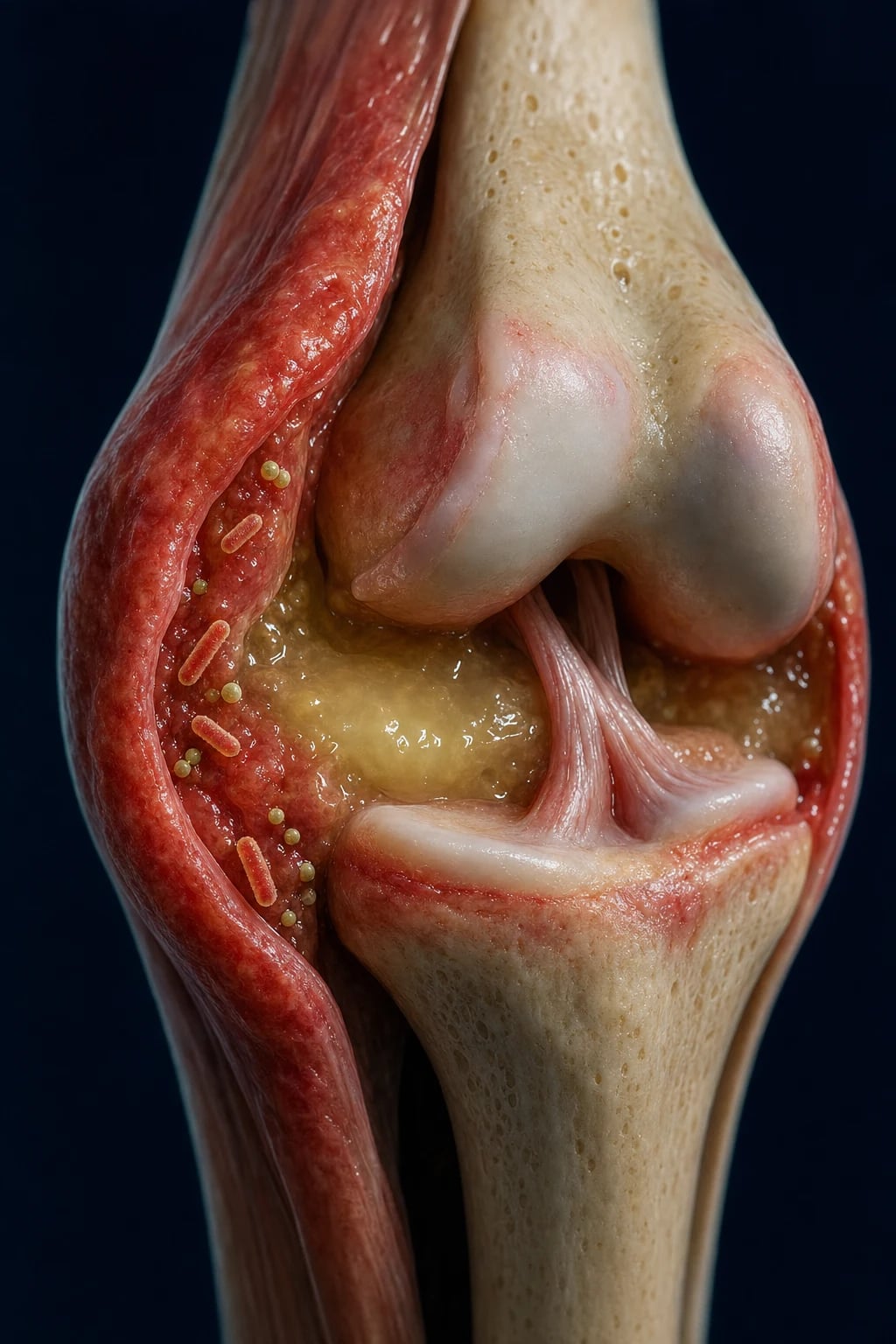
Septic arthritis is classified along three clinically useful axes — organism, route, and host/joint context — because each shifts the differential, the empiric antibiotic, and the surgical pathway.[1][2]
By organism (nongonococcal)
- Staphylococcus aureus (incl MRSA) — commonest across all ages
- Streptococcus pyogenes, S. pneumoniae, Group B strep (neonates)
- Gram-negatives — E. coli, Pseudomonas (IVDU), Klebsiella (diabetes)
- Coagulase-negative staphylococci (S. epidermidis) — prosthetic joints
- Salmonella — sickle-cell disease; unusual joints in immunocompromised
By organism (gonococcal)
- Neisseria gonorrhoeae — disseminated gonococcal infection (DGI)
- Young sexually active adults; scanty pus, cultures often negative
- Migratory polyarthralgia + tenosynovitis + pustular dermatitis triad
- Diagnose with NAAT (urine, cervical, urethral, pharyngeal, rectal)
- Ceftriaxone + azithromycin/doxycycline; treat partners and screen HIV/syphilis
By route
- Haematogenous — commonest; bacteria seed the vascular synovium
- Direct inoculation — penetrating trauma, intra-articular injection, surgery
- Contiguous spread — cellulitis, osteomyelitis, septic bursitis
- Prosthetic joint — peri-operative (early) vs haematogenous (late)
By host/joint
- Native joint — adults (knee > hip > shoulder) and children (hip)
- Prosthetic joint — early (under 3 mo), delayed (3 to 24 mo), late (over 24 mo)
- Polyarticular — RA, immunosuppression, endocarditis, gonococcal
- Paediatric/neonatal — occult hip, Group B strep, H. influenzae if unvaccinated
Epidemiology & Risk Factors
The annual incidence in Western populations is 4 to 10 per 100,000, but this average conceals enormous stratification. In patients with rheumatoid arthritis the incidence rises roughly threefold; in joint prostheses it reaches 2 to 4 per 1,000 joint-years (and 1 to 2% of all primary arthroplasties); in intravenous drug users and the immunocompromised it is again several-fold higher. The incidence in older adults (over 80) is roughly double that of the general adult population.[1][2][9]
The age distribution is bimodal: a peak in children and young adults (especially neonates and toddlers, where the hip is the dominant joint), and a second, larger peak in adults over 60. Males are affected slightly more often than females, except in young adults where gonococcal disease is commoner in women (often during menstruation or pregnancy).[2][8]
Septic arthritis — the numbers that matter
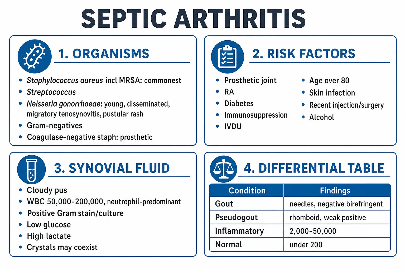
The risk factors form a short, examinable list — each works by either increasing bacterial exposure (skin breakdown, injection, bacteraemia), reducing host defences (immunosuppression, diabetes, malignancy, HIV), or changing the joint environment (prosthetic material, prior joint damage, RA synovium).[1][2]
- Prosthetic joint — the single largest risk factor; foreign material supports biofilm and resists host clearance.
- Rheumatoid arthritis — damaged synovium, prior steroid/biologic exposure; risk multiplied and the diagnosis is delayed because sepsis mimics a flare.
- Diabetes mellitus — impaired neutrophil function, peripheral neuropathy and skin ulcers.
- Immunosuppression — glucocorticoids, anti-TNF agents, methotrexate, malignancy, HIV.
- Intravenous drug use — bacteraemia with S. aureus and Pseudomonas; unusual axial joints (sternoclavicular, sacroiliac, spinal).
- Age over 80, skin infection/ulcer, recent intra-articular injection or surgery, alcohol excess, haemodialysis.
- Sexually active young adults — N. gonorrhoeae; risk amplified during menstruation, pregnancy, and complement deficiency (C5–C9).[1][2][8]
Pathophysiology
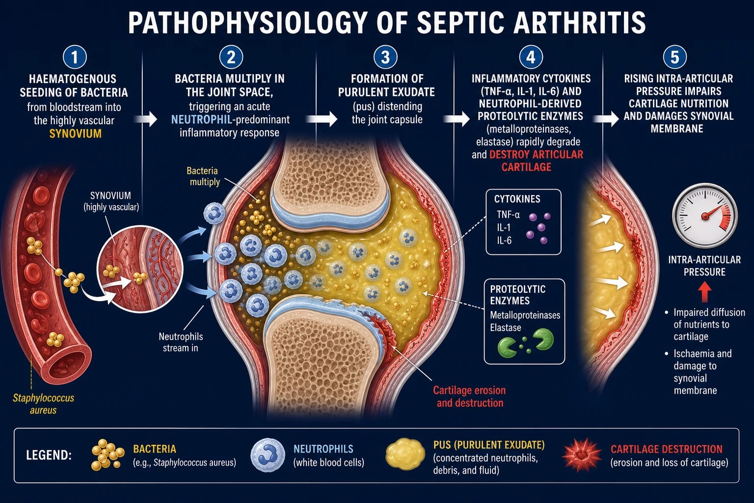
The pathogenesis of septic arthritis is best understood as a three-step cascade — arrival, multiplication, and destruction — followed by irreversible structural failure. Understanding the cascade tells you why time is cartilage.[2][5]
Step 1 — Arrival. Bacteria reach the joint by one of three routes. Haematogenous seeding is by far the commonest: bacteria lodge in the highly vascular synovial membrane, which lacks a limiting basement membrane, so organisms pass directly from capillaries into the joint space and synovial fluid. Less often the route is direct inoculation (penetrating trauma, intra-articular injection, arthrotomy) or contiguous spread from overlying cellulitis, a septic bursa, or adjacent osteomyelitis — particularly in the paediatric hip, where the metaphyseal circulation is intracapsular.[2][7]
Step 2 — Multiplication and immune failure. Once inside the joint, bacteria evade host defences in three ways. S. aureus expresses virulence factors — protein A (binds the Fc portion of IgG, evading opsonisation), microcapsular polysaccharide, alpha-toxin and PNAG (poly-N-acetylglucosamine) — that resist complement and neutrophil killing. In prosthetic joints, organisms form a biofilm (slime layer of exopolysaccharide) on the implant surface, embedding bacteria in a metabolically quiescent state that is essentially impervious to antibiotics and host immunity.[2][6]
Step 3 — Purulent destruction. An acute neutrophil-predominant response floods the joint. Activated neutrophils and synoviocytes release proteolytic enzymes — matrix metalloproteinases (MMP-1, MMP-3, MMP-13), elastase, collagenase and cathepsins — and a cytokine storm (TNF-alpha, IL-1, IL-6, IL-8) that degrade articular cartilage within 24 to 48 hours. Cartilage, which is avascular and depends on synovial fluid for nutrition, does not regenerate: once destroyed, it is gone for good.[2]
Step 4 — Structural failure. Rising intra-articular pressure from accumulating pus compresses the epiphyseal and capsular vasculature, impairing cartilage nutrition and producing avascular necrosis of the epiphysis — a particular catastrophe in the paediatric hip. Pannus, adhesions and capsular fibrosis follow, producing permanent stiffness; in late disease, secondary osteoarthritis supervenes. Pus under pressure can rupture the capsule, producing a sinus or spread to surrounding soft tissues.[2][7]
Clinical Presentation
The classic presentation of native-joint septic arthritis in an adult is unmistakable: an acute monoarthritis developing over hours to days, with the joint hot, red, swollen, exquisitely tender, held in the position of comfort (slight flexion for the knee, flexion-abduction-external rotation for the hip), with severe pain on any active or passive movement — passive movement is the cardinal sign, more reliable than fever.[1][5]
Joint distribution. The knee is involved in about half of cases, then the hip (deeper, more occult — pain referred to the groin, thigh or knee, and a child may simply refuse to weight-bear), shoulder, wrist, ankle; small joints are uncommon. Polyarticular disease (around 10 to 20% of adults) occurs in rheumatoid arthritis, endocarditis, severe immunosuppression and disseminated gonococcal infection.[2][9]
Systemic features. Fever, rigors, malaise and sweats are common but fever is absent in up to 40% — its absence does not exclude sepsis, especially in the elderly, diabetic and immunocompromised. Septic shock may be the presenting syndrome in fulminant disease.[1][5]
Atypical presentations examiners deliberately probe:[1][2]
- Elderly or immunocompromised — pain may be muted, fever absent, confusion or falls the only clue; a high index of suspicion is required.
- Rheumatoid arthritis — a single joint disproportionately hot, swollen or painful compared with others; do not assume a flare. Aspirate.
- Prosthetic joint — new or increasing joint pain, stiffness, wound drainage or sinus tract; systemic features are often absent in chronic infection.[6][9]
- Diabetic with neuropathy — presentation may be that of an indolent swollen foot or ankle; Charcot-like in tempo.
- Disseminated gonococcal infection (DGI) — a young, sexually active adult with the tenosynovitis-dermatitis-arthritis syndrome: migratory polyarthralgia, asymmetric tenosynovitis (especially extensor tendons of hands/wrists/ankles), and a painless pustular rash on the distal extremities (often confused with insect bites). Two-thirds present with this pattern; the remainder present with purulent monoarthritis.[8]
- Paediatric hip — a limping or non-weight-bearing child, often held flexed and externally rotated; pseudoparalysis in the infant. The diagnosis is largely clinical + Kocher criteria + imaging because aspiration of the deep hip requires ultrasound.[3][7]
Differential Diagnosis
The differential of an acute monoarthritis is short and the synovial fluid almost always resolves it — but crystals do NOT exclude infection, and around 5 to 20% of culture-positive septic joints also contain crystals. Every acutely hot joint is aspirated and the fluid sent for cell count, Gram stain, culture AND crystal microscopy, regardless of how classical the alternative looks.[1][2]
Septic arthritis
- Cloudy or purulent fluid
- WBC typically 50,000 to 200,000+, neutrophil-predominant (but lower does not exclude)
- Positive Gram stain (about 75% of Gram-positive, 50% overall) and culture; low glucose, raised lactate
- Urgent washout + IV antibiotics 4 to 6 weeks; never assume without aspirating
Gout
- Negatively birefringent NEEDLE-shaped monosodium urate crystals (intracellular)
- WBC 15,000 to 50,000+, neutrophilic
- First MTP (podagra) classical; serum urate may be normal during the attack
- Acute: NSAIDs, colchicine, oral or intra-articular steroids; chronic: allopurinol
Pseudogout (CPPD)
- Weakly POSITIVELY birefringent RHOMBOID crystals
- WBC variable, neutrophilic
- Knee and wrist; elderly; chondrocalcinosis on X-ray
- NSAIDs, colchicine, steroids; treat underlying cause (hyperparathyroidism, haemochromatosis)
Reactive arthritis
- Asymmetric oligoarthritis 1 to 4 weeks after STI (Chlamydia) or GI infection (Salmonella, Shigella, Campylobacter, Yersinia)
- Enthesitis, conjunctivitis, urethritis — classic triad; keratoderma blennorrhagicum
- Sterile synovial culture; HLA-B27 association
- NSAIDs first-line; intra-articular steroids; antibiotics do not change the arthritis course
Other / mimics
- RA flare or psoriatic arthritis flare (often polyarticular, but aspirate a suspicious single joint)
- Trauma and haemarthrosis (haemophilia, anticoagulation, ACL tear — history of injury)
- Adjacent osteomyelitis, septic bursitis (movement preserved in bursitis), cellulitis
- Viral arthritis (parvovirus B19, rubella, hepatitis B/C — usually polyarticular and self-limiting)
- Lyme arthritis (endemic area, target erythema migrans, ELISA/Western blot)
- Tuberculous arthritis (chronic, indolent, 'caries sicca', positive AFB culture)
Clinical & Bedside Assessment
Bedside assessment runs in parallel with resuscitation — a septic joint may coexist with systemic sepsis, and the patient must be safe to be moved to theatre before they are sent.[2][5]
- ABCDE and sepsis screen — airway, breathing (oxygen saturation, respiratory rate), circulation (heart rate, blood pressure, perfusion, capillary refill, urine output), disability (mental status), exposure of the joint and any source. If the patient meets Sepsis Six criteria, deliver oxygen, take cultures, give IV fluids and antibiotics, monitor lactate and urine output, and prepare for theatre.[5]
- The joint exam — compare with the opposite side. Look for erythema, swelling, warmth, effusion; feel for a boggy effusion and exquisite tenderness; the cardinal sign is severe pain on passive movement in any direction — far more restrictive than a flare. Active and passive range of motion are both markedly limited. Hold position noted (slight flexion at the knee, flexion-abduction-external rotation at the hip).
- Single vs polyarticular, and distribution — note tenosynovitis, pustules or rash (gonococcal); check all joints, the spine and the sacroiliac joints.
- Source hunt — skin ulcers, cellulitis, abscess, injection sites (IVDU), indwelling lines, prosthetic material, recent surgery/injection, sexually transmitted infection risk, dental infection, endocarditis stigmata (Osler nodes, Janeway lesions, splinter haemorrhages, new murmur).
- Risk-factor screen — prosthetic joint, RA, diabetes, immunosuppression, age over 80, alcohol, haemodialysis, sickle cell.
- Mnemonic for the bedside rule is the same one for the whole disease:
SEPTIC joint — the six non-negotiable steps
SEPTIC
Any acute hot, swollen, very tender single joint = septic until proven otherwise
IV vancomycin/flucloxacillin + Gram-negative cover AFTER aspiration
Synovial WBC over 50,000, neutrophil-predominant, low glucose
Urgent surgical washout — delay over 48 hours destroys cartilage
Gram stain, culture, blood cultures; gonococcal NAAT; aspirate FIRST
Septic arthritis can coexist with gout or CPPD — always send culture
Investigations
The single most important investigation is urgent arthrocentesis performed before antibiotics are given. The fluid is sent for cell count and differential, Gram stain, culture, and crystal microscopy — and — crucially — crystal microscopy does not stop you sending culture. The synovial WBC, percentage of neutrophils, Gram stain, and culture together generate the likelihood of infection.[1][2]
Synovial fluid interpretation
Synovial fluid is categorised by appearance, WBC, and differential into normal, non-inflammatory, inflammatory, and purulent. The septic range typically shows 50,000 to 200,000 WBC per cubic millimetre with a neutrophil predominance of at least 75%, low glucose (less than serum minus 0.5 mmol/L), raised lactate, and low mucin clot. But: a partially treated patient, gonococcal disease, immunocompromise, or prosthetic joint may all present with synovial WBC well below 50,000 — a low count never excludes infection in the right clinical context.[1][5]
Synovial fluid — likelihood by WBC count
Test
- Gram stain
- Culture (aerobic and anaerobic; hold for at least 5 to 14 days)
- Cell count + differential
- Crystal microscopy (compensated polarised light)
- Glucose, lactate, mucin clot, protein
Septic-arthritis performance
- Gram stain positive in about 75% of Gram-positive, but only about 50% overall
- Culture positive in more than 90% of nongonococcal, only 25 to 50% of gonococcal
- WBC over 50,000 with neutrophils over 75% — likelihood ratio 7.7; over 100,000 — LR 28
- Crystals present in 5 to 20% of culture-positive septic joints — culture must still be sent
- Low glucose, raised lactate support purulent; PCR/16S in culture-negative cases
The likelihood ratios (Margaretten, JAMA 2007) underpin the algorithm: synovial WBC under 25,000 has LR 0.32; at least 25,000 LR 2.9; over 50,000 LR 7.7; over 100,000 LR 28. A polymorphonuclear count of at least 90% has LR 3.4 (under 90% LR 0.34). These numbers anchor risk-stratification when culture is pending.[1]
Bloods, imaging and the irritable paediatric hip
- Blood cultures (two sets, before antibiotics) — positive in around 50% of nongonococcal septic arthritis; essential because the organism may be isolated from blood even when synovial culture is negative (especially in children).
- FBC, CRP, ESR, U&E, LFT, glucose, lactate, coagulation — baseline and to track response. CRP falls within 7 to 10 days of effective therapy; a persistently raised CRP suggests persistent infection or complication.
- HIV and STI screen if gonococcal disease is suspected — NAAT for N. gonorrhoeae (and C. trachomatis) from urine, cervix, urethra, pharynx and rectum; NAAT is more sensitive than culture for DGI.[8]
- X-ray — baseline to exclude osteomyelitis, foreign body, fracture, chondrocalcinosis; usually normal early.
- Ultrasound — first-line for the deep hip effusion (especially in children) and to guide aspiration of any difficult joint; quick, sensitive, no radiation.
- MRI — most sensitive for early cartilage/osteomyelitis, soft-tissue collection, and prosthetic joint infection; the gold standard for complicated or culture-negative presentations.
- CT — for the sternoclavicular, sacroiliac or spinal joints in IVDU, where aspiration is deep and risky.
Kocher criteria — the irritable paediatric hip
In children, distinguishing septic arthritis of the hip from transient (toxic) synovitis is the central question, because the latter is managed expectantly and the former needs urgent surgery. Kocher (1999) derived a four-variable prediction algorithm from a large retrospective series at Boston Children's Hospital, validated externally with caution.[3][4]
Kocher probability — predicted risk of septic arthritis
Management — Resuscitation
The resuscitation phase parallels the diagnostic phase; you do not delay either for the other.[2][5]
- ABCDE if septic — high-flow oxygen if hypoxic or shocked; two large-bore cannulae; IV crystalloid 30 mL/kg bolus for septic shock, repeated to mean arterial pressure at least 65 mmHg and urine output at least 0.5 mL/kg/h.
- Cultures then empirical antibiotics within 1 hour for septic shock — blood cultures before antibiotics, synovial fluid before antibiotics if at all possible (the diagnostic yield drops sharply after the first dose).
- Analgesia — IV paracetamol, opioid (e.g. morphine 0.1 mg/kg titrated); avoid NSAIDs in acute kidney injury.
- Splint the joint in the position of comfort — slight flexion for the knee, neutral for the wrist — but mobilise early once inflammation settles.
- Urgent orthopaedic referral for washout — this is not something the physician alone completes. Septic arthritis of the hip in a child is a same-hour theatre emergency.[2][5][7]
Management — Definitive & Stepwise
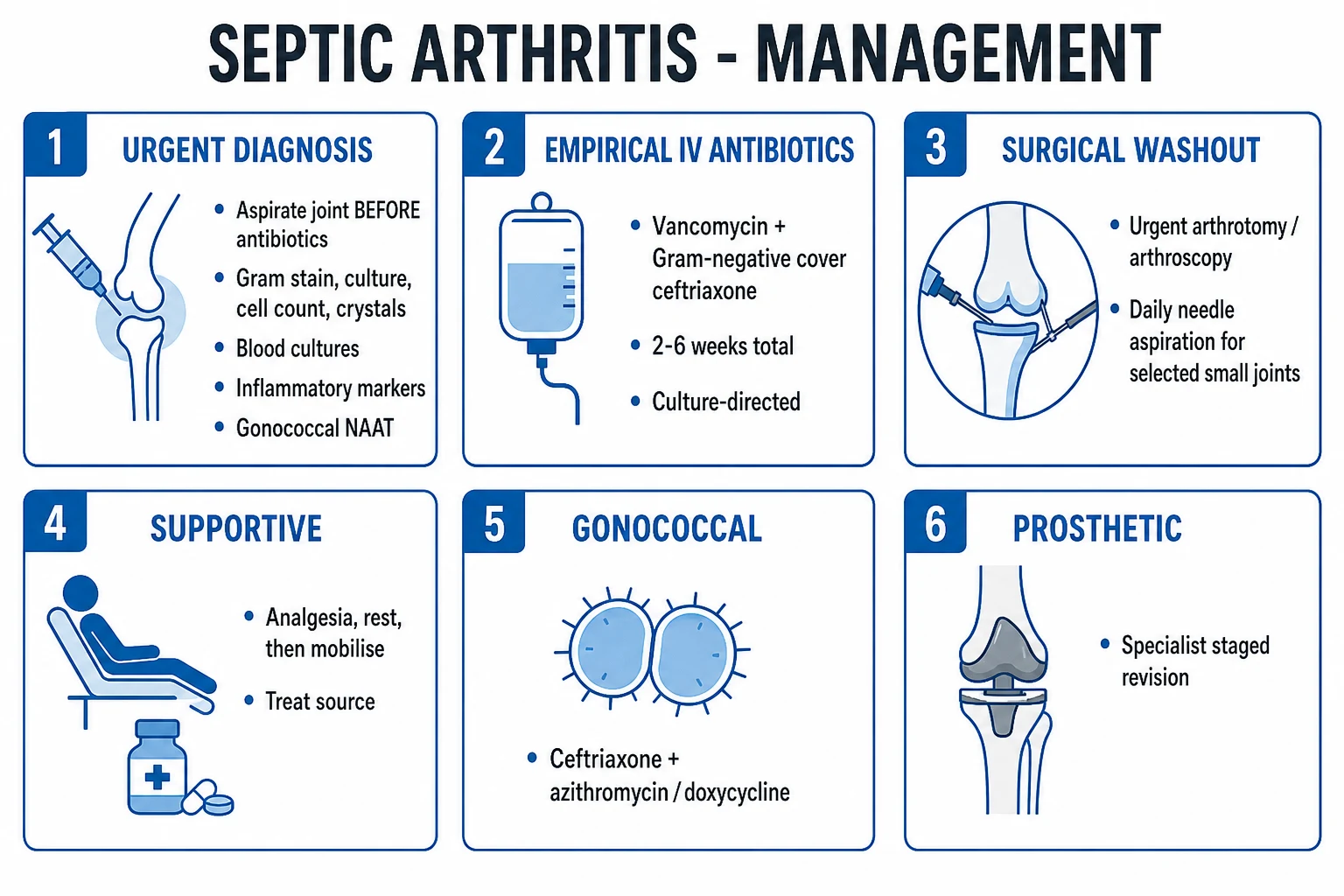
Definitive management has four pillars: (1) surgical drainage or washout, (2) empirical IV antibiotics then culture-directed, (3) analgesia, rest then early mobilisation, and (4) source control and comorbidity optimisation. Both surgery and antibiotics are needed — neither is sufficient alone.[2][10]
Pillar 1 — Surgical drainage
- Urgent surgical washout or drainage (arthrotomy or arthroscopy) is the mainstay for large joints, especially the knee, hip and shoulder. Arthroscopy (where available) is comparable to open arthrotomy in outcome for the knee and allows inspection and lavage under vision. Repeated daily needle aspiration is an option for small or selected joints (wrist, interphalangeal) and in centres without immediate theatre access, but is inferior for the knee and unsuitable for the hip.
- Hip septic arthritis — urgent open arthrotomy in children and adults; the deep joint cannot be adequately drained by arthroscopy or needle, and pus under pressure causes avascular necrosis of the femoral head within hours.[3][7]
- Repeat washout is indicated if the patient fails to improve (persistent fever, rising CRP, continued purulent effusion) within 48 to 72 hours.
Pillar 2 — Antibiotics (after aspiration)
Empirical adult IV regimen
- Flucloxacillin 2 g IV QID (or clindamycin 600 mg IV QDS if penicillin-allergic) — covers methicillin-sensitive S. aureus and streptococci
- PLUS ceftriaxone 2 g IV OD (Gram-negative cover, including gonococcal in the sexually active young adult)
- ADD vancomycin 1 to 1.5 g IV BD (or teicoplanin) if MRSA suspected (prior MRSA, IVDU, healthcare exposure, severe sepsis, prosthetic joint)
- Consider gentamicin or piperacillin-tazobactam if Pseudomonas risk (IVDU, burns, immunocompromise)
- Narrow to organism and sensitivities as soon as available
Targeted therapy (typical)
- MSSA, streptococci — flucloxacillin 2 g IV QID (or cefazolin 2 g IV TDS)
- MRSA — vancomycin 1 to 1.5 g IV BD (target trough 15 to 20 mg/L) or linezolid 600 mg IV BD
- Gram-negatives — ceftriaxone 2 g IV OD or cefepime; Pseudomonas — ceftazidime or piperacillin-tazobactam ± gentamicin
- Gonococcal — ceftriaxone 1 g IV/IM OD PLUS azithromycin 1 g PO single dose (or doxycycline 100 mg BD 7 days)
- Coag-neg staph / prosthetic — vancomycin ± rifampicin (biofilm-active); specialist input
Duration
- Nongonococcal native joint — typically 2 weeks IV then 2 to 4 weeks oral (4 to 6 weeks total)
- Gonococcal — 7 days IV/IM ceftriaxone then 7 days oral; total 1 to 2 weeks; responds rapidly
- Prosthetic joint — usually 6 weeks IV then prolonged oral suppressive; depends on surgical strategy
- Children — typically 2 to 3 weeks IV then 2 to 3 weeks oral, guided by CRP and clinical response
- Step down to oral when afebrile, CRP falling, and culture-directed oral agent available
The rationale for the duration is biological: bacteria in synovial fluid are cleared by the joint, but organisms adherent to cartilage and in biofilm require prolonged therapy. CRP should fall by half within 1 to 2 weeks of effective therapy; a rising or plateauing CRP mandates repeat washout, reculture, and reconsideration of organism/resistance.[2][10]
Pillars 3 and 4 — Rest, mobilisation, source
- Rest then early mobilisation — splint in comfort for the first 24 to 48 hours, then physiotherapy for range of motion and strengthening to prevent stiffness and contracture.
- Source control — drain or debride any skin/soft-tissue, urinary or endovascular source; remove infected lines; manage endocarditis if present.
- Comorbidity optimisation — glycaemic control, nutrition, stop smoking, reduce immunosuppression where feasible, treatment of any underlying RA flare (cautiously).
- Gonococcal disease — screen and treat partners; test for chlamydia, syphilis, HIV; report to public health.[8]
Specific Subtypes & Scenarios
Disseminated gonococcal infection (DGI)
DGI is the commonest cause of septic arthritis in young, sexually active adults in many series. Two clinical patterns exist, often overlapping.[8]
- Tenosynovitis-dermatitis-arthritis syndrome (about two-thirds) — migratory polyarthralgia, asymmetric tenosynovitis (extensor tendons of hands, wrists, ankles), and a painless pustular rash on the distal extremities (often on an erythematous base, easily mistaken for insect bites or vasculitis). Systemic features are mild.
- Purulent mono- or oligoarthritis (about one-third) — looks like classic septic arthritis, often knee, wrist or ankle. Synovial culture is positive in only 25 to 50%, NAAT is the diagnostic test of choice.[8]
Risk amplifiers: menstruation, pregnancy, complement deficiency (especially terminal C5–C9), HIV, and untreated genital infection. Treatment: ceftriaxone 1 g IV/IM OD for 7 days (or 7 days IV/IM then 7 days oral cefixime 400 mg BD), PLUS azithromycin 1 g PO single dose (or doxycycline 100 mg BD for 7 days) for chlamydia co-infection. Test of cure and partner notification are mandatory; screen for HIV, syphilis, hepatitis B and C. Joint washout is rarely required in DGI — unlike staphylococcal disease, the arthritis usually responds rapidly to antibiotics.[8]
Prosthetic joint infection (PJI)
PJI is classified by time from implantation — and the timing predicts the organism, the source, and the surgical strategy.[6]
Early (under 3 months)
- Acquired at surgery or peri-operatively
- Organisms: S. aureus, Gram-negatives (Pseudomonas, Enterobacterales)
- Acute presentation — pain, warmth, effusion, wound drainage, sinus
- Often debridement + retention with module exchange (if implant stable and symptoms under 3 weeks)
Delayed (3 to 24 months)
- Indolent, low-virulence organisms acquired at surgery
- Coagulase-negative staphylococci (S. epidermidis), Cutibacterium acnes
- Persistent pain, loosening, sinus — systemic features often absent
- Usually two-stage exchange with antibiotic-loaded cement spacer
Late (over 24 months)
- Haematogenous from a distant source (dental, urinary, skin)
- Same organisms as native joint sepsis — S. aureus, streptococci, Gram-negatives
- Acute onset in a previously well joint
- Two-stage exchange or, if unfit, long-term suppressive antibiotics
Diagnosis of PJI uses the MSIS (Musculoskeletal Infection Society) criteria — sinus tract communicating with the joint, two positive cultures with the same organism, or a combination of elevated serum CRP/Esr, elevated synovial WBC (over 3,000) and neutrophil percentage (over 80%), positive histology, and a single positive culture.[6]
Management principles (IDSA 2013):
- Acute (early or haematogenous) PJI with a stable implant and short symptom duration — debridement, antibiotics and implant retention (DAIR) with exchange of the modular components, plus 4 to 6 weeks IV then prolonged oral suppressive antibiotics.
- Chronic PJI — two-stage exchange: removal of all infected material and cement, placement of an antibiotic-loaded cement spacer, 4 to 6 weeks of organism-directed IV antibiotics, then reimplantation when CRP/ESR normalise. Single-stage exchange is an option in selected centres and organisms.
- Lifelong suppressive antibiotics — for patients unfit for surgery, with a difficult organism, or as a bridge to definitive surgery.
- Biofilm — organisms in biofilm are essentially irradiable only by surgery; rifampicin (for staphylococci) and fluoroquinolones penetrate biofilm and are used in combination once the implant is stable.[6][9]
Polyarticular septic arthritis
Polyarticular disease (around 10 to 20% of adults) occurs in rheumatoid arthritis, immunosuppression, endocarditis, severe sepsis, and disseminated gonococcal infection. S. aureus dominates. Outcomes are worse than monoarticular disease. Every hot joint must be aspirated, cultured and (where appropriate) washed out.[2][9]
Paediatric and neonatal septic arthritis
In infants and children the hip is the commonest and most dangerous joint (because the deep capsular anatomy conceals the effusion and pus under pressure causes femoral head necrosis within hours).[7]
- Neonate — Group B streptococcus and Gram-negatives; hip commonly affected; presentation is pseudoparalysis (the infant does not move the limb), irritability, refusal to feed, sometimes sepsis. Ultrasound is the diagnostic modality (the hip is too deep to examine reliably).
- Toddler/child — S. aureus commonest; S. pneumoniae and Kingella kingae (in under-fives) in the conjugate-vaccine era; H. influenzae type b where unvaccinated; Salmonella in sickle-cell disease; N. gonorrhoeae in neonates born to infected mothers (ophthalmia neonatorum association). The Kocher criteria stratify the irritable hip; three or more predictors mandate urgent ultrasound-guided aspiration under anaesthetic.
- Antibiotics — initial empirical: cefuroxime or flucloxacillin plus cefotaxime/ceftriaxone in the neonate; narrow to organism. Duration typically 2 to 3 weeks IV then 2 to 3 weeks oral, totalling 4 to 6 weeks.[7]
Septic arthritis and sickle-cell disease
Salmonella is over-represented in osteomyelitis and septic arthritis in sickle-cell disease (classically diaphyseal osteomyelitis, but septic arthritis also occurs); S. aureus remains the commonest organism overall. Initial empirical therapy must cover both — ceftriaxone plus flucloxacillin/vancomycin, narrowed to culture.[2]
IV-drug-use septic arthritis
S. aureus (often MRSA) and Pseudomonas aeruginosa dominate, with unusual sites — sternoclavicular, sacroiliac, spinal (discitis/vertebral osteomyelitis), and symphyseal joints. Endocarditis must be actively excluded (multiple blood cultures, echocardiography). Empirical therapy must cover MRSA and Pseudomonas — vancomycin + an anti-pseudomonal beta-lactam (e.g. piperacillin-tazobactam or ceftazidime) ± aminoglycoside.[2]
Complications & Pitfalls
- Permanent articular cartilage destruction — the central, time-critical complication; secondary osteoarthritis follows within months to years.
- Avascular necrosis — particularly of the femoral head in paediatric hip sepsis, and of other epiphyses where pus raises intra-articular pressure above capillary perfusion.
- Joint stiffness, contracture, limb deformity and limb-length discrepancy (in children with growth-plate injury).
- Amputation in severe or refractory cases, especially in prosthetic joint infection, diabetes, or polyarticular disease with septic shock.
- Osteomyelitis — contiguous spread from joint to adjacent metaphysis (and vice versa); particularly in the paediatric hip where the metaphysis is intracapsular.
- Septicaemia, septic shock, multi-organ failure, death — mortality 7 to 15% in adults; higher in the elderly, RA, polyarticular disease, and prosthetic joint.
- Prosthetic loosening and failure — requiring revision surgery.
- Recurrence or relapse — particularly if source control is incomplete or immunosuppression persists.[2][9]
- Treating a septic joint as a gout flare because urate crystals are present — they coexist in 5 to 20%.
- Treating an RA flare in a single hot joint without aspirating — sepsis is the single most common fatal error in RA management.
- Withholding antibiotics while waiting for aspiration that is delayed by hours — give within 1 hour if septic.
- Relying on a negative Gram stain to exclude sepsis — Gram stain is only about 50% sensitive overall.
- Assuming a prosthetic joint is uninfected because systemic features are absent — chronic PJI is often afebrile.
- Forgetting to screen for endocarditis in staphylococcal bacteraemic arthritis, especially in IVDU.
- Mishandling the paediatric hip — failing to apply Kocher criteria, or delaying ultrasound-guided aspiration.[3][7]
Prognosis & Disposition
Outcome is determined by four factors: (1) the time to treatment, (2) the organism, (3) the host, and (4) the joint.[1][2][9]
- Time to treatment — delay of more than 24 to 48 hours from symptom onset to washout/antibiotics markedly worsens outcome; mortality and joint destruction climb linearly.
- Organism — S. aureus (especially MRSA) and Gram-negatives have the worst outcomes; N. gonorrhoeae the best (rapid response, less destructive).
- Host — older age, rheumatoid arthritis, diabetes, immunosuppression, prosthetic joint, polyarticular disease, and prior joint damage all worsen prognosis.
- Joint — hip worse than knee; shoulder and spine notoriously difficult.[2][9]
Mortality is 7 to 15% in modern series; up to 50% of survivors retain some joint dysfunction.[2][9]
- Admit under joint orthopaedic + medical/infectious-diseases care.
- Theatre for washout, repeat washout if not improving in 48 to 72 hours.
- Step down to oral antibiotics when afebrile, CRP falling, and culture-directed oral agent available.
- Outpatient follow-up with inflammatory markers, physiotherapy, and rheumatology/orthopaedic review for residual joint damage; consideration of arthroplasty for joint destruction in survivors.
- Safety-net: any patient readmitted with fever, joint pain or rising CRP within 3 months is assumed to have recurrent/persistent infection until proven otherwise.
Special Populations
Children
- Hip commonest and most dangerous; apply Kocher criteria
- Ultrasound-guided aspiration under anaesthetic; urgent arthrotomy for confirmed hip sepsis
- Organisms: S. aureus, Group B strep (neonate), K. kingae (under-5), S. pneumoniae, H. influenzae if unvaccinated, Salmonella (sickle cell)
- Weight-based antibiotics: flucloxacillin 50 mg/kg IV QDS (max 2 g), cefotaxime/ceftriaxone, vancomycin 15 mg/kg IV; 4 to 6 weeks total
Rheumatoid arthritis
- Risk multiplied; sepsis mimics a flare — aspirate any disproportionately hot single joint
- Often on anti-TNF or methotrexate; culture may be atypical
- Mortality higher; aggressive early washout and prolonged antibiotics
- Coordinate with rheumatology — hold biologics during sepsis
Prosthetic joint
- Coagulase-negative staph, polymicrobial, biofilm; MSIS criteria for diagnosis
- Specialist surgical pathway: DAIR, one- or two-stage exchange, lifelong suppressive
- Rifampicin + fluoroquinolone for biofilm once implant stable
- Never a simple 'washout and 6 weeks' — needs MDT planning
Diabetes / elderly
- Fever may be absent; muted inflammatory response; atypical organisms (Gram-negatives)
- Neuropathic Charcot joint can mimic sepsis — aspirate if any doubt
- Aggressive glycaemic control; review antiplatelet/anticoagulant for theatre
- Higher mortality; lower threshold for ICU
Pregnancy / immunocompromised
- Pregnancy amplifies gonococcal risk; use pregnancy-safe antibiotics (ceftriaxone, flucloxacillin; avoid gentamicin if possible, avoid doxycycline)
- HIV and anti-TNF increase risk and may blunt inflammation; broad empiric cover
- Listeria and Salmonella in advanced immunosuppression
- Multidisciplinary with obstetric medicine / ID
Evidence, Guidelines & Regional Differences
- Mathews 2010 (Lancet) and Carpenter 2011 (Acad Emerg Med) remain the canonical reviews for native-joint septic arthritis in adults. Both emphasise synovial WBC and percentage of neutrophils as the most powerful laboratory predictors, surgical drainage as the mainstay, and vancomycin plus Gram-negative cover as the empirical regimen when MRSA is plausible.[1][2][5]
- IDSA 2013 (Osmon, CID) — prosthetic joint infection guideline — defines DAIR, one- and two-stage exchange, and suppressive therapy; recommends rifampicin plus fluoroquinolone for staphylococcal biofilm once the implant is stable.[6]
- GEIO-SEIMC 2024 (Benito) — Spanish/SEIMC guideline for adults and children — reaffirms early aspiration, multidisciplinary management, and 4 to 6 weeks of antibiotic therapy for native joint sepsis.[10]
- Therapeutic Guidelines (eTG) and ASID recommend flucloxacillin 2 g IV QID + ceftriaxone 2 g IV OD as empirical cover for native-joint sepsis, with vancomycin substituted for flucloxacillin if MRSA risk or penicillin allergy; 4 to 6 weeks total. Gonococcal: ceftriaxone 1 g IV/IM OD plus azithromycin 1 g PO.[2][8]
- Empirical therapy in India often begins with cloxacillin/cefazolin + an aminoglycoside or third-generation cephalosporin, with vancomycin for healthcare-associated or known MRSA. Salmonella must be covered empirically in sickle-cell disease (ceftriaxone covers it). Tuberculous arthritis is in the differential in endemic areas — chronic monoarthritis, "caries sicca", positive AFB culture, and Poncet's reactive arthritis (tuberculides) is a distinct entity. Aedes-borne arboviruses (Chikungunya) produce an explosive polyarthritis that mimics sepsis in the acute phase — usually distinguishable by the typical fever-rash-polyarthritis triad and a self-limiting course.[2][10]
Landmark evidence to cite:[1][3][6]
- Margaretten 2007 JAMA — systematic review of 14 studies, 6,242 patients: synovial WBC at least 25,000 LR 2.9; over 50,000 LR 7.7; over 100,000 LR 28. Polymorphs at least 90% LR 3.4. Risk factors confirmed: age, diabetes, RA, joint surgery, prosthesis, skin infection, HIV. Joint pain sensitivity 85%, fever only 57%.[1]
- Kocher 1999 JBJS Am — derived the four-variable prediction algorithm for the irritable paediatric hip; 99.6% predicted probability with all four positive.[3]
- Sultan 2010 JBJS Br — external validation; added CRP at least 20 mg/L as fifth predictor; showed the rule overestimates in low-prevalence settings.[4]
- Osmon 2013 CID (IDSA) — prosthetic joint infection guideline: DAIR, one-/two-stage exchange, suppressive; biofilm-aware antibiotics.[6]
- Benito 2024 (GEIO-SEIMC) — contemporary adult and paediatric guidance; reaffirms early aspiration and 4 to 6 weeks of therapy.[10]
Exam Pearls
- "Any acute hot, swollen, tender joint = septic until proven otherwise — aspirate BEFORE antibiotics."[1]
- Commonest organism Staphylococcus aureus (all ages); MRSA in at-risk; coag-neg staph in prosthetic.
- Neisseria gonorrhoeae in young sexually active — migratory polyarthralgia + tenosynovitis + pustular rash; diagnose with NAAT; treat with ceftriaxone + azithromycin.[8]
- Organisms by host: neonate = Group B strep; child = S. aureus, K. kingae, S. pneumoniae, H. influenzae (unvaccinated); sickle cell = Salmonella; IVDU = Pseudomonas and S. aureus (MRSA), at unusual joints (sternoclavicular, sacroiliac, spine); prosthetic = coagulase-negative staph.[2][7]
- Synovial fluid: WBC over 50,000, neutrophil-predominant, positive Gram stain/culture, low glucose, raised lactate. Knee is the commonest joint.
- Crystals do NOT exclude sepsis — septic arthritis coexists with gout or CPPD in 5 to 20%.
- Kocher criteria (1999): fever, non-weight-bearing, ESR at least 40, WBC more than 12,000 — 0/1/2/3/4 predictors give under 0.2%, 3%, 40%, 93%, 99.6% probability of septic hip in children; Sultan added CRP at least 20.[3][4]
- Treatment: urgent surgical washout + empirical IV antibiotics — flucloxacillin 2 g IV QID (clindamycin if allergic, vancomycin 1 g IV BD if MRSA) + ceftriaxone 2 g IV OD, narrowed to culture, 4 to 6 weeks total.[2]
- Hip in children = same-hour theatre emergency — ultrasound-guided aspiration, urgent open arthrotomy; delay destroys the femoral head.
- Aspirate before antibiotics; blood cultures in 50%; FBC, CRP, ESR; gonococcal NAAT.
- Prosthetic joint infection = coag-neg staph; biofilm; MSIS criteria; DAIR or two-stage exchange; rifampicin + fluoroquinolone once implant stable.[6]
- Delay over 24 to 48 hours destroys cartilage; mortality 7 to 15%; up to half retain joint damage.[2]
- Margaretten LRs by synovial WBC: at least 25,000 = 2.9; over 50,000 = 7.7; over 100,000 = 28; polymorphs at least 90% = 3.4.[1]
Exam application bank (NEET-PG / INICET)
One-line answer
Septic arthritis is a medical and surgical emergency — direct microbial invasion of a joint space (usually bacterial and haematogenous) causing purulent synovial inflammation, rapid articular cartilage destruction, sepsis and death if untreated. Incidence is 4 to 10 per 100,000 in the West and rises steeply with prosthetic joints, rheumatoid arthritis, diabetes, immunosuppression, IV drug use and age over 80. The knee is the commonest joint (about half of cases). Staphylococcus aureus is the commonest organism in all ages; Neisseria gonorrhoeae in young sexually active adults (migratory polyarthralgia, tenosynovitis, pustular rash); Group B streptococcus in neonates; Haemophilus influenzae and Streptococcus pneumoniae in unvaccinated children; Salmonella in sickle-cell disease; Pseudomonas and Gram-negatives in IV drug users; coagulase-negative staphylococci in prosthetic joints. Present
Worked stems (answer without another resource)
Stem 1 — Classic presentation. Map symptoms to mechanism; name the first investigation and first treatment step with dose/route if drug therapy is standard. [1]
Stem 2 — Unstable / complicated. List red flags that force immediate resuscitation, theatre, ICU, antidote, or reperfusion — and what you do in the first 15 minutes. [1]
Stem 3 — Atypical group. Elderly, pregnancy, child, or immunocompromised: how presentation and thresholds change. [1]
Stem 4 — Differential trap. Name the three closest mimics and one discriminator for each. [1]
Stem 5 — Disposition. Who goes home with safety-netting, who is admitted, who needs HDU/ICU/theatre, and what follow-up is mandatory. [1]
Rapid viva checklist
- Definition + classification
- Pathophysiology chain
- Bedside signs / criteria
- Score with exact components (if any)
- Emergency bundle
- Definitive therapy with doses
- Complications of disease and of treatment
- Special populations
- Guideline/trial name if classic
- Three exam traps
Coverage self-check
If you cannot answer any stem above from this page alone, re-read the matching section — the page is intended to be self-sufficient for final-prof and NEET-PG/INICET questions on Septic Arthritis.
References
- [1]Margaretten ME, Kohlwes J, Moore D, Bent S. Does this adult patient have septic arthritis? JAMA, 2007.PMID 17405973
- [2]Mathews CJ, Weston VC, Jones A, Field MS, Coakley G. Bacterial septic arthritis in adults Lancet, 2010.PMID 20206778
- [3]Kocher MS, Zurakowski D, Kasser JR. Differentiating between septic arthritis and transient synovitis of the hip in children: an evidence-based clinical prediction algorithm J Bone Joint Surg Am, 1999.PMID 10608376
- [4]Sultan J, Hughes PJ. Septic arthritis or transient synovitis of the hip in children: the value of clinical prediction algorithms J Bone Joint Surg Br, 2010.PMID 20798450
- [5]Carpenter CR, Schuur JD, Everett WW, Pines JM. Evidence-based diagnostics: adult septic arthritis Acad Emerg Med, 2011.PMID 21843213
- [6]Osmon DR, Berbari EF, Bertrand AR, et al. Diagnosis and management of prosthetic joint infection: clinical practice guidelines by the Infectious Diseases Society of America Clin Infect Dis, 2013.PMID 23223583
- [7]Montgomery NI, Epps HR. Pediatric Septic Arthritis Orthop Clin North Am, 2017.PMID 28336043
- [8]Rice PA. Gonococcal arthritis (disseminated gonococcal infection) Infect Dis Clin North Am, 2005.PMID 16297736
- [9]Nair R, Bhasin V, Ho G Jr. Septic Arthritis and Prosthetic Joint Infections in Older Adults Infect Dis Clin North Am, 2017.PMID 29079156
- [10]Benito N, Treviño M, Gómez-Martínez MA, et al. Executive summary: Guidelines for the diagnosis and treatment of septic arthritis in adults and children, developed by the GEIO (SEIMC), SEIP and SECOT Enferm Infecc Microbiol Clin (Engl Ed), 2024.PMID 37919201