Rheumatology · Rheumatology
Systemic Lupus Erythematosus
Also known as SLE · lupus · disseminated lupus erythematosus
Systemic lupus erythematosus (SLE) is a multisystem autoimmune disease driven by loss of self-tolerance, autoantibody formation (ANA, anti-dsDNA, anti-Sm), immune complex deposition, and type I interferon activation. 9:1 female predominance, peak 15-45 years, worse in African, Hispanic, Asian populations. Classification: EULAR/ACR 2019 (ANA entry + additive weighted criteria, score 10+). Manifestations span skin (malar rash sparing nasolabial folds, discoid, photosensitivity), joints (non-erosive, Jaccoud), kidneys (lupus nephritis ISN/RPS class I-VI), serosae (pericarditis, pleuritis), CNS (neuropsychiatric lupus), blood (cytopenias, antiphospholipid), and heart (Libman-Sacks endocarditis). Management: hydroxychloroquine for ALL patients; NSAIDs and short-course steroids for flares; mycophenolate, cyclophosphamide, azathioprine for organ disease; belimumab, anifrolumab, rituximab for refractory. Treat-to-target: remission or low disease activity.
On this page & tools
Your progress
Saved locally on this device.
Practise this topic
Exam tags
Red flags
Overview & Definition
Systemic lupus erythematosus (SLE) is the prototype of a multisystem autoimmune connective-tissue disease, characterised by the production of a wide array of autoantibodies directed against nuclear and cytoplasmic antigens, the formation and tissue deposition of immune complexes, and consequent chronic relapsing-remitting inflammation across virtually every organ system. The clinical expression ranges from mild cutaneous and articular disease at one extreme to life-threatening renal, neuropsychiatric, and haematological involvement at the other, and the disease course is marked by flares and remissions over decades. The 2019 EULAR/ACR classification criteria frame the disease around a single obligatory entry criterion — a positive antinuclear antibody (ANA) at a titre of 1:80 or higher on HEp-2 cells — followed by additive weighted clinical and immunological domains, with a cumulative score of at least 10 required for classification.[2]
The hallmark immunological disturbance is loss of self-tolerance to nuclear antigens. Apoptotic debris that should be cleared silently instead exposes DNA, RNA, and nucleosomes to the immune system, driving autoantibody formation by autoreactive B cells, sustained T-cell help, and an exaggerated type I interferon (IFN) signature generated by plasmacytoid dendritic cells. Immune complexes of anti-dsDNA, anti-Sm, and related antibodies deposit in glomeruli, serosae, skin, and blood-vessel walls, activating complement and recruiting neutrophils — the basis for the low C3/C4 that tracks active disease, and for the type III hypersensitivity vasculitic pattern shared across organs.[3]
The prognosis of SLE has been transformed since the 1950s, when five-year survival was roughly 50 percent. Today five-year survival exceeds 95 percent and ten-year survival 90 percent in developed settings, driven by hydroxychloroquine use, better immunosuppression, dialysis, and infection prophylaxis. The leading causes of death have shifted accordingly: in early disease, active lupus, nephritis, infection, and central nervous system disease predominate; in late disease, accelerated atherosclerosis and cardiovascular disease, infection, end-stage renal disease (ESRD), and malignancy dominate.[1][3]
Classification
SLE must be distinguished from a family of related lupus phenotypes and from other connective-tissue diseases. The four canonical lupus subtypes differ in target organ, antibody profile, and prognosis, and a fifth entity — overlap syndromes including mixed connective-tissue disease (MCTD) — bridges SLE with systemic sclerosis, polymyositis, and rheumatoid arthritis. [1]
Systemic SLE
- Multisystem involvement (skin, joint, kidney, CNS, blood, serosa)
- ANA positive in over 95 percent; anti-dsDNA and anti-Sm specific
- 9 to 10 times more common in women; African, Hispanic, Asian descent
- Classification: EULAR/ACR 2019 or SLICC 2012
- Treatment: hydroxychloroquine plus immunosuppressives and biologics
Discoid lupus (DLE)
- Chronic cutaneous form; discoid lesions above the neck (scalp, face, ears)
- Follicular plugging, scarring, atrophy, depigmentation; permanent alopecia
- ANA positive in only about 25 percent; anti-dsDNA negative usually
- Under 5 percent progress to systemic SLE over time
- Treatment: topical steroids, hydroxychloroquine, strict photoprotection
Subacute cutaneous LE
- Photosensitive annular or papulosquamous rash on upper trunk and extensor arms
- Non-scarring; highly associated with anti-Ro/SSA antibodies
- May be drug-induced (terbinafine, griseofulvin, calcium-channel blockers, TNF inhibitors)
- About half meet SLE criteria, but severe systemic disease is uncommon
- Treatment: hydroxychloroquine, sun protection, topical steroids
Drug-induced lupus
- Triggers: procainamide, hydralazine, isoniazid, minocycline, anti-TNF agents
- Anti-histone antibodies positive (over 95 percent)
- Anti-dsDNA negative (usually); kidney and CNS spared
- Resolves within weeks of stopping the offending drug
- Anti-TNF-induced variant may be anti-dsDNA positive but is still clinically mild
Neonatal lupus
- Maternal anti-Ro/SSA or anti-La/SSB crosses the placenta
- Congenital heart block (third-degree) is the hallmark (1 to 2 percent risk)
- Transient annular rash, cytopenias, hepatitis, cholestasis in the neonate
- Mothers may have SLE, Sjogren syndrome, or be asymptomatic carriers
- Hydroxychloroquine in pregnancy reduces heart block recurrence risk
The three classification criteria systems used in SLE, in historical order, are the 1997 ACR revised criteria (4 of 11 criteria), the 2012 SLICC criteria (4 of 17 criteria with at least one clinical and one immunological, or biopsy-proven lupus nephritis plus ANA or anti-dsDNA), and the 2019 EULAR/ACR criteria (ANA entry plus weighted additive score of at least 10). The 2019 system has the highest sensitivity and specificity and is the current research and clinical standard, although it should be remembered that classification criteria are designed for research enrolment and serve only as an aid at the bedside.[2][4][12]
Epidemiology & Risk Factors
SLE shows a striking female predominance of 9 to 1 in the childbearing years, attenuating to about 2 to 1 after menopause — a pattern that implicates oestrogen as a permissive hormone, supported by the higher disease activity seen with oestrogen-containing oral contraceptives and by disease flares in pregnancy. Ethnicity powerfully modulates both incidence and severity: people of African, Afro-Caribbean, Hispanic, and South or East Asian descent have a two- to four-fold higher prevalence and an earlier, more aggressive disease course, with higher rates of lupus nephritis and cardiovascular damage. The reasons include a higher prevalence of high-risk HLA alleles, lower complement component levels, and socioeconomic determinants of access to care.[1][3]
The principal risk factors and triggers cluster into genetic, hormonal, environmental, and infectious groups: [1]
Genetic
- HLA-DR2 and HLA-DR3 increase risk roughly two-fold
- Complement deficiencies (C1q, C1r/s, C2, C4) are the strongest single-gene risk factors
- Over 60 susceptibility loci identified by GWAS (IRF5, STAT4, TNFAIP3, BLK)
- 10 percent of patients have a first-degree relative with SLE
- Monozygotic twin concordance about 25 percent (dizygotic 2 percent)
Hormonal
- Oestrogen is permissive; disease flares with OCPs, pregnancy, IVF
- Prolactin and hyperprolactinaemia associated with activity
- Disease milder in men but more organ-damaging when it occurs
- Klinefelter syndrome (47,XXY) markedly increases male risk
Environmental
- Ultraviolet light triggers cutaneous and systemic flares (the classic photosensitivity)
- Smoking worsens disease activity, impairs hydroxychloroquine efficacy, and raises cardiovascular risk
- Silica dust, hair dyes, and smoking are occupational and chemical triggers
- Drug triggers for drug-induced lupus (procainamide, hydralazine, isoniazid, minocycline, anti-TNF)
Infectious
- Epstein-Barr virus (EBV) infection is near-universal in SLE and may break tolerance via molecular mimicry
- Molecular mimicry and bystander activation link infection to first flares
- Live vaccines may trigger flares in active disease
- Reactivation of latent viruses (CMV, VZV) on immunosuppression drives morbidity
Pathophysiology
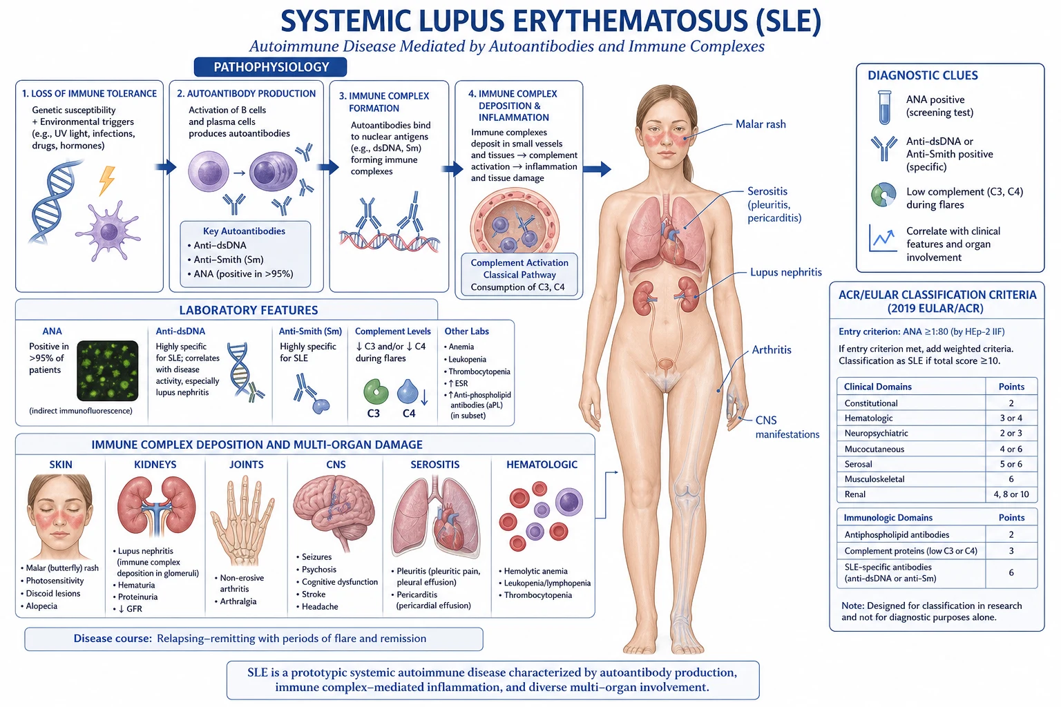
The pathogenesis of SLE can be conceptualised as a self-amplifying loop with seven interconnected nodes, each a target for therapy:[3]
-
Defective clearance of apoptotic debris. Apoptotic cells normally expose phosphatidylserine and are silently cleared by macrophages. In SLE, this clearance is impaired (linked to C1q deficiency and DNase I defects), so nuclear antigens — DNA, RNA, histones, nucleosomes — accumulate and become immunogenic. [1]
-
Aberrant type I interferon response. Immune complexes of autoantibody with DNA or RNA are internalised by plasmacytoid dendritic cells via Fc receptors and reach endosomal Toll-like receptors (TLR7 for RNA, TLR9 for DNA), driving massive release of IFN-alpha. The resulting type I interferon signature is one of the most consistent transcriptional abnormalities in SLE and underlies the fever, fatigue, and immune activation of active disease. Anifrolumab (anti-IFN-alpha receptor) blocks this node. [1]
-
Loss of T-cell tolerance and help. CD4-positive T cells provide help to autoreactive B cells, while regulatory T cells are functionally impaired. This shifts the balance from tolerance to antibody production. [1]
-
B-cell activation and autoantibody production. Autoreactive B cells survive negative selection and mature into plasma cells producing high-affinity anti-dsDNA, anti-Sm, anti-Ro, anti-La, anti-RNP, anti-histone, and antiphospholipid antibodies. The cytokine BAFF/BLyS supports B-cell survival and is the target of belimumab. [1]
-
Immune complex formation and deposition (type III hypersensitivity). Circulating immune complexes of autoantibody with nuclear antigen deposit in glomeruli, dermal vessels, serosae, and the choroid plexus, activating the classical complement pathway (hence low C3 and C4) and the membrane attack complex, recruiting neutrophils and causing the vasculitic tissue injury of nephritis, serositis, and rash. [1]
-
Cytokine and cellular amplification. Beyond IFN-alpha, IL-6, IL-17, IL-23, and BAFF amplify B- and T-cell responses, while neutrophil extracellular traps (NETs) from low-density granulocytes deposit their own DNA and antimicrobial peptides in tissues, perpetuating the cycle. [1]
-
Organ-specific damage. Direct antibody effects (anti-Ro/SSA crossing the placenta causing congenital heart block; antiphospholipid antibodies causing thrombosis; anti-dsDNA cross-reacting with neuronal and glomerular antigens) layer on top of immune-complex injury. The net result is the multisystem clinical picture.[3][7]
Clinical Presentation
SLE is the great imitator. Presentation ranges from insidious fatigue and arthralgia to an explosive multisystem illness with nephritis, psychosis, and cytopenias. The classical patient is a young woman of reproductive age with constitutional upset, a photosensitive rash, and non-erosive arthritis, but every organ system can dominate the picture. Below the manifestations are organised by system, mirroring how examiners test them. [1]
Constitutional features
Fatigue, low-grade fever, weight loss, malaise, and generalised lymphadenopathy are common and non-specific. Fatigue is the most disabling symptom for many patients and does not correlate with serological activity. Fever in SLE is usually low-grade and accompanies a flare, but a high fever or a markedly raised CRP must prompt a search for infection — the leading cause of early death — before the fever is attributed to active lupus. Hepatosplenomegaly may be present in active disease. [1]
Mucocutaneous features
The skin is involved in about 85 percent of patients at some point, and dermatological manifestations are weighted in every classification system.[2]
- Malar (butterfly) rash — the classic acute cutaneous lupus. Erythema over the bridge of the nose and cheeks, sparing the nasolabial folds (the key discriminator from rosacea and seborrhoeic dermatitis, which involve them), photosensitive, transient, and non-scarring.
- Discoid lupus (DLE) — coin-shaped, scaly, follicular-plugged lesions, typically on the scalp, face, and ears, that heal with scarring, atrophy, and permanent alopecia. Only about 5 percent of DLE patients develop systemic SLE.
- Subacute cutaneous lupus (SCLE) — annular, polycyclic, or papulosquamous, non-scarring, photosensitive rash on the upper trunk and extensor forearms, strongly associated with anti-Ro/SSA antibodies and often drug-induced (terbinafine, calcium-channel blockers, anti-TNF).
- Photosensitivity — an exaggerated rash response to UV light, reported in over half of patients and a known flare trigger.
- Oral and nasal ulcers — usually painless, located on the hard palate or nasal septum, and easy to miss without a careful look.
- Alopecia — diffuse non-scarring (lupus telogen effluvium) or scarring (discoid scalp disease).
- Livedo reticularis and periungual erythema — reflect underlying vasculopathy, often coexisting with antiphospholipid antibodies.
- Raynaud phenomenon, chilblain lupus, and bullous lupus are less common but characteristic. [1]
Musculoskeletal features
SLE arthritis
- Symmetrical, migratory, small joints of hands and wrists
- Non-erosive on X-ray (distinguishes from rheumatoid arthritis)
- Morning stiffness, synovitis, tenderness
- Jaccoud arthropathy: reducible swan-neck and ulnar deviation
- Deformities from tendon and ligament laxity, not bone erosion
Rheumatoid arthritis
- Symmetrical, additive, small joints
- Erosive on X-ray (marginal erosions)
- Rheumatoid factor and anti-CCP positive
- Fixed, irreducible deformities (ulnar deviation, boutonniere)
- No malar rash, no anti-Sm, no nephritis
Arthralgia and arthritis are the most common presenting symptom (over 90 percent). The arthritis is non-erosive (distinguishing SLE from rheumatoid arthritis) but can cause Jaccoud arthropathy — reducible swan-neck and ulnar-deviation deformities from capsular and ligamentous laxity rather than bone destruction. Myalgia is common; frank myositis occurs in about 10 percent. Avascular necrosis (especially of the femoral head) is a long-term complication, driven partly by the disease but more by corticosteroid use. [1]
Renal involvement — lupus nephritis
Lupus nephritis develops in 40 to 60 percent of SLE patients and is the strongest predictor of mortality. The 2003 ISN/RPS classification (revised 2018) divides lupus nephritis into six classes that determine treatment intensity:[9][11]
Class I — Minimal mesangial
- Normal on light microscopy
- Immune deposits on immunofluorescence only
- No treatment beyond hydroxychloroquine
- Clinically silent
Class II — Mesangial proliferative
- Mesangial hypercellularity on light microscopy
- Mild proteinuria, microscopic haematuria
- No nephrotic syndrome
- Treat underlying SLE; ACE inhibitor for proteinuria
Class III — Focal proliferative
- Under 50 percent of glomeruli involved
- Active/chronic lesions; subendothelial deposits
- Proteinuria, haematuria, possible nephritic syndrome
- Induction: mycophenolate or cyclophosphamide plus corticosteroids
Class IV — Diffuse proliferative
- Over 50 percent of glomeruli involved; worst prognosis
- Subendothelial deposits, wire loops, crescents
- Nephritic and nephrotic features, hypertension, rising creatinine
- Aggressive induction with mycophenolate or cyclophosphamide; worst prognosis
Class V — Membranous
- Diffuse thickening of glomerular basement membrane
- Subepithelial deposits; **nephrotic syndrome** is typical
- May coexist with class III or IV (mixed)
- High thrombosis risk; renin-angiotensin blockade plus immunosuppression
Class VI — Advanced sclerosis
- Over 90 percent globally sclerosed glomeruli
- End-stage, irreversible
- No role for immunosuppression; dialysis or transplant
- Dialysis-dependent; recurrence post-transplant is uncommon if quiescent
Clinically, lupus nephritis presents with proteinuria (over 0.5 g/day, or a urine protein-to-creatinine ratio over 50 mg/mmol), haematuria and red cell casts, rising creatinine, hypertension, oedema, and a low C3 and C4 with high anti-dsDNA. A renal biopsy is mandatory at first sign of significant proteinuria or active sediment to confirm class, grade activity and chronicity, and guide intensity of treatment.[9][15]
Cardiovascular features
Pericarditis is the most common cardiac manifestation (about 25 percent), presenting with positional chest pain and a pericardial rub. Myocarditis is rarer but life-threatening, causing heart failure and arrhythmias. Libman-Sacks endocarditis — non-bacterial verrucous vegetations on the mitral and aortic valves — is the classic valvular lesion and a source of embolic stroke, especially when antiphospholipid antibodies coexist. Premature atherosclerosis is now the leading cause of late mortality: young women with SLE have a fifty-fold higher risk of myocardial infarction than age-matched controls, driven by chronic inflammation, corticosteroids, hypertension, and renal disease. [1]
Pulmonary features
Pleuritis with or without pleural effusion is the most common pulmonary manifestation (up to 50 percent). Acute lupus pneumonitis, interstitial lung disease, and pulmonary hypertension are less common. Shrinking lung syndrome — elevated diaphragms with reduced lung volumes but no parenchymal disease — is a characteristic but rare complication thought to arise from diaphragmatic myopathy. Diffuse alveolar haemorrhage is a medical emergency presenting with haemoptysis, falling haemoglobin, and diffuse infiltrates; it requires urgent pulse methylprednisolone, cyclophosphamide, and ventilatory support. [1]
Neuropsychiatric lupus (NPSLE)
NPSLE encompasses 19 named syndromes defined by the ACR, ranging from cognitive dysfunction and headache at the mild end to seizures, psychosis, transverse myelitis, and stroke at the severe end. The most common manifestations in practice are cognitive dysfunction, mood disorder, headache, seizures, and cerebrovascular disease. Acute confusional state, psychosis (distinguished from steroid-induced psychosis by onset before or early in steroid treatment), transverse myelitis, optic neuritis, chorea, and cranial neuropathy are rarer but clinically critical. The pathogenesis is mixed — vasculopathy, antiphospholipid-mediated thrombosis, antibody-mediated neuronal injury (anti-NR2), and cytokine effects — which is why major NPSLE events are treated with immunosuppression, while antiphospholipid-related stroke is treated with anticoagulation.[3]
Haematological features
Cytopenias are among the most common laboratory findings. Leukopenia (under 4000 per microlitre) and lymphopenia (under 1500 per microlitre) are weighted in the classification criteria. Anaemia of chronic disease is usual; autoimmune haemolytic anaemia with a positive direct Coombs test occurs in about 10 percent. Thrombocytopenia (under 100,000) may be the presenting feature and overlaps with immune thrombocytopenic purpura (ITP). Antiphospholipid antibodies (lupus anticoagulant, anticardiolipin, anti-beta-2-glycoprotein I) are present in 30 to 40 percent and confer a marked risk of venous and arterial thrombosis and recurrent pregnancy loss — the antiphospholipid syndrome (APS) overlap.[10]
Gastrointestinal features
GI involvement is less prominent but important. Mesenteric vasculitis presents with abdominal pain and can mimic surgical emergencies. Pancreatitis, hepatitis (lupoid hepatitis overlaps with autoimmune hepatitis), protein-losing enteropathy, and ascites (from serositis or nephrotic syndrome) are recognised. [1]
Atypical presentations
ATYPICAL
Male or late-onset SLE (after age 50) is insidious, with more serositis and pulmonary disease and less nephritis and rash, and a higher index of suspicion is needed. Paediatric-onset SLE is more aggressive, with higher rates of nephritis, haematological involvement, and cumulative damage. Acute catastrophic antiphospholipid syndrome presents with rapid multiorgan thrombosis and has a high mortality. [1]

Differential Diagnosis
SLE enters the differential of almost any multisystem illness in a young woman, and conversely many conditions mimic SLE. The key is to distinguish SLE from its mimics by serology and pattern of organ involvement, and to remember that infection is the most dangerous mimic of a lupus flare.[1][3]
Rheumatoid arthritis
- Erosive, symmetric small-joint polyarthritis
- Rheumatoid factor and anti-CCP positive
- No malar rash, no anti-Sm, no nephritis
- Morning stiffness over an hour; fixed deformities
- Distinguishing SLE: non-erosive, anti-dsDNA positive, extra-articular features
Mixed connective-tissue disease
- Overlap features: Raynaud, swollen hands, arthritis, myositis, oesophageal dysmotility
- Anti-U1-RNP in high titre is obligatory
- Pulmonary hypertension is the major killer
- Generally more responsive to corticosteroids than scleroderma
- Distinguishing SLE: anti-RNP only, anti-dsDNA negative, less nephritis
Systemic sclerosis
- Raynaud, skin thickening, digital ulcers, telangiectasia
- Anti-centromere or anti-Scl-70 (topoisomerase I)
- ILD, pulmonary hypertension, renal crisis
- ANA may be positive; anti-dsDNA negative
- Distinguishing SLE: no sclerodactyly, anti-dsDNA positive, nephritis pattern
Sjogren syndrome
- Dry eyes and dry mouth (sicca); parotid enlargement
- Anti-Ro/SSA and anti-La/SSB
- ANA positive in a speckled pattern; may meet SLE criteria
- Distinguishing SLE: anti-dsDNA negative, low complement uncommon, salivary gland biopsy
Adult-onset Still disease
- Spiking fevers, evanescent salmon-pink rash, arthritis
- Ferritin markedly elevated; ANA and RF negative
- Distinguishing SLE: double-negative autoantibodies, fever pattern, rash on pressure areas
Infection mimics
- Endocarditis, EBV, HIV, hepatitis B/C, parvovirus B19 — all ANA-positive and multisystem
- Subacute bacterial endocarditis: murmur, splinter haemorrhages, immune-complex nephritis
- Distinguishing SLE: positive blood cultures, viral serology, normal complement; treat infection first
Malignancy
- Lymphoma, leukaemia, paraneoplastic syndromes — fever, cytopenias, adenopathy
- Distinguishing SLE: bone marrow and tissue biopsy; SLE antibodies negative in malignancy
For specific manifestations, narrower differentials apply: malar rash must be distinguished from rosacea (nasolabial involvement, pustules), seborrhoeic dermatitis, and photosensitive drug eruptions; arthritis from viral, rheumatoid, and reactive arthritis; nephritis from post-streptococcal and IgA nephropathy and antiphospholipid nephropathy; serositis from viral or bacterial pericarditis; and cytopenias from primary haematological disease. [1]
Clinical & Bedside Assessment
A focused SLE assessment at the bedside is built around the systems most commonly involved, with particular attention to the kidneys, blood pressure, skin, and serosae. [1]
History should probe for photosensitivity (rash after sun exposure), Raynaud phenomenon, oral ulcers (often painless and noticed only on direct questioning), alopecia, joint pain with morning stiffness, pleuritic chest pain, palpitations (pericarditis), headaches, seizures, cognitive change, miscarriages and thromboses (antiphospholipid overlap), drug exposure (hydralazine, procainamide, minocycline, anti-TNF), family history of autoimmune disease, and obstetric history. Always ask about a pregnancy test in women of reproductive age before starting any teratogen. [1]
Examination is multisystem and structured: [1]
- Vital signs: blood pressure (hypertension signals renal involvement), temperature (fever — distinguish flare from infection), and oxygen saturation.
- Skin: malar rash (spare nasolabial folds), discoid lesions (scalp, ears, face — scarring and atrophy), alopecia, livedo reticularis, periungual erythema, vasculitic ulcers, oral ulcers (hard palate, buccal mucosa), Raynaud.
- Lymph nodes and abdomen: lymphadenopathy, hepatosplenomegaly, ascites.
- Musculoskeletal: symmetrical synovitis of small joints, Jaccoud deformities (swan-neck, ulnar deviation — reducible), muscle tenderness.
- Respiratory: pleural rub, effusion (dullness, reduced breath sounds), basal crackles of interstitial disease.
- Cardiovascular: pericardial rub, murmurs of Libman-Sacks endocarditis, signs of heart failure (myocarditis).
- Neurological: mental state and cognition, focal signs (stroke, transverse myelitis), cranial nerves (optic neuritis, neuropathy), peripheral nerves (mononeuritis multiplex), and fundoscopy (cytoid bodies — retinal vasculitis).
- Bedside urine dipstick: protein and blood — the single most important screening test for nephritis in clinic. [1]
Investigations
Investigation in suspected or established SLE serves four purposes: to confirm the diagnosis (autoantibody profile), to measure disease activity (complement, anti-dsDNA, ESR, CRP, urine, blood count), to define organ involvement (urine, biopsy, imaging), and to screen for comorbidity and treatment toxicity (renal function, lipids, glucose, bone density, viral serology, retinal screening). [1]
Autoantibody strategy
ANA (screening)
- Sensitivity 95 to 98 percent; the entry criterion for EULAR/ACR 2019
- HEp-2 immunofluorescence at 1:80 or higher
- Low specificity — positive in other CTDs, infection, malignancy, healthy people
- A negative ANA makes SLE very unlikely (under 5 percent)
- Pattern (homogeneous, speckled, peripheral) does not reliably predict specificity
Anti-dsDNA (activity)
- Specificity high (over 95 percent); sensitivity about 70 percent
- Correlates with disease activity and especially with lupus nephritis
- Serial titres monitor response to treatment
- Farr assay most specific; ELISA more sensitive but less specific
Anti-Smith (most specific)
- Most specific antibody for SLE (over 99 percent)
- Sensitivity only about 30 percent
- Does not fluctuate with disease activity (so not for monitoring)
- Confirms the diagnosis when present
Anti-Ro/SSA and anti-La/SSB
- Subacute cutaneous lupus; Sjogren overlap
- Neonatal lupus and congenital heart block (anti-Ro crosses placenta)
- ANA may be negative in anti-Ro-positive disease (ANA-negative lupus)
- Photosensitivity and annular rash
Anti-RNP (U1)
- High titre defines mixed connective-tissue disease (MCTD)
- May be positive in SLE; less renal and CNS disease than pure SLE
Anti-histone
- Over 95 percent sensitive for drug-induced lupus
- May be positive in idiopathic SLE (so not perfectly specific)
- Combine with anti-dsDNA negative and renal/CNS sparing
Antiphospholipid antibodies
- Lupus anticoagulant, anticardiolipin (IgG/IgM), anti-beta-2-glycoprotein I
- Present in 30 to 40 percent of SLE; defines APS overlap
- Confers thrombosis and pregnancy-loss risk; requires lifelong anticoagulation after an event
- Sydney 2006 criteria require a clinical event plus persistent antibody on two occasions 12 weeks apart
Complement and inflammatory markers
C3 and C4 are low in active SLE, especially with nephritis, reflecting immune-complex consumption. Serial C3/C4 and anti-dsDNA are the most useful laboratory monitors of activity — falling complement and rising anti-dsDNA herald a flare. ESR is elevated during flares, sometimes dramatically; CRP is usually normal or mildly raised, and a markedly raised CRP suggests infection or serositis rather than lupus activity alone — a critical distinction in the febrile patient.[1][3]
Full blood count and biochemistry
FBC commonly shows leukopenia, lymphopenia, anaemia of chronic disease, autoimmune haemolytic anaemia (positive direct Coombs, raised reticulocytes, low haptoglobin), and thrombocytopenia. UEC assesses renal function; LFT may show transaminitis or hypoalbuminaemia. Urinalysis looking for protein, blood, and casts is essential and often the first sign of nephritis. Urine protein-to-creatinine ratio quantifies proteinuria; 24-hour urine is reserved when the spot ratio is borderline. [1]
Imaging and biopsy
- Chest X-ray for pleural effusion, cardiomegaly, interstitial change, alveolar infiltrate.
- Echocardiogram for pericardial effusion, valvular vegetations (Libman-Sacks), pulmonary hypertension, and LV function (myocarditis).
- CT or MRI brain for neuropsychiatric lupus — stroke, demyelination, transverse myelitis.
- Joint X-ray to confirm the non-erosive pattern (distinguishing from rheumatoid arthritis).
- Renal biopsy for lupus nephritis: ISN/RPS class, activity and chronicity indices, immunofluorescence (full-house complement deposition).
- Skin biopsy with immunofluorescence shows the lupus band (deposition of IgG, IgM, and complement at the dermo-epidermal junction) in lesional and sun-exposed non-lesional skin.
- Lumbar puncture in suspected NPSLE to exclude infection before attributing neurological change to lupus. [1]
Activity, damage, and classification instruments
The 2019 EULAR/ACR classification requires a positive ANA entry criterion, then additive weighted criteria across constitutional, haematological, neuropsychiatric, mucocutaneous, serosal, musculoskeletal, and renal domains, plus immunological criteria (low complement, SLE-specific antibodies). A cumulative score of at least 10 with at least one clinical criterion classifies SLE. The 1997 ACR criteria require four of 11 (malar rash, discoid rash, photosensitivity, oral ulcers, arthritis, serositis, renal disorder, neurological disorder, haematological disorder, immunological disorder, ANA). The 2012 SLICC criteria require at least four of 17 with at least one clinical and one immunological criterion, or biopsy-proven lupus nephritis plus ANA or anti-dsDNA.[2][4][12]
Disease activity is measured by SLEDAI (SLE Disease Activity Index — score over 6 indicates active disease; a rise of over 3 defines a flare), BILAG (British Isles Lupus Assessment Group, by organ system), and SELENA-SLEDAI (a safety-of-estrogens version used in trials). Cumulative damage is scored with the SLICC/ACR Damage Index, which captures non-reversible changes from both disease and treatment. [1]
Management — Resuscitation
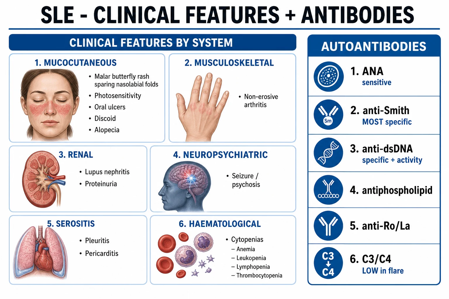
Life-threatening SLE presentations are medical emergencies requiring hospital admission, urgent immunosuppression, and simultaneous exclusion of infection. [1]
The febrile, deteriorating SLE patient presents a particularly difficult question: flare or infection? A markedly raised CRP (over 50 or 100), a positive blood culture, or a clear focus (chest, urine, line) favours infection and mandates antibiotic treatment first; a low or normal CRP with falling complement and rising anti-dsDNA favours active lupus. When in doubt, treat both with broad antibiotics plus corticosteroids, then narrow as data return. [1]
Macrophage activation syndrome (MAS), the SLE variant of haemophagocytic lymphohistiocytosis, is an emergency characterised by fever, cytopenias, ferritin over 1000, hypertriglyceridaemia, hypofibrinogenaemia, and hepatosplenomegaly, often triggered by infection or a flare. Treatment is high-dose corticosteroids, with ciclosporin and etoposide (HLH-94 protocol) in refractory cases. [1]
Catastrophic antiphospholipid syndrome (CAPS) presents with rapid multiorgan thrombosis and is treated with triple therapy: anticoagulation, high-dose corticosteroids, and plasma exchange or IVIG; rituximab or eculizumab in refractory cases.[10]
Management — Definitive & Stepwise
Definitive management is built on three principles: (1) hydroxychloroquine for all patients, (2) corticosteroids and immunosuppression titrated to organ threat and activity, and (3) a treat-to-target aim of clinical remission or low disease activity (LLDAS — Lupus Low Disease Activity State). All three are reinforced across the 2019 EULAR and 2020 EULAR/ERA-EDTA recommendations.[1][11]
Foundation measures for all patients
Hydroxychloroquine (HCQ) 200 to 400 mg daily (5 mg per kg of actual body weight) is the cornerstone for every SLE patient without contraindication. It reduces flares, thrombosis, dyslipidaemia, diabetes, organ damage, and overall mortality, and is safe in pregnancy and lactation. Baseline and annual ophthalmological screening for retinal toxicity is mandatory (the risk rises after five years of use, in renal impairment, and at higher daily doses).[1]
Lifestyle measures include strict sun protection (broad-spectrum SPF 50+ sunscreen, hat, protective clothing), smoking cessation (smoking impairs hydroxychloroquine efficacy and raises cardiovascular risk), vaccination (annual influenza, COVID-19, pneumococcal, HPV, recombinant zoster; live vaccines avoided on immunosuppression), cardiovascular risk reduction (lipids, blood pressure, diabetes), bone protection (calcium, vitamin D, bisphosphonate on chronic steroids), family planning (avoid oestrogen-containing oral contraceptives in antiphospholipid-positive women; avoid teratogens), and psychosocial support. [1]
Mild disease (skin, joint, constitutional, serositis, mild cytopenia)
- NSAIDs (e.g., naproxen 500 mg twice daily, ibuprofen 400 to 600 mg three times daily) for arthritis and serositis, with caution in renal disease.
- Short-course low-dose prednisolone (5 to 15 mg daily for flares, tapering over weeks) — minimise cumulative exposure.
- Topical corticosteroids and topical calcineurin inhibitors (tacrolimus, pimecrolimus) for cutaneous lesions.
- Methotrexate 10 to 25 mg weekly (with folic acid) for refractory arthritis or skin disease.
- Azathioprine 1.5 to 2.5 mg per kg per day for maintenance or steroid-sparing in musculoskeletal and mild systemic disease. [1]
Moderate to severe disease (organ-threatening)
Glucocorticoids
- Prednisolone 0.5 to 1 mg per kg per day for significant flares
- IV methylprednisolone 500 mg to 1 g daily for 3 to 5 days for severe organ-threatening disease
- Taper to the lowest effective dose; aim for under 7.5 mg daily maintenance
- Toxicity: diabetes, osteoporosis, infection, avascular necrosis, cataract, hypertension
Mycophenolate mofetil
- 2 to 3 g per day orally (target 2 g)
- First-line for induction of class III, IV, V lupus nephritis; also maintenance
- Side effects: cytopenias, infection, teratogenic (contraindicated in pregnancy)
- Equivalent to cyclophosphamide for induction (ALMS, Ginzler trials)
Cyclophosphamide
- Low-dose Euro-Lupus regimen: 500 mg IV every 2 weeks for 6 doses
- High-dose NIH regimen: 0.5 to 1 g per m squared monthly for 6 months
- Reserved for severe proliferative nephritis, CNS lupus, severe vasculitis, alveolar haemorrhage
- Side effects: haemorrhagic cystitis, infertility, infection, malignancy, cytopenias
Azathioprine
- 1.5 to 2.5 mg per kg per day orally
- Maintenance of lupus nephritis after cyclophosphamide induction; steroid-sparing
- Pregnancy-compatible (with monitoring)
- Check TPMT activity before starting to avoid fatal myelosuppression
Methotrexate
- 10 to 25 mg once weekly (oral or subcutaneous) with folic acid
- Refractory arthritis, skin disease, serositis
- Teratogenic; avoid in renal impairment; liver and FBC monitoring
Calcineurin inhibitors
- Tacrolimus 2 to 4 mg daily or voclosporin 23.7 mg twice daily
- Voclosporin approved for lupus nephritis on AURORA-1 trial data
- Ciclosporin 2.5 to 5 mg per kg per day for membranous (class V) nephritis
- Monitor renal function, blood pressure, and drug levels
Belimumab
- Anti-BAFF monoclonal antibody; 10 mg per kg IV at weeks 0, 2, 4 then monthly (or subcutaneous)
- Approved for active autoantibody-positive SLE including nephritis (BLISS-52 and BLISS-76)
- Add-on to standard therapy; reduces disease activity and steroid dose
- Generally well tolerated; small infection risk
Anifrolumab
- Anti-type I interferon receptor monoclonal antibody; 300 mg IV monthly
- Approved for moderate to severe SLE (TULIP-1 and TULIP-2 trials)
- Reduces disease activity and oral steroid requirement
- Side effects: herpes zoster reactivation (vaccinate first)
Rituximab
- Anti-CD20 B-cell-depleting antibody; 1 g IV at days 0 and 14 or 375 mg per m squared weekly x4
- Off-label for refractory SLE; LUNAR trial in nephritis was negative
- Useful for cytopenias (ITP, AIHA), severe skin disease, nephritis unresponsive to standard therapy
- Infusion reactions, infection, progressive multifocal leukoencephalopathy (rare), hypogammaglobulinaemia
BLISS-52 — Navarra 2011
Key finding
Belimumab 10 mg per kg added to standard therapy significantly improved SLE response (SSI responder index) at 52 weeks versus placebo in 867 patients, with a favourable safety profile. Established belimumab as the first approved biologic for SLE in over 50 years.
TULIP-2 — Morand 2020
Key finding
Anifrolumab 300 mg IV monthly significantly improved disease activity (BICLA response 47.8 percent vs 31.5 percent placebo), reduced oral steroid use, and reduced flare rate in 362 patients with moderate to severe SLE. Confirmed the type I interferon pathway as a drug target.
AURORA-1 — Rovin 2021
Key finding
Voclosporin 23.7 mg twice daily added to mycophenolate and rapid steroid taper achieved a higher renal response at 52 weeks (40.8 percent vs 19.3 percent placebo) in 357 patients with active lupus nephritis. Established voclosporin as a steroid-sparing, biopsy-class-independent treatment for lupus nephritis.
Lupus nephritis induction and maintenance
Class III or IV proliferative lupus nephritis requires induction then maintenance:[11][13][14]
- Induction (3 to 6 months): mycophenolate mofetil 2 to 3 g daily or low-dose IV cyclophosphamide (Euro-Lupus regimen) plus corticosteroids (IV methylprednisolone pulses then oral prednisolone 0.5 to 1 mg per kg tapering). Add-on belimumab or voclosporin improves response. The landmark ALMS and Ginzler trials showed mycophenolate to be non-inferior — and in some groups superior — to cyclophosphamide, with a better side-effect profile.
- Maintenance (years): mycophenolate mofetil 2 g daily or azathioprine 1.5 to 2.5 mg per kg per day, with hydroxychloroquine throughout and low-dose prednisolone (under 7.5 mg daily) if needed. Azathioprine is preferred in pregnancy planning.
- Class V membranous nephritis: mycophenolate or calcineurin inhibitor plus corticosteroid; high thrombosis risk warrants anticoagulation in nephrotic patients.
- Class VI advanced sclerosis: dialysis or transplantation — immunosuppression is futile.
- Rituximab is reserved for refractory nephritis (LUNAR was negative for primary endpoint but subgroup and real-world data support use). [1]
Cutaneous lupus
Strict photoprotection is essential. Topical corticosteroids and topical calcineurin inhibitors are first-line. Hydroxychloroquine is the systemic agent of choice; quinacrine may be added for refractory disease. Methotrexate, mycophenolate, or thalidomide (with strict contraception) are reserved for refractory cases. [1]
Specific Subtypes & Scenarios
Lupus nephritis — ISN/RPS classes in depth
The ISN/RPS 2003 classification (revised 2018) determines treatment intensity. Classes I and II are mesangial and require only hydroxychloroquine and a renin-angiotensin blocker for proteinuria. Classes III and IV (focal and diffuse proliferative) are the most aggressive and demand induction-maintenance immunosuppression. Class V (membranous) presents with nephrotic syndrome and high thrombosis risk. Class VI (advanced sclerosis) is end-stage and dialysis-dependent. The activity and chronicity indices on biopsy add further prognostic detail — high activity is reversible with treatment; high chronicity is not.[9][15]
Neuropsychiatric lupus
Treatment is dictated by the specific syndrome. Major events — seizures, psychosis, transverse myelitis, optic neuritis, acute confusional state — are treated with immunosuppression (pulse methylprednisolone then high-dose oral corticosteroid plus cyclophosphamide, with mycophenolate or azathioprine for maintenance) once infection is excluded. Minor events — headache, cognitive dysfunction, mood disorder — are managed symptomatically with analgesia, cognitive behavioural therapy, and antidepressants. Antiphospholipid-related stroke is treated with lifelong anticoagulation, not immunosuppression. MRI and lumbar puncture help to exclude mimics (infection, posterior reversible encephalopathy syndrome, progressive multifocal leukoencephalopathy from rituximab).[3]
Antiphospholipid syndrome overlap
About one-third of SLE patients have antiphospholipid antibodies, and roughly half of those develop clinical APS over time. The Sydney 2006 criteria define APS as a clinical event (venous or arterial thrombosis, or pregnancy morbidity) plus a persistent antiphospholipid antibody (lupus anticoagulant, anticardiolipin, or anti-beta-2-glycoprotein I) on two occasions at least 12 weeks apart. Management is lifelong anticoagulation after a thrombotic event (warfarin is preferred; direct oral anticoagulants are inferior in APS and should be avoided).[10]
Drug-induced lupus
Triggered by procainamide, hydralazine, isoniazid, minocycline, methyldopa, chlorpromazine, and anti-TNF agents. Characterised by anti-histone antibodies (over 95 percent), anti-dsDNA negative (usually), renal and CNS sparing, and resolution within weeks of withdrawing the offending drug. Anti-TNF-induced lupus is an exception that may be anti-dsDNA positive but is still clinically mild. Short-course corticosteroids may be needed for symptomatic control. [1]
Neonatal lupus
Caused by transplacental passage of maternal anti-Ro/SSA or anti-La/SSB antibodies. The most serious manifestation is congenital heart block (third-degree), with a mortality of 15 to 30 percent; other features include a transient annular rash (especially periocular), cytopenias, hepatitis, and cholestasis. Hydroxychloroquine through pregnancy reduces the recurrence risk in subsequent pregnancies. Most mothers are eventually diagnosed with SLE or Sjogren syndrome, and all anti-Ro-positive women require serial fetal echocardiography from 16 weeks. [1]
Complications & Pitfalls
Renal
- End-stage renal disease (class IV nephritis the main driver)
- Nephrotic syndrome and thrombosis (class V)
- Hypertension and accelerated cardiovascular risk
- Lupus recurrence post-transplant uncommon if quiescent
Cardiovascular
- Premature atherosclerosis — leading late cause of death
- Myocardial infarction risk fifty-fold higher in young women
- Pericardial tamponade, valve disease (Libman-Sacks)
- Pulmonary hypertension
Infectious
- Leading cause of early death (immunosuppression plus complement consumption)
- Encapsulated organisms (pneumococcus, meningococcus) with splenic hypofunction and immunosuppression
- Herpes zoster (especially on anifrolumab and rituximab)
- Opportunistic infection (PJP, CMV) on high-dose immunosuppression
Neurological
- Seizures, stroke, cognitive impairment
- Antiphospholipid-mediated thrombosis and chorea
- Cyclophosphamide-related infertility and infection
Musculoskeletal
- Avascular necrosis (femoral head; from steroids)
- Osteoporosis (steroids and disease)
- Jaccoud arthropathy (functional impairment)
Haematological
- Severe cytopenias (ITP, AIHA)
- Thrombosis (antiphospholipid)
- Macrophage activation syndrome
Drug toxicity
- Corticosteroid: diabetes, osteoporosis, avascular necrosis, infection, cataract
- Hydroxychloroquine: retinal toxicity (bull's-eye maculopathy)
- Mycophenolate: cytopenias, teratogenicity
- Cyclophosphamide: haemorrhagic cystitis, infertility, malignancy
- Belimumab: infection; rituximab: hypogammaglobulinaemia and PML
Prognosis & Disposition
SLE survival has improved from roughly 50 percent at five years in the 1950s to over 95 percent at five years and over 90 percent at ten years in developed settings, driven mainly by hydroxychloroquine use, better immunosuppression, dialysis, and infection prophylaxis. The leading causes of death have shifted over time: in early disease, active lupus (especially nephritis and NPSLE), infection, and serositis predominate; in late disease, accelerated cardiovascular disease, infection, ESRD, and malignancy dominate.[1][3]
Predictors of poor outcome include class IV nephritis with persistent proteinuria, hypertension and reduced GFR at presentation, high chronicity index on biopsy, antiphospholipid syndrome overlap, younger age at onset, African or Hispanic ethnicity, lower socioeconomic status, and high cumulative corticosteroid exposure. Hydroxychloroquine use and early control of disease activity to a low-disease-activity state are the modifiable factors that most improve prognosis. [1]
Disposition depends on activity and organ involvement: most patients are managed in the outpatient rheumatology clinic with shared-care nephrology, dermatology, cardiology, and obstetrics as needed. Indications for hospital admission include severe flare (renal, CNS, alveolar haemorrhage, severe cytopenia), infection, suspected CAPS or MAS, and diagnostic uncertainty. Nephrology referral is indicated for any new or worsening proteinuria, rising creatinine, or active urinary sediment; dialysis and transplantation are the endgame for class VI or refractory nephritis, with recurrence post-transplant uncommon if disease is quiescent. [1]
Special Populations
Pregnancy and SLE
Pregnancy should be planned during disease remission for at least six months to minimise flares, pre-eclampsia, fetal loss, and neonatal lupus. The principles are:[1]
- Continue hydroxychloroquine throughout — it reduces flares, pre-eclampsia, and neonatal heart block.
- Low-dose aspirin (75 to 150 mg daily) for all SLE pregnancies from early gestation, to reduce pre-eclampsia.
- Add low-molecular-weight heparin if antiphospholipid-positive (prophylactic) or after prior thrombosis or recurrent loss (therapeutic), continued through pregnancy and for six weeks postpartum.
- Avoid teratogens: mycophenolate, cyclophosphamide, methotrexate, leflunomide, warfarin (beyond six weeks), and thalidomide. Stop these at least three months before conception (mycophenolate) and substitute azathioprine or calcineurin inhibitor.
- Screen anti-Ro/SSA and anti-La/SSB in all SLE pregnancies; if positive, serial fetal echocardiography from 16 to 26 weeks for congenital heart block. Hydroxychloroquine reduces recurrence risk in anti-Ro-positive mothers with prior affected offspring.
- Belimumab and anifrolumab: insufficient data — discontinue at conception (azathioprine or calcineurin inhibitor substituted).
- Corticosteroids: avoid high doses if possible (steroids raise the risk of gestational diabetes, hypertension, premature rupture of membranes); prednisolone is preferred over dexamethasone (the latter crosses the placenta).
- Flares in pregnancy are treated with corticosteroids and azathioprine or calcineurin inhibitor. [1]
Antiphospholipid-positive pregnancy
Prophylactic or therapeutic low-molecular-weight heparin plus low-dose aspirin throughout pregnancy and six weeks postpartum is the standard, individualised by clinical history (prior thrombosis: therapeutic; prior loss alone: prophylactic). This combination reduces fetal loss from roughly 50 to 70 percent down to under 20 percent.[10]
Children and adolescents
Paediatric-onset SLE is more aggressive, with higher rates of nephritis, haematological involvement, and cumulative damage. Dosing is weight-based (hydroxychloroquine 5 mg per kg, mycophenolate 600 mg per m squared twice daily, azathioprine 1.5 to 2.5 mg per kg), with particular attention to growth, puberty, vaccination, and transition to adult care. [1]
Late-onset SLE (over age 50)
Insidious onset with more serositis and pulmonary involvement and less nephritis and rash. Differential includes malignancy, drug-induced lupus (common in older patients on multiple medications), and infection. Management is more conservative — lower-intensity immunosuppression with attention to comorbidity and drug interactions. [1]
Renal impairment
Dose-adjust hydroxychloroquine (caution above eGFR 30), avoid NSAIDs entirely, and dose-adjust mycophenolate, azathioprine, and cyclophosphamide. Calcineurin inhibitors need level monitoring. ESRD patients on dialysis need continuation of hydroxychloroquine and ongoing management of disease activity. [1]
Patients on immunosuppression and biologics
Screen and treat latent tuberculosis (interferon-gamma release assay), hepatitis B and C, and HIV before starting. Vaccinate before immunosuppression (pneumococcal, influenza, hepatitis B, recombinant zoster, HPV; avoid live vaccines once immunosuppressed). Monitor for infection throughout, with a low threshold for cultures and imaging. [1]
Evidence, Guidelines & Regional Differences
The 2019 EULAR/ACR classification criteria (Aringer, Arthritis Rheumatol 2019) are the current international standard for SLE classification, with the highest sensitivity and specificity of any system. The 2019 EULAR management recommendations (Fanouriakis, Ann Rheum Dis 2019) and the 2020 EULAR/ERA-EDTA lupus nephritis recommendations (Fanouriakis, Ann Rheum Dis 2020), with a 2025 update for kidney involvement (PMID 41107121), form the treatment backbone worldwide.[1][2][11][17]
The Australian Rheumatology Association and New Zealand rheumatology societies endorse the EULAR framework with national biologic access schemes. Indigenous Australians have among the highest SLE prevalence and severity worldwide, with high rates of lupus nephritis and early cardiovascular mortality.
Landmark trials
ALMS — Appel 2009
Key finding
In 370 patients with class III to V lupus nephritis, mycophenolate mofetil 3 g daily was non-inferior to IV cyclophosphamide for induction, with similar response and a more favourable safety profile. Established mycophenolate as a first-line induction option alongside cyclophosphamide.
Ginzler 2005 NEJM
Key finding
In 140 patients with class III to V lupus nephritis, mycophenolate mofetil 2 to 3 g daily was superior to monthly IV cyclophosphamide for induction of remission (22 percent vs 6 percent complete remission at 24 weeks), with fewer severe infections. A pivotal trial establishing mycophenolate as first-line.
LUNAR — Rovin 2012
Key finding
Rituximab added to mycophenolate and corticosteroid in 144 patients with class III or IV lupus nephritis **did not meet its primary endpoint** of superior renal response versus placebo. Rituximab remains off-label but is used for refractory nephritis and for cytopenias.
Controversies
- When to biopsy in lupus nephritis — many advocate biopsy at first sign of significant proteinuria to confirm class before committing to immunosuppression.
- Role of calcineurin inhibitors (voclosporin, tacrolimus) — voclosporin was approved on AURORA-1 (2021) but cost and long-term safety in diverse populations remain under study.
- Anifrolumab zoster risk — vaccinate before starting.
- DOACs versus warfarin in APS — DOACs are inferior for thrombotic APS and should be avoided; warfarin remains standard.
- Stopping hydroxychloroquine — almost never; the drug reduces flares, thrombosis, damage, and mortality, and is safe in pregnancy. [1]
Exam Pearls
LUPUS HANDS
Exam application bank (NEET-PG / INICET)
One-line answer
Systemic lupus erythematosus (SLE) is a multisystem autoimmune disease driven by loss of self-tolerance, autoantibody formation (ANA, anti-dsDNA, anti-Sm), immune complex deposition, and type I interferon activation. 9:1 female predominance, peak 15-45 years, worse in African, Hispanic, Asian populations. Classification: EULAR/ACR 2019 (ANA entry + additive weighted criteria, score 10+). Manifestations span skin (malar rash sparing nasolabial folds, discoid, photosensitivity), joints (non-erosive, Jaccoud), kidneys (lupus nephritis ISN/RPS class I-VI), serosae (pericarditis, pleuritis), CNS (neuropsychiatric lupus), blood (cytopenias, antiphospholipid), and heart (Libman-Sacks endocarditis). Management: hydroxychloroquine for ALL patients; NSAIDs and short-course steroids for flares; mycophenolate, cyclophosphamide, azathioprine for organ disease; belimumab, anifrolumab, rituximab for re
Worked stems (answer without another resource)
Stem 1 — Classic presentation. Map symptoms to mechanism; name the first investigation and first treatment step with dose/route if drug therapy is standard. [1]
Stem 2 — Unstable / complicated. List red flags that force immediate resuscitation, theatre, ICU, antidote, or reperfusion — and what you do in the first 15 minutes. [1]
Stem 3 — Atypical group. Elderly, pregnancy, child, or immunocompromised: how presentation and thresholds change. [1]
Stem 4 — Differential trap. Name the three closest mimics and one discriminator for each. [1]
Stem 5 — Disposition. Who goes home with safety-netting, who is admitted, who needs HDU/ICU/theatre, and what follow-up is mandatory. [1]
Rapid viva checklist
- Definition + classification
- Pathophysiology chain
- Bedside signs / criteria
- Score with exact components (if any)
- Emergency bundle
- Definitive therapy with doses
- Complications of disease and of treatment
- Special populations
- Guideline/trial name if classic
- Three exam traps
Coverage self-check
If you cannot answer any stem above from this page alone, re-read the matching section — the page is intended to be self-sufficient for final-prof and NEET-PG/INICET questions on Systemic Lupus Erythematosus.
[1] [1]Why does anti-dsDNA track disease activity when anti-Smith does not?
Anti-dsDNA titres rise and fall with immune-complex formation — they deposit in glomeruli and serosae during active disease, so their titre reflects current immune activation and especially renal activity. Anti-Smith antibodies, by contrast, persist at a stable level regardless of disease activity because they reflect a fixed pool of anti-Sm B-cell clones. Use anti-Smith to confirm the diagnosis and serial anti-dsDNA (with complement) to monitor activity.
References
- [1]Fanouriakis A, Kostopoulou M, Alunno A, et al. 2019 update of the EULAR recommendations for the management of systemic lupus erythematosus Ann Rheum Dis, 2019.PMID 30926722
- [2]Aringer M, Costenbader K, Daikh D, et al. 2019 European League Against Rheumatism/American College of Rheumatology Classification Criteria for Systemic Lupus Erythematosus Arthritis Rheumatol, 2019.PMID 31385462
- [3]Tsokos GC Systemic lupus erythematosus N Engl J Med, 2011.PMID 22129255
- [4]Petri M, Orbai AM, Alarcon GS, et al. Derivation and validation of the Systemic Lupus International Collaborating Clinics classification criteria for systemic lupus erythematosus Arthritis Rheum, 2012.PMID 22553077
- [5]Navarra SV, Guzman RM, Gallacher AE, et al. Efficacy and safety of belimumab in patients with active systemic lupus erythematosus: a randomised, placebo-controlled, phase 3 trial Lancet, 2011.PMID 21296403
- [6]Rovin BH, Furie R, Latiris K, et al. Efficacy and safety of rituximab in patients with active proliferative lupus nephritis: the Lupus Nephritis Assessment with Rituximab study Arthritis Rheum, 2012.PMID 22231479
- [7]Morand EF, Furie R, Tanaka Y, et al. Trial of Anifrolumab in Active Systemic Lupus Erythematosus N Engl J Med, 2020.PMID 31851795
- [8]Rovin BH, Teng YKO, Ginzler EM, et al. Efficacy and safety of voclosporin versus placebo for lupus nephritis (AURORA 1): a double-blind, randomised, multicentre, placebo-controlled, phase 3 trial Lancet, 2021.PMID 33971155
- [9]Weening JJ, DAgati VD, Schwartz MM, et al. The classification of glomerulonephritis in systemic lupus erythematosus revisited Kidney Int, 2004.PMID 14717922
- [10]Miyakis S, Lockshin MD, Atsumi T, et al. International consensus statement on an update of the classification criteria for definite antiphospholipid syndrome (APS) J Thromb Haemost, 2006.PMID 16420554
- [11]Fanouriakis A, Kostopoulou M, Cheema K, et al. 2019 Update of the Joint European League Against Rheumatism and European Renal Association-European Dialysis and Transplant Association (EULAR/ERA-EDTA) recommendations for the management of lupus nephritis Ann Rheum Dis, 2020.PMID 32220834
- [12]Hochberg MC Updating the American College of Rheumatology revised criteria for the classification of systemic lupus erythematosus Arthritis Rheum, 1997.PMID 9324032
- [13]Ginzler EM, Dooley MA, Aranow C, et al. Mycophenolate mofetil or intravenous cyclophosphamide for lupus nephritis N Engl J Med, 2005.PMID 16306519
- [14]Appel GB, Contreras G, Dooley MA, et al. Mycophenolate mofetil versus cyclophosphamide for induction treatment of lupus nephritis J Am Soc Nephrol, 2009.PMID 19369404
- [15]Hahn BH, McMahon MA, Wilkinson A, et al. American College of Rheumatology guidelines for screening, treatment, and management of lupus nephritis Arthritis Care Res (Hoboken), 2012.PMID 22556106
- [16]Furie R, Khamashta M, Merrill JT, et al. Type I interferon inhibitor anifrolumab in active systemic lupus erythematosus (TULIP-1): a randomised, controlled, phase 3 trial Lancet Rheumatol, 2019.PMID 38229377
- [17]Fanouriakis A, Kostopoulou M, Andersen J, et al. EULAR recommendations for the management of systemic lupus erythematosus with kidney involvement: 2025 update Ann Rheum Dis, 2026.PMID 41107121