Rheumatology · General Medicine
Systemic Sclerosis (Scleroderma)
Also known as Systemic sclerosis · Scleroderma · SSc · CREST syndrome · Limited cutaneous SSc · Diffuse cutaneous SSc
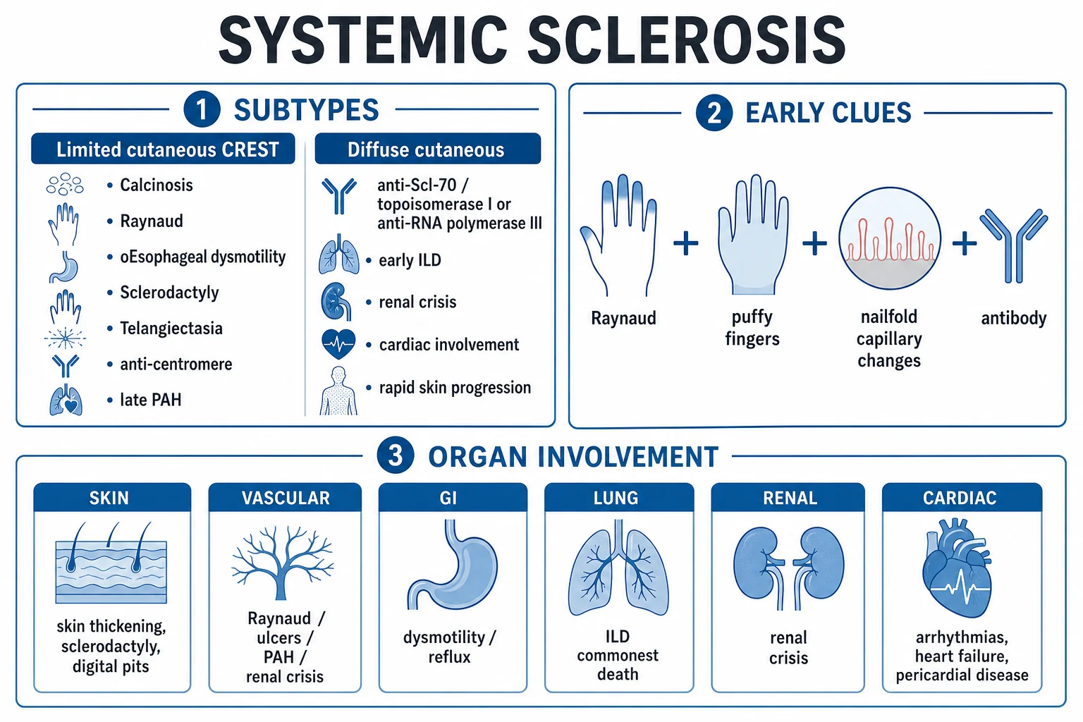
Systemic sclerosis (scleroderma) is a chronic multisystem autoimmune connective tissue disease defined by a pathophysiological triad of microvascular vasculopathy, immune dysregulation and generalised fibrosis (excess collagen deposition) of the skin and internal organs, with disease-specific autoantibodies (anti-centromere, anti-Scl-70/topoisomerase I, anti-RNA polymerase III). The two principal subtypes are limited cutaneous (CREST — Calcinosis, Raynaud, oEsophageal dysmotility, Sclerodactyly, Telangiectasia; anti-centromere; late pulmonary arterial hypertension) and diffuse cutaneous (anti-Scl-70 or anti-RNA polymerase III; early interstitial lung disease, renal crisis, rapidly progressive skin). Raynaud phenomenon is the first manifestation in over 90 percent. It is the rheumatic disease with the highest case fatality — interstitial lung disease and pulmonary arterial hypertension are the leading causes of death, and scleroderma renal crisis is the classical emergency demanding an urgent ACE inhibitor.
On this page & tools
Your progress
Saved locally on this device.
Practise this topic
Exam tags
Red flags
Overview & Definition
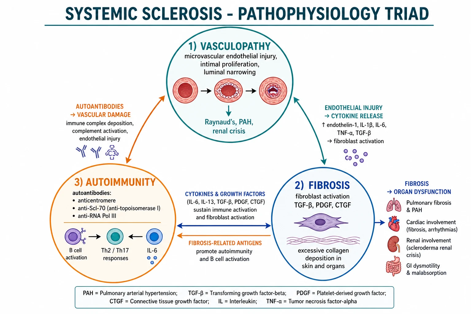
Systemic sclerosis (SSc, scleroderma) is a chronic, multisystem autoimmune connective tissue disease of unknown cause defined by a pathophysiological triad — microvascular vasculopathy, immune dysregulation and generalised tissue fibrosis (excessive deposition of type I and type III collagen and extracellular matrix by activated myofibroblasts).[2][9] The disease targets the skin (producing the eponymous hard, hidebound sclerosis) and virtually every internal organ — most lethally the lungs (interstitial lung disease and pulmonary arterial hypertension), kidneys (scleroderma renal crisis), heart (myocardial fibrosis) and gastrointestinal tract (dysmotility and malabsorption). It is not a primary skin disease; the skin is simply the most visible manifestation of a systemic fibrosing vasculopathy.
The clinical art in systemic sclerosis is not the late, hidebound patient — anyone can recognise end-stage scleroderma. It is recognising the early phenotype while disease is still modifiable: Raynaud phenomenon plus puffy fingers plus abnormal nailfold capillaroscopy plus a disease-specific antibody (the so-called very early diagnosis of systemic sclerosis, or VEDOSS, criteria).[1] Once this triad appears, the patient must be risk-stratified by subtype and antibody: limited cutaneous disease with anti-centromere favours late-onset pulmonary arterial hypertension; diffuse cutaneous disease with anti-Scl-70 favours early interstitial lung disease; anti-RNA polymerase III identifies the subgroup at highest risk of scleroderma renal crisis and of synchronous malignancy. Because no therapy reverses established fibrosis, early organ screening (annual pulmonary function tests and echocardiography) and aggressive vasodilation and immunosuppression are how outcome is improved.[7][9]
Two emergencies dominate the disease and decide survival: (1) scleroderma renal crisis — new-onset malignant hypertension with acute kidney injury, demanding an urgent ACE inhibitor (captopril) even when the creatinine is rising and dialysis is needed; and (2) interstitial lung disease — the leading cause of death, presenting with insidious exertional dyspnoea and dry cough, needing immunosuppression and antifibrotic therapy. ACE inhibitors are life-saving in renal crisis, and high-dose corticosteroids (over 15 mg/day prednisolone equivalent) are dangerous because they precipitate it.[9]
Classification
Classification of systemic sclerosis serves two purposes: to distinguish it from mimics (localised scleroderma, nephrogenic systemic fibrosis, eosinophilic fasciitis) and to stratify prognosis and therapy within the disease. The two axes are extent of skin involvement and autoantibody. [1]
The 2013 ACR/EULAR classification criteria
The 2013 ACR/EULAR classification criteria (replacing the 1980 criteria, which were insensitive to early disease) use a point-based scoring system. A patient is classified as having definite systemic sclerosis if the total score is 9 or more.[5]
2013 ACR/EULAR classification — scoring table
The criterion that skin thickening proximal to the metacarpophalangeal joints is sufficient alone (9 points) is the single most exam-rewarded rule: such a patient has systemic sclerosis by definition. Note that this criterion is the sum of all — proximal skin thickening excludes the additive distal fingertip items, but in its absence, the additive domains accumulate. [1]
The subtype classification (extent of skin involvement)
The clinical classification by the extent of skin thickening is the bedrock of prognosis:[2]
Limited cutaneous SSc (lcSSc)
- Skin thickening confined to the face and DISTAL to elbows/knees (hands, face, feet, distal forearms)
- Long history of Raynaud (often years to decades) before skin disease
- Anti-centromere antibody in 50-70 percent
- CREST phenotype: Calcinosis, Raynaud, Esophageal dysmotility, Sclerodactyly, Telangiectasia
- Late-onset pulmonary arterial hypertension is the main threat (after 10-20 years)
- Better prognosis than diffuse disease
Diffuse cutaneous SSc (dcSSc)
- Skin thickening PROXIMAL to elbows/knees and on the trunk
- Raynaud onset within 1 year of skin disease (often concurrent or after)
- Anti-Scl-70 (topoisomerase I) or anti-RNA polymerase III
- Rapidly progressive skin (peak modified Rodnan skin score at 1-3 years), then often slow softening
- High risk of EARLY interstitial lung disease, renal crisis and cardiac disease
- Tendon friction rubs common
- Worse prognosis
SSc sine scleroderma
- SSc-specific internal organ involvement (PAH, ILD, GI) plus Raynaud and a disease-specific antibody
- But NO skin thickening (scleroderma 'without the sclerosis')
- Nailfold capillaroscopy is abnormal
- Often presents late, with isolated PAH or ILD
Overlap syndrome
- SSc features plus another defined connective tissue disease (polymyositis/dermatomyositis, SLE, RA, Sjogren)
- Anti-PM/Scl, anti-U1-RNP (MCTD) typical
- Treat the dominant organ threat
The CREST acronym
The CREST syndrome describes the cardinal features of limited cutaneous disease, and remains the most exam-rewarded mnemonic in scleroderma:[2]
CREST — limited cutaneous systemic sclerosis
CREST
Subcutaneous calcium deposits (calcium hydroxyapatite) on fingertips, over joints and extensor surfaces; painful, may ulcerate and extrude chalky material
Vasospasm of digital arteries — triphasic pallor then cyanosis then erythema; the first manifestation in over 90 percent of all SSc
Loss of smooth muscle in the lower two-thirds of the oesophagus causing absent peristalsis, lower sphincter incompetence, reflux and strictures
Thickened, tight, shiny skin of the fingers distal to the MCP joints, with reduced flexion and contractures
Dilated mat-shaped capillary lesions on face, lips, hands and oral mucosa — a marker of vascular disease
Autoantibody-phenotype correlations
Each disease-specific autoantibody predicts the clinical course. They are largely mutually exclusive — a patient has one dominant SSc antibody, not several.[1][2]
Autoantibody-phenotype map
Epidemiology & Risk Factors
Systemic sclerosis is uncommon but not rare, with a prevalence of approximately 50-300 per million and an annual incidence of around 10-20 new cases per million population per year. There is geographic and ethnic variation, with somewhat higher rates reported in the United States and Australia and lower rates in Japan and northern Europe.[9]
SSc — key numbers
The female-to-male ratio is approximately 4:1, and the peak age of onset is 30-50 years (childhood disease is rare and is usually localised scleroderma rather than systemic). Male sex, older age at onset and African ancestry are associated with worse prognosis, partly through a higher frequency of diffuse cutaneous disease and renal crisis.[2]
Genetic susceptibility accounts for only a minority of risk. The strongest associations are with the HLA-DRB1 and HLA-DQB1 haplotypes, with IRF5, STAT4, CD247 and other immune-gene loci contributing. Specific autoantibodies cluster with specific HLA alleles (for example, anti-topoisomerase I with HLA-DRB1*11), but the absolute risk to relatives is low.[3]
Environmental and occupational triggers are well documented. The strongest association is with silica dust (miners, stonemasons, sandblasters) — relative risk in the order of 3-25 fold. Other recognised exposures include organic solvents (vinyl chloride, trichloroethylene — the original "vinyl chloride disease" in chemical workers), epoxy resins, bleomycin (which produces a scleroderma-like syndrome), taxane chemotherapy, radiation therapy (which can localise or worsen skin disease in the irradiated field), and contaminated L-tryptophan (the eosinophilia-myalgia syndrome of the 1980s, related to contaminated supplements). Viral triggers (cytomegalovirus, parvovirus B19) have been proposed but not confirmed. Cigarette smoking accelerates vascular disease and is the single most modifiable risk factor for digital ischaemia.[3][4]
Drug-induced and paraneoplastic forms are increasingly recognised. Anti-RNA polymerase III antibody positivity carries a substantially elevated risk of a synchronous solid malignancy (especially breast and lung), implying that in this subgroup the disease may be a paraneoplastic phenomenon — age-appropriate cancer screening is recommended at diagnosis and for several years thereafter.[1]
Pathophysiology
The pathogenesis of systemic sclerosis is pathophysiologically a triad of microvascular vasculopathy, immune dysregulation, and generalised fibrosis, which together drive every clinical feature from Raynaud to renal crisis. Although the initiating trigger is unknown, the cascade proceeds in three reinforcing loops.[3][9]
1. Microvascular vasculopathy — the initiating event
The first demonstrable abnormality in systemic sclerosis is injury to the endothelium of small arteries, arterioles and capillaries, producing a disease of the microcirculation before fibrosis is detectable. Endothelial cells undergo apoptosis, the intimal layer proliferates, the basement membrane reduplicates, and the capillary bed is progressively lost (rarefaction). The surviving capillaries dilate to compensate, producing the giant (ectatic) capillaries and microhaemorrhages seen on nailfold capillaroscopy — the pathognomonic early finding.[6]
The injured endothelium shifts the balance of vascular mediators: it produces more endothelin-1 (a potent vasoconstrictor and mitogen) and less nitric oxide and prostacyclin (vasodilators and antiproliferatives). The result is the fixed structural narrowing of the digital and visceral microvasculature plus the superimposed vasospasm that produces Raynaud phenomenon and recurrent tissue ischaemia. The same vascular lesion, when it occurs in the pulmonary arteries, renal cortex and coronaries, drives pulmonary arterial hypertension, scleroderma renal crisis and myocardial ischaemia.[4][9]
2. Immune dysregulation and autoantibodies
Perivascular CD4 T-cell infiltrates with a Th2-skewed cytokine profile (IL-4, IL-13), along with B-cell hyperactivity and autoantibody production, characterise the immune phase. The disease-specific autoantibodies (anti-centromere, anti-topoisomerase I/Scl-70, anti-RNA polymerase III) are highly valuable as biomarkers but their direct pathogenic role remains debated. The cytokines released — most importantly transforming growth factor-beta (TGF-beta) and connective tissue growth factor (CTGF/CCN2) — are the principal drivers of fibroblast activation. Interleukin-6 is also markedly elevated and is a therapeutic target (tocilizumab).[3]
3. Fibroblast activation and fibrosis (the irreversible end-stage)
The defining effector of systemic sclerosis is the conversion of quiescent dermal and visceral fibroblasts into activated myofibroblasts by TGF-beta, CTGF/CCN2, PDGF and IL-6. Myofibroblasts acquire smooth-muscle contractile properties (alpha-smooth muscle actin) and overproduce type I and type III collagen, fibronectin and other extracellular matrix proteins. This matrix accumulates in the dermis (skin thickening), lung interstitium (interstitial lung disease), gut wall (dysmotility), vascular intima (vascular narrowing) and heart (myocardial fibrosis), producing the irreversible end-organ damage of late disease.[3]
A key insight is that myofibroblasts become autonomous — they continue to produce matrix even when the initiating immune signal has been silenced, an epigenetic "imprinting" analogous to that of fibroblast-like synoviocytes in rheumatoid arthritis. This is why established fibrosis is currently irreversible and why no drug has been shown to reverse it. Therapy therefore targets the upstream processes (vasodilation, immunosuppression) and the organ consequences (PAH, ILD, renal crisis), and the antifibrotic nintedanib slows rather than reverses fibrosis.[7][8]
Clinical Presentation
Systemic sclerosis can present in almost any way, but the classical sequence is Raynaud phenomenon first (often years before other features), followed by puffy fingers, then skin thickening, then organ involvement. The order, tempo and organ pattern are dictated by subtype and antibody. [1]
Raynaud phenomenon — the first sign
Raynaud phenomenon is the first manifestation in over 90 percent of patients with systemic sclerosis and may precede skin disease by years to decades in limited cutaneous disease, or appear concurrently with skin thickening in diffuse disease.[4] The classical description is the triphasic colour response of the fingers to cold or stress: pallor (vasospastic ischaemia), then cyanosis (deoxyhaemoglobin in static blood), then erythema (reactive hyperaemia on rewarming). Primary Raynaud (benign, common in young women) lacks the nailfold capillary changes, digital ulcers, pitting scars, or autoantibodies that mark secondary Raynaud — and any Raynaud onset after age 30, or with these accompanying features, demands an autoimmune workup.[4]
Skin and digital features
The skin evolves through three phases:[2]
- Oedematous (puffy fingers) phase — early non-pitting swelling of the fingers, often the first objective skin sign; this is the highest-yield window for diagnosis.
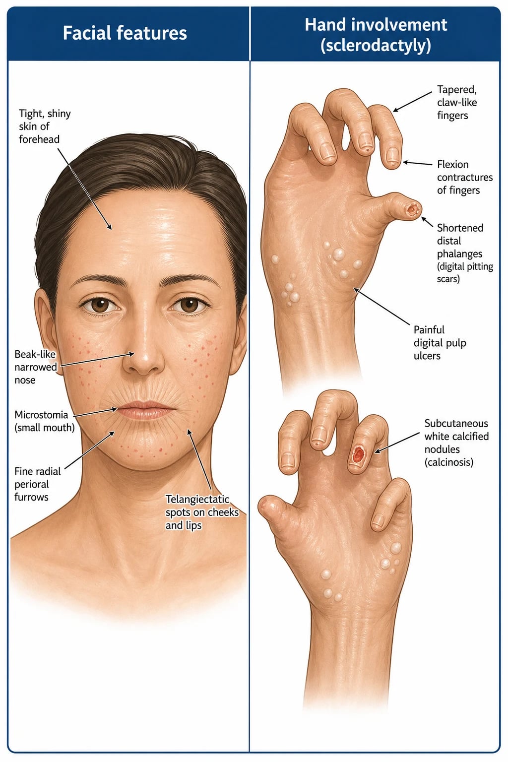
- Fibrotic (indurated, hidebound) phase — the skin becomes hard, tight, shiny and bound to underlying structures; sclerodactyly (thickening distal to the MCP joints), microstomia (reduced oral aperture), perioral radial furrowing (rhagades), loss of hair and sweat, and hyper- or hypopigmentation.
- Atrophic phase — late thinning of the skin, but the underlying sclerosis persists; in dcSSc the skin often softens after the peak (1-3 years), which can be misread as improvement. [1]
Digital features include digital pitting scars (small, depressed atrophic scars at the fingertips, the residue of ischaemic microinfarcts), ischaemic digital ulcers (painful, slow to heal, prone to infection), calcinosis cutis (hard, chalky subcutaneous calcium deposits that ulcerate and extrude white material), acral osteolysis (resorption of the terminal phalanges, producing shortened fingertips), and in severe cases digital gangrene and auto-amputation. [1]
Telangiectasia — mat-shaped dilated capillaries on the face, lips, hands, palms and oral mucosa — is a hallmark of limited disease and is often called mat telangiectasia to distinguish it from the spider naevi of chronic liver disease. [1]
Gastrointestinal features
The gastrointestinal tract is involved in up to 90 percent of patients and is the most common organ manifestation overall. The lower two-thirds of the oesophagus (smooth muscle) loses peristalsis and the lower sphincter becomes incompetent, producing gastro-oesophageal reflux, dysphagia, oesophagitis, strictures and Barrett's oesophagus (with a markedly increased risk of oesophageal adenocarcinoma).[2]
In the stomach, gastric antral vascular ectasia (GAVE, "watermelon stomach") produces chronic iron-deficiency anaemia from occult bleeding, with the characteristic endoscopic appearance of linear red stripes converging on the antrum. In the small bowel, dysmotility produces small intestinal bacterial overgrowth (SIBO) — the leading cause of diarrhoea, steatorrhoea and malabsorption in SSc — and may progress to intestinal pseudo-obstruction and rarely pneumatosis intestinalis. The colon and anorectum may be atonic, with wide-mouthed diverticula, incontinence and constipation. Primary biliary cholangitis (PBC) overlaps with limited disease, especially with anti-centromere antibody.[9]
Pulmonary features — the lethal organs
The lungs are the leading cause of death in systemic sclerosis, through two distinct mechanisms: [1]
- Interstitial lung disease (SSc-ILD) — affects approximately 40 percent of patients; presents with insidious exertional dyspnoea and dry cough; bibasal fine Velcro-like crackles on auscultation; the non-specific interstitial pneumonia (NSIP) pattern is commonest (basal subpleural ground-glass, reticulation, traction bronchiectasis) and carries a better prognosis than the usual interstitial pneumonia (UIP) pattern (honeycombing). Strongly associated with anti-Scl-70 (topoisomerase I) antibody.[8]
- Pulmonary arterial hypertension (SSc-PAH) — affects approximately 10-15 percent, mostly in limited cutaneous disease and with anti-centromere antibody; presents with progressive exertional dyspnoea, fatigue, exertional syncope, chest pain and signs of right heart failure (elevated JVP, right ventricular heave, loud pulmonary component of S2, tricuspid regurgitation, peripheral oedema). May also arise in the context of left heart disease (diastolic dysfunction from myocardial fibrosis) or chronic ILD hypoxia, but WHO Group 1 PAH is the classical SSc pattern.[9]
Isolated or disproportionate reduction in DLCO (with relatively preserved FVC) and a raised FVC/DLCO ratio (over 1.6) are the screening red flags for pulmonary vascular disease even before symptoms.[7]
Renal features — the scleroderma renal crisis
Scleroderma renal crisis is the classical emergency, occurring in approximately 5-10 percent of patients, almost always in early diffuse cutaneous disease and strongly associated with anti-RNA polymerase III antibody.[9] It presents with new-onset severe (malignant) hypertension, acute kidney injury, microangiopathic haemolytic anaemia (with schistocytes on blood film), thrombocytopenia, raised renin, retinopathy and encephalopathy, and may produce flash pulmonary oedema. It is precipitated or worsened by high-dose corticosteroids (over 15 mg/day prednisolone equivalent), which is why steroids must be used sparingly in SSc. The treatment is an urgent ACE inhibitor — see Management.
Cardiac features
Myocardial fibrosis (replacement fibrosis, microvascular ischaemia and conduction system involvement) produces diastolic dysfunction, systolic heart failure, arrhythmias (atrial and ventricular) and conduction defects. Pericardial effusion is common but usually small and asymptomatic; tamponade is rare. Cardiac MRI with late gadolinium enhancement is the most sensitive test for subclinical myocardial fibrosis. Cardiac involvement is an independent predictor of mortality.[9]
Musculoskeletal features
Inflammatory myositis (overlap with polymyositis/dermatomyositis, especially anti-PM/Scl), flexion contractures of the fingers, tendon friction rubs (a palpable/audible leathery crepitus over extensor tendons — a specific and prognostically adverse finding in dcSSc), acro-osteolysis, juxta-articular erosive arthritis, and generalised bone pain from malabsorption-related osteomalacia all occur.[2]
Atypical presentations
- Elderly male with anti-RNA polymerase III — rapid-onset diffuse skin and high risk of renal crisis within months; synchronous malignancy must be excluded.
- Very early diagnosis (VEDOSS) — Raynaud with puffy fingers and abnormal nailfold capillaries but no established skin thickening; this is the highest-yield window for screening.
- SSc sine scleroderma — isolated PAH or ILD with no skin disease.
- Overlap syndromes and mixed connective tissue disease (MCTD) — anti-U1-RNP, arthritis, myositis, Raynaud.
- Paediatric — usually localised scleroderma (morphea, linear scleroderma, en coup de sabre); juvenile systemic sclerosis is rare. [1]
Differential Diagnosis
The differential diagnosis of "hard skin" is broad, and the dangerous omission is to label a mimic as systemic sclerosis (or vice versa). The key discriminators are Raynaud, nailfold capillaroscopy, SSc-specific antibodies and internal organ involvement.[2]
Localised scleroderma (morphea/linear)
- Skin-only plaques (morphea) or linear bands (linear scleroderma)
- No Raynaud, no internal organ involvement, no SSc-specific antibodies, normal nailfold capillaroscopy
- Linear scleroderma in children causes growth arrest (limb-length discrepancy, facial hemiatrophy / Parry-Romberg)
- Treated with topical/intralesional steroid, UVA1 phototherapy, methotrexate for active disease
Nephrogenic systemic fibrosis (NSF)
- Exposure to GADOLINIUM-based contrast in advanced (stage 4-5) CKD or post-transplant
- Symmetric fibrotic 'woody' plaques on extremities, SPARING THE FACE
- No Raynaud, no SSc-specific antibodies, normal nailfold capillaroscopy
- Histology: thickened dermis with CD34+ spindle cells and TRAMP staining
Chronic graft-versus-host disease (GVHD)
- Prior haematopoietic stem cell transplant
- Lichenoid then sclerotic skin involvement
- No Raynaud, no SSc-specific antibodies
- Treated with immunosuppression
Eosinophilic fasciitis (Shulman disease)
- Acute onset of symmetric woody induration of limbs, SPARING HANDS AND FACE
- Peripheral blood EOSINOPHILIA, hypergammaglobulinaemia
- Grooving of superficial veins (the 'groove sign' or 'sign of the vein')
- Deep fascial thickening on MRI; biopsy shows fascial inflammation
- Rapid response to corticosteroids; no Raynaud, no SSc antibodies
Diabetic cheiroarthropathy
- Long-standing type 1 diabetes mellitus
- 'Prayer sign' — inability to oppose the palms
- Limited joint mobility, tight waxy skin of the fingers
- No Raynaud, no SSc-specific antibodies, normal nailfold capillaroscopy
Scleromyxoedema, scleredema of Buschke, amyloidosis, porphyria cutanea tarda
- Scleromyxoedema: IgG lambda paraprotein, mucin deposition, papular lesions
- Scleredema adultorum (Buschke): post-streptococcal or diabetic, upper back and neck, self-limited
- Amyloidosis: macroglossia, periorbital purpera, restrictive cardiomyopathy
- Porphyria cutanea tarda: photosensitivity, blisters, hypertrichosis, high serum iron/ferritin
Other causes of Raynaud
- Primary (benign, young women, no ulceration, no antibodies)
- SLE, Sjogren, dermatomyositis/polymyositis, cryoglobulinaemia, cold agglutinin disease
- Buerger disease (thromboangiitis obliterans), atherosclerosis
- Vibration injury, drugs (beta-blockers, ergotamine, bleomycin, interferon, cisplatin)
The single safest bedside discriminator is the combination of Raynaud phenomenon + abnormal nailfold capillaroscopy + a disease-specific antibody — this triad is essentially diagnostic of systemic sclerosis in the right clinical setting.[6]
Clinical & Bedside Assessment
The long-case examination of systemic sclerosis centres on the skin, hands, face, nailfold capillaroscopy, chest and cardiovascular system. The goal is to stage the disease, identify the subtype, detect organ involvement and quantify skin thickening. [1]
The modified Rodnan skin score (mRSS)
The modified Rodnan skin score is the validated, internationally used skin-thickening outcome measure in systemic sclerosis.[9] It is assessed by pinching the skin at 17 body sites and grading the thickness from 0 to 3:
Modified Rodnan skin score (mRSS)
The mRSS tracks disease activity over time, predicts internal organ involvement (high early scores predict renal crisis and ILD), and is the primary outcome in skin disease trials. [1]
Skin, hand and face examination
Inspect and palpate the fingers (puffy fingers, sclerodactyly, digital pitting scars, ischaemic ulcers, calcinosis, reduced flexion, fixed flexion contractures, acro-osteolysis), the face (microstomia with reduced oral aperture measured by the inter-incisor distance, perioral radial furrowing (rhagades), telangiectasia, loss of wrinkles, beak-like nose, mask-like facies), and the skin generally (salt-and-pepper dyspigmentation, hidebound areas, calcinosis over extensor surfaces). The oral aperture shrinks — patients struggle to fit food in or to brush their teeth. [1]
Nailfold capillaroscopy — the early diagnostic test
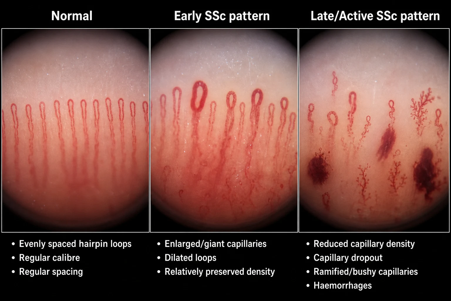
Nailfold capillaroscopy is the single most useful early test in suspected SSc. Using a dermatoscope, ophthalmoscope (+40 D lens) or dedicated videocapillaroscope with immersion oil on the nailfold, the capillaries of the proximal nailfold are examined. The scleroderma pattern has three components:[6]
- Giant (ectatic/dilated) capillaries — homogeneously enlarged loops, the hallmark.
- Microhaemorrhages — dark blobs from capillary rupture.
- Capillary loss (rarefaction, dropout, avascular areas) — reduced capillary density, late and prognostically adverse. [1]
The disease evolves through early (giant capillaries, no dropout), active (giant capillaries, microhaemorrhages and dropout) and late (predominantly dropout, avascular areas and bushy/ramified capillaries) patterns.[6]
Chest and cardiovascular examination
Auscultate for fine basal Velcro-like crackles (interstitial lung disease), a pulmonary flow murmur (pulmonary hypertension), a loud pulmonary component of S2 (P2), a tricuspid regurgitation murmur (functional, secondary to right ventricular dilation), a right ventricular heave, elevated JVP and peripheral oedema (right heart failure). Look for pericardial rubs and auscultate for arrhythmia. Palpate the apex (displaced in left heart failure). [1]
Other key bedside assessments
- Blood pressure at every visit — renal crisis surveillance; new or rising BP in early dcSSc is a red flag.
- Tendon friction rubs — palpate/auscultate over the extensor tendons (knee, elbow) with passive movement; a leathery crepitus is highly specific for active dcSSc and prognostically adverse.
- Functional and nutritional assessment — grip, oral aperture, ability to bring hand to mouth, weight loss, hand function, HAQ-DI, patient global. Malnutrition is common and under-recognised.
- Musculoskeletal examination — proximal muscle weakness (myositis), joint synovitis and contractures. [1]
Investigations
Investigations in systemic sclerosis serve three purposes: to confirm the diagnosis and subtype (autoantibodies, capillaroscopy), to screen and stage organ involvement (PFTs, HRCT, echocardiography, renal function, GI studies) and to monitor disease progression and therapy. The diagnosis is clinical, supported by serology and organ screening — there is no single diagnostic test. [1]
Autoantibodies — the serological backbone
Antinuclear antibody (ANA) is positive in over 95 percent of patients — typically with a speckled, nucleolar or centromere pattern. The disease-specific antibodies are mutually exclusive and predict the clinical phenotype (see the Classification table above).[1][2]
SSc-specific autoantibodies
A patient with a clinical picture of systemic sclerosis and a negative disease-specific antibody panel should be re-tested for the rarer specificities (anti-Th/TO, anti-U3-RNP) and reconsidered for a mimic. [1]
Nailfold capillaroscopy
See the Clinical Assessment section. The scleroderma pattern (giant capillaries, microhaemorrhages, dropout) has a sensitivity of over 90 percent for established SSc and is the most useful early diagnostic test in Raynaud.[6]
Pulmonary function tests (PFTs) — annual screening
PFTs are the cornerstone of ILD and PAH screening and should be performed at baseline and annually in all patients:[7][8]
- FVC reduced (and FVC/DLCO ratio preserved or low) — points to interstitial lung disease (restrictive defect).
- DLCO disproportionately reduced (with relatively preserved FVC) and FVC/DLCO ratio raised (over 1.6) — points to pulmonary vascular disease / PAH, even before symptoms.
- A DLCO under 60 percent predicted is a strong independent predictor of PAH.
- A 10 percent or greater fall in FVC or DLCO between visits mandates HRCT and echocardiography. [1]
High-resolution CT (HRCT) of the chest
HRCT is the imaging test of choice for interstitial lung disease and shows the pattern (NSIP vs UIP), extent (the prognostically important quantitative extent of over 20 percent is high-risk) and progression over time.[8]
- NSIP (commonest in SSc): basal and subpleural ground-glass opacification, reticulation, traction bronchiectasis, sparing of the immediate subpleural band at the base (useful vs UIP), relatively preserved architecture.
- UIP: basal, subpleural, honeycombing, traction bronchiectasis; carries a worse prognosis and is more frequently associated with anti-Scl-70. [1]
Echocardiography — annual screening
Echocardiography screens for pulmonary hypertension, right ventricular size and function, pericardial effusion, left ventricular diastolic dysfunction and valvular disease. A tricuspid regurgitant jet velocity over 2.8 m/s, right atrial area over 18 cm2, right ventricular systolic dysfunction and a ventricular septal flattening are screening red flags for PAH. In high-risk limited disease, the DETECT algorithm (a two-step score using PFTs, biomarkers, ECG and echo) is more sensitive than echo alone.[7]
Right heart catheterisation is mandatory to confirm PAH (mean pulmonary artery pressure 20 mmHg or higher, pulmonary capillary wedge pressure 15 mmHg or lower, pulmonary vascular resistance over 3 Wood units = pre-capillary PAH) and to exclude post-capillary causes.[9]
Renal function and renal crisis workup
In the clinic, blood pressure, serum creatinine and urinalysis are performed at every visit (in early dcSSc, more frequently — weekly to monthly). In a suspected renal crisis, send: [1]
- Urea and electrolytes, creatinine, eGFR (rising).
- Full blood count and blood film — microangiopathic haemolytic anaemia (schistocytes/helmet cells), thrombocytopenia.
- LDH and haptoglobin — intravascular haemolysis (LDH high, haptoglobin low).
- Urinalysis — mild proteinuria and microscopic haematuria (the kidney is NOT the primary inflammatory target — the lesion is vascular — so heavy nephritic or nephrotic sediment suggests a different or additional process).
- Plasma renin — markedly elevated (the basis for ACE inhibitor therapy). [1]
Gastrointestinal investigations
Upper GI endoscopy for reflux oesophagitis, Barrett's oesophagus, stricture and GAVE (watermelon stomach); barium swallow, oesophageal manometry or video fluoroscopy for dysmotility; hydrogen breath test or jejunal aspirate culture for SIBO; faecal calprotectin and faecal fat for malabsorption; liver biochemistry and anti-mitochondrial antibody for PBC overlap. [1]
Other investigations
- Acute-phase markers (ESR, CRP) — modestly elevated in active disease, very high in overlap or myositis; persistent elevation predicts worse outcome.
- Cardiac MRI with late gadolinium enhancement — sensitive for myocardial fibrosis; 24-hour ECG (Holter) for arrhythmia; NT-proBNP and troponin as PAH and cardiac screening biomarkers.
- Hand X-rays — acro-osteolysis, calcinosis, erosive arthritis.
- Right heart catheterisation — confirms PAH. [1]
Management — Resuscitation
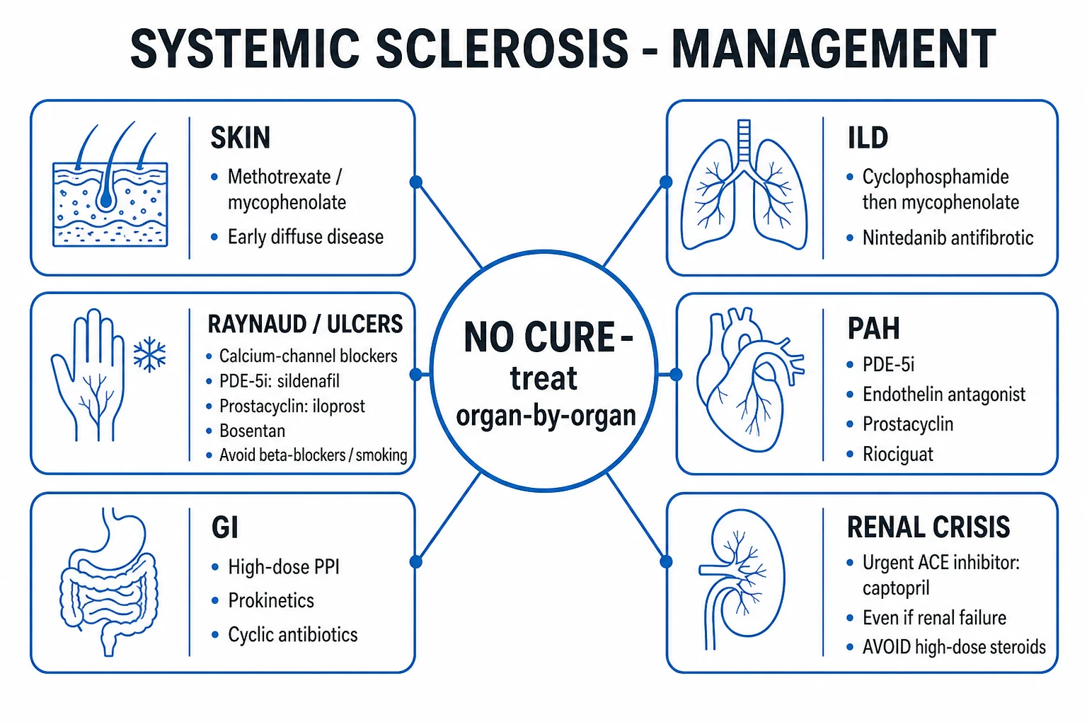
Systemic sclerosis is not an ABCDE-resuscitation disease in the classic sense, but several acute, time-critical scenarios demand immediate action. The two most important are scleroderma renal crisis (urgent ACE inhibitor) and severe Raynaud/digital ischaemia (urgent vasodilation). [1]
Acute scenarios that must be addressed before chronic therapy: [1]
-
Scleroderma renal crisis — urgent ACE inhibitor. New-onset severe (malignant) hypertension with acute kidney injury and microangiopathic haemolytic anaemia in early diffuse SSc is renal crisis until proven otherwise. Start an ACE inhibitor immediately — captopril 6.25-12.5 mg PO, titrated rapidly up to 25-50 mg three times daily — even if the creatinine is rising and even if dialysis is needed. The ACE inhibitor is life-saving and must not be withheld for fear of worsening AKI; expect the creatinine to worsen before it improves. Add an angiotensin-receptor blocker (ARB, e.g., losartan) only if ACE inhibitor alone is insufficient. Avoid intravenous vasodilators that worsen renal perfusion. Renal replacement therapy (haemodialysis) is often needed temporarily and may be recoverable. AVOID high-dose corticosteroids (over 15 mg/day prednisolone equivalent) — they precipitate and worsen renal crisis.[9]
-
Acute severe Raynaud / critically ischaemic digit. Admit, warm the patient, give intravenous prostacyclin (iloprost 0.5-2 ng/kg/min over 6 hours daily for 3-5 days), add or escalate oral vasodilators (calcium-channel blocker plus PDE-5 inhibitor), provide adequate analgesia (the pain of digital ischaemia is severe), consider a digital sympathetic block or botulinum toxin injection, and involve vascular surgery for a critically ischaemic digit. Treat or exclude infection in an ulcerated digit. [1]
-
Infected digital ulcer. Send wound swab and blood cultures; treat with antistreptococcal/antistaphylococcal antibiotics (e.g., flucloxacillin 500 mg PO QDS or IV; consider MRSA cover with vancomycin if risk factors); debride necrotic tissue; escalate vasodilators as above; surgical review for osteomyelitis or amputation. [1]
-
Severe PAH with right heart failure. Oxygen, diuretics (furosemide), refer to a specialist pulmonary hypertension centre, consider parenteral prostanoids (epoprostenol IV) and PAH-specific combination therapy. [1]
-
Acute severe ILD with respiratory failure. Oxygen, treat superimposed infection, escalate immunosuppression (cyclophosphamide or rituximab), consider antifibrotic therapy (nintedanib) and lung transplant referral. [1]
Management — Definitive & Stepwise
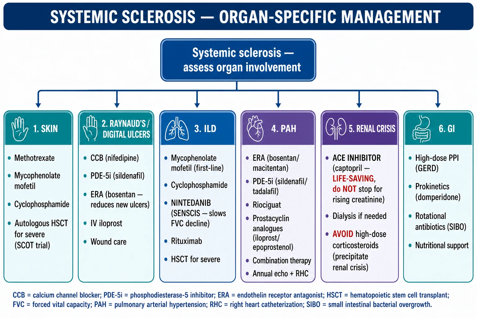
There is no cure for systemic sclerosis. Therapy is organ-targeted and stratified by subtype and antibody, using the 2023 EULAR update as the current framework.[7] No therapy reverses established fibrosis; the goals are to control vascular disease, suppress immune-driven progression, protect organs, and manage complications.
Skin disease
For early diffuse cutaneous disease with rapidly progressive skin thickening (rising mRSS), immunosuppression is offered to attenuate progression:[7]
- Methotrexate 10-25 mg weekly PO/SC (with folic acid) — evidence modest; useful when inflammatory arthritis or myositis coexist.
- Mycophenolate mofetil (MMF) 1 g BD, titrated to 2-3 g/day — widely used, also for ILD.
- Cyclophosphamide — used more for ILD; rarely for skin alone.
- D-penicillamine — historically used as an antifibrotic; now largely abandoned because of toxicity and lack of efficacy.
- Autologous haematopoietic stem cell transplant (AHSCT) — for selected patients with early, severe, rapidly progressive dcSSc unresponsive to standard immunosuppression (see Evidence section).
- Physiotherapy, hand therapy and contracture prevention — essential to preserve function; UVA1 phototherapy is useful for localised skin disease and morphoea. [1]
Raynaud phenomenon and digital ulcers — the stepwise ladder
Management of Raynaud and digital ulceration is stepwise, escalating from lifestyle measures through oral vasodilators to intravenous therapy:[4][7]
Raynaud / digital ulcer — stepwise ladder
AVOID beta-blockers (worsen peripheral vasospasm) and sympathomimetic decongestants. Smoking cessation is non-negotiable. Statins and low-dose aspirin are often added for antiplatelet and endothelial effects, though evidence is modest. [1]
Gastrointestinal management
- Gastro-oesophageal reflux and dysmotility — high-dose proton-pump inhibitor (omeprazole 20-40 mg OD, pantoprazole, esomeprazole) plus a prokinetic (metoclopramide 10 mg TDS, domperidone 10 mg TDS; macrolides such as erythromycin for refractory gastroparesis). Consider fundoplication cautiously — gastroparesis may worsen post-fundoplication.
- Strictures — endoscopic dilation.
- SIBO and malabsorption — cyclic rotating antibiotics (e.g., rifaximin 550 mg BD for 2 weeks, alternating with doxycycline, metronidazole or ciprofloxacin), nutritional support (elemental diet, PEG/jejunostomy in severe cases), vitamin and mineral replacement (especially vitamin D, B12, iron, calcium).
- GAVE (watermelon stomach) — endoscopic argon plasma coagulation; repeated sessions as needed; rarely surgery.
- PBC overlap — ursodeoxycholic acid 13-15 mg/kg/day. [1]
Interstitial lung disease
The goal is to attenuate progression of the inflammatory component and slow fibrosis:[7][8]
- Mycophenolate mofetil 2-3 g/day — first-line for SSc-ILD, non-inferior to cyclophosphamide with less toxicity (Scleroderma Lung Study II).
- Cyclophosphamide 600 mg/m2 IV monthly for 6 months, then maintenance with MMF — for progressive or severe disease (Scleroderma Lung Study I).
- Rituximab 1 g IV days 0 and 14 (repeat 6-monthly) — for refractory or rapidly progressive disease; depletes B cells.
- Nintedanib 150 mg BD PO — an antifibrotic tyrosine-kinase inhibitor that slows the rate of FVC decline in SSc-ILD (SENSCIS trial); now widely used alongside immunosuppression. Monitor LFTs; the main side-effects are diarrhoea and transaminitis.
- Lung transplantation for end-stage disease in selected patients.
- Vaccination (pneumococcal, influenza, COVID-19) and prompt treatment of infection; prophylaxis against Pneumocystis jirovecii when on cyclophosphamide or rituximab plus steroids. [1]
Pulmonary arterial hypertension
PAH therapy is specialist-led and now uses upfront combination therapy targeting the three key pathways:[9]
- Endothelin-receptor antagonists (ERAs) — bosentan 62.5-125 mg BD; ambrisentan 5-10 mg OD; macitentan 10 mg OD. Monitor LFTs (bosentan) and haemoglobin; contraindicated in pregnancy.
- PDE-5 inhibitors — sildenafil 20 mg TDS or 20-80 mg TDS; tadalafil 40 mg OD.
- Soluble guanylate cyclase stimulator — riociguat 1.0-2.5 mg TDS (note: riociguat is contraindicated in SSc-ILD because of increased risk of respiratory hospitalisation, and must not be combined with PDE-5 inhibitors).
- Prostacyclin analogues — inhaled iloprost, subcutaneous/inhaled treprostinil, intravenous epoprostenol (the latter for severe WHO functional class IV disease).
- Supportive therapy — oxygen to maintain saturation over 90 percent, diuretics for right heart failure, oral anticoagulation in selected severe cases, digoxin, avoidance of pregnancy, pneumococcal, influenza and COVID-19 vaccination.
- Lung transplantation for refractory disease. [1]
Scleroderma renal crisis — the ACE-inhibitor rule
Scleroderma renal crisis is treated with an ACE inhibitor — start immediately on suspicion:[9]
- Captopril 6.25-12.5 mg PO, titrated rapidly to 25-50 mg TDS (the short half-life allows precise titration in the unstable patient).
- Switch to a longer-acting ACE inhibitor (e.g., lisinopril, ramipril) once stable.
- Add an angiotensin-receptor blocker (ARB) if ACE inhibitor alone insufficient.
- Renal replacement therapy (haemodialysis) as required — many patients recover sufficient renal function to come off dialysis over weeks to months; ACE inhibitor should be continued indefinitely even after recovery.
- AVOID high-dose corticosteroids (over 15 mg/day prednisolone equivalent) — they precipitate renal crisis.
- Treat the malignant hypertension — slow, controlled BP reduction (not abrupt), with intravenous labetalol or nicardipine if oral therapy insufficient (avoid aggressive overshoot).
- Plasma exchange and eculizumab have been tried in atypical or refractory cases. [1]
Musculoskeletal and other organ therapy
- NSAIDs (caution renal function and GI risk) for arthralgia; low-dose steroids (under 15 mg/day — and avoid if possible, especially in early dcSSc); methotrexate or MMF for inflammatory arthritis/myositis; physiotherapy for contractures.
- Diuretics and ACE inhibitor for heart failure; treat arrhythmia; pericardiocentesis for tamponade.
- Intravenous immunoglobulin may help refractory myositis and is steroid-sparing. [1]
Autologous haematopoietic stem cell transplant (AHSCT)
For selected patients with early (under 2-4 years), severe, rapidly progressive dcSSc unresponsive to standard immunosuppression, AHSCT improves long-term event-free and overall survival compared with cyclophosphamide alone, at the cost of significant early treatment-related mortality (around 5-10 percent at one year). The ASTIS and SCOT trials underpin this option; it is offered only in specialist transplant centres and is contraindicated in advanced organ failure (severe PAH, severe ILD, renal crisis, cardiac involvement) where the toxicity is unjustified.[7]
Specific Subtypes & Scenarios
- Limited cutaneous SSc (lcSSc/CREST) — onset long after Raynaud; skin confined to face and distal to elbows/knees; anti-centromere; slow course; threat of late-onset PAH after 10-20 years; PBC overlap; better prognosis than dcSSc.
- Diffuse cutaneous SSc (dcSSc) — onset of skin within 1 year of Raynaud; skin proximal to elbows/knees and trunk; anti-Scl-70 or anti-RNA polymerase III; rapid early skin peak (mRSS peak 1-3 years, then often softening); risk of early ILD, renal crisis and cardiac disease; tendon friction rubs common; worse prognosis.
- SSc sine scleroderma — PAH, ILD or GI disease with SSc-specific antibodies and capillaroscopy changes but no skin thickening; often presents late with isolated PAH or ILD.
- Scleroderma renal crisis — malignant hypertension + AKI + microangiopathic haemolytic anaemia; over 80 percent in anti-RNA polymerase III or early diffuse disease; precipitated by high-dose steroids; treated by ACE inhibitor.
- SSc-ILD — commonest cause of death (over one third); NSIP predominant; anti-Scl-70 association; screened by PFTs and HRCT; treated by MMF/cyclophosphamide/rituximab and nintedanib.[8]
- SSc-PAH — commonest in lcSSc; screened annually by echo and PFTs; confirmed by right heart catheter; treated with combination PAH therapy.
- Overlap syndromes — SSc with myositis (anti-PM/Scl), SLE, RA, or Sjogren; MCTD (anti-U1-RNP).
- Localised scleroderma (morphea, linear scleroderma, en coup de sabre, Parry-Romberg) — skin and underlying tissue only, no internal organ involvement, no Raynaud; treated with topical/intralesional steroid, UVA1 phototherapy and methotrexate for active or linear disease.
Complications & Pitfalls
Life-threatening complications include interstitial lung disease (the commonest cause of death), pulmonary arterial hypertension (second commonest), scleroderma renal crisis (the classical emergency), cardiac involvement (myocardial fibrosis, arrhythmia, sudden death), severe GI malabsorption and malnutrition, and digital amputation.[9]
Gastrointestinal complications — refractory GAVE bleeding, oesophageal stricture, SIBO and malabsorption (with vitamin D, B12, iron, calcium deficiency), intestinal pseudo-obstruction, pneumatosis intestinalis, Barrett's oesophagus with adenocarcinoma, and PBC overlap. [1]
Digital complications — ischaemic ulcers, osteomyelitis, gangrene and auto-amputation. [1]
The cancer association — anti-RNA polymerase III is associated with synchronous solid malignancy (especially breast and lung); cancer screening is recommended at diagnosis and for 2-5 years thereafter. [1]
The course of diffuse cutaneous SSc
Classic pitfalls: [1]
- Missing renal crisis because the ACE inhibitor was withheld fearing AKI — the creatinine will rise before it falls; do not stop the ACE inhibitor.
- Precipitating renal crisis with high-dose corticosteroids (over 15 mg/day prednisolone equivalent).
- Missing ILD because PFTs were not done — annual PFTs and HRCT for any decline.
- Missing PAH because the echo looked normal — a low isolated DLCO and raised FVC/DLCO ratio are screening red flags; right heart catheter confirms.
- Labelling NSF or eosinophilic fasciitis as SSc — gadolinium exposure and the groove sign discriminate.
- Giving beta-blockers for Raynaud — they worsen peripheral vasospasm.
- Overlooking malnutrition — weight loss in SSc is common, multifactorial and under-treated.
- Forgetting the cancer association in anti-RNA polymerase III patients. [1]
Prognosis & Disposition
Systemic sclerosis has the highest case fatality of any rheumatic disease. Overall 10-year survival is approximately 60-70 percent, with a substantial survival gap between subtypes: limited cutaneous disease approximately 70-80 percent and diffuse cutaneous disease approximately 50-60 percent at 10 years.[9]
SSc — survival and prognosis
Poor-prognosis markers: diffuse subtype, older age at onset, male sex, diffuse early skin thickening with a high mRSS, anti-Scl-70 or anti-RNA polymerase III, tendon friction rubs, elevated ESR/CRP, low DLCO (under 60 percent predicted), early ILD, renal crisis, and cardiac involvement.[9]
Disposition: most patients are managed by a rheumatology-led multidisciplinary team in close partnership with pulmonology, cardiology/pulmonary hypertension, nephrology, gastroenterology, dermatology, hand therapy, dietetics and psychology. Annual pulmonary function tests and echocardiography are the backbone of organ surveillance. Patients should be admitted for renal crisis, severe PAH with right heart failure, respiratory failure from ILD, an acutely ischaemic digit, or severe malnutrition. A named rheumatology nurse and flare plan is the safety net. [1]
Special Populations
- Pregnancy — pregnancy in SSc is higher risk than in the general population, but is generally feasible in stable disease without severe organ involvement. AVOID ACE inhibitors (fetopathy — renal agenesis, oligohydramnios, neonatal hypotension). Pulmonary arterial hypertension is a contraindication to pregnancy (maternal mortality up to 30-50 percent). Early diffuse disease (within the first 4 years) carries a higher risk of renal crisis. Multidisciplinary perinatal care (rheumatology, obstetric medicine, anaesthesia) is essential; regional anaesthesia preferred (microstomia and skin thickening complicate intubation). Disease may flare postpartum. Drugs compatible with pregnancy include low-dose steroids, hydroxychloroquine, azathioprine, intravenous immunoglobulin and (sometimes) sulfasalazine.[9]
- Paediatric / localised scleroderma — linear scleroderma in children causes growth arrest of bone and soft tissue (limb-length discrepancy, facial hemiatrophy / Parry-Romberg syndrome); MRI to monitor deep involvement; methotrexate and systemic steroids for active disease; physiotherapy and occupational therapy.
- Elderly male with anti-RNA polymerase III — high risk of renal crisis within months of onset; BP surveillance; age-appropriate malignancy screening.
- Overlap syndromes and MCTD — tailor immunosuppression to the dominant feature (myositis, ILD, arthritis).
- Pre-operative assessment — assess airway (microstomia, limited neck movement), cardiac and pulmonary status; avoid hypothermia (worsens Raynaud); avoid vasopressors that worsen digital ischaemia; regional anaesthesia where possible; careful BP control (renal crisis risk); PPI cover for reflux under anaesthesia.
- Renal impairment — avoid NSAIDs and nephrotoxins; avoid high-dose steroids; adjust methotrexate dose; the ACE inhibitor is still required for renal crisis even in established CKD.
- Cancer-associated SSc (anti-RNA polymerase III) — age-appropriate malignancy screening at diagnosis and for 2-5 years thereafter (breast, lung, haematological).
Evidence, Guidelines & Regional Differences
Landmark shifts in classification and management: [1]
- 2013 ACR/EULAR classification criteria (replacing the 1980 criteria) — more sensitive to early disease; skin thickening proximal to MCP is sufficient alone (9 points).[5]
- EULAR 2023 update for the treatment of systemic sclerosis — the current European framework: prostacyclin analogues for severe Raynaud and digital ulcers; bosentan for the prevention of new digital ulcers; PDE-5 inhibitors for digital ulcers; MMF or cyclophosphamide for SSc-ILD; riociguat contraindicated in SSc-ILD; AHSCT for selected severe early dcSSc.[7]
Landmark trials: [1]
- Scleroderma Lung Study I (SLS I) — oral cyclophosphamide modestly improved FVC and skin score vs placebo in SSc-ILD.
- Scleroderma Lung Study II (SLS II) — mycophenolate mofetil non-inferior to cyclophosphamide with less toxicity and better tolerability, establishing MMF as first-line.
- SENSCIS — nintedanib slows the rate of FVC decline in SSc-ILD (approximately 40 ml/year benefit).[8]
- ASTIS and SCOT — autologous haematopoietic stem cell transplant improves long-term event-free and overall survival vs cyclophosphamide, at the cost of significant early treatment-related mortality.[7]
- DETECT — a two-step screening algorithm for PAH in high-risk SSc, more sensitive than echo alone.
EULAR (Europe, 2023 update) — the current overarching framework: organ-targeted therapy; MMF first-line for SSc-ILD; prostacyclin analogues and PDE-5 inhibitors for Raynaud/digital ulcers; bosentan to prevent new ulcers; AHSCT for selected severe early dcSSc; ACE inhibitor for renal crisis.[7]
EUSTAR (EULAR Scleroderma Trials and Research) is the major international observational registry that has defined organ involvement patterns, prognosis and treatment response in thousands of patients, and underpins much of the modern evidence base.[1]
Vaccination in immunosuppressed SSc: pneumococcal (PCV15/20 then PPSV23), annual influenza, COVID-19, hepatitis B, recombinant zoster (Shingrix) — give before starting biologics where possible; avoid live vaccines once on significant immunosuppression. [1]
Exam Pearls
- Raynaud is the FIRST manifestation in over 90 percent of SSc; Raynaud + puffy fingers + abnormal nailfold capillaroscopy + SSc-specific antibody = very early SSc (VEDOSS).
- CREST = Calcinosis, Raynaud, oEsophageal dysmotility, Sclerodactyly, Telangiectasia (limited cutaneous phenotype).
- Anti-centromere = limited/CREST/PAH; anti-Scl-70 (topoisomerase I) = diffuse/ILD; anti-RNA polymerase III = renal crisis + cancer-associated.
- ILD is the commonest cause of death; screen with PFTs (FVC, DLCO) and HRCT.
- Scleroderma renal crisis: malignant hypertension + AKI; URGENT ACE inhibitor (captopril); AVOID high-dose steroids (over 15 mg/day).
- Nailfold capillaroscopy scleroderma pattern = giant capillaries + microhaemorrhages + dropout.
- Modified Rodnan skin score (mRSS): grades 0-3 across 17 sites, max 51 — the validated skin-thickening outcome.
- Bosentan (endothelin-receptor antagonist) is licensed to PREVENT NEW digital ulcers (not to heal existing ones).
- PAH: FVC/DLCO ratio over 1.6 and low isolated DLCO are screening red flags; confirm by right heart catheter.
- Nintedanib slows FVC decline in SSc-ILD (SENSCIS); riociguat is contraindicated in SSc-ILD.
- AVOID beta-blockers in SSc (worsen Raynaud); smoking cessation is essential.
- AHSCT for selected severe early dcSSc (ASTIS, SCOT) — significant early mortality but improved long-term survival.
- Female predominance 4:1; peak onset 30-50 years; highest case fatality of any rheumatic disease.
- NSF (gadolinium in CKD), eosinophilic fasciitis (groove sign, eosinophilia) and morphea (skin only) are the classic mimics.
- GAVE (watermelon stomach) is the leading cause of GI bleeding in SSc; treat with endoscopic argon plasma coagulation. [1]
Exam application bank (NEET-PG / INICET)
One-line answer
Systemic sclerosis (scleroderma) is a chronic multisystem autoimmune connective tissue disease defined by a pathophysiological triad of microvascular vasculopathy, immune dysregulation and generalised fibrosis (excess collagen deposition) of the skin and internal organs, with disease-specific autoantibodies (anti-centromere, anti-Scl-70/topoisomerase I, anti-RNA polymerase III). The two principal subtypes are limited cutaneous (CREST — Calcinosis, Raynaud, oEsophageal dysmotility, Sclerodactyly, Telangiectasia; anti-centromere; late pulmonary arterial hypertension) and diffuse cutaneous (anti-Scl-70 or anti-RNA polymerase III; early interstitial lung disease, renal crisis, rapidly progressive skin). Raynaud phenomenon is the first manifestation in over 90 percent. It is the rheumatic disease with the highest case fatality — interstitial lung disease and pulmonary arterial hypertension ar
Worked stems (answer without another resource)
Stem 1 — Classic presentation. Map symptoms to mechanism; name the first investigation and first treatment step with dose/route if drug therapy is standard. [1]
Stem 2 — Unstable / complicated. List red flags that force immediate resuscitation, theatre, ICU, antidote, or reperfusion — and what you do in the first 15 minutes. [1]
Stem 3 — Atypical group. Elderly, pregnancy, child, or immunocompromised: how presentation and thresholds change. [1]
Stem 4 — Differential trap. Name the three closest mimics and one discriminator for each. [1]
Stem 5 — Disposition. Who goes home with safety-netting, who is admitted, who needs HDU/ICU/theatre, and what follow-up is mandatory. [1]
Rapid viva checklist
- Definition + classification
- Pathophysiology chain
- Bedside signs / criteria
- Score with exact components (if any)
- Emergency bundle
- Definitive therapy with doses
- Complications of disease and of treatment
- Special populations
- Guideline/trial name if classic
- Three exam traps
Coverage self-check
If you cannot answer any stem above from this page alone, re-read the matching section — the page is intended to be self-sufficient for final-prof and NEET-PG/INICET questions on Systemic Sclerosis (Scleroderma).
References
- [1]Di Battista M, Lepri G, Codullo V, et al. Systemic sclerosis: one year in review 2025 Clin Exp Rheumatol, 2025.PMID 40737082
- [2]Jerjen R, Nikpour M, Krieg T, et al. Systemic sclerosis in adults. Part I: Clinical features and pathogenesis J Am Acad Dermatol, 2022.PMID 35131402
- [3]Rosendahl AH, Schonborn K, Krieg T. Pathophysiology of systemic sclerosis (scleroderma) Kaohsiung J Med Sci, 2022.PMID 35234358
- [4]Herrick AL, Wigley FM. Raynaud's phenomenon Best Pract Res Clin Rheumatol, 2020.PMID 32007400
- [5]van den Hoogen F, Khanna D, Fransen J, et al. 2013 classification criteria for systemic sclerosis: an American college of rheumatology/European league against rheumatism collaborative initiative Ann Rheum Dis, 2013.PMID 24092682
- [6]Smith V, Ickinger C, Hysa E, et al. Nailfold capillaroscopy Best Pract Res Clin Rheumatol, 2023.PMID 37419757
- [7]Del Galdo F, Lescoat A, Conaghan PG, et al. EULAR recommendations for the treatment of systemic sclerosis: 2023 update Ann Rheum Dis, 2025.PMID 39874231
- [8]Maher TM. Interstitial Lung Disease: A Review JAMA, 2024.PMID 38648021
- [9]Denton CP, Khanna D. Systemic sclerosis Lancet, 2017.PMID 28413064