EM · Ectopic pregnancy
Ectopic pregnancy
Also known as Tubal pregnancy · Ruptured ectopic · Extrauterine pregnancy
The ectopic pregnancy — the implantation outside the uterine cavity, most commonly in the fallopian tube; the rupture causes the catastrophic intra-abdominal haemorrhage and is the leading cause of the pregnancy-related death in the first trimester. The clinical presentation (the abdominal pain, the vaginal bleeding, the missed period, the shoulder-tip pain from the diaphragmatic irritation, the collapse), the risk factors (the PID, the previous ectopic, the IVF, the tubal surgery, the IUD), the diagnosis (the quantitative beta-hCG, the transvaginal ultrasound), the management (the resuscitation, the surgical salpingectomy for the ruptured, the methotrexate for the stable unruptured). ACEM-primary, globally tagged.
On this page & tools
Your progress
Saved locally on this device.
Target exams
Red flags
Related topics
The ectopic pregnancy is the implantation of the fertilised ovum outside the uterine cavity, most commonly in the ampulla of the fallopian tube, and the rupture of the tubal ectopic is one of the most time-critical emergencies in the emergency department — the patient who is bleeding internally can exsanguinate within minutes. The Fellowship candidate must know the clinical presentation (every woman of reproductive age with abdominal pain has an ectopic until the beta-hCG is negative or the intrauterine pregnancy is confirmed), the diagnostic pathway (the beta-hCG, the transvaginal US, the discriminatory zone), and the management (the resuscitation, the surgical salpingectomy for the ruptured, the methotrexate for the stable unruptured).[1][2]
Definition and the epidemiology
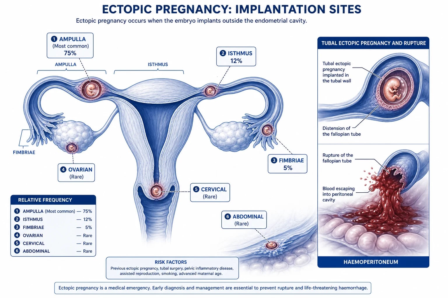
The ectopic pregnancy is the implantation of the fertilised ovum outside the uterine cavity. The tubal ectopic (the ampulla, the isthmus, the fimbria, the interstitial/cornual) accounts for over 95 per cent of all ectopics. The non-tubal sites include the ovary, the cervix, the Caesarean scar, and the abdomen. The incidence is approximately 1 to 2 per cent of all pregnancies, and it is the leading cause of the pregnancy-related death in the first trimester. The risk factors include the previous ectopic (the recurrence risk is 10-fold), the pelvic inflammatory disease (the chlamydia, the gonorrhoea), the tubal surgery (the sterilisation, the tubal reconstruction), the assisted reproduction (the IVF), the intrauterine device (the IUD prevents the intrauterine pregnancy but does not prevent the ectopic), the smoking, and the maternal age over 35. [1]
The clinical presentation
The classic triad is the abdominal pain, the vaginal bleeding, and the amenorrhoea (the missed period). The pain is typically unilateral, lower abdominal, and may be severe if the ectopic has ruptured. The shoulder-tip pain (the referred pain to the shoulder tip from the diaphragmatic irritation by the free intra-peritoneal blood) is the sign of the significant haemoperitoneum. The vaginal bleeding is typically light and dark (the "prune juice"). The collapse or the dizziness on standing indicates the significant blood loss. The examination may show the pallor, the tachycardia, the hypotension (the signs of the hypovolaemic shock), the abdominal tenderness (the lower quadrant, the rebound, the guarding), the cervical motion tenderness (the "chandelier sign"), the adnexal mass or fullness, and the pallor of the vaginal fornices (from the haemoperitoneum). [1]
Differential diagnosis — the woman of reproductive age with abdominal pain
Ectopic pregnancy
- Abdominal pain, vaginal bleeding, missed period; the positive beta-hCG
- The empty uterus on the TVS with the free fluid; the adnexal mass
- The resuscitation, the surgery (ruptured) or the methotrexate (stable)
- The leading cause of the first-trimester maternal death
Miscarriage
- Vaginal bleeding, the cramping; the open cervical os
- The intrauterine pregnancy on the US (or the retained products)
- The expectant, the medical (misoprostol), or the surgical (ERPC) management
- The stable patient unless the heavy bleeding
Ovarian cyst
- The rupture or the torsion; the sudden unilateral pain
- The US shows the cyst; the beta-hCG is negative
- The analgesia, the observation; the surgery for the torsion
- The follicular or the corpus luteum cyst
PID
- Bilateral lower abdominal pain, the fever, the discharge
- The high vaginal swab; the raised WCC and CRP
- The ceftriaxone 500 mg IM plus doxycycline 100 mg BD plus metronidazole 400 mg BD
- The tubo-ovarian abscess in the severe case
Appendicitis
- The right lower quadrant pain, the fever, the nausea
- The normal beta-hCG; the US or the CT shows the appendix
- The surgical referral; the appendicectomy
- The most common surgical emergency in pregnancy
The investigations
The urine or serum beta-hCG is the first test — every woman of reproductive age with abdominal pain has a pregnancy test. A negative urine beta-hCG effectively excludes the ectopic (except in the very early pregnancy with the beta-hCG below the detection threshold, or in the rare hook effect of the very high level). The quantitative serum beta-hCG is used for the discriminatory zone: a beta-hCG over 1500 IU/L with an empty uterus on the transvaginal US is the ectopic until proven otherwise. The transvaginal ultrasound is the diagnostic test — it identifies the intrauterine pregnancy (the gestational sac with the yolk sac at 5 to 6 weeks), the adnexal mass (the complex mass separate from the ovary), the free fluid in the pouch of Douglas (the blood from the rupture), and the rare interstitial or cervical ectopic. The full blood count (the haemoglobin, the WCC), the group and hold (or the crossmatch 4 units if the rupture is suspected), the blood gas (the lactate, the base deficit for the occult shock), and the bloods (the U&E, the LFTs, the coagulation) complete the workup.[1]
Management — the drug doses and the approach
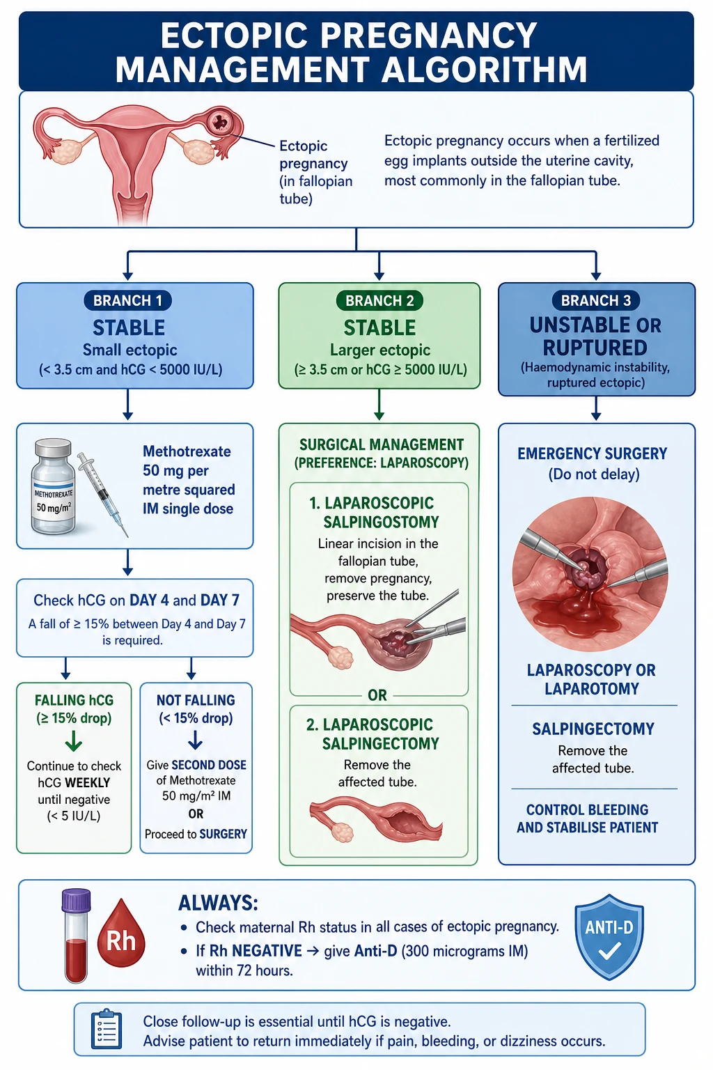
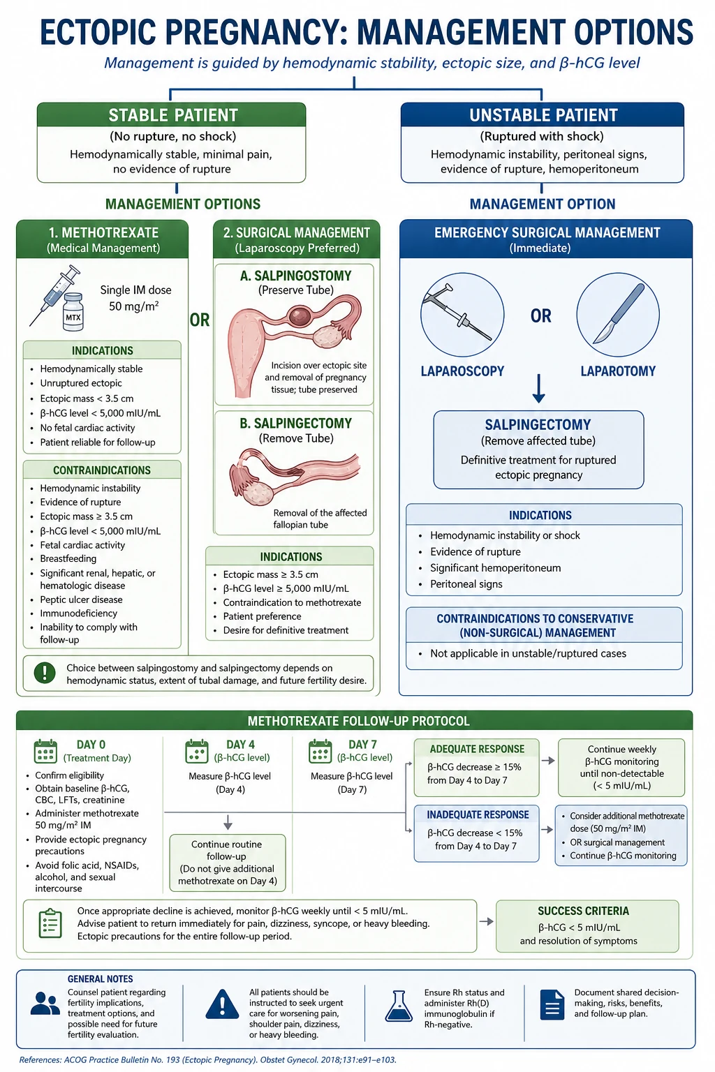
The management depends on the haemodynamic stability and the findings on the ultrasound. [1]
[1]The ectopic drug doses
The methotrexate and the medical management
The methotrexate is the folate antagonist that inhibits the trophoblastic cell division. It is given as a single intramuscular injection of 50 mg per metre squared and is effective in approximately 85 to 90 per cent of the appropriately selected patients. The selection criteria for the methotrexate: the haemodynamically stable patient, the beta-hCG under 5000 IU/L (the success rate falls with the higher levels), the ectopic mass under 3.5 cm, the absence of the fetal cardiac activity, the no contraindication to the methotrexate (the renal impairment, the hepatic impairment, the thrombocytopenia, the breastfeeding, the peptic ulcer), and the reliable patient who returns for the follow-up. The follow-up: the beta-hCG on day 4 and day 7 (the day 7 should be lower than the day 4), then weekly until the zero. The methotrexate failure (the rising beta-hCG, the rupture) is the indication for the surgery.[2]
The surgical management
The laparoscopic salpingectomy (the removal of the affected tube) is the standard operation for the ruptured ectopic and for the methotrexate failure. The laparoscopic salpingostomy (the linear incision on the tube, the evacuation of the ectopic, the preservation of the tube) is used when the contralateral tube is diseased and the fertility preservation is the priority. The laparotomy is reserved for the haemodynamically unstable patient who cannot tolerate the pneumoperitoneum of the laparoscopy. The interstitial (cornual) ectopic and the cervical ectopic require the specialised surgical approach and may need the uterine artery embolisation or the hysterectomy. [1]
Complications and prognosis
The complications are the haemorrhagic shock (from the tubal rupture, the intra-abdominal bleeding — the life-threatening emergency that requires the simultaneous resuscitation and the surgical control), the recurrence (the 10-fold increased risk of the ectopic in the future pregnancy — the patient is counselled to present early for the ultrasound in the next pregnancy), the infertility (from the tubal damage, the adhesions, and the salpingectomy — the subfertility rate after one ectopic is 30 to 40 per cent), the infection (the post-operative pelvic infection), the Rh sensitisation (the Rh-negative mother needs the anti-D immunoglobulin 500 IU IM within 72 hours to prevent the haemolytic disease of the newborn in the future pregnancy), the persistent trophoblast (the residual trophoblastic tissue after the salpingostomy that produces the beta-hCG — treated with the methotrexate), and the psychological impact (the grief, the anxiety about the future fertility, and the depression — the patient is offered the counselling and the follow-up). The prognosis is good if the ectopic is diagnosed before the rupture; the mortality is 0.2 per cent (the leading cause of the first-trimester maternal death). The methotrexate success rate is 85 to 90 per cent in the appropriately selected patient; the failure is the indication for the surgery. [1]
Special populations
The IVF pregnancy has a higher ectopic rate (2 to 5 per cent) and the heterotopic pregnancy (the simultaneous intrauterine and the ectopic) occurs at 1 in 100 — the intrauterine pregnancy does NOT exclude the ectopic in the IVF patient with the abdominal pain. The IUD user who becomes pregnant has a 5 per cent ectopic rate — the pregnancy with the IUD in situ is the ectopic until proven otherwise. The previous Caesarean section patient is at a higher risk of the Caesarean scar ectopic (the implantation in the scar of the previous Caesarean — a high-risk ectopic that may cause the catastrophic uterine rupture). [1]
Evidence and regional guidelines
The contemporary framework is the NICE ectopic pregnancy guideline and the RCOG green-top guideline for the diagnosis and the management.[1][2]
ANZ practice note. The ectopic pregnancy is managed with the beta-hCG, the transvaginal US, the surgical referral for the ruptured (the laparoscopic salpingectomy), and the methotrexate 50 mg/m squared IM for the stable unruptured. The anti-D 500 IU IM is given to the Rh-negative mother. The early surgical referral and the simultaneous resuscitation are the standard for the ruptured ectopic. [1]
Pathophysiology and the natural history
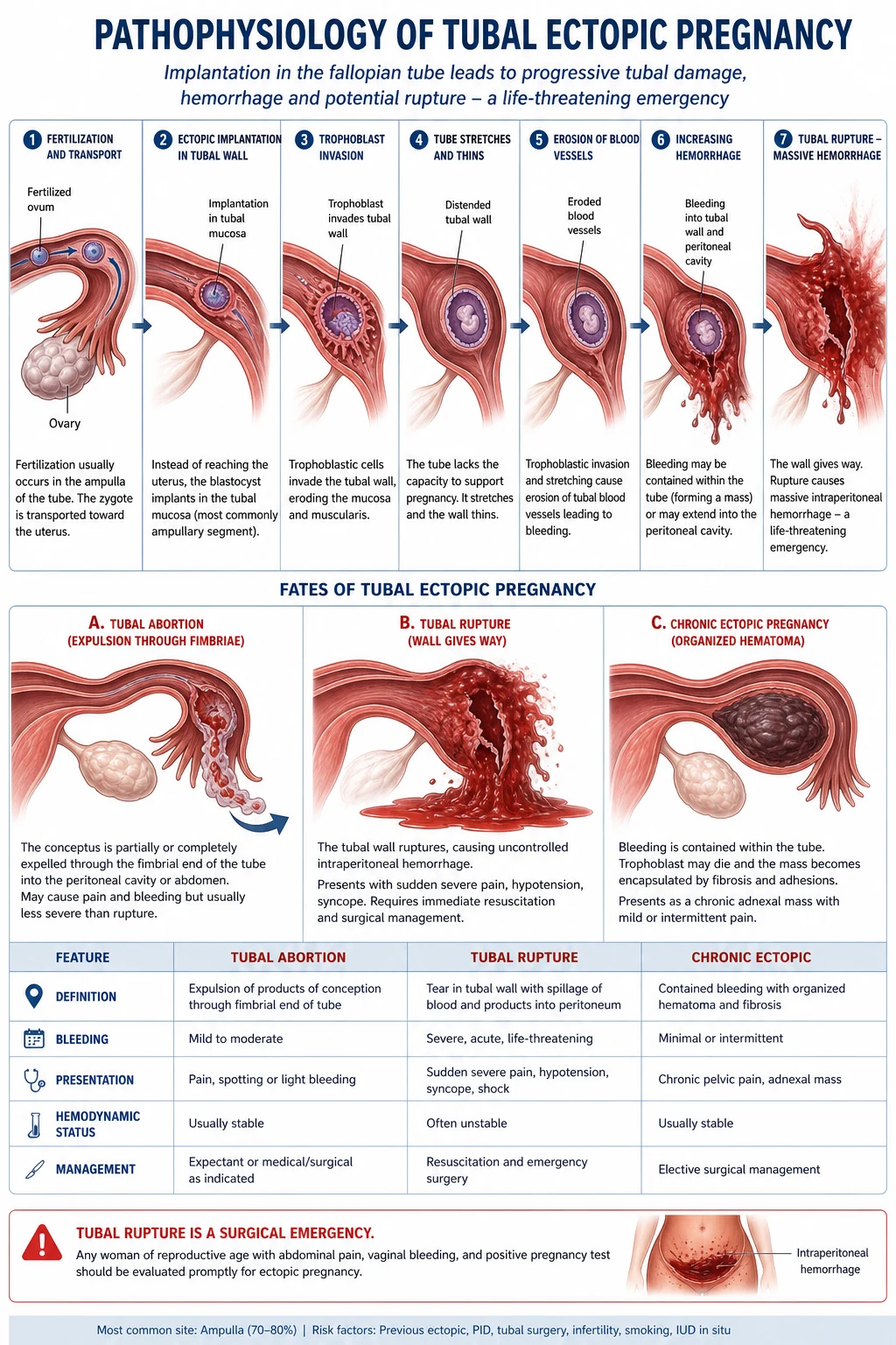
The fertilised ovum normally implants in the endometrial lining of the uterine cavity six to seven days after the fertilisation. In the ectopic pregnancy the embryo implants in a site that cannot accommodate the growing trophoblast — most often the tubal mucosa, which lacks the decidual reaction that normally limits the trophoblastic invasion. The trophoblast therefore erodes the tubal wall directly into the muscularis and the vasculature. The natural history follows three trajectories: the tubal rupture (the most dangerous — the erosion into the tubal branch of the uterine artery produces the catastrophic haemoperitoneum, often 1 to 2 litres within minutes), the tubal abortion (the expulsion of the conceptus through the fimbrial end into the peritoneal cavity, producing the self-limiting bleeding and the cul-de-sac haematocele), and the spontaneous resolution (the resorption of a small, non-viable pregnancy with the falling beta-hCG — the basis of the expectant management). The ampullary ectopic (the commonest site, 80 per cent) tends to rupture later at 8 to 12 weeks because the ampulla is the widest and most distensible segment; the isthmic ectopic (12 per cent) ruptures earlier at 6 to 8 weeks because the isthmus is the narrowest and least distensible segment and has the highest concentration of the vasculature; the interstitial (cornual) ectopic (2 per cent) ruptures latest at 12 to 16 weeks but produces the most catastrophic haemorrhage because it implants in the highly vascular interstitial portion of the tube within the muscular wall of the uterus.[8]
Ampullary (80%)
- The commonest tubal site; the widest, most distensible segment
- Ruptures later — 8 to 12 weeks
- The moderate haemorrhage; the laparoscopic salpingectomy is the standard
- The best tubal-preservation outcomes after the salpingostomy
Isthmic (12%)
- The narrowest segment; the least distensible; the richest vasculature
- Ruptures early — 6 to 8 weeks
- The brisk haemorrhage; the high risk of the rapid exsanguination
- The segmental salpingectomy often required
Interstitial/cornual (2%)
- Implants within the muscular uterine wall; the late presentation
- Ruptures late — 12 to 16 weeks but the catastrophic haemorrhage
- The uterine artery branch erosion; the cornual resection or the hysterectomy
- The highest mortality of all ectopic sites
Cervical (<1%)
- Implants in the cervical canal; the painless heavy vaginal bleeding
- The medical management with the methotrexate or the uterine artery embolisation
- The dilatation and curettage is CONTRAINDICATED — the catastrophic bleeding
- The high risk of the hysterectomy for the haemorrhage control
Caesarean scar (<1%)
- Implants in the scar of the previous Caesarean; the rising incidence
- The catastrophic uterine rupture risk; the placenta accreta overlap
- The systemic plus the local methotrexate; the uterine artery embolisation
- The high risk of the hysterectomy
The diagnostic algorithm — the woman of reproductive age with abdominal pain or bleeding
The ectopic pregnancy diagnostic pathway
- The pregnancy test first. Every woman of reproductive age (the menarche to the menopause, including the tubal ligation and the IUD patient) with the abdominal pain, the vaginal bleeding, the syncope, or the gastrointestinal symptoms has a urine beta-hCG. The positive test triggers the ectopic pathway.
- The haemodynamic assessment. The airway, the breathing, the circulation — the tachycardia, the hypotension, the cool peripheries, the delayed capillary refill, the altered mental state indicate the haemorrhagic shock. The two large-bore cannulae, the bloods, the group and crossmatch 4 units, and the simultaneous surgical referral.
- The quantitative serum beta-hCG. If the urine is positive, send the serum beta-hCG and apply the discriminatory zone — a level over 1500 IU/L with the empty uterus on the TVS is the ectopic until proven otherwise.
- The transvaginal ultrasound. The TVS identifies the intrauterine gestational sac (the yolk sac confirms the true IUP — the pseudosac of the ectopic has no yolk sac and no double decidual ring), the adnexal mass (the complex mass separate from and moving independently of the ovary), the free fluid in the pouch of Douglas, and the fetal cardiac activity in the adnexa (the definitive sign).
- The categorisation. The patient is categorised as the confirmed ectopic (the extrauterine gestational sac with the yolk sac or the fetal pole), the pregnancy of unknown location (the PUL — the positive beta-hCG with no visible intrauterine or extrauterine pregnancy), or the ruptured ectopic (the haemodynamic instability with the free fluid).
- The disposition. The ruptured/unstable — the simultaneous resuscitation and the theatre. The unruptured stable with the high-risk features — the surgery. The unruptured stable meeting the criteria — the methotrexate. The PUL with the low risk — the serial beta-hCG at 48 hours (the viable IUP rises 49 per cent or more, the failing pregnancy falls, the ectopic rises sub-optimally — the sub-50 per cent rise in 48 hours is the ectopic until proven otherwise).
The pregnancy of unknown location (PUL)
The pregnancy of unknown location is the positive beta-hCG with the transvaginal US that shows neither the intrauterine nor the extrauterine pregnancy. It occurs in 8 to 10 per cent of the early-pregnancy scans. The PUL is managed by the serial beta-hCG at 48 hours: a rise of 49 per cent or more in 48 hours is consistent with the viable intrauterine pregnancy; a fall of 21 per cent or more in 48 hours is consistent with the failing pregnancy (most often the miscarriage); the sub-optimal rise (between 35 and 50 per cent over 48 hours) or the plateau is the ectopic until proven otherwise and warrants the repeat scan, the laparoscopy, or the methotrexate in the high-risk patient. The risk stratification uses the M6 model (the beta-hCG, the progesterone, the endometrial thickness) to predict the high-risk PUL. The patient with the PUL is given the safety net advice: to return immediately if the abdominal pain, the shoulder-tip pain, the fainting, or the heavy bleeding occurs.[6]
[1]The single-dose versus the two-dose methotrexate
The methotrexate is given as either the single-dose protocol (the 50 mg/m² intramuscularly on day 0, with the beta-hCG checked on day 4 and day 7 — the day 7 must fall by at least 15 per cent from the day 4, otherwise the second dose is given) or the two-dose protocol (the 50 mg/m² IM on day 0 and day 4, with the beta-hCG on day 6 and day 8). The meta-analysis by Alur-Gupta and colleagues showed that the two-dose protocol has the higher overall success rate (the 92 per cent versus the 88 per cent for the single-dose) particularly in the higher beta-hCG ranges (over 3500 IU/L), but with the greater side-effect burden. The single-dose remains the standard first-line in most units; the two-dose is reserved for the higher-risk patient (the beta-hCG over 3500, the larger mass).[3][4][5]
Single-dose MTX
- The 50 mg/m² IM on day 0; the day-4 and day-7 beta-hCG
- The success rate 85 to 90 per cent in the well-selected patient
- The day-7 must fall by 15 per cent from the day-4 or the repeat dose is given
- The first-line in most units; the lower side-effect burden
Two-dose MTX
- The 50 mg/m² IM on day 0 and day 4; the day-6 and day-8 beta-hCG
- The success rate 92 per cent overall; the advantage in the higher beta-hCG
- The preferred protocol for the beta-hCG over 3500 IU/L
- The greater side-effect burden — the stomatitis, the gastritis, the transaminitis
Multidose MTX
- The methotrexate 1 mg/kg IM on days 1, 3, 5, 7 alternating with the folinic acid
- The highest success rate but the most side effects and the most visits
- The largely historical; the reserved for the refractory cases
- The leucovorin rescue reduces the toxicity
Expectant management
- For the small, non-viable, falling beta-hCG ectopic
- The beta-hCG under 1500 IU/L and the falling trend
- The serial beta-hCG until the negative; the close follow-up
- The 70 per cent success in the strictly selected patient
The trials and the evidence base
The systematic review — surgery versus methotrexate versus expectant (Mol et al. 2008)
Systematic review and meta-analysis
Population: Tubal ectopic pregnancy; the haemodynamically stable patient
Comparator: Across-modality comparison
Key finding
The laparoscopic salpingostomy and the systemic methotrexate were equally effective for the primary resolution; the methotrexate had the higher rate of the persistent trophoblast requiring the further treatment; the subsequent fertility was equivalent across the modalities
The DEMET trial — methotrexate versus expectant management in the low-risk PUL (van Mello et al. 2013)
Multicentre randomised controlled trial
Population: The ectopic pregnancy or the PUL with the low risk of complications; the beta-hCG under 1500 IU/L
Comparator: The expectant management with the serial beta-hCG
Key finding
No significant difference in the resolution rate between the methotrexate and the expectant management; the expectant management avoided the unnecessary treatment in the majority
The two-dose versus single-dose methotrexate meta-analysis (Alur-Gupta et al. 2019)
Meta-analysis
Population: The haemodynamically stable unruptured ectopic pregnancy
Comparator: The single-dose methotrexate protocol
Key finding
The two-dose protocol had the higher overall success rate (92 versus 88 per cent), with the greatest advantage in the beta-hCG over 3500 IU/L; the two-dose had the higher side-effect rate
The anti-D immunoglobulin — the Rh-negative mother
The ectopic pregnancy, like any event that exposes the Rh-negative mother to the fetal Rh-positive red cells, can cause the Rh sensitisation — the maternal IgG antibody response against the D antigen that causes the haemolytic disease of the fetus and newborn in the subsequent pregnancy. The anti-D immunoglobulin 500 IU (100 micrograms) intramuscularly is given to every Rh-negative woman with the ectopic pregnancy, ideally within 72 hours of the surgical intervention or the rupture (the Kleihauer-Betke test is used for the events over 20 weeks to quantify the fetomaternal haemorrhage and the dose, but the ectopic is the first-trimester event and the single 250 to 500 IU dose is sufficient). The omission of the anti-D in the Rh-negative woman with the ectopic is the avoidable error — the sensitisation causes the preventable perinatal loss in the next pregnancy.[7]
The anti-D and the Rh sensitisation
The interstitial (cornual) and the rare ectopic — the high-risk variants
The interstitial (cornual) ectopic implants in the interstitial portion of the tube — the segment that traverses the muscular wall of the uterus. It is surrounded by the myometrium and the rich anastomosis of the uterine and the ovarian arteries, so it grows larger before the rupture (12 to 16 weeks) and produces the catastrophic haemorrhage when it does. The diagnosis is the interstitial line sign on the ultrasound — the myometrial mantle of less than 5 mm around the gestational sac, with the sac located lateral to the round ligament. The management is the cornual resection (the open or the laparoscopic) or the systemic plus the local methotrexate; the rupture often requires the hysterectomy for the haemorrhage control. The cervical ectopic implants below the internal os and causes the painless heavy vaginal bleeding — the dilatation and curettage is absolutely CONTRAINDICATED because it provokes the catastrophic haemorrhage from the non-contractile cervical tissue. The ovarian ectopic is rare and is managed with the ovarian wedge resection or the oophorectomy. The abdominal ectopic (the peritoneal, the omental, the bowel) is the rarest and the most dangerous — it can grow to the late gestation and is managed with the laparotomy and the careful removal.[8]
The disposition and the safety netting
The discharged patient (the PUL with the low risk, the falling beta-hCG) must have the explicit safety-net advice — the return immediately if the abdominal pain, the shoulder-tip pain, the dizziness or the fainting, the worsening bleeding, or the syncope occurs. The patient on the methotrexate returns for the day-4 and the day-7 beta-hCG and is warned about the abdominal pain between the day-3 and the day-7 — the separation pain from the tubal abortion, which is usually self-limiting, but the severe pain, the tachycardia, the hypotension, or the peritonism mandates the reassessment for the rupture. The methotrexate failure (the beta-hCG rising or the less-than-15 per cent fall between the day-4 and the day-7) is the indication for the second dose or the surgery. The persistent trophoblast (the plateau or the rising beta-hCG after the salpingostomy) is treated with the single-dose methotrexate. [1]
[1] [1] [1]The risk factors and the recurrence counselling
The risk factors for the ectopic pregnancy cluster around the tubal damage and the altered tubal transport. The previous ectopic confers the 10-fold recurrence risk (the 10 to 15 per cent recurrence after one ectopic, the 25 to 30 per cent after two). The pelvic inflammatory disease (the chlamydia, the gonorrhoea) causes the tubal mucosal damage and the adhesions — the chlamydia IgG serology is the marker of the tubal factor infertility. The tubal surgery (the sterilisation and the reversal, the salpingostomy for the hydrosalpinx) damages the mucosa. The assisted reproduction (the IVF, the ovulation induction) increases the ectopic rate to 2 to 5 per cent and the heterotopic rate to 1 in 100 — the embryo migrates from the uterus into the tube. The intrauterine device prevents the intrauterine pregnancy but does not prevent the ectopic — the IUD user who conceives has a 5 per cent ectopic rate. The smoking impairs the tubal motility through the nicotine effect on the ciliary beat. The diethylstilboestrol (the DES) exposure in utero causes the uterine and the tubal anomalies. The maternal age over 35 and the endometriosis are the additional factors. The counselling after the ectopic: the patient is advised to present early in the next pregnancy for the ultrasound at 6 to 7 weeks to confirm the intrauterine location; the future fertility is 30 to 40 per cent reduced after one ectopic.[7]
The recurrence and the fertility after the ectopic
The ED approach — the team-based resuscitation
The patient with the suspected ruptured ectopic is the time-critical emergency managed by the simultaneous resuscitation and the surgical referral — never the sequential approach. The ED approach: the call for help (the senior ED physician, the obstetric and gynaecology team, the anaesthetist, the theatre), the two large-bore cannulae (14 to 16 gauge), the bloods (the FBC, the U&E, the group and crossmatch 4 units, the coagulation, the beta-hCG quantitative), the balanced crystalloid 500 mL bolus titrated to the mean arterial pressure over 65 mmHg, the tranexamic acid 1 g IV within three hours of the bleeding onset, the O-negative blood if the crossmatch is not ready within 15 minutes (the massive transfusion protocol if the ongoing haemorrhage — the ratio of the red cells to the fresh frozen plasma to the platelets of 1:1:1), the permissive hypotension (the systolic over 90 mmHg) to reduce the bleeding before the surgical control, the warming to prevent the lethal triad (the hypothermia, the acidosis, the coagulopathy), and the urgent theatre for the laparoscopic salpingectomy. The anti-D 500 IU IM is given to the Rh-negative woman. The psychological support — the ectopic is the pregnancy loss, and the patient and the partner need the sensitive communication and the bereavement support.[1]
[1] [1]Exam pearls
- Every woman of reproductive age with abdominal pain has an ectopic until the beta-hCG is negative or the IUP is confirmed.
- The discriminatory zone: the beta-hCG over 1500 IU/L with the empty uterus on the TVS is the ectopic.
- The shoulder-tip pain = the significant haemoperitoneum.
- The ruptured ectopic: the simultaneous resuscitation and the theatre.
- The methotrexate 50 mg/m squared IM for the stable unruptured under 3.5 cm with the beta-hCG under 5000.
- The anti-D 500 IU IM for the Rh-negative mother within 72 hours.
- The heterotopic pregnancy: the IUP does NOT exclude the ectopic in the IVF patient.
- The previous Caesarean section — the scar ectopic is the high-risk variant.
- The pseudosac has no yolk sac and no double decidual ring — the yolk sac (not the gestational sac) confirms the IUP.
- The PUL: the 48-hour beta-hCG ratio — the rise 49 per cent = viable IUP, the fall 21 per cent = failing, the indeterminate = ectopic until proven otherwise.
- The single-dose methotrexate: the day-7 must fall by 15 per cent from the day-4 or the repeat dose; the separation pain between the day-3 and the day-7 is the tubal abortion — observe if stable, reassess if unstable.
- The cervical ectopic: the dilatation and curettage is CONTRAINDICATED — the catastrophic cervical haemorrhage.
- The interstitial (cornual) ectopic: the late rupture at 12 to 16 weeks with the catastrophic haemorrhage; the interstitial line sign (the myometrial mantle under 5 mm).
- The hook effect: the very high beta-hCG gives the falsely low or negative result — dilute the serum 1 in 100.
- The obstetric haemorrhage needs the fibrinogen over 2 g/L — the cryoprecipitate for the hypofibrinogenaemia; the tranexamic acid 1 g IV early.
- The methotrexate failure: the rising beta-hCG or the less-than-15 per cent fall between the day-4 and the day-7 — the repeat dose or the surgery.
- The IUD user who conceives has a 5 per cent ectopic rate — the pregnancy with the IUD in situ is the ectopic until proven otherwise.
- The previous ectopic: the 10 to 15 per cent recurrence; counsel to present early for the ultrasound in the next pregnancy. [1]
Exam practice
SAQ — Ruptured ectopic with haemorrhagic shock: the simultaneous resuscitation and the theatre
10 minutes · 10 marks
A 31-year-old woman (G3P1) at 8 weeks by her last menstrual period is brought to the ED after collapsing at home. She complains of severe lower abdominal pain and right shoulder-tip pain. She is pale, diaphoretic, and confused. HR 132, BP 78/42 (MAP 54), RR 26, SpO2 97 per cent on room air. The abdomen is rigid with guarding and rebound. The bedside urine beta-hCG is positive. The haemoglobin is 68 g/L and the venous lactate is 5.6 mmol/L.
SAQ — The transvaginal ultrasound in the suspected ectopic: the discriminatory zone and the pseudosac
10 minutes · 10 marks
A 29-year-old woman at 7 weeks by her last menstrual period presents with two days of the right-sided lower abdominal pain and the light dark vaginal bleeding. She is haemodynamically stable (HR 96, BP 108/68). The abdomen is soft with the right adnexal fullness and the cervical motion tenderness. The quantitative serum beta-hCG is 2400 IU/L. The transvaginal ultrasound is performed in the ED.
Red flags
[1]References
- [1]Abdullah NA, Mahmud SN, Ahmed DM, et al. Epidemiological study of Ectopic Pregnancy at Sulaimani Maternity Teaching Hospital, Iraq: A cross-sectional study Medicine (Baltimore), 2026.PMID 42216416
- [2]Adawe MMM, Elisha DE, Ngene NC, et al. Conservative Methotrexate Management of Ovarian Ectopic Pregnancy in a Resource-Constrained Setting: A Rare Case Report Int Med Case Rep J, 2026.PMID 42164425
- [3]Mol F, Mol BW, Ankum WM, et al. Current evidence on surgery, systemic methotrexate and expectant management in the treatment of tubal ectopic pregnancy: a systematic review and meta-analysis Hum Reprod Update, 2008.PMID 18522946
- [4]Lipscomb GH, Givens VM, Meyer NL, et al. Comparison of multidose and single-dose methotrexate protocols for the treatment of ectopic pregnancy Am J Obstet Gynecol, 2005.PMID 15970826
- [5]Alur-Gupta S, Cooney LG, Senapati S, et al. Two-dose versus single-dose methotrexate for treatment of ectopic pregnancy: a meta-analysis Am J Obstet Gynecol, 2019.PMID 30629908
- [6]van Mello NM, Mol F, Verhoeve HR, et al. Methotrexate or expectant management in women with an ectopic pregnancy or pregnancy of unknown location and low serum hCG concentrations? A randomized comparison Hum Reprod, 2013.PMID 23081873
- [7]Lipscomb GH, Givens VA, Meyer NL, et al. Previous ectopic pregnancy as a predictor of failure of systemic methotrexate therapy Fertil Steril, 2004.PMID 15136080
- [8]van Mello NM, Mol F, Mol BW, et al. Conservative management of tubal ectopic pregnancy Best Pract Res Clin Obstet Gynaecol, 2009.PMID 19299204